Solem et al.: Automated iron determination, comparison of chromogens 697 J. Clin. Chem. Clin. Biochem.
Vol. 26, 1988, pp. 697-703
© 1988 Walter de Gruyter & Co.
Berlin · New York
Automated Determination of Serum (Plasma) and Urine Iron:
A Comparative Investigation of Chromogens Improved Tripyridyltriazine Micromethod
By E. Solem
Klinisch-chemisches Labor, Zentrum der Kinderheilkunde der Universitätskliniken Frankfurt am Main U. B. Seiffert
• Zentrallabor, Zentrum der Inneren Medizin der Universitätskliniken Frankfurt am Main
l| U. Jalner, M. Kaul
l
Klinisch-chemisches Labor, Zentrum der Kinderheilkunde der Universitätskliniken Frankfurt am Main L. J. Behnken
Bioscientia Institut für Laboruntersuchungen Ingelheim GmbH, Mainz K. Fröhlich
Medizinisch-technische Untersuchungsstelle der Bundesversicherungsanstalt für Angestellte, Frankfurt am Main and
M. Zirker
Zentrallabor, Zentrum der Inneren Medizin der Universitätskliniken Frankfurt am Main
(Received March 28/August 1, 1988)
Summary: Four different, well known colour reagents for iron determination were tested in a citrate buffer
at pH 2.0 under the same automated standard conditions and compared with a manual reference method on human serum and plasma samples. Specific results were obtained only with the chromogen tripyridyl-s- triazine. A micromethod was developed, which is generally well suited for automated, direct serum iron determinations, with respect to good flow properties, simple reagent composition, high reagent stability, rapid colour development, stable colour complex and high specificity. This method was run on either a Gilford System 203 S, Gilford Impact 400 or a Greiner G-400 analyser and adapted to the Technicon SMAC II, Technicon RA 1000, Eppendorf Epos and Abbott Spectrum automated systems. The tripyridyl-s-triazine method permits the determination of ferrioxainine iron in urine after a simple sample pretreatment.
introduction , , * * j j r -
several other automated methods for serum iron de- The first automated method for serum iron measure- termination have been introduced in the routine lab- rnent was developed on a continuous flow system with oratory (2—9). These methods were mainly based on the chromogen tripyridyl-s-triazine (1). Subsequently, the colour reagents bathophenanthroline sulphonate
J. Clin. Chem. Clin. Biochem. / Vol. 26,1988 / No. 11
698 Solem et al.: Automated iron determination, comparison of chromogens
and ferrozine. The latter chromogen has a somewhat
higher molar absorptivity than tripyridyl-s-triazine and bathophenanthroline sulphonate. Later, ferene- S, a chromogen with improved molar absorptivity and the same features as ferrozine, with respect to slow colour development and interference by copper (10), was introduced for serum iron determination (11). The relatively slow colour development in the ferrozine reaction was found to be a limiting factor in some analysers (6). Interference by copper can be eliminated with thiosemicarbazide, which forms an uncoloured complex with copper (10). Reaction of thiosemicarbazide with ascorbic acid impairs reagent stability. If the test is not performed at sufficiently acidic pH, interference by released haemoglobin iron may arise (10). Such difficulties may easily impair the specificity of the measurement, in particular in the clinically important, abnormally low range. We searched for an improved, automated method for serum iron determination, which overcomes these problems.
Materials and Methods Chemicals
Bathophenanthroline suJphonate (4,7-diphenyl-l. 10-phenan- throline disulphonic acid sodium salt), TPTZ (2,4,6-tripyridyl- s-triazine), ferrozine (3-(2-pyridyl)-5,6-bis(4-phenylsulphonic acid)-l,2,4-triazine sodium salt) and ferene-S (3-(2-pyridyl)-5,6- bis(2-(5-furyl-sulphonic acid))-l ,3,4-triazine disodium salt) were from Sigma Chem. Corp., Duishofen, FRG. Other analytical grade reagents were obtained from E. Merck Darmstadt, FRG.
The iron standard was either from Boehringer Mannheim, FRG (17.9 μιηοΐ/ΐ) or prepared from a stock solution, containing 895.5 μηιοΙ/1 iron. The stock solution was made by dissolving reduced iron in approx 6 mol/1 HC1, followed by dilution to 1 1 with distilled water.
Serum and plasma samples
Control sera were obtained from E. Merck, Darmstadt, FRG (Seronorm), from Beckman, Munich, FRG (Decision Level 1 —3) and from Behringwerke, Marburg, FRG (Kontrollogen- LP).
Serum or plasma samples from patients with normal and path- ological iron values were used. Sampling was performed be- tween 8 and 9 a. m. under fasting conditions. Tubes containing ammonium heparinate were used for obtaining plasma.
Instrumentation
In the present investigation, serum (plasma) iron and urine desferal iron determinations were performed either on a Gilford System 203 S, a Gilford Impact 400 Analyser (Gilford Instru- ments, Oberlin, OH, USA) or a Greiner G-400 analyser (Greiner Instruments GmbH, Birkenfeld, FRG). Only the tripyridyl-s- triazine method was adaptable to the Greiner G-400 (at 37 °C).
Atomic absorption spectrophotometric determinations of iron in urine were performed on a Perkin-Elmer 2380 system (Perkin- Elmer Corp., Norwalk, Ct., USA).
Reagents
The reagent for sample blanks (reagent 1) consisted of 0.24 mol/1 aqueous citric acid monohydrate (50 g/1 + 1 drop of chloroform) and 5.6 mmol/1 ascorbic acid. The colour reagent (reagent 2) consisted of 1.6 mmol/l of one of the reagents, tripyridyl-s-triazme, bathophenanthroline sulphonate, ferrozine or ferene-S dissolved in reagent 1. Total-'bilirubin was measured on a Greiner G-40 with the dichloroaniline method (kit from Greiner No. 4057).
Method for serum (plasma) examinations
The methods were run with sample and reagent blanks at 25 °C on the Gilford Systems with four different standards (2.2, 4.5, 9.0 and 17.9 μτηοΐ/ΐ) and at 37 °C on the Greiner G-400 with or without standards. Details for the programming of these systems are listed in table 1. The measurements were performed at a wavelength of 593, 533, 562 and 593 nm with tripyridyl-s- triazine, bathophenanthroline sulphpnatej ferrozine and ferene- S, respectively. The tripyridyks-triazine/citrate method was found to be linear up to at least 260 μιηοΐ/l. For the examination of urine samples, higher standards were used (see below). The systems were accepted for use when they showed linearity for the different standards (automatically checked) and/or a within- run precision of 2% or less in a control serum (Decision Level 2). Otherwise, a cleaning of the system with 0.1 rnol/I HC1 and/
or a photometer check (lamp drift) was performed, After an initial cleaning with HC1, problems of contamination normally do not occur in selective analysers such as the Greiner G-400 and comparable systems.
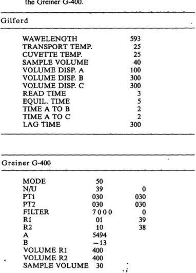
Tab. 1. Data on the programming of the Gilford systems and the Greiner G-400.
Gilford
WAWELENGTH TRANSPORT TEMP.
CUVETTE TEMP.
SAMPLE VOLUME VOLUME DISP. A VOLUME DISP. B VOLUME DISP. C READ TIME EQUIL. TIME TIME A TO B TIME A TO C LAG TIME
59325 4025 300100 3003 52 3002
Greiner G-400 MODEN/U PT1PT2 FILTER RlR2 AB
VOLUME Rl VOLUME R2 SAMPLE VOLUME
5039 030030 7 0 0 0
0110 5494-13 400400
30
0300 0300 3938
• i
J. Clin. Chem. Clin. Biochem. / Vol. 26,1988 / No. 11
TrdfpUDikf 1988
Aus Tradition in Halle 4
Die Themen des diesjährigen Medica-Treffpunktes Merck sind:
Gerätesysteme
EASY ST, der kleine Analyzer mit den Methoden des großen Labors und das maßgeschnei- derte Packungskonzept für den AU 5000 von Olympus.
D Klinische Chemie IMAC, Immunoassays so ein- fach wie die klassische klinische Chemie, als erster Parameter dieser neuen Technologie die PMN-Elastase. Kontrpllseren Qualitrol® und Creatinin als enzymatischer Test.
D Mikrobiologie
Anaerocult®, das Paket für die Anaerobiose, Bactident® Test- stäbchen, unser Programm für die Candida-Diagnostik und Fertignährböden.
Zelldiagnostik
Kryofix®, Fixiermittel für Gefrier- schnitte mit und ohne Mikrowel- lenstimulation. TissuGnost®, immunhistochemische Färbe- reagenzien und LeucoGnost®
zur Leukämiedifferenzierung.
Außerdem präsentieren wir an unserem Stand die Diagnostica Merck PC-Liste und den Merck- Katalog auf Diskette sowie Compas, das PC-gestützte Infor- mationssystem für das Labor.
Besuchen Sie uns in Düssel- dorf, dort liegt auch wieder unser Diagnostica-Spectrum für Sie bereit.
DIAGNOSTICA
MERCK
E. Merck
Frankfurter Straße 250 D-6100 Darmstadt 1
HPLC-electrochemical detection with and without BIOSENSORS
Electrochemical Detector EP 30
highly sensitive determination of clinical relevant compounds O catecholamines
in blood plasma and urine by using the new Biometra glassy carbon detector cell and the Biometra-catecholamines- kits
O acetylcholine choline
glucose gaBactose ethanol methanol
l V "JA
in the pmole range ~^^
by combination of the Biometra-BIOSENSORS (with immobilized enzymes) and electro- chemical detection using the Biometra platinum detector cell
Analyse these substances with the same system simply by chan- ging the BIOSENSOR cartridge the company which introduced BIOSENSORS into HPLC
Biometra
biomedizinische Analytik GmbH Wagenstieg 5 · D-3400 Göttingen
Tel. (05 51) 37 10 32-34 · Fax (05 51) 4 76 55 Telex 965213
m
Gewinnen Sie mitt den PiVuN-EicGStase-Assuys deei WettBairf gegen doe Zeof
1C
Die PMN-Elastase ist einer der sen- sitivsten Parameter zur Diagnose einer Entzündung. Die bekannte 2 h-Version ermöglicht die Bestim- mung aus Plasma, Stenalmark- un- Synovialflüssigkeit und weiterem Untersuchungsmaterial. Ab Oktobe 1988 steht der homogene Enzym- immunoassay PMN-Elastase IMAC zur Verfügung, er liefert aus dem Plasma innerhalb von 10 Minuten, bei manueller Methode innerhalb von 20 Minuten, die Entscheidungs hilfe für den Kliniker.
DIAGNOSTICA
MERCK
E. Merck
Frankfurter Straße 250 D-6100 Darmstadt 1
(95)
Bereits 150 Laboratorien in Deutschland arbeiten mit BM/Hitachi 717 Systemen.
Dieser Erfolg ist unsere beste Empfehlung.
BMI Hitachi 717
Sit) ttiibt>n Anspruch mit f l V.·*/»/
Molecular, Cellular and Clinical Endocrinology D
Proceedings of the Seventh Workshop on Vitamin D Rancho Mirage, California, U.S.A. April 1988
Editors A. W. Norman · K. Schaefer · H.-G. Grigoleit - D. v. Herrath 1988.17 cm χ 24 cm. XL, 1072 pages. Numerous illustrations
Hardcover. DM 390,- ISBN 3110114771
The quality of the science presented at the Seventh Workshop on Vitamin D was outstanding. Major developments were made in understanding of Vitamin D on many research frontiers, including those of the chemists, biochemists, physiologists as well as clinicians who attended the meeting and who are actively conducting research in various aspects of the Vitamin D endocrine system.
From the Contents
Chemistry .of Vitamin D Seco-Steroids · Vitamin D Metabolism and Catabolism · Biochemistry and Regulation of Hydroxylases · Receptors for 1,25 (OH)? D
3(Biochemistry and Molecular Biology) · Cell Differentiation/Hematopoiesis/Immu- nology · Gene Regulation by 1,25 (OH)
2D
3· Biological Actions of Vitamin D Metab- olites Calbindins (Biochemistry, Molecular Biology, Biological Actions) · Author Index · Key Word Index · Cell Line Index
Also available:
Vitamin D
Uremic Bone Disease: 1974. XXTV, 779 pages. DM 205,- Basic Research: 1979. XXVIII, 1318 pages. DM 220,-
Calcium Metabolism: 1982. XXVIII, 1286 pages. DM 285,-
Chemical, Biochemical and Clinical Update: 1985. XXXXHI, 1249 pages. DM 340,-
Prices are subject to change without notice
w
DEG Gmyter · Berlin · New York
Verlag Walter de Gmyter & Co., Genthiner Str. 13, D-1000 Berlin 30, Tel.: (030) 26005-0, Telex 184027 Walter de Gruyter, Inc., 200 Saw Mill River Road, Hawthorne, N.Y. 10532, Tel. (914) 7 47-0110/Telex 646677
(98)
Solem et al.: Automated iron determination, comparison of chromogens 699 Reference method
A manual reference method with the chromogen bathophen- anthroline sulphonate with deproteinisation (kit from Boehrin- ger Mannheim, FRG No. 124214), which is comparable with the recommended method of The International Committee for Standardization in Hematology (12) was used.
Methods for urine examination
Standards of 17.9, 90.0, 179.1, and 358.2 μιηοΐ/ΐ and the same reagents and programming as for serum and plasma determi- nations (s. above) were used.
Pretreatment of urine samples
The urine samples were diluted 1:3.5 with a solution containing citric acid (0.57 mol/1) and ascorbic acid (0.48 mol/1); 0.5 ml of this diluted solution was placed in a capped micro reaction vessel and kept in a heat block (Eppendorf 3401, Eppendorf Ger tebau, Hamburg, FRG) at 56 °C for 15 min. After this pretreatment, the urine sample was assayed automatically as described above. If the total amount of ferrioxamine iron and transferrin iron in serum is to be determined, serum can be pretreated in the same manner.
0.8η ^/OOOOOO
0.2
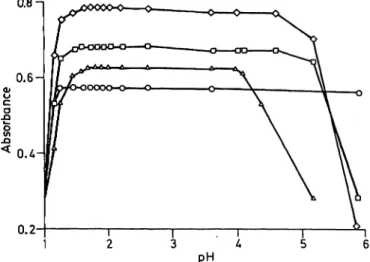
Fig. L Colour development and intensity of the four tested chromogens at different pHs by the use of citrate/HCl/
NaOH buffer.
Ο = Ferene-S D = Ferrozine
Δ = 2,4,6-Tripyridyl-s-triazine ο = Bathophenanthroh'ne sulphonate
Results
Investigation of different chromogens
The reaction time for total colour development was found to be 30 s for tripyridyl-s-triazine, 2 min for bathophenanthroline sulphonate and ferrozine and l h for ferene-S, examined at 25 °C and at a pH of 1.85 (see tab. 2). The four different chromogens were tested under the same standard conditions, using 0.24 mol/1 citric acid/-HCl-NaOH buffer. Figure 1 indi- cates that tripyridyl-s-triazine has a constant colour intensity in the pH range between 1.6 and 4.0 and bathophenanthroline sulphonate in the pH range 1.3 to at least 6.0. The chromogens ferrozine and ferene- S were found to have a somewhat less constant colour intensity between pH 1.6 and 4.6 and between pH 1.7 and 4.6, respectively. All reagents were found to be
Tab. 2. Some features of the four different chromogens under the same standard conditions (citrate buffer, pH 2.0).
Colour reagent
Tripyridyl- s-triazine Bathophen- anthroline sulphonate Ferrozine Ferene-S
Reaction Solu- . time bility (min)
0.5 + 2.0 +
2.0 + 60.0 +
Molar absorptivity under test conditions (m2/mol) 220660 219210
282360 360020
stable for at least 2 months at 4 —8 °C and 1 week at room temperature. The colour complexes of tripyri- dyl-s-triazine and ference-S were found to be stable for at least 72 h, and for bathophenanthroline sul- phonate and ferrozine for at least 36 and 24 h, re- spectively. The interference of copper in the tripyridyl- s-triazine/citrate method was found to be less than 2.0% in a plasma with various concentrations of added copper chloride, as described in detail elsewhere
(13).
Correlations with the reference method Sixty different plasma samples from patients were tested with tripyridyl-s-triazine, bathophenanthroline sulphonate, ferrozine and ferene-S and the results compared with those from the reference method. In figure 2 a, it is shown that a good correlation was obtained with the tripyridyl-s-triazine method. No interference was observed in this method in icteric, lipaemic or haemolytic plasmas. These findings show clearly that the tripyridyl-s-triazine/citrate method is specific and sensitive for iron determination in human blood. This method detects iron concentrations of less than 0.2 μπιοΐ/ΐ. Distinctly turbid, lipaemic sera may impair precision and should be pretreated with a commonly available cleaning agent. Interference with the determination of haemoglobin iron arise at 37 °C on the Greiner system in plasmas with a haemoglobin content of 0.8 g/1 or more. In the other systems, haemoglobin contents up to 5.0 g/1 did not interfere, as demonstrated in table 3. In figure 2 it is shown J. Clin. Chem. Clinr Biochem. / Vol. 26,1988 / No. 11
700
Solem et al.: Automated iron determination, comparison of chromogens 7060 50
^ 20cο
ι—ι
10
10 20 30 40 50 60
Iron (reference method) [μηηοΐ/ϋ 70 10 20 30 40 50 60
Iron (reference method) [μηηοΐ/ϋ 70
70 60 50
toE
1 30 Φc ωω
- 20 g
10
10 20 30 40 50 60
Iron (reference method) [μιτιοΙ/Π 70 10 20 30 40 50 60
Iron (reference method) [μπιοΐ/ΐ] 70
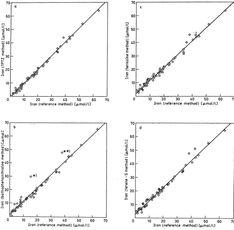
Fig. 2. Correlation data of the four different chromogens with the manual reference method in plasma samples from patients.
The chromogens were tested under the same standard conditions (citrate buffer pH 2.0).
a) 2,4,6-Tripyridyl-s-triazine (TPTZ)
Total range: n = 60; r = 0.996; y = 0.1035 χ - 0.508 Range 0.7-10.2 μιηοΐ/l: η = 25; r = 0.916; y = 0.948 χ + 0.068 Range 10.3-63.4 μτηοΐ/l: n = 35; r = 0.995; y = 1.045 χ - 0.854 b) Bathophenanthroline
Total r nge: n = 60; r = 0.983; y = 1.002 χ Range 0.7-10.2 μιηοΐ/ΐ: n = 25; r = 0.874; y = 1.154 χ + Range 10.3-13.4 μηιοΐ/ΐ: n = 35; r = 0.974; y = 1.03 χ +
*) plasma sample with a bilirubin content of 73.5 μτηοΙ/1.
**) plasma sample with a bilirubin content of 287.3 μιηοΐ/ΐ.
c) Ferrocine
Total range: η = 60; r = 0.992; y = 0.977 χ -ί Range 0.7-10.2 μπιοΐ/l: η = 25; r = 0.898; y
Range 10.3-63.4 μιηοΐ/l: n = 35; r = 0.990; y d) Ferene-S
Total r nge: n = 59; r
1.066 χ 1.005x
1.187 0.776 0.093
2.247 2.078 1.285 Range 0.7-10.2 μιηοΐ/ί: η = 24; r = 0.879; y = Ϊ!θ16°χ
Range 10.3-63.4 μιηοΐ/l: n = 35; r = 0.989; y = 1.023 χ 0.992; y = 0.997„x + 2.835
3.030 1.933
J. Clin. Chem. Clin. Biochem. / Vol. 26,1988 / No. 11
Solem et al.: Automated iron determination, comparison of chromogens 701 Tab. 3. Determination of iron in haemolytic plasma samples.
From a blood sample, erythrocytes were haemolysed in distilled water and diluted to different concentrations.
Separate fractions of the plasma sample were mixed with the same volume of one of the haemolysate dilu- tions. The expected value was determined after addition of the same volume of distilled water to a plasma fraction. Haemoglobin was determined with the cyan- methaemoglobin method on a Gilford Stasar III spec- tral photometer (Gilford Instruments, Oberlin, OH, USA).
Tab. 4. List of results (μηιοΐ/ΐ) of the four tested methods under the same standard conditions in plasma samples in the pathologically decreased range.
Hb concentration (g/D
0.621.25 2.505.05
Expected value (μπιοΐ/ΐ) 12.212.2 12.212.2
Found value (μηιοΐ/ΐ) 12.512.2 12.012.3
that the other chromogens have a considerably posi- tive error, particularly in the pathologically decreased range, as also demonstrated in table 4. In plasmas with iron concentrations above 10.2 μιηοΐ/ΐ the cor- relation with the reference method was found to be improved, and the difference between the 4 different chromogens was found to be almost balanced. Two plasma samples with elevated total bilirubin concen- tration were found to have a falsely positive error in the bathophenanthroline sulphonate/citrate method at pH 2.0. These plasma samples (fig. 2b * and **) had bilirubin concentrations of 73.5 and 278.3 μήΐοΐ/ΐ, respectively, indicating that bilirubin interferes with this method. The latter finding was confirmed in a pooled plasma from 12 different icteric patients. In this pooled plasma, the following data were obtained:
total bilirubin = 155.6 μιηοΐ/ΐ, iron concentration in the tripyridyl-s-triazine method = 17.6 μιηοΐ/ΐ, iron concentration in the bathophenanthroline sulphonate method == 24.7 μιηοΐ/l, positive error in the batho-
21 34 65 78 109 1112 1314 1615 1718 2019 2122 2324 25
Reference method 0.701.80 2.002.30 2.703.00 3.803.90 4.304.50 4.805.70 5.906.10 6.607.20 7.207.70 7.908.20 8.208.20 8.609.00 10.20
Tripyridyl- s-triazine 0.402.30 2.002.00 2.502.50 6.404.50 2.505.20 3.405.70 5.006.10 3.907.00 8.105.70 8.406.60 7.909.00 8.609.30 9.90
Bathophen- anthroline sulphonate 2.000.50 3.903.20 4.105.00 9.104.80 4.503.90 9.706.10 7.206.80 7.907.50 7.908.20 12.709.70 11.30 10.60 14.50 11.109.30
Ferrozine
4.503.40 4.702.70 5.708.40 5.605.70 6.605.00 5.009.50 9.108.10 7.709.90 9.007.90 11.50 12.90 10.709.70 13.10 12.20 13.10
Ferene-S
5.003.90 5.60 7.00— 6.407.50 6.806.40 4.307.20 10.90 10.60 8.608.60 9.309.30 12.509.30 13.30 10.409.50 13.30 13.60 13.80
phenanthroline sulphonate method = 40.3%. By in- creasing (decreased plasma/reagent ratio) the pH in the direct bathophenanthroline sulphonate method from pH 2.0 to pH 4.0, the interference by bilirubin was abolished. Thus, the bathophenanthroline sul- phonate/citrate method seems to be specific for iron measurement at the latter pH. No significant differ- ence in the iron concentration could be detected be- tween human serum and plasma.
Tab. 5. Data on the intra-assay precision of the tripyridyl-s-triazine method in different control sera.
Control serum Decision L 1 (Beckman) Decision L 2 (Beckman) Decision L 3 (Beckman) Kontrollogen-LP (Behringwerke) Serononn (E. Merck)
Charge No.
C 401202 C 401203 C 401 204
623208 162
Assigned value1
(μιηοΐ/ΐ) 21.1 28.8 41.5 35.52
26.3
η
30 30 30 30 30
Found mean value (μιηοΐ/ΐ) 22.5 32.2 47.0 41.2 26.4
SD
0.25 0.30 0.34 0.89 0.48
CV (%) 1.11 0.93 0.72 2.17 1.82
1 bathophenanthroline sulphonate without deproteinization.
2 ferrozine without deproteinization.
J. Clin. Chem. Clin. Biochem. / Vol. 26,1988 / No. 11
702 Solem et al.: Automated iron determination, comparison of chromogens Accuracy and precision in control sera
The best precision in the tripyridyl-s-triazine method was obtained in non-lyophilized sera with a CV rang- ing from 0.72 to 1.11% (within one run), as shown in table 5. The day-to-day variation and mean accu- racy for Seronorm were 2.4% and + 0.5%, respec- tively.
Adaptation to other automated systems The tripyridyl-s-triazine/citrate method was adapted to a Technicon SMAC II and Technicon RA 1000 (13), Eppendorf Epos and Abbot Spectrum auto- mated analysers. The method can be run at any com- monly used temperature. In the latter two systems, some modifications are necessary1).
500
'
Μ
t 200
•J 100
100 200 300 400
Urine iron (atomic absorption) L<umol/lD 500
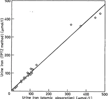
Fig. 3. Correlation data of the 2,4,6-tripyridyl-s-triazine (TPTZ) method with the atomic absorption spectro- photometric method in urine samples,
n = 26; r = 0.992; y = 0.950 χ + 5.834 Ferrioxamine iron in urine
In order to release all desferal-bound iron during sample pretreatment at 56 °C, different molarities of citric acid and ascorbic acid were investigated. At 0.57 mol/1 citric acid and 0.48 mol/1 ascorbic acid, all ferrioxamine-bound iron was released for the tripyr- idyl-s-triazine reaction, as determined in urines with known ferrioxamine iron content. This pretreatment can also be used for serum samples, if both ferriox- amine iron and transferrin iron are to be determined.
') Data on the programming of the Eppendorf Epos and Ab- bott Spectrum systems are available from the author.
The correlation data between the tripyridyl-s-triazine method and the atomic absorption method in urine samples is demonstrated in figure 3. These findings are in accordance to the data obtained in iron-free urine samples after addition of desferal and iron (II) chloride at various concentrationsr(not shown).
Discussion
The present investigation clearly shows that our tri- pyridyl-s-triazine/citrate method is specific and sen- sitive for direct iron measurements in human blood, and even in plasmas with iron values of 0.2 μιηοΐ/ΐ or less. The high specificity of tripyridyl-Sr-triazine is con- sistent with an earlier report (14). Moreover, the sim- ple reagent composition, high reagent stability and the instant colour development in the tripyridyl-s- triazine method are features of great practical impor- tance, which facilitate an adaptation to other auto- mated systems. It has previously been pointed out that tripyridyl-s-triazine, in contrast to ferrozine, is quite insoluble in water (14--15). This seems to be the main reason why tripyridyl-s-triazine has found only limited use. Under our standard conditions, at an acidic pH, tripyridyl-s-triazine was totally soluble within few minutes.
The reaction time of l h found with ferene-S is some- what longer than reported by Eskelinen (11). Colour development within approx. 2 min in the bathophen:
anthroline sulphonate- and ferrozine methods and within approx. 30 s for tripyridyl-s-triazine is there- fore an obvious advantage. In our automated systems, a relatively good precision was obtained with ferene- S, even after an incubation time of only 5 min. The high sensitivity of ferene-S is only of limited value under these conditions. On the other hand, a reaction time of more than 5 to 10 min limits the usef llness of a method in an automated system. The positive error in the ferrozine and ferene-S method indicates interference by copper, in accordance with an earlier report (11). We have no explanation at present for the interference of bilirubin at pH 2.0 in the direct bathophenanthroline sulphonate/citrate method. In view of this interference, the bathophenanthroline sul- phonate method is not suitable as a micromethod under our standard conditions. In contrast to batho- phenanthroline sulphonate, tripyridyl-s-triazine is available at low cost.
Both the reducing and chelating effect of citric acid and the rapid release of iron from transferrin at the low pH are advantageous. The coloured ferroin com- plexes also seem to be stabilized in our method, giving a constant colour development hva relatively wide J. Clin. Chem. Clin. Biochem. / Vol. 26,1988 / No. 11
Sol em et al.: Automated iron determination, comparison of chromogens 703
pH-range, in contrast to HCl-glycine-Tween buffer at
the same pH (16). By the use of less sensitive photo- metric equipment, the latter finding enables a decrease in the ratio, sample/reagent volume, and hence an increase in pH.
The wide linear range of the ferrioxamine iron deter- mination in urine samples with a high iron content is advantageous. During desferal treatment, an increase in the serum iron concentration occurs (17). Without pretreatment of the serum samples, our method will release some ferrioxamine iron. Since desferal partly binds transferrin iron (18), a decrease in the serum transferrin iron during desferal treatment can be ex-
pected. The quantity of released ferrioxamine iron may vary widely under different standard conditions.
Direct serum iron determinations during desferal ther- apy might thus be rather uncertain. In contrast to an earlier report (19), we found no evidence for the sensitivity of tripyridyl-s-triazine to desferal under our standard conditions. Pretreatment of samples permits the determination of the total ferrioxamine iron and transferrin iron in serum.
Acknowledgement
We wish to thank Mrs. Helma Maigatter and Mrs. Christine Meyer for their skilful technical assistance.
References
1. Young, D. S. & Hicks, J. M. (1965) J. Clin. Pathol. 18, 98-102.
2. Kauppinen, V. & Gref, G.-C. (1967) Scand. J. Clin. Lab.
Invest. 20, 24-28.
3. Giovannieilo, T. J., DiBenedetto, G., Palmer, D. W. &
Peters, T. Jr. (1968) J. Lab. Clin. Med. 77, 874-883.
4. Werkman, H. P. T., Trijbels, J. M. R, van Munster, P. J. J., Schretlen, E. D. A. M. & Moerkerk, C. (1971) Clin. Chim.
Acta 31, 395-401.
5. Yee, H. Y. & Zin, A. (1971) Clin. Chem. 77, 950-953.
6. Bjerve, K. S. (1976) Scand. J. Cün. Lab. Invest. 36, 673-677.
7. Levinson, S. S. (1980) Clin. Chem. 26, 671-672.
8. Viollier, . ., Gschwind, H. & Schläpfer, P. (1980) Lab.
Med. 4, 240-244.
9. Mori, L., Bekkering, A., Traini, J. & Vanderlinden, L.
(1981) Clin. Chem. 27, 1441-1444.
10. Ceriotti, F. & Ceriotti, G. (1980) Clin. Chem. 26, 327- 11. Eskelinen, S., Haikonen, M. & Räisänen, S. (1983) Scand.331.
J. Clin. Lab. Invest. 43, 453-455.
12. International Committee for Standardization in Hematol- ogy (1971) Blood 37, 598-600.
13. Seiffert, U. B., Solem, E. & Zirker, M. (1988) Lab. Med.
72, 174-178.
14. Collins, P. R & Diehl, H. (1959) Anal. Chem. 31, 1862- 1867.
15. Stookey, L. L. (1970) Anal. Chem. 42, 779-781.
16. Ichida, T., Osaka, T. & Kojima, K. (1968) Clin. Chim. Acta 22,271-275.
17. Wöhler, R (1964) Acta Haematol. 32, 321 -337.
18. Keberle, H. (1964) Ann. N. Y. Acad. Sei. 779, 758-768.
19. van Stekelenburg, G. J., Valk, C. & de Boer, G. J. (1982) Clin. Chem. 28, 2328-2329.
PD Dr. E. Solem Laboratorien
Zentrum der Kinderheilkunde der Universitätskliniken Theodor-Stern-Kai 7
D-6000 Frankfurt am Main 70
J. Clin. Chem. Clin. Biochem. / Vol. 26,1988 / No. 11