Short communication/Kurzmitteilung 431 J. Clin. Chem. Clin. Biochem.
Vol. 18, 1980, pp. 431-432
S H O R T C O M M U N I C A T I O N / K U R Z M I T T E I L U N G
A Sensitive Method for Determination
of Conjugated Catecholamines in Blood Plasma By Marianne Nagel and H.-J. Schumann
Pharmakologisches Institut des Universit tsklinikum Essen
(Received October 29,1979/March 3,1980)
Summary: Conjugated catecholamines in blood plasma of blood donors were determined from the difference between the total and the free catecholamines. Total catecholamines were measured after complete hydrolysis of the conjugated amines (heating of acidified samples for 40 min at 95 °C). Free catecholamines were determined in untreated plasma. Deter- minations were performed radioenzymatically. We combined and modified the procedures of Peuler & Johnson (1977) Life Sei 27, 625-636) and Da Prada & Z rcher (1976) Life Sei 29, 1161—1174). The resulting method was less laborious and cheaper.
Eine empfindliche Methode zur Bestimmung der konjugierten Katecholamine im Plasma
Zusammenfassung: Die konjugierten Katecholamine im Plasma von Blutspendern wurden als Differenz zwischen dem Gehalt an gesamten und freien Katecholaminen bestimmt. Nach abge- schlossener Hydrolyse der konjugierten Amine durch 40min ti- ges Erhitzen der sauren Proben auf 95 °C wurden die gesamten Katecholamine gemessen. Die freien Katecholamine wurden im unbehandelten Plasma gemessen. Wir kombinierten und modi- fizierten die radioenzymatischen Bestimmungsmethoden von Peuler & Johnson (1977) Life Sei 27, 625-636) and Da Prada
&Z rcher (1976) Life Sei 19,1161-1174) und erhielten ein einfacheres und billigeres Verfahren.
introduction
Conjugated catecholamines (sulfurie and glucuronic acid con- jugates) are present in urine and blood (1, 2). Their presence in blood is often ignored, although they greatly exceed the free catecholamines. Thus, the greater proportion of the amines is disregarded when the estimation of the tone of the sympathetic nervous system in connexion with illnesses like hypertension or neurological disorders is based solely on the amount of the free catecholamines in blood. It therefore seems advisable to measure the conjugated as well as free catecholamines, in order to avoid misinterpretations. The methods for estimation of the conjugated amines in blood used hitherto are either too insensi- tive or unreliable; either the amines undergo varying degrees of destruction during hydrolysis, or hydrolysis is incomplete. The method presented here overcomes these difficulties.
Experimental
Blood samples drawn from blood donors of either sex (20-40 years) immediately after loss of 500 ml blood were mixed with a solution (10 ml/1 blood) containing 0.26 mol/1 dithiothreitol and 0.5 mol/1 EGTA1), cooled to 0 °C, and centrifuged to obtain the plasma. One part of the supernatant was used to measure free catecholamines (a). Another part (500 μΐ) was mixed with 500 μΐ 0.6 mol/1 perchloric acid containing 2 g/1 EGTA. After sedimentation of the precipitated protein the supernatant was heated to 95 °C in Eppendorf tubes for various periods of time. After cooling, the samples were used to deter- mine the total (= free + conjugated) amount of noradrenaline, adrenaline and dopamine (b).
Catecholamines were determined after enzymatic O-methylation, using radioactive S-adenosyl-L-methionine-methyl[3H] as methyl donor. The procedure is a simplification of previously published methods (3, 4). The incubation mixtures were as follows: 25 μΐ sample hydrolysed (a) or unhydrolysed (b), 10 μ! 0.01 mol/1 HC1 and 25 μΐ of a mixture consisting of 17.5 μΐ Tris buffer (2 mol/1 (a) or 0.3 mol/1 (b), 93 mmol/1 MgCl2, 32 mmol/1 EGTA), 2 μΐ catechol-O-methyltransferase (prepared according to (5) and further purified by Sephadex G 200 chromatography) 37 kBq S-adenosyl-methionine- methyl[3H] (18-55.5 Bq/mmol), 0.05 mg dithiothreitol and H2O up to 25 μΐ. We used 0.3 mol/1 perchloric acid or H2O as blanks. For quantitative analysis, 100 pg noradrenaline, adrenaline and dopamine in 10 μΐ 0.01 mol/1 HC1 were added to each sample in duplicate. After incubation at 37 °C for l h the reaction was stopped by cooling in ice-cold water. Borate buffer (150 μΐ, 0.5 mol/1 pH 8), tetraphenylborate (50 μΐ,
15 g/l H2O) and carrier (noradrenaline, adrenaline and dopa- mine, 0.5 g/1 0.01 mol/1 HC1) were added. The amines were extracted into 5 ml diethylether. After freezing the aqueous layer the ether was transferred totally to a second set of tubes containing 0.25 ml 0.1 mol/1 HC1. After shaking and centrifuga- tion the-ether phase was discarded and the aqueous phase was washed with 5 ml butyl acetate saturated with H2O and eva- porated. The residue was dissolved in 50 μΐ of a mixture of 80 ml methanol and 20 ml 0.001 mol/1 HC1 and spotted on Kieselgel plates with fluorescent indicator ( 5 X 1 0 cm, 3 spots/
plate). The tubes were rinsed with 25 μΐ of the above solution.
After chromatography in chloroform, ethanol, ethylamine (70%) (16 ml, 3 ml, 2 ml) the spots with normetanephrine, metanephrine and methoxytyramine were scraped into separate scintillation vials. Normetanephrine and metanephrine were oxidized by adding NH4OH (1 ml, 0.05 mol/1). Scintillation cocktail (6 ml; 4 g 2,5-diphenyloxazole, 50 mg 1,4-di (2-(5- phenyloxazolyl)) benzene per liter toluene) was added and vanillin extracted into the organic phase. Methoxytyramine was dissolved in 0.5 ml 0.5 mol/1 HC1 and 7.5 ml Quickscint 212 (Zinsser) were added.
The radioactivity was measured in a MARK II Nuclear Chicago liquid scintillation counter. The amount of free and total noradrenaline, adrenaline and dopamine and that of the con- jugated part (= total -free) were determined.
For control purposes (see text) two dimensional chromato- graphy was performed on Kieselgel plates. The usual alkaline solvent was used for the first dimension. For the second dimension, the solvent was: /so-butanol, methanol, 1 mol/1 formic acid (10 ml, 20 ml, 20 ml) (6).
EGTA = (ethylene-bis(oxyethylenenitrilo)tetraaceticacid
0340-076X/80/0018-0431 $2.00
© by Walter de Gruyter & Co. · Berlin · New York
432
Short communication/KuizmitteilungResults and Discussion
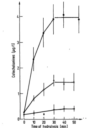
During hydrolysis the amount of detectable catecholamines increased. As can be seen from figure 1 hydrolysis was complete after 40 min. Of the catecholamines added before hydrolysis 97.1 ± 4.7% (noradrenaline), 95.8 ± 9% (adrenaline), and 103.4 ± 5.5% (dopamine) (5Γ ± 5χ, η = 10) were recovered.
Table 1 shows the amounts of free and total (= free + con- jugated) catecholamines in plasma samples from 10 different
blood donors.
In order to affirm that the high levels of total noradrenaline, adrenaline, and dopamine found after hydrolysis were not due to methodical errors, we tested whether the amine spots were overlapped by unknown substances and whether the blank values were elevated after heating the samples. We used two- dimensional chromatography for separation of the methylated catecholamines from putative unknown contaminants that might contribute to the radioactivity. 76-105% of the original amine content was found after two-dimensional chromato- graphy. To determine blank values with a hydrolyzed sample catechol-O-methyltransferase was inhibited by CaCl2, added to the buffer in place of and at the same molarity as MgCl2. Ca"1"1"
diminished the blank values as compared to those with Mg4"4". In the case of noradrenaline and adrenaline, addition of a hydro- lyzed sample to the Ca** reaction mixture did not raise the blank values. The blank value for dopamine increased, but the increase was negligible compared with the number of counts obtained for dopamine after hydrolysis: 13 counts per minute compared to several hundreds of the sample (13 counts per minute = 0.5 pg).
Our results contradict those of Buu & Kuchel (2), who stated that acidic hydrolysis at elevated temperatures is not suitable for radioenzymatic determinations of catecholamines. Their
Tab. 1. Free and total (free + conjugated) catecholamines in plasma from blood donors immediately after loss of 500 ml blood.
10 20 30 40 50 Time of hydrolysis [min]
Fig. 1. Rise of detectable catecholamines with increasing time of hydrolysis.
Values are mean ± S.E.M. of ten different plasma samples.
• Dopamine ο Noradrenaline
• Adrenaline
Age Sex (a) 20 931 9 37 921 d 23 d26 d 35 d39 d 40 d39 d Mean
± S.E.M.
Noradrenaline (ng/1) free total
79 738 416 683 356 892 213 468 330 1106 345 631 315 805 561 3017 439 844 206 903 326 1008
± 43 ± 230 conjugated:
67.6%
Adrenaline (ng/1) free total
49 835 76 80 92 563 79 446 125 431 81 149 9.0 1330 81 224 116 210 68 204 87.5 447-
± 7 ±122 conjugated:
80.4%
Dopamine (ng/1) free total 26 3533 53 7877 27 8413 34 7108 29 3095 27 3746 30 4422 35 5017 29 9423 36 2128 32.6 5476
±2.5 ±800 conjugated:
99.3%
Plasma samples for determination of total noradrenaline, adrenaline and dopamine were heated at 95 °C for 40 min.
procedure of hydrolysis consists of concentrating the samples containing 0.3 mol/1 perchloric acid by lyophilisation. We tried their method and found it insufficiently controllable. As judged from the counts per minute for the internal standard, it appeared that in some cases most of the catecholamines were lost during the concentration process. In others the amounts of conjugated catecholamines were less than those found with our method. In other words: The tune between complete hydrolysis and the start of wholesale amine destruction is not defined, whereas we determined the end of hydrolysis for our procedure precisely.
Our method yielded complete hydrolysis of conjugated catechol- amines without detectable destruction. It was combined with a very sensitive radioenzymatic method for the quantitative analysis of plasma catecholamines based upon previously published procedures that were combined. The result was a cheaper and less laborious method without loss of its original sensitivity.
Acknowledgements
We are obliged to Dr. W. Lubold who provided us with blood samples and to Mrs. U. Chebbifoi her skilful technical assistance.
References
1. Haggendahl, J. (1964), Acta PhysioL Scaiid. 59, 255-260.
2. Buu, N. T. & Kuchel, O. (1977), J. Lab. Clin. Med. 90, 680- 3. Peuler, J. D. & Johnson, G. A. (1977), Life Sei. 21, 625-685.
4. Da Prada, M. & Z rcher, G. (1976), Life Sei. 7P, 1161-636.
1174.
5. Axelrod, J. & Tomchick, R. (1958), J. Biol. Chem. 233, 702-705,
6. Fleming, R. M. & Clark, W. G. (1970), J. Chrpmatogr. 52, 305-312.
Prof. Dr. med. H. J. Schumann
Direktor des Pharmakologischen Instituts Hufelandstra e 55
D-4300 Essen
J. Clin. Chem. Clin. Biochem. / Vol..18,1980 / No. 7