Characterisation of atypical
Rho GTPases of the RhoBTB family and their binding partners
Inaugural-Dissertation zur
Erlangung des Doktorgrades
der Mathematisch-Naturwissenschaftlichen Fakultät der Universität zu Köln
vorgelegt von
Kristína Schenková aus Nitra, Slowakei
Köln 2010
Berichterstatter/in: Prof. Dr. Angelika A. Noegel
Prof. Dr. Jürgen Dohmen
Tag der mündlichen Prüfung: 30. Juni 2010
Die vorliegende Arbeit wurde in der Zeit von November 2006 bis Mai 2010 unter
Anleitung von Prof. Dr. Angelika A. Noegel und der Betreuung von PD Dr. Francisco
Rivero am Institut für Biochemie I der Medizinischen Fakultät der Universität zu Köln und
Centre for Biomedical Research, Hull York Medical School, University of Hull,
Großbritannien angefertigt.
Acknowledgement
I would like to thank all people that that by any means contribute to the successful completion of this dissertation thesis. Especially I would like to thank:
- PD Dr. Francisco Rivero for the opportunity to work in his group on the interesting topic, for sharing his knowledge and experience, for never ending helpfulness, for long discussions and for the opportunity to present my research at various international meetings.
- Prof. Dr. Angelika A. Noegel for the opportunity to conduct my work in her renowned institute.
- Prof. Dr. Jürgen Dohmen, Prof. Dr. Matthias Hammerschmidt and Prof. Dr. Ludwig Eichinger for their willingness and time to read this thesis and participate in the committee during my exam.
- Prof. Dr. Pontus Aspenström for providing RhoBTB constructs and one cullin construct, Prof. Dr. Reinhard Fässler for providing kindlin constructs, Dr. Manabu Furukawa for providing cullin constructs, Prof. Dr. Martin Bähler for myosin construct and to Dr. Michael Gmachl for providing the ubiquitin construct.
- All my colleagues and co-workers in the Institute for Biochemistry I, Medical Faculty, University of Cologne and in the Centre for Biomedical Research, Hull York Medical School, University of Hull for help and advices, especially to Anja and Nils. Thank you for the great time in Hull ☺!
- My family for support.
- Vlasta for encouragement, emotional support and faith in me.
Table of contents
1 INTRODUCTION... 8
1.1 The Ras superfamily of small GTPases... 8
1.2 The Rho-family... 9
1.3 RhoBTB proteins... 10
1.3.1 Structure of RhoBTB proteins... 11
1.3.1.1 The GTPase domain... 11
1.3.1.2 The proline-rich region... 12
1.3.1.3 The BTB domain... 13
1.3.1.4 The C-terminal region... 14
1.3.2 Expression of RhoBTB proteins... 14
1.3.3 Function of RhoBTB proteins... 15
1.3.3.1 RhoBTB, cell growth, and apoptosis... 15
1.3.3.2 RhoBTB and chemokine expression... 16
1.3.3.3 RhoBTB and vesicle transport... 16
1.3.3.4 RhoBTB and the actin filament system... 17
1.3.4 RhoBTB in human diseases... 18
1.4 RhoBTB proteins and proteasome-dependent degradation ... 19
1.4.1 General overview of the proteasome-dependent degradation pathway ... 19
1.4.2 Cullin dependent E3 ligases ... 21
1.4.3 RhoBTB proteins as adaptors of Cul3-dependent ubiquitin ligases ... 22
1.5 MUF1/LRRC41 ... 23
1.5.1 The SOCS-box ... 24
1.5.2 The LRR ... 25
1.6 The kindlin protein family ... 26
1.6.1 The role of kindlins in integrin activation ... 27
1.6.2 Expression and localisation of kindlins ... 28
1.6.3 Kindlin in human diseases ... 29
1.7 Uev1 ... 29
1.8 Aims of the study ... 31
2 MATERIAL AND METHODS ... 32
2.1 Material ... 32
2.1.1 Cell lines and strains ... 32
2.1.2 Vectors ... 32
2.1.3 Oligonucleotides for siRNA ... 32
2.1.4 Oligonucleotides for RT-PCR ... 33
2.1.5 Oligonucleotides for PCR ... 33
2.1.6 Constructs ... 34
2.1.7 Enzymes ... 35
2.1.8 Antibodies and fluorescent dyes ... 36
2.1.8.1 Primary antibodies ... 36
2.1.8.2 Secondary antibodies ... 36
2.1.8.3 Fluorescent dyes ... 36
2.1.9 Inhibitors ... 36
2.1.10. Transfection reagents ... 37
2.1.11. Antibiotics ... 37
2.1.12 Molecular weight markers ... 37
2.1.13 Chemicals ... 38
2.1.14 Kits ... 38
2.1.15 Laboratory material ... 38
2.2 Sterilisation ... 39
2.3 Cell culture methods ... 39
2.3.1 Defrosting of mammalian cell stocks ... 39
2.3.2 Passaging of mammalian cells ... 40
2.3.3 Transfection of mammalian cells ... 40
2.3.4 Drug treatment ... 40
2.3.5 Determination of protein stability ... 40
2.3.6 Gene silencing ... 41
2.3.7 Cryostocks preparation ... 41
2.4 Bacterial culture methods ... 42
2.4.1 Media for bacterial cells cultivation ... 42
2.4.2 Preparation of E. coli XL-1 blue competent cells ... 42
2.4.3 Transformation of E. coli XL-1 blue competent cells ... 43
2.4.4 Preparation of glycerol stocks ... 43
2.5 Yeast two-hybrid system ... 43
2.5.1 Media for yeast cells cultivation ... 43
2.5.2 Modified lithium acetate method for yeast transformation ... 44
2.5.3 Test of galactosidase activity ... 44
2.6 Immunohistochemistry ... 45
2.6.1 Fixation and permeabilisation of mammalian cells ... 45
2.6.2 Immunodetection of proteins in the cells ... 45
2.6.3 Immunostaining of mitochondria ... 45
2.6.4 Immunostaining of microtubules ... 46
2.6.5 Immunostaining of actin filaments ... 46
2.6.6 Transferrin uptake ... 46
2.6.7 Microscopy and image processing ... 46
2.7 Biochemical methods ... 47
2.7.1 Lysis of mammalian cells ... 47
2.7.2 Immunoprecipitation of proteins with Myc-epitope or GFP-epitope tag ... 47
2.7.3 SDS-polyacrylamide gel electrophoresis (SDS-PAGE) ... 48
2.7.4 Staining of polyacrylamide gels with Coomassie-Brilliant-Blue R 250 ... 49
2.7.5 Transfer of proteins to membrane (Western blot) ... 49
2.7.6 Staining of proteins bound to the membranes ... 50
2.7.7 Immunodetection of proteins bound to the membrane ... 50
2.7.8 Subcellular fractionation of mammalian cells ... 50
2.7.9 Ubiquitination assay ... 51
2.7.10 Protein expression and purification ... 52
2.7.11 Expression and purification of GST-Ubiquitin (Ub) ... 52
2.7.12 BCA (bicinchoninic acid) protein assay ... 53
2.7.13 GTP binding assay ... 53
2.8 Molecular biology methods ... 54
2.8.1 Isolation of plasmid DNA by the alkaline method ... 54
2.8.2 Isolation of plasmid DNA for transfection of mammalian cells ... 54
2.8.3 Determination of plasmid DNA concentration ... 55
2.8.5 Polymerase chain reaction (PCR) ... 56
2.8.6 Reverse transcription PCR (RT-PCR) ... 56
2.8.7 Elution of DNA-fragment from agarose gel ... 57
2.8.8 Restriction reaction ... 58
2.8.9 Dephosphorylation of DNA 5´- ends ... 58
2.8.10 Ligation of vector and DNA-fragment ... 58
2.8.11 DNA-sequencing ... 58
3 RESULTS ... 59
3.1 Characterisation of RhoBTB proteins ... 59
3.1.1 Subcellular localisation of RhoBTB3 ... 59
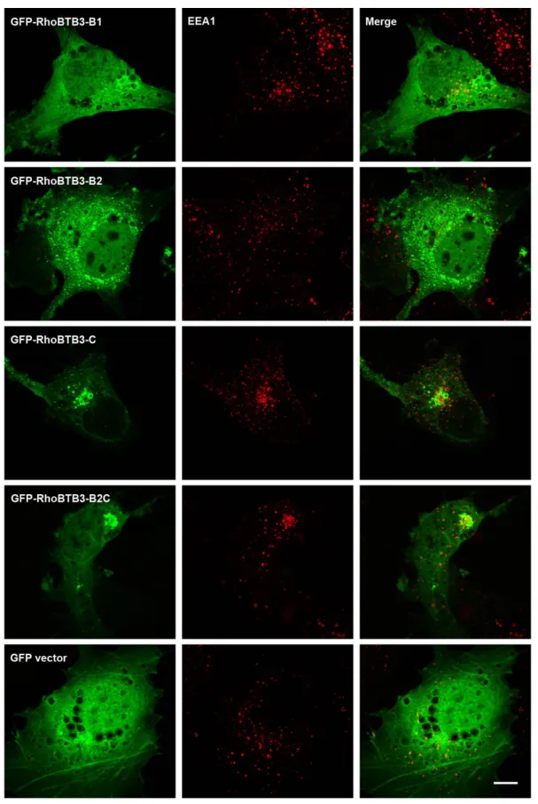
3.1.1.1 RhoBTB3 partially localises at early endosomes through the C-terminal domain... 60
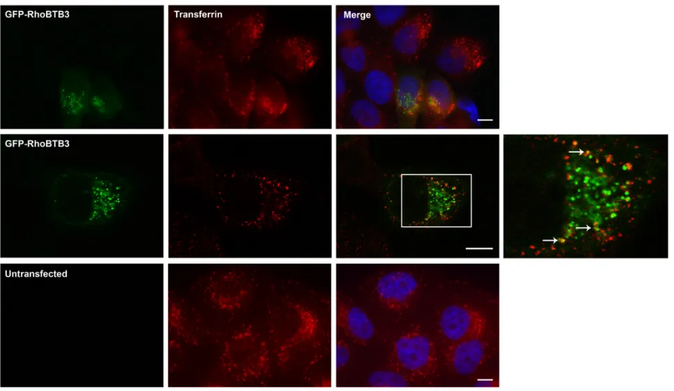
3.1.1.2 GFP-RhoBTB3 occasionally co-localises with transferrin ... 61
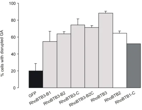
3.1.1.3 Overexpression of RhoBTB3 disrupts the Golgi apparatus ... 64
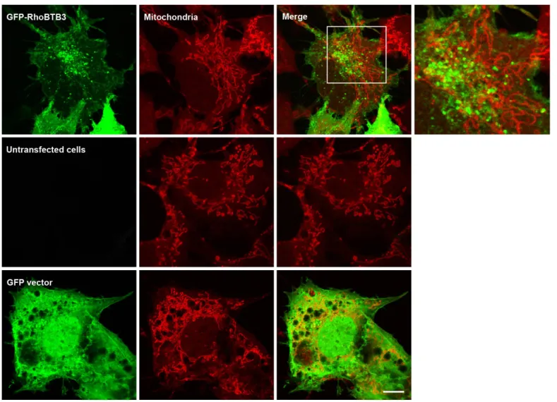
3.1.1.4 RhoBTB3 does not localise to the mitochondria ... 67
3.1.1.5 Interaction of RhoBTB3 with the cytoskeleton ... 69
3.1.1.5.1 Interaction of RhoBTB3 with microtubules ... 69
3.1.1.5.2 Co-localisation of RhoBTB2 and RhoBTB3 does not depend on an intact microtubule network ... 69
3.1.1.5.3 Interaction of RhoBTB3 with the actin cytoskeleton ... 72
3.1.2 The GTPase domain of RhoBTB3 does not bind GTP ... 72
3.1.3 Interaction of RhoBTB3 with Cul3 ... 73
3.1.3.1 RhoBTB3 interacts with endogenous Cul3 ... 73
3.1.3.2 Dimerisation of RhoBTB is Cul3-independent ... 74
3.1.4 Proteasomal degradation of RhoBTB3 is prevented by intramolecular interaction ... 75
3.1.5 RhoBTB3 is ubiquitinated by a Cul3-dependent ligase ... 76
3.2 Characterisation of MUF1/LRRC41, a binding partner of RhoBTB GTPases ... 77
3.2.1 Computational characterisation of MUF1 ... 78
3.2.2 Lrrc41 mRNA is ubiquitously expressed ... 80
3.2.3 Localisation of MUF1 ... 80
3.2.4 MUF1 interacts with all three RhoBTB proteins in vivo ... 84
3.2.5. RhoBTB3 has probably multiple binding sites on MUF1 ... 88
3.2.6. MUF1 has probably multiple binding sites on RhoBTB3 ... 89
3.2.7. MUF1 interacts with RhoBTB3 in a Cul3 and Cul5 independent manner ... 90
3.2.8 MUF1 is degraded in the proteasome in a Cul5 independent manner ... 91
3.2.9 MUF1 is able to homodimerise ... 94
3.2.10 Characterisation of MUF1 interaction with potential binding partners ... 94
3.2.10.1 MyoIXb as a potential binding partner of MUF1 ... 95
3.2.10.2 RBPMS as a potential binding partner of MUF1 ... 96
3.2.11 Dimerisation of cullins ... 97
3.3 Kindlin, an interaction partner of RhoBTB3 ... 100
3.3.1 RhoBTB2 and RhoBTB3 interact with kindlin-1 and kindlin-2 ... 100
3.3.2 RhoBTB3 has probably multiple binding sites on kindlin-1 ... 101
3.3.3 RhoBTB3 partially co-localises with kindlin-1 and kindlin-2 ... 103
3.3 Uev1a, an interaction partner of RhoBTB3 ... 104
4 DISCUSSION ... 106
4.1 Subcellular localisation of RhoBTB3 ... 106
4.2 RhoBTB3 is an adaptor of Cul3-dependent ligase complexes ... 108
4.3 Characterisation of MUF1 ... 111
4.4 Expression and subcellular localisation of MUF1 ... 111
4.5 MUF1 as an adaptor for Cul5 ubiquitin ligase ... 112
4.6 MUF1 is a binding partner of RhoBTB proteins ... 113
4.7 MUF1 as a substrate of Cul3-RhoBTB3 ubiquitin ligase ... 114
4.8 Heterodimerisation of Cul3 and Cul5 ... 116
4.9 A possible model of MUF1 function and degradation ... 117
4.10 Kindlin is a binding partner of RhoBTB proteins ... 119
4.11 Uev1a is a binding partner of RhoBTB proteins ... 121
5 ABSTRACT ... 123
6 ZUSAMMENFASSUNG ... 125
7 REFERENCES ... 127
8 ABBREVIATIONS ... 138
Erklärung ... 140
Curriculum vitae ... 141
Lebenslauf ... 142
1 Introduction
1.1 The Ras superfamily of small GTPases
The Ras superfamily, named after the most studied oncogene in human carcinogenesis, Ras, represents a group of small guanosine triphosphatases (GTPases), which comprises over 150 members in humans but can be found in all eukaryotes (Colicelli 2004, Wennerberg et al. 2005). The common feature of these proteins (with few exceptions) is their ability to bind and hydrolyse GTP due to the presence of a ~20 kDa G-domain.
The G-domain consists of a six-stranded β-sheet and five α-helices and contains four to five conserved G-box motif elements (G1-G5), which are responsible for binding GTP.
The so-called switch domains I and II bind γ-phosphate oxygens of GTP and after GTP hydrolysis and release of γ-phosphate, the switch domains relax into the GDP-specific conformation (Bourne et al. 1991).
Ras proteins act as molecular switches, cycling between an active GTP-bound state and an inactive GDP-bound state. Guanine nucleotide exchange factors (GEFs) and GTPase activating proteins (GAPs) regulate the activation/inactivation cycle (Figure 1.1). The dissociation of GDP from the inactive GDP-bound form is promoted by an upstream signal and conversion to the GTP-bound state is catalysed by GEFs. In the GTP-bound state, small GTPases are active and interact with downstream effector proteins.
Hydrolysis of GTP to GDP is very slow and is accelerated by GAPs. In addition, GDP- dissociation inhibitors (GDIs) regulate cycling of Rho and Rab GTPases between cytosol and membranes by capturing them in both GTP- and GDP-bound states (Colicelli 2004, Takai et al. 2001).
Figure 1.1: Regulation of activity of small GTPases. In the active state, the GTPase binds GTP and interacts with effectors of signalisation. GAPs accelerate the hydrolysis of bound GTP. In GDP-bound state, small GTPases are inactive. GEFs catalyse the release of GDP. Due to the higher cytosolic concentration of GTP than GDP, the GTPase can again bind GTP. Taken from Colicelli (2004).
The Ras superfamily can be subdivided into five families according to the sequence and known functions of their members: Ras (Rat sarcoma oncoproteins), Rho (Ras homologous proteins), Rab (Ras-like proteins in brain), Ran (Ras-like nuclear protein), Arf (ADP-ribosylation factor) and Miro (mitochondrial Rho) (Colicelli 2004, Wennerberg et al. 2005). Members of the Ras superfamily are involved in a variety of cellular processes like gene expression (Ras, Rho), regulation of cell proliferation, differentiation and survival (Ras), actin organisation and cell cycle progression (Rho), vesicular transport and trafficking of proteins (Rab, Arf), transport between nucleus and cytoplasm and microtubule organisation (Ran) (Colicelli 2004, Takai et al. 2001, Wennerberg et al. 2005).
1.2 The Rho-family
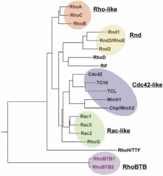
The Rho family is characterised by an insertion (so-called Rho insert) of usually 13 residues with high sequence variability between the fifth ß-strand and the fourth α-helix in the GTPase domain (Valencia et al. 1991). To date, 21 proteins of the Rho family have been described in vertebrates (Figure 1.2): Cdc42-like (Cdc42, TC10, TCL, Chp/Wrch2, Wrch1), Rac-like (Rac1-3, RhoG), Rho-like (RhoA-C), Rnd (Rnd1-2, Rnd3/RhoE), RhoD (RhoD und Rif), RhoH/TTF and RhoBTB (RhoBTB1-3) (Wennerberg and Der 2004).
RhoBTB3 is very often not considered as a member of the Rho family because of its divergent GTPase domain. Members of the Rho family are present from lower eukaryotes up to mammals and have not been identified in eubacteria and archaea.
The most studied Rho GTPases are RhoA, Rac1 and Cdc42. The members of the Rho, Rac
and Cdc42 subfamilies are involved in regulation of cytoskeleton reorganisation in
response to extracellular signals. Rho proteins are responsible for the formation of stress
fibres and focal adhesions, Rac proteins for the formation of lamellipodia and Cdc42
proteins are involved in filopodia formation. They also have been implicated in many other
cytoskeleton-dependent processes like cell growth (G1 cell cycle progression), cytokinesis,
morphogenesis, cell-cell interaction, cell polarity and cell migration. In addition, Rho
proteins are involved in cellular processes such a membrane trafficking, endocytosis
and gene expression (Jaffe and Hall 2005, Takai et al. 2001). Other Rho GTPases have
been also identified in cytoskeleton-dependent processes like loss or formation of stress
fibres (Rnd1, Rnd3, RhoD and Rif), focal adhesions (Rnd1, Rnd3, and RhoD), cell
migration and cell-cell adhesion (Rnd3), formation of Cdc42-independent filopodia (Rif)
and cell migration and cytokinesis (RhoD) (Vega and Ridley 2007). For others, like Rnd2 and RhoBTB1, 2 and 3 an effect on the actin cytoskeleton has not been observed.
Figure 1.2: Phylogenetic tree of the Rho family of small GTPases. The family can be divided into six subfamilies: RhoA-related, Rac-related, Cdc42-related, Rnd proteins, RhoH/TTF and RhoBTB proteins. Note that RhoBTB3 is not shown in this tree because of its divergent GTPase domain. Taken from Burridge and Wennerberg (2004).
Some of the members of the Rho family are referred to as atypical Rho GTPases because their structure and functional characteristics differ from those of the classical ones.
The atypical GTPases are Rnd proteins, RhoH, Wrch1, Chp/Wrch2 and RhoBTB (Aspenström et al. 2007). One of the most striking features that make these Rho GTPases atypical is the difference in the cycling between the GTP- and the GDP-bound state. For example, RhoH has been shown to be constitutively in the GTP-bound state (Li et al.
2002), as well as Rnd1 and Rnd3 (Chardin 2006). Wrch1 is predominantly in the GTP- bound state (Saras et al. 2004, Shutes et al. 2004). RhoBTB proteins seem to be even more different – RhoBTB2 does not bind GTP (Chang et al. 2006) and it was shown recently that RhoBTB3 can bind and hydrolyse ATP (Espinosa et al. 2009).
1.3 RhoBTB proteins
The RhoBTB subfamily constitutes the more recent addition to the Rho family. It was
identified during the study of Rho-related protein-encoding genes in Dictyostelium
discoideum (Rivero et al. 2001). In humans, the RhoBTB subfamily is composed of three
isoforms: RhoBTB1, RhoBTB2 (also named DBC2, deleted in breast cancer 2) and RhoBTB3. RhoBTB1 and RhoBTB2 are very similar to each other (79% similarity) and to the Drosophila orthologue (Dm RhoBTB), whereas RhoBTB3 (43% similarity to human RhoBTB1 and RhoBTB2) and the Dictyostelium discoideum orthologue (RacA) are the most divergent members. Orthologues of RhoBTB have been found in numerous eukaryotes, but they are absent in fungi and plants.
1.3.1 Structure of RhoBTB proteins
RhoBTB proteins consist of a GTPase domain followed by a proline-rich region, a tandem of two BTB domains and a C-terminal domain (Figure 1.3).
Figure 1.3: Domain architecture of RhoBTB proteins. The GTPase domain of RhoBTB3 is barely recognisable as such. The first BTB domain is interrupted by an insertion of variable length. Only RhoBTB3 has a CAAX motif at the C-terminus. The simplified phylogenetic tree on the left illustrates the relationship among the proteins. Figures denote overall percentage similarity between branches, but the degree of similarity is higher when the comparisons are restricted to particular domains (not shown). Asterisks denote the positions of mutations in RhoBTB2 found in tumours. Hs: Homo sapiens, Dm: Drosophila melanogaster, Dd: Dictyostelium discoideum. Taken from Berthold et al. (2008a).
1.3.1.1 The GTPase domain
The GTPase domain is perhaps the region where most divergence is found among members of the RhoBTB subfamily. Early analyses revealed that this domain is typically Rac-like in RacA and divergent, but recognisable as Rho-related in RhoBTB1 and RhoBTB2 as well as in Dm RhoBTB (Rivero et al. 2001).
The GTPase domain of RhoBTB proteins other than RhoBTB3 and RacA also contains
a Rho insert, which is characteristic for Rho proteins. This insert is longer than usual
(18 residues or more) and rich in charged residues. Moreover, the GTPase domain of these
RhoBTB proteins contains two insertions and one deletion, as well as a few deviations
from the GTPase consensus of most Rho GTPases (Rivero et al. 2001). The deletion
the deleted residues is the glutamine equivalent to Q61 in Ras. Also of importance, the glycine residue equivalent to G12 in Ras appears substituted by asparagine in RhoBTB1 and RhoBTB2 or threonine in Dm RhoBTB. Because these two residues are essential for GTP hydrolysis, these proteins would predictably display impaired enzyme activity.
Indeed, using a blot overlay approach, Chang et al. (2006) have shown that the GTPase domain of RhoBTB2 appears not to bind GTP at all.
In RhoBTB3, the GTPase domain appears extensively erased, to the point that it is virtually unrecognisable as a GTPase. Only a short stretch at the end of the domain can be reliably aligned to the GTPase domain of other subfamily members. Interestingly, it has been shown recently that RhoBTB3 can bind and hydrolyse ATP (Espinosa et al. 2009).
Small GTPases usually contain the sequence NKXD in the G4 box motif within the GTPase domain and the aspartic acid confers specificity for guanosine (Hwang and Miller 1987, Zhong et al. 1995). In RhoBTB3 this aspartic acid is mutated to asparagine and this is the crucial residue for ATP binding and hydrolysis (Espinosa et al.
2009).
In phylogenetic analyses, the GTPase domain of RacA groups together with GTPases of the Rac subfamily and all relevant residues for nucleotide binding and enzymatic activity are conserved (Rivero et al. 2001). In RacA the so-called Rho insert is shorter (6 amino acids) than the usual 13 amino acids of most Rac proteins. As far as it has been examined, the GTPase domain of RacA behaves like other Rac proteins (see section 1.3.3.4).
1.3.1.2 The proline-rich region
The proline-rich region links the GTPase to the first BTB domain. Sequences rich in proline are very common recognition motifs involved in protein-protein interactions.
Among the modules that bind proline-rich regions are the SH3 (Src homology 3) domain,
the WW domain, the Ena/VASP homology 1 domain, profilin, the GYF domain, ubiquitin
enzyme variant (Uev), and the cytoskeleton-associated protein glycine-rich domain (Kay
et al. 2000, Li 2005). The SH3 domain is often present in proteins involved in signal
transduction and cytoskeleton organisation. The proline-rich region of some RhoBTB
proteins could act as a SH3 domain binding site (Figure 1.4). Nevertheless, albeit
the sequence analysis strongly suggests that the proline-rich region of several RhoBTB
proteins is a potential SH3 domain-binding site, this still needs to be verified
experimentally.
DdRacA SVPIPPVMPPAGKAPWIDIITS- HsRhoBTB1 PLLQAPFLPPKAPPPVIKIPECP HsRhoBTB2 PLLQAPFLPPKPPPPIIVVPDPP DmRhoBTB PLLQAPFRPPKPPPPEVTVMVG- HsRhoBTB3 HGIRPPQLEQPEKMPVLKAEAS-
Figure 1.4: Alignment of proline-rich region sequences of RhoBTB proteins. The PxxP motif (where x denotes any amino acid) described initially as the core binding motif of the SH3 domain can be found in RhoBTB1, RhoBTB2, DmRhoBTB and Rac1. In RhoBTB3 this region is very poorly preserved. Subsequent analyses have defined proline-rich motifs for a number of different SH3 domains more precisely as +xFPxFP (class I ligands), FPxFPx+ (class II ligands) and R/KxxK/R (class III ligands); where F is a hydrophobic and + is in most cases a basic residue) (Kay et al. 2000, Li 2005, Mayer 2001). RhoBTB1 and RhoBTB2 have a conserved class II motif and DmRhoBTB has class III motif. Proline residues are marked in grey background. Class II motif and RxxK sequence are underlined.
1.3.1.3 The BTB domain
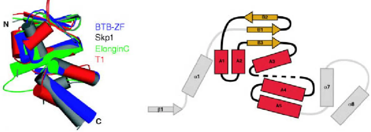
The BTB domain (Broad-Complex, Tramtrack, Bric à brac) was named after Drosophila transcription factors where this domain was first described. It is also known as a poxvirus and zinc finger domain (POZ). The BTB domain is an evolutionarily conserved domain that is widespread among eukaryotes. In humans, nearly 200 different proteins bear BTB domains, in most cases in combination with other domains. The BTB domain is a protein- protein interaction domain participating in homomeric and heteromeric associations with other BTB domains (Aravind and Koonin 1999) and RhoBTB proteins are also capable of forming homodimers and heterodimers (Berthold et al. 2008b). The crystal structure of some BTB domains has been solved. The BTB core fold consists of a 95 amino acid globular cluster of 5 α-helices flanked by 3 short β-strands (Figure 1.4). The BTB domain of the RhoBTB proteins contains an N-terminal extension that folds into one α-helix and one β-strand, and this extension mediates the formation of dimers and oligomers (Stogios et al. 2005).
More recently, it was shown that proteins containing BTB domains are components of cullin 3 (Cul3)-dependent ubiquitin ligase complexes (Furukawa et al. 2003, Geyer et al. 2003, Pintard et al. 2003, Xu et al. 2003) (see section 1.4.2). The role of RhoBTB proteins as components of Cul3-dependent complexes will be also discussed below (see section 1.4.3).
The BTB domains of RhoBTB have some special features. A tandem of two BTB domains
as in RhoBTB is not found within the BTB protein family. Moreover, the first BTB
domain is bipartite, being interrupted by an extension of unknown function that is rich in
charged residues.
Figure 1.4: Structure of the BTB domain. The BTB core folds from the structures of BTB-Zinc-Finger (BTB-ZF), Skp1, Elongin C and T1 are shown on the left hand panel. The core of the BTB fold consists of three conserved β-strands (B1 to B3) and five α-helices (A1 to A5). The 'long form' of BTB proteins has additionally one α-helix α1 and one β-strand β1. Skp1 protein has two additional α-helices at the carboxyl terminus (α7 and α8). Taken from Stogios et al. (2005).
1.3.1.4 The C-terminal region
Following the second BTB domain, there is a region conserved in all members of the RhoBTB subfamily that may constitute a novel domain, but has not been found so far in any other protein apart from RhoBTB. The core of the C-terminal domain consists of approximately 80 amino acids that predictably folds as four consecutive α-helices. The last helix ends close before the prenylation signal of RhoBTB3, but prolongs further in a predicted β-strand in RhoBTB1, RhoBTB2, and Dm RhoBTB (Ramos et al. 2002).
RhoBTB are atypical Rho GTPases – in contrast to the classical Rho family members they do not undergo any known post-translational modification, and almost all of them lack the C-terminal CAAX prenylation motif that is recognised by a set of enzymes that introduce a post-translational modification, isoprenylation, responsible for the targeting of the modified protein to membranes. Only RhoBTB3 ends with the CAAX prenylation motif. Closely upstream of this motif is an additional cysteine residue, which suggests that RhoBTB3 might be also palmitoylated (Ramos et al. 2002).
1.3.2 Expression of RhoBTB proteins
The expression of both, mouse and human RHOBTB genes has been studied using different
approaches. All three RHOBTB genes are ubiquitously expressed although with notable
differences in the pattern of tissue levels among the three genes. Interestingly, while
RHOBTB1 and RHOBTB3 showed high expression levels in many tissues examined,
RHOBTB2 was very weakly expressed in both, human and mouse tissues. RHOBTB genes
are also expressed in foetal tissues (Nagase et al. 1998a, Nagase et al. 1998b, Ramos et al.
2002). In addition, the expression of one or more RHOBTB genes has been reported in numerous human and mouse cell lines using RT–PCR. RHOBTB3 was found expressed in human cancer cell lines: in leukaemia cell lines, in cervical carcinoma and in colorectal carcinoma (Ramos et al. 2002).
Expression of RHOBTB2 has been also studied during mammogenesis. St-Pierre et al.
(2004) found that during mammary gland development in mice, RHOBTB2 transcripts are expressed at low but constant levels. However, attempts to study the spatial pattern of the expression of RHOBTB2 in the mammary gland using in situ hybridisation were inconclusive because of undetectable mRNA levels.
1.3.3 Function of RhoBTB proteins
Over the past years it has been shown that RhoBTB is involved in several cellular processes. However, the connection between these processes is largely unknown. The role of RhoBTB proteins as components of the Cul3-dependent complexes will be discussed below (see section 1.4.3).
1.3.3.1 RhoBTB, cell growth, and apoptosis
Overexpression of RhoBTB2 in the breast cancer cell line T-47D (a cell line that lacks RhoBTB2 transcripts) effectively suppressed cell growth in vitro (Hamaguchi et al. 2002).
More recently, Freeman and co-workers have shown that the overexpression of RhoBTB2 leads to a short-term increase in cell cycle progression and proliferation, but long-term expression has a negative effect on proliferation (Freeman et al. 2007). The growth arrest effect of RhoBTB2 has been explained by the downregulation of cyclin D1. Cyclin D1 is upstream of cyclin E, and the overexpression of any of both prevented the growth arrest effect of RhoBTB2 (Yoshihara et al. 2007). The effect on cyclin D1 is only partially dependent on proteasomal degradation. Moreover, it has not been investigated whether cyclin D1 is degraded by Cul3-dependent complexes through direct binding to RhoBTB2.
The downregulation of cyclin D1 is essential for the cell proliferation suppression effect of
RhoBTB2, but this works for T-47D cells and not for 293 cells. It therefore appears that
the regulation of cyclin D1 is not a universal tumour suppressive mechanism used by
RhoBTB2. The explanation has been put forward that resistance to RhoBTB2 in some cell
lines may be achieved by rapid destruction of the protein through 26S proteasome-
mediated degradation (Collado et al. 2007). Further support for the roles in cell cycle
the E2F1 transcription factor (Collado et al. 2007). E2F1 is a member of a class of E2F implicated in the transcription of genes necessary for DNA replication and cell cycle progression and can also promote apoptosis (DeGregori and Johnson 2006). RhoBTB2 levels increase upon initiation of prophase and decrease at telophase, and this effect depends on E2F1 (Freeman et al. 2007). RhoBTB2 levels also increase during drug- induced apoptosis in an E2F1-dependent manner, and the downregulation of RHOBTB2 delays the onset of apoptosis (Freeman et al. 2007). In agreement with an implication in this process, RhoBTB was found in Drosophila as one of several genes whose expression was significantly upregulated in a DNA microarray analysis aimed at identifying genes associated with cell death induced by the steroid hormone ecdysone (Lee et al. 2003).
However, the role of RhoBTB as a possible cell death regulator was not investigated further.
1.3.3.2 RhoBTB and chemokine expression
It was shown recently that downregulation of RHOBTB2 by RNA interference in primary lung epithelial cells causes a decrease in mRNA expression of CXCL14 (a chemokine that controls leukocyte migration and angiogenesis). The same effect was observed in keratinocytes. Apparently, this effect is independent of Cul3-mediated protein degradation (McKinnon et al. 2008).
1.3.3.3 RhoBTB and vesicle transport
Chang et al. (2006) have addressed the potential role of RhoBTB2 in vesicle transport in a fluorescent recovery after photobleaching analysis with the help of a vesicular stomatitis virus glycoprotein (VSV-G) fused to GFP. VSV-G is extensively used to study anterograde transport from the endoplasmic reticulum (ER) to the Golgi apparatus (GA). Knockdown of endogenous RhoBTB2 hindered the ER to GA transport and resulted in the altered distribution of the fusion protein.
More recently, it was reported that RhoBTB3 binds the GTP-bound conformation of Rab9
through the second BTB domain and the C-terminal domain (Espinosa et al. 2009). Rab9
localises to late endosomes and vesicles travelling towards the Golgi apparatus (GA). It is
required for the trafficking of the mannose 6-phosphate receptors (MPRs) from late
endosomes to the GA (Lombardi et al. 1993, Riederer et al. 1994). Using siRNA depletion
of RhoBTB3 Espinosa et al. (2009) demonstrated that RhoBTB3 is required for retrograde
transport of MPRs to the GA. Interestingly, these authors did not observe any alteration in
transport of VSV-G from ER to GA as it was reported for RhoBTB2 (Chang et al. 2006).
By measuring ATPase activity of purified proteins Espinosa et al. (2009) demonstrated that Rab9 enhances ATP hydrolysis of RhoBTB3. Moreover, RhoBTB3 binds TIP47 (tail- interacting protein of 47 kDa), that is recruited by Rab9 from the cytosol to late endosomes to package MPRs cargo for transport (Carroll et al. 2001, Ganley et al. 2004). Espinosa et al. (2009) proposed a model in which Rab9 activates RhoBTB3 on the GA, which removes TIP47 from the vesicles and permits membrane fusion of vesicles with the GA.
Further, in support of a role in vesicle trafficking, RhoBTB has been identified as one of the genes that suppress the neuromuscular junction overgrowth phenotype induced in Drosophila larvae by the expression of a dominant negative form of the N-ethylmaleimide sensitive factor (NSF) (Laviolette et al. 2005). NSF is an ATPase that participates in vesicle trafficking through binding to the SNARE complex and is also important for the regulation of receptor trafficking (Zhao et al. 2007).
1.3.3.4 RhoBTB and the actin filament system
Although very atypical, RhoBTB proteins are members of the Rho family, therefore,
the first aspect that was investigated was their effect on the organisation of the actin
filament system. Aspenström et al. (2004) observed only a moderate influence on
the morphology and actin organisation of porcine aortic endothelial cells upon the ectopic
expression of RhoBTB1 and RhoBTB2. Not surprisingly, neither RhoBTB1 nor RhoBTB2
were found to interact with the GTPase-binding domain of WASP, PAK1, or Rhotekin,
three well-known effectors of many typical Rho GTPases. Confirming that, at least in
metazoa, RhoBTB proteins do not play a major role in the organisation of the actin
filament system, Dm RhoBTB was found among the proteins whose depletion had no
effect on lamellae morphology in Drosophila S2 cells (Rogers et al. 2003). Unlike
metazoan RhoBTB, the Dictyostelium orthologue RacA may be directly implicated in
the regulation of the actin cytoskeleton. The GTPase domain of RacA, which is very
closely related to members of the Rac subfamily, is able to interact with the Rac-binding
domain of WASP and kinases of the PAK family in yeast two-hybrid assays (de la Roche
et al. 2005, Han et al. 2006, Park et al. 2004). Unlike metazoan RhoBTB, RacA is
susceptible to regulation by RhoGEF and RhoGAP, and in vitro interaction with
a RhoGEF, GxcDD, has been reported recently (Mondal et al. 2007). RacA probably
represents a “primitive” cytoskeleton-regulating stage of the RhoBTB subfamily that was
replaced in the evolved metazoan RhoBTB proteins by roles in cell proliferation and vesicle trafficking.
1.3.4 RhoBTB in human diseases
Since the first report proposing RHOBTB2 as a tumour suppressor gene, evidence is accumulating in support of members of the RhoBTB subfamily being implicated in tumorigenesis. The RHOBTB2 gene was identified as the gene homozygously deleted at region 8p21 in breast cancer samples (Hamaguchi et al. 2002). This is a region commonly associated with loss of heterozygosity (LOH) in a wide range of cancers. Alterations found in RHOBTB genes in tumour tissues and cell lines are summarised in Table 1.1 and are also depicted in the Figure 1.3.
Tumour type
Genomic alterations (% cases)
Mutation and effect Decreased
expression (% cases)
Reference
RHOBTB1 Head and neck
23% LOH (n=52 tumours)
None n = 52 tumours
37%
(n=46 tumours)
(Beder et al. 2006)
RHOBTB2/DBC2 Breast 3.5% HD
(n=200 tumours)
E5 G>A D299N No growth inhibition when re- expressed
E9 C>A P647T Unknown effect E5 A>C D368A Unknown effect
n = 65 tumours + 65 cell lines of breast and lung cancer
42%
(n=19 cell lines)
(Hamaguchi et al.
2002)
Lung NA E5 T>G Y284D Abolished binding to Cul3 n = 65 cell lines of breast and lung cancer
50%
(n=14 cell lines)
(Hamaguchi et al.
2002)
(Wilkins et al. 2004) Breast
(sporadic)
NA Promoter –238G>A Altered expression?
Promoter –121C>T Altered expression?
5’ UTR +48G>A Altered expression?
n = 100 tumours
NA (Ohadi et al. 2007)
Breast (familial)
NA None
n = 17 tumours
NA (Ohadi et al. 2007)
Stomach 29% LOH (n=95 tumours)
E5 C>T R275W Unknown effect n=95 tumours
NA (Cho et al. 2007)
Bladder 42% LOH (n=54 tumours) 38% LOH (n=32 cell lines)
E5 G>C E349D Unknown effect E7 G>A G561S* Unknown effect
(only cases with SSCP mobility shift were sequenced)
75%
(n=12 cell lines)
(Knowles et al. 2005)
Table 1.1: Alterations found in RHOBTB genes in tumour tissues and cell lines. Only changes identified either as mutations or as polymorphisms that could result in functional alterations are shown in the table. The genomic structure of the RHOBTB genes is described in Ramos et al. (2002). Note that in the table exons are numbered from the first transcribed exon, whereas in Ramos et al. exon 1 was considered the exon with the ATG codon. RHOBTB1 is placed in 10q21.2, RHOBTB2/DBC2 in 8p21.3 and RHOBTB3 in 5q15. HD - homologous deletion; LOH - loss of heterozygosity; NA - not analysed; SSCP - single strand conformation polymorphism; *Polymorphism with possible effect. Modified from Berthold et al. (2008a).
In some of these studies decreased expression of RHOBTB1 and RHOBTB2 in tumour samples was observed (Beder et al. 2006, Hamaguchi et al. 2002, Knowles et al. 2005).
Although we still have limited information on the status of RHOBTB genes in tumours, the picture that emerges from the reports is one of rare mutations but common reduced or extinguished expression. This observation can be made extensive to the third family member, RHOBTB3. Berthold et al. (2008b) determined the expression of RHOBTB3 in an array of tumour tissues and their matched normal tissues and have found a moderate but significant decrease of RHOBTB3 expression in the breast, kidney, uterus, lung, and ovary tumours.
Recently, RhoBTB3 was proposed by Kurian et al. (2009) as a blood biomarker for psychosis that are accompanied by hallucinations, as decreased expression of RhoBTB3 mRNA in patients with high hallucinations states was observed.
1.4 RhoBTB proteins and proteasome-dependent degradation 1.4.1 General overview of the proteasome-dependent degradation pathway
Until 1980’s, protein degradation was an unexplored area of cell biology. This process was considered as an unspecific phenomenon. Today it is clear that destruction of proteins is as important as their synthesis to achieve homeostasis in living organisms. The key molecule in this process is ubiquitin, a small (8.6 kDa) globular protein. It is highly conserved among eukaryotes (e.g. human and yeast ubiquitin differ only in three amino acids), but is absent in eubacteria and archaea. It is necessary to mention that besides degradation of proteins, ubiquitination is also involved in other non-proteolytical cellular processes like histone modification (Shukla et al. 2009) and viral budding (Patnaik et al.
2000, Schubert et al. 2000, Strack et al. 2000).
Degradation of unwanted or misfolded proteins by the proteasome-dependent degradation pathway is accomplished in two consecutive steps: 1) substrate tagging by a polyubiquitin chain and 2) recognition of the polyubiquitinated protein by the 26S proteasome. First, the ubiquitin-activating enzyme E1 activates ubiquitin in an ATP-requiring reaction to generate a high-energy E1-thiol-ester~ubiquitin intermediate E1-S~Ub (Haas et al. 1982).
This activated thiol moiety is transferred to the ubiquitin-conjugating enzyme E2 (also
called ubiquitin-carrier protein, UBC), forming a high-energy thiol ester intermediate E2-
S~Ub (Hershko et al. 1983). Activated ubiquitin is then transferred from E2 to
the substrate by the third enzyme called ubiquitin-ligase E3 (Hershko et al. 1983). It is the
E3, which specifically interacts with different substrates. E3s mediate this transfer by different mechanism and can be grouped in several classes. The two best known classes are HECT (homologous to the E6-AP COOH terminus) ligases and RING (Really Interesting New Genes) finger-containing ligases. In the case of HECT ligases, activated ubiquitin is transferred first to the E3 before it is conjugated to the ligase-bound substrate. RING finger-containing ligases catalyse the direct transfer of the ubiquitin to the substrate (Glickman and Ciechanover 2002) (Figure 1.5).
Figure 1.5: Proteasome-dependent degradation pathway. E1 activates ubiquitin in an ATP-requiring reaction. Activated ubiquitin is then transferred to E2 and from E2 to the substrate by the third enzyme called ubiquitin-ligase E3. RING finger-containing ligases catalyse the direct transfer of ubiquitin from E2 to a lysine-residue on the substrate protein. HECT-domain E3 enzyme transfers ubiquitin from E2 to a cysteine- residue on E3 and then to a lysine-residue in the substrate protein. Taken from Jung et al. (2009).
The ubiquitin molecule has seven lysine residues (K6, K11, K27, K29, K33, K48 and K63)
and it has been shown that for formation of ubiquitin chains in vivo all lysine residues are
used (Peng et al. 2003). Ubiquitin is attached to the protein substrate by a covalent
isopeptide bond between the C-terminal glycine of ubiquitin and the ε-NH2 group of
a lysine residue of the substrate protein (Hershko et al. 1980). A polyubiquitin chain is
formed by subsequent attachment of the C-terminal glycine of the next ubiquitin to
the lysine of the previous ubiquitin. The minimal and sufficient length of polyubiquitin
chain that serves as a tag for degradation in the proteasome is four ubiquitin molecules (Thrower et al. 2000). Up to date, the best described and characterised polyubiquitin chains are the Lys48- and Lys63-linked chains. Lys48-linked polyubiquitin chains constitute the major signal for targeting the substrates to the proteasome for degradation (Pickart 1997). Lys63-linked ubiquitination was believed to be involved in proteasome-independent processes such as DNA repair (Spence et al. 1995) and receptor endocytosis (Acconcia et al. 2009). However, nowadays there is also evidence that polyubiquitin chains linked through Lys63 to the protein may also serve as a signal for proteasome-dependent degradation (Kim et al. 2007, Saeki et al. 2009).
Ubiquitin-mediated degradation is a very complex process that is undoubtedly crucial for proper cell function. It has been implicated in a variety of cellular processes, like cell-cycle regulation, signal transduction, transcription factor regulation (Glickman and Ciechanover 2002, Weissman 2001), the quality control of newly synthesised proteins (ERAD) (Hirsch et al. 2009) and immune response (Bhoj and Chen 2009). Dysfunction of components of this pathway leads to the development of diseases like cystic fibrosis, atherosclerosis, diabetes, several neurodegenerative illnesses (e.g. Parkinson’s disease, Alzheimer’s disease) and has been linked also with the process of aging (Jung et al. 2009).
1.4.2 Cullin (Cul)-dependent E3 ligases
Cullins (of which there are 7 in mammals) function as scaffolding proteins that bring together the ubiquitin-conjugating enzyme and substrate-recognition components.
The crystal structure revealed that they are composed of three repeats of five helix bundles and a globular C-terminal domain (Angers et al. 2006, Goldenberg et al. 2004, Zheng et al.
2002). Cullins belong to the group of RING-finger containing ligases. The core ligase of
a Cul-dependent complex consists of a cullin protein that binds the RING-finger protein
Roc1 (which recruits the ubiquitin-conjugating enzyme) through its C-terminus and
a linker protein through its N-terminus. An adaptor protein then acts as a bridge between
the linker protein and the substrates. The complex is positively regulated by covalent
attachment of the Nedd8 ubiquitin-like protein to the cullin subunit. Each cullin family
member interacts with a specific linker. The Cul1 complex contains the Skp1 linker and
an F-box-containing adaptor (called SCF complex: Skp1/Cul1/F-box). The Cul2 and Cul5
complexes contain the linker Elongin C (along with Elongin B) and a SOCS-box-
containing protein (called ECVcomplex: EloB/C-Cul2-VHL-box protein or ECS complex:
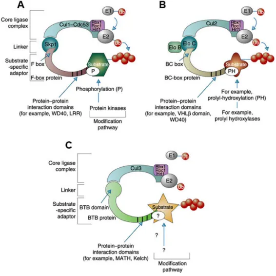
protein that functions simultaneously as linker and adaptor (Petroski and Deshaies 2005) (Figure 1.6). Interestingly, comparison of the structure of the BTB domain with Skp1 and Elongin C demonstrated that Skp1 and Elongin C are in fact BTB proteins that display a similar interface for interaction with the corresponding cullin, despite a low degree of primary sequence conservation (Stogios et al. 2005).
Figure 1.6: Cullin-dependent ubiquitin ligases share a similar architecture. A) The archetypal SCF complex contains Cul1, Skp1 and Rbx1/Roc1/Hrt1 and an F-box protein. B) ECV/ECS complex contains Cul2 (or Cul5), Elongin B/C, Rbx1/Roc1/Hrt1 and a BC-box protein. C) In the ubiquitin-ligase complex containing Cul3, Rbx1/Roc1/Hrt1 and BTB-domain protein, the BTB protein associates directly with Cul3 through its BTB domain without the help of a linker protein. Taken from Krek (2003).
1.4.3 RhoBTB proteins as adaptors of Cul3-dependent ubiquitin ligases
The identification of the BTB domain as adaptor in Cul3-dependent ubiquitin ligase
complexes prompted Wilkins and co-workers to investigate whether RhoBTB2 may also
take part in the formation of such complexes (Wilkins et al. 2004). They identified the N-
terminal region of murine Cul3 as an interacting partner of RhoBTB2 in a yeast two-hybrid
screening (Wilkins et al. 2004). RhoBTB2 interacts specifically with Cul3, but not with other cullin family members in vivo. The interaction was mapped to the first BTB domain in a series of pull-down experiments with deletion constructs. Wilkins et al. (2004) also provided evidence that RhoBTB2 is itself a substrate for Cul3-based ubiquitin ligase complexes, as treatment with proteasomal inhibitor MG132 or shRNA ablation of Cul3 resulted in increased levels of RhoBTB2, and RhoBTB2 was polyubiquitinated by Cul3 complexes in vitro. RhoBTB3, similar to RhoBTB2 interacts with Cul3 through the first BTB domain and upon proteasomal inhibition accumulates in the cells (Berthold et al.
2008b). RHOBTB2 was proposed as a candidate tumour suppressor gene based on the fact that its re-expression in T-47D (a breast cancer cell line that lacks RHOBTB2 transcripts) caused growth inhibition, whereas the expression of the somatic mutant D299N did not have the same effect (Hamaguchi et al. 2002). This mutation is placed in the first BTB domain immediately before the insertion. In fact, it is interesting that almost all missense mutations found in the RHOBTB2 locus reside in the first BTB domain of the protein (Figure 1.3). The question arises whether one or more of those mutations result in impaired interaction with Cul3. This has been investigated by Wilkins et al. (2004) who found that the Y284D mutant, but not the D299N and D368A mutants, failed to co-immunoprecipitate with Cul3, and consequently, had a longer half life than the wild-type protein. The Y284D mutation resides in the dimerisation interface of the first BTB domain and could prevent proper folding. Analogous mutants have been shown to abrogate function by impairing folding of the BTB domain, for example, in the transcription factor PLZF (Melnick et al.
2000).
1.5 MUF1/LRRC41
To date, no substrates have been shown to be degraded by RhoBTB-Cul3-dependent ligase complexes. Previous experiments performed in our laboratory identified a list of potential binding partners of RhoBTB3 that includes MUF1, kindlin-2 and Uev1a (S. Ramos, personal communication). However, these interactions have not been investigated extensively and are the object of this thesis.
MUF1/LRRC41 (leucine-rich repeat containing 41) is a largely uncharacterised protein
that has an N-terminal SOCS-box motif and a C-terminal leucine-rich repeat (LRR) region
(Figure 1.7A). It has been shown that MUF1 interacts with Elongin B/C through the BC-
box subdomain of the SOCS-box. This interaction mediates binding to Cul5 and
the MUF1-Elongin B/C-Cul5-Rbx1 complex has ubiquitin ligase activity (Kamura et al.
2001). The expression, subcellular localisation and function of MUF1 have not been addressed so far.
Figure 1.7: Domain composition of MUF1. A) Schematic representation of MUF1. Numbering of amino acids is in respect to the murine MUF1. The SOCS-box is at the N-terminus of MUF1 and the leucine-rich repeat (LRR) region is C-terminal. B) Sequence alignment of MUF1 SOCS-box with representative SOCS- box sequences and SOCS-box consensus. Amino acids at position A42-P71 of MUF1 are shown. Red arrows indicate amino acid residues crucial for interaction with EloB/C. SOCS3, SH2 domain-containing SOCS-box protein 3; WSB1, WD repeat-containing SOCS-box protein 1; ASB2, ankyrin repeat-containing SOCS-box protein 2; RAR, Ras-related SOCS-box protein; SSB1, SPRY domain-containing SOCS-box protein.
Modified from Kamura et al. (2001). C) The LRR of murine MUF1. The conserved regions LxxLxLxxN/CxL are highlighted in grey.
1.5.1 The SOCS-box
The SOCS-box is a structural motif that has been initially characterised in the members of the suppressors of cytokines signalling (SOCS) family (Hilton et al. 1998). SOCS proteins have been originally identified as negative regulators of the JAK-STAT signalling pathway induced by cytokine stimulation (Endo et al. 1997, Naka et al. 1997, Starr et al. 1997). All SOCS proteins (SOCS1-7, CIS) share structural similarities: an N-terminal region of
IGEVACGALDGSDPSCLGLPALEASQRFRSISTLELFTVPLSTEAALTLCHLLSSWV SLESLTLSYNGLGSNIFRLLDSLRALSGQAGCRLRALHLSDLFSPLPILELTRAIVR ALPLLRVLSIRVDHPSQRDNPAVPENAGPPGHIVGDEEIPENCLEQLEMGFPRGAQP APLLCSVLKASGSLQQLSLDSATFASPQDFGLVLQTLKEHNLSLKRLSFHDMNLADC QSEVLFLLKNLTLQEITFSFCRLFEKRPVQFLPEMVAAMKGNSTLKGLRLPGNRLGN AGLLALADVFSEDSSSSLCQLDISSNCIKPDGLLEFAKRLERWGRGAFGHLRLFQNW LDQDAVTAREAIRRLRATCHVVSDSWDSTQAFADYVSTM
variable length is followed by an SH2 domain and the SOCS-box, which is located at the C-terminus (Hilton et al. 1998, Starr et al. 1997). The SOCS-box is a ~40 amino acid motif composed of two well conserved blocks separated by 2 – 10 non-conserved residues (Figure 1.7B). The C-terminal conserved region is a cullin box motif, and the N-terminal conserved region is a consensus BC-box, a ~10 amino acid degenerate sequence motif with the consensus sequence (A,P,S,T)LxxxCxxx(A,I,L,V), where leucine at position 2 and cysteine at position 6 are highly conserved. SOCS-box proteins bind to the Elongin C through the BC-box (Kamura et al. 1998, Zhang J.G. et al. 1999), which is structurally similar to Skp1 and the BTB fold as already mentioned (Stogios et al. 2005). Binding of Elongin C to the BC-box is mediated by interaction with the highly conserved leucine at position 2 (Stebbins et al. 1999) and point mutations at conserved residues within the BC- box abolish this interaction (Kamura et al. 1998, Kamura et al. 2001). Elongin C interacts with ubiquitin like protein Elongin B (Garrett et al. 1994) and the whole complex can bind to Cul2 or Cul5, depending on the specific cullin box (Kamura et al. 2004). The ECV complex (EloB/C-Cul2-VHL-box) and the ECS complex (EloB/C-Cul5-SOCS box) resemble SCF complex and have ubiquitin ligase activity (De Sepulveda et al. 2000, Iwai et al. 1999, Kamizono et al. 2001, Kamura et al. 2001). A large number of SOCS-box containing proteins accommodate additional domains like WD-40 repeats, SPRY domains and ankyrin repeats (Hilton et al. 1998, Masuhara et al. 1997). In all these proteins the SOCS-box domain is C-terminal. With an N-terminal SOCS-box, MUF1 seems to be unique.
1.5.2 The LRR
The leucine-rich repeat (LRR) is a structural motif that has been implicated in mediating protein-protein interactions. It has been identified in thousands of proteins in eukaryotes, bacteria, viruses and archaea. LRRs are usually 20-29 residues long sequences repeating multiple times within the protein (2 to 52 times). LRRs are usually composed of a so-called conserved segment, which shows significant similarities in all known LRR containing proteins, and a variable segment whose sequence varies among the members of different subfamilies (Bella et al. 2008). The sequence of the conserved region is LxxLxLxx
N/
CxL, where x denotes any amino acid. Later it has been shown that there is certain variability in the conserved region and leucine can be substituted by valine, isoleucine or phenylalanine;
asparagine/cysteine can be substituted by either serine or threonine (Kajava 1998).
adopts a curved solenoid structure where the LRRs form β-sheets on the concave side of the solenoid and are connected by loops with α-helices that align on the convex side (Figure 1.8A) (Kobe and Deisenhofer 1993). Since the 3D structure of RI has been solved, nearly 90 structures of other LRR proteins have been determined indicating that all LRRs adopt a more or less perfect solenoid conformation (Bella et al. 2008).
LRR containing proteins have been implicated in wide range of cellular processes as cell adhesions, signalling, extracellular matrix assembly, platelet aggregation, neuronal development, RNA processing, invasion of pathogenic bacteria to the cell and immune response (Bella et al. 2008). Mutations in genes encoding LRR proteins have been associated with several human diseases such as schizophrenia, Parkinson’s disease and Crohn’s disease (Matsushima et al. 2005).
Figure 1.8: 3D structure of LRR proteins. A) 3D structure of ribonuclease inhibitor (RI). The LRR domains are shown in cyan, the other domains are shown in magenta. Taken from Kobe and Kajava (2001).
B) 3D structure of decorin core protein (DCN). DCN is capable of homodimerisation, where the surface on the concave side is used as a homodimerisation interface. Taken from Bella et al. (2008).
It is the concave side of LRR domain that is mainly involved in protein-protein interactions and moreover, it has been demonstrated that proteins containing a LRR region form very often homo- and heterodimers (Figure 1.8B) (e.g. Toll-like receptor ectodomain, Ran GTPase-activating protein, monocyte differentiation antigen CD14) or even larger assemblies like tetramers (Yersinia outer membrane protein M, YopM) (Bella et al. 2008).
Examination of the leucine rich repeat region of mouse MUF1 revealed at least four conserved 11 amino acids long LxxLxLxx
N/
CxL motifs (Figure 1.7C).
1.6 The kindlin protein family
The kindlin family consists of three members: kindlin-1, kindlin-2 and kindlin-3 (Siegel
et al. 2003). Kindlins are encoded by the FERMT (fermitin family homologue) genes
FERMT1, FERMT2 and FERMT3. The first identified member of this family was kindlin-
2, which was identified among serum-inducible genes in serum-starved quiescent human fibroblasts and was initially named Mig-2 (mitogen-induced gene 2) (Wick et al. 1994).
Kindlins are composed of a FERM (4.1, ezrin, radixin, moesin) domain, which is subdivided into four subdomains named F0, F1, F2 and F3 (Figure 1.9). The N-terminally localised F0 subdomain adopts an ubiquitin-like fold. The F1 subdomain is interrupted by an insert that has a similar size in kindlin-1 and kindlin-2, but is shorter in kindlin-3 (Goult et al. 2009). The F2 subdomain is interrupted by a pleckstrin homology (PH) domain and the F3 subdomain contains a phosphotyrosine binding fold (PTB) that is capable of recognising β integrin tails (Kloeker et al. 2004, Shi et al. 2007).
Figure 1.9: Domain structure of kindlins. Kindlins are composed of a FERM (4.1, ezrin, radixin, moesin) domain, which is subdivided into subdomains named F0, F1, F2 and F3. The F2 subdomain is interrupted by a pleckstrin homology domain (PH). This insertion does not interfere with FERM domain function.
A nuclear localisation signal has been described in the F0 subdomain of kindlin-2.
1.6.1 The role of kindlins in integrin activation
All three kindlins have been shown to bind directly to cytoplasmic tails of β1 and β3 subunits of integrins (Figure 1.10) and have been identified as key regulators of integrin activation, together with talin (Harburger et al. 2009, Moser et al. 2008, Ussar et al. 2008).
Integrins are heterodimeric transmembrane receptors that are composed of α and β
subunits. There are 18 α and 8 β subunits in mammals that combine with each other to
form 24 specific dimers. Each integrin subunit has a large extracellular domain, a short
transmembrane domain and a cytoplasmic tail (Hynes 2002). Inactive integrins assume
a bent conformation and are in a low affinity binding state for ligands; they shift to a high
affinity state during activation (Moser et al. 2009). Integrins are localised in focal
adhesions and interact with proteins of the extracellular matrix (ECM) through their
extracellular domain and, via talin, with the actin cytoskeleton through their cytoplasmic
tail (Zhang et al. 2008). Through these interactions integrins constitute a fundamental
connection for bi-directional communication between the ECM and the intracellular
environment. Talin, similarly to kindlins, bears a FERM domain composed of F0, F1, F2
and F3 subdomains. The F3 subdomain contains a PTB that is the major integrin binding
site (Wegener et al. 2007). Both talin and kindlin are crucial for integrin activation by
the membrane proximal NPxY motif and induces separation of integrin tails, whereas kindlin binds to the membrane distal NxxY motif (Moser et al. 2009).
Figure 1.10: A model of kindlin binding to β integrin cytoplasmic tails. Kindlin prefers to interact with a membrane distal (T/S)TxxNxxY amino acid sequence but interaction with the membrane proximal NPxY motif has been also reported. The N-terminal region corresponds to the F0 subdomain Taken from Meves et al. (2009).
1.6.2 Expression and localisation of kindlins
Human kindlin genes show distinct differences in expression. Expression of kindlin-1 is restricted to the skin epithelial cells (keratinocytes) and intestinal epithelial cells (Lai- Cheong et al. 2008, Siegel et al. 2003, Ussar et al. 2006), while kindlin-2 is ubiquitously expressed with the exception of the hematopoietic cells, where kindlin-3 was found (Ussar et al. 2006). Kindlin-1 and kindlin-2 localise predominantly to focal adhesions in keratinocytes (Kloeker et al. 2004, Siegel et al. 2003, Tu et al. 2003, Ussar et al. 2006) but cytoplasmic localisation of kindlin-1 has also been reported, including the perinuclear area (Kloeker et al. 2004, Lai-Cheong et al. 2008, Siegel et al. 2003). Localisation of kindlin-2 to stress fibres has also been reported (Tu et al. 2003). Kindlin-2 is the only member of the kindlin family that contains a nuclear localisation signal (Ussar et al. 2006) and endogenous kindlin-2 was visualised in the nuclei of smooth muscle cells (Kato et al.
2004). Contrary to this, kindlin-1 but not kindlin-2 was found in the nucleus of normal
human keratinocytes (Lai-Cheong et al. 2008). Mouse kindlin-3 localises to the podosomes
of macrophages and dendritic cells that are integrin-dependent adhesion sites in
hematopoietic cells (Ussar et al. 2006).
1.6.3 Kindlin in human diseases
All three kindlins have been implicated in human diseases and cancer. This family is named after dermatologist Theresa Kindler who described a patient with traumatic bulla formation, atrophy of the skin and congenital poikiloderma. Today this disease is named Kindler syndrome, an autosomal recessive genodermatosis caused by loss-of-function mutations in the FERMT1 gene. These mutations are predicted to reduce expression and function of kindlin-1. It was reported that patients with Kindler syndrome have a predisposition to non-melanoma skin cancer and expression of kindlin-1 and kindlin-2 is altered in many cancer cell lines. Mutations in the FERMT3 gene cause so called LAD-III syndrome (leukocyte adhesion deficiency), which is characterised by bleeding problems and life-threatening infections (Lai-Cheong et al. 2009).
1.7 Uev1a
Uev1a (ubiquitin-conjugating E2 enzyme variant, also called CROC1, CIR1, UBE2V) was originally identified as a signal transducing molecule able to activate the human FOS proto-oncogene promoter (Rothofsky and Lin 1997). Uev1a resembles ubiquitin- conjugating enzymes in sequence as well as structure, but lacks the ability to transfer ubiquitin to the substrate due to the absence of a catalytic cysteine residue (Sancho et al.
1998). Uev1 interacts with Ubc13 (Andersen et al. 2005), which is the only ubiquitin- conjugating enzyme identified so far that mediates the assembly of Lys63-linked polyubiquitination chains (Hofmann and Pickart 1999). For this process both of the proteins are required; neither Ubc13 nor Uev1a alone is able to promote Lys63 polyubiquitination chain formation (Hofmann and Pickart 1999, McKenna et al. 2001).
The crystal structure of Uev1a has been solved recently. Based on this, a structural model of the Ub-Ubc13-Uev1a~Ub tetramer has been proposed that elucidates the mechanism how specificity in forming Lys63-linked polyubiquitin chains is achieved (Hau et al.
2006).
An increasing line of evidence supports the role of Ubc13-Uev1a complex in activation of
NFκB (Deng et al. 2000, Shi and Kehrl 2003, Wang et al. 2001). One of the pathways that
lead to the activation of NFκB is TNFα (tumour necrosis factor α) signalling. Briefly,
the proinflammatory cytokine TNFα binds to the TNFR (TNFα receptor), which causes
aggregation of the receptor and allows binding of TRADD (TNFR-associated death
domain protein). TRADD recruits multiple adaptor proteins like TRAF (TNF-receptor-
associated factor) and RIP (receptor interacting protein 1) that subsequently recruit IκB kinase complex (IKK). IKK is composed of two kinase subunits IKKα and IKKβ and one regulatory subunit IKKγ (also called NEMO, NFκB essential modifier). Activated IKKβ phosphorylates IκBα (inhibitor of NFκB complex) that serves as a signal for SCF-ligases to ubiquitinate IκBα. Proteasomal degradation of IκBα frees the NFκB dimer, which translocates to the nucleus where it binds to the promoter or enhancer regions of target genes (Hayden and Ghosh 2004). It has been shown that the family of TRAF proteins, which contain a RING-domain, have ubiquitin-ligase activity and upon binding of the Ubc- Uev1a complex catalyse the synthesis of Lys63-linked polyubiquitin chains (Deng et al.
2000). This complex ubiquitinates RIP (Ea et al. 2006) as well as NEMO (Andersen et al.
2005), which leads to the activation of IKK (Figure 1.11).
Figure 1.11: Model depicting the function of Ubc13-Uev1a ubiquitin conjugating enzyme in NFκB signalling pathway. TNFα binds to the TNFR that promotes the interaction between TRADD, RIP1 and TRAF2/6. Ubc13-Uev1a is involved in ubiqitination of NEMO and RIP by forming Lys63-linked polyubiquitin chains. Activated IKKβ phosphorylates IκBα. Phosphorylated IκBα is modified by Lys48- linked polyubiquitin chain that results in its degradation in proteasome. Activated NFκB dimer translocates to the nucleus. Taken from Syed (2006).