Characterization of the Ubiquitin/Nedd8 E3 Ligase Activity of the Mdm2/MdmX complex
Inaugural–Dissertation zur
Erlangung des Doktorgrades
der Mathematisch-Naturwissenschaftlichen Fakultät der Universität zu Köln
vorgelegt von
Rajesh Kumar Singh
aus
Bhojpur Bihar, Indien Köln, 2006
Referees/Berichterstatter: Prof. Dr. Mats Paulsson Prof. Dr. Maria Leptin
Tag der mündlichen Prüfung: 15.02.2007
Acknowledgements
I feel profound privilege to thank my supervisor Prof. Dr. Martin Scheffner for all his help during the materialization of the work presented in this thesis. His excellent guidance, valuable suggestions, constructive criticism and constant encouragement are not only praiseworthy, but also unforgettable. Discussions with him have always lent me new ideas to proceed with. My sincere apologies to him for keeping him busy with my thesis even on weekends. Once again, thanks Martin for everything !!!
My thanks to Prof. Dr. Mats Paulsson, Prof. Dr. Maria Leptin, and Prof. Dr. Siegfried Roth for being a part of my doctoral thesis committee.
I also owe my thanks to Dr. Shahri Raasi, Dr. Saravanakumar Iyappan, Konstantin Matentzoglu, and Amarendra Pegu for their critical inputs in the completion of my thesis. My special thanks to Hans-Peter Wollscheid for translating the abstract herein in German language.
I am thankful to the rest of my labmates and colleagues, Alejandro, Dietmar, Elvira, Adrian, Thomas and Nicole, for their help as and when required. I also thank Armin Benz and Alexander Waniek for their contribution to this work.
I would also take this opportunity to thank Dr. Michael D’silva and Dr. Ulrike Kogel for providing a great company and unforgettable friendship. Their elaborate emails describing the requirements for my thesis are really appreciable.
It would be unfair if I do not acknowledge the Cologne-, Essen-, and Konstanz-Indian Gangs for all the fun I had with them. I really enjoyed the time spent with all of you. Thank you guys!
A word of praise will not be sufficient to acknowledge Dr. Brigitte v.Wilcken-Bergman for her timely help to solve all the administrative problems.
My indebtedness to my parents for their constant support and numerous sacrifice are beyond expression. Their incredible love and affection has invariably been the source of my encouragement and motivation.
Last but not the least, the financial support and opportunity provided by the Graduate School of Genetics and Functional Genomics, Cologne, is highly acknowledged.
Konstanz, 12thDec’ 2006 (Rajesh Kumar Singh)
Abbreviations
°C Degree celcius mRNA messenger-RNA
ADP Adenosine Di-Phosphate mu murine
AMP Adenosine Mono-Phosphate n nano
ATP Adenosine Tri-Phosphate N Normal
bp base pairs ng nanogram
cDNA complementary DNA nm nanometer
ddH2O double-distilled water mA miliAmpere
DMSO Dimethylsulphoxide mM miliMolar
DNA Deoxyribonucleic acid nt nucleotide
DTT Dithiothreitol OD Optical Density
E1 Ubiquitin-activating enzyme ONPG Orthonitrophenyl-β-
E2 Ubiquitin-conjugating enzyme D- galactosidase
E3 Ubiquitin-protein ligase PBS Phosphate buffer EDTA Ethylenediamine tetraacetic acid saline
FBS Fetal bovine serum PCR Polymerase chain
HA Haemagglutinin reaction
hr hour RNA Ribonucleic acid
hrs hours rpm revolution per minute
i.e. id est or that is s second
IgG Immunoglobulin G TAE Tris-acetated-EDTA
IP immunoprecipitation Ub ubiquitin
kDa Kilodalton V Volt
LiAc Lithium Acetate wt wild-type
M Molar µ micro
min minute µl microlitre
ml mililitre µg microgram
Table of Contents
1 Introduction...1
1.1 The ubiquitin-proteasome system ...1
1.1.1 The ubiquitin conjugation pathway...1
1.1.1.1 HECT E3s ...3
1.1.1.2 RING and RING finger like E3s...4
1.1.2 The Proteasome...6
1.2 p53 and its family members ...7
1.3 Mdm2 ...9
1.3.1 Mdm2 gene structure and protein domains...9
1.3.2 Structural requirements for p53 ubiquitination and degradation ...11
1.3.3 The p53-Mdm2 feedback loop...12
1.3.3.1 The p53-Mdm2 loop under non-stressed conditions...12
1.3.3.2 The p53-Mdm2 loop upon stress...13
1.3.4 p53 independent functions of Mdm2 ...15
1.4 MdmX ...15
1.5 The Nedd8 conjugation pathway ...17
2 Aim of the study...19
3 Materials and Methods...20
3.1 Materials...20
3.1.1 Solutions and Media...20
3.1.2 Cell Strains and Cell lines...21
3.1.2.1 Bacterial Strains ...21
3.1.2.2 Yeast Strains ...21
3.1.2.3 Mammalian cell lines ...21
3.1.3 Antibodies ...21
3.1.4 Expression Vectors ...22
3.1.5 cDNA library...22
3.1.6 Primers ...22
3.1.7 Plasmids constructed during this study...24
3.1.8 Other plasmids used in this study ...30
3.2 Methods...30
3.2.1 PCR and Cloning ...30
3.2.1.1 Polymerase Chain Reaction (PCR) ...30
3.2.1.2 Site directed mutagenesis...30
3.2.1.3 Restriction digestion ...30
3.2.1.4 Agarose Gel Electrophoresis...31
3.2.1.5 Purification of DNA from Agarose gels ...31
3.2.1.6 Ligation ...31
3.2.1.7 Transformation of DNA into E. coli by CaCl2 method...31
3.2.1.8 Estimation of DNA concentration...32
3.2.2 Maintenance of bacterial cultures and mammalian cell lines ...32
3.2.2.1 Bacterial cultivation and preparation of glycerol stocks...32
3.2.2.2 Maintenance of mammalian cell lines...32
3.2.2.3 Freezing of cell lines in liquid nitrogen ...32
3.2.3 Transfection of mammalian cell lines...32
3.2.3.1 Transfection using Lipofectamine 2000 ...33
3.2.3.2 Transfection using DOTAP ...33
3.2.4 Protein assays for mammalian cells ...33
3.2.4.1 In vivo degradation assays for p53 and MdmX...33
3.2.4.2 In vivo ubiquitination and neddylation assays ...33
3.2.4.3 β galactosidase assay...34
3.2.4.4 Bradford assay...34
3.2.4.5 SDS-polyacrylamide gel electrophoresis (SDS-PAGE) ...34
3.2.4.6 Western blotting...34
3.2.4.7 Immunoprecipitation (IP)...35
3.2.4.8 Immunoflorescence...35
3.2.5 In vitro biochemical assays ...36
3.2.5.1 GST-tagged protein purification from bacteria...36
3.2.5.2 Purification of Nedd8 E1 from H1299 cells ...36
3.2.5.3 In vitro ubiquitination assays ...36
3.2.5.4 In vitro neddylation assays...37
3.2.5.5 GST-pulldown assays ...37
3.2.5.6 Fixation, amplification and drying of polyacrylamide gel containing radioactive proteins. ...37
3.2.6 Yeast two-hybrid interaction system...37
3.2.6.1 Competent cell formation (low efficiency)...37
3.2.6.2 Transformation...38
3.2.6.3 Reporter assay ...38
3.2.6.4 Expression and purification of proteins from yeast cells...38
3.2.7 Yeast two-hybrid screening ...38
3.2.7.1 Competent cell formation (high efficiency)...38
3.2.7.2 High efficiency transformation ...38
4 Results...40
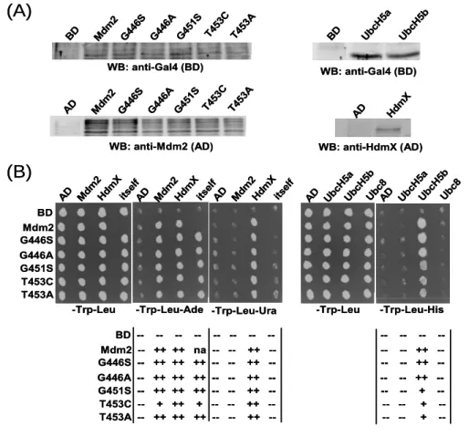
4.1 Walker A motif in Mdm2 and MdmX ...40
4.1.1 Several Walker A mutants of Mdm2 are impaired in their E3 ligase activity .40 4.1.2 MdmX rescues the E3 ligase activity of the ligase defective Mdm2 Walker A mutants ………..42
4.1.3 Mutation of the Walker A motif of HdmX does not impair its rescue activity44 4.1.4 Mdm2 Walker A mutants were highly ubiquitinated by wt-Mdm2...46
4.1.5 MdmX rescues the E3 activity of Mdm2 Walker A mutants in H1299 cells ..46
4.1.6 Mdm2 Walker A mutants oligomerize with wt-MdmX, wt-Mdm2, and themselves...48
4.1.7 Interaction of Mdm2 Walker A mutants with E2s...51
4.1.8 Interaction of RNA and nucleotides with the Mdm2 Walker A mutants...53
4.2 Residues of Mdm2 and MdmX involved in oligomers formation ...55
4.2.1 Dimerisation mutants have impaired E3 activity...55
4.2.2 Rescue of the E3 activity of the Mdm2 dimerisation mutants by MdmX ...56
4.2.3 Mdm2 dimerisation mutants are highly ubiquitinated by wt-Mdm2 ...60
4.2.4 Oligomer formation and E2 binding ability of the Mdm2 dimerisation mutants ………..61
4.2.5 Effect of Walker A and dimerisation mutants of Mdm2 on MdmX ubiquitination and degradation...63
4.3 Neddylation...64
4.3.1 The in vivo neddylation assay ...65
4.3.2 Mdm2 is the only Nedd8 E3 ligase for p53 ...65
4.3.3 Nedd8 is attached to its substrate by forming an isopeptide bond...66
4.3.4 Nedd8 attachment to p53 by Mdm2 Walker A and dimerisation mutants...67
4.3.5 Effect of Nedd8 on p53 ubiquitination and vice versa...68
4.3.6 MdmX Neddylation ...73
4.3.7 p73 Neddylation...75
4.3.8 Dominant negative mutants for Neddylation pathway ...77
4.4 Screening of interacting partners of MdmX by the yeast two-hybrid system...78
5 Discussion ...81
5.1 Walker A motif Mdm2 mutants and p53 ...81
5.2 Walker A motif Mdm2 mutants and MdmX...85
5.3 Mdm2 and its interaction with UbcH5 family members...86
5.4 Walker A motif of Mdm2 and its RNA binding ability...87
5.5 The dimerisation mutants of Mdm2...87
5.6 Mdm2 as substrate for ubiquitination ...88
5.7 Neddylation...89
5.8 Significance of the results obtained with respect to molecular approaches in the treatment of cancer...94
6 References ...96
7 Zusammenfasung ...105
8 Abstract ...106
9 Erklaerung ...107
10 Lebenslauf...108
1 Introduction
Protein degradation is commonly accepted to be involved in the regulation of many fundamental processes within cells. Three decades ago, lysosomes were known to degrade most of the extracellular proteins and some intracellular proteins in the presence of certain stimuli in an unspecific manner. However, there were indications that at least some intracellular proteins can also be degraded by lysosomal free cell lysates in a controlled manner and that this type of degradation was ATP-dependent. During those days, it was hard for scientists to believe that the catabolism (i.e. degradation) of a protein should need energy (in the form of ATP), since hydrolysis of a peptide bond per se is an exergonic process. The existence of a non-lysosomal degradation system was further evident by the biochemical fractionation and in vitro reconstitution of a degradation machinery that was able to degrade several abnormal/misfolded proteins independent of lysosomes1. Subsequently, the components of this degradation machinery were identified and termed collectively as the ubiquitin-proteasome system.
1.1 The ubiquitin-proteasome system
In the ubiquitin-proteasome system (UPS), ubiquitin, a 76 amino acid long polypeptide, is first covalently attached to another protein, which is followed by the recognition and degradation of the ubiquitin attached protein by a protein complex having inherent protease activity, termed “proteasome”2; 3; 4. Modification by ubiquitin (ubiquitylation or ubiquitination) was the first example of a protein attached to another protein by a covalent bond. Since then, hundreds of proteins involved in a broad array of basic cellular processes have been unravelled to be modified by ubiquitin and degraded by the proteasome. Cell cycle5, DNA repair6; 7, endocytosis of cell surface receptors8, differentiation and development are only few examples of basic cellular processes requiring a functional UPS for their proper activity. Considering the vast array of proteins modified by ubiquitin, it is not surprising that deregulation of the UPS has been implicated in the pathogenesis of many diseases9; 10; 11. 1.1.1 The ubiquitin conjugation pathway
Ubiquitination to a substrate is achieved by a cascade of enzymatic reactions involving at least three different enzymes, E1, E2, and E3 (Fig 1)12. The reaction starts with the sequential binding of Mg2+-ATP and ubiquitin to the ubiquitin activating enzyme (E1) to form an adenylate ubiquitin intermediate from which ubiquitin is transferred to the catalytic cysteine of E1 via the formation of a thioester bond. Hence, one E1 molecule binds two ubiquitin
E1 E2E3
Substrate E3 E2
SubstrateE2E3
Substrate
RING E3s HECT E3s
Ubiquitin
26S Proteasome Mg2+
1 ATP
2
3
4a
4b
5
5
6 7
E2E3 E2E2E3E3
E1 E1 E1
Substrate E3 Substrate E3 E2
E2 E2
SubstrateE2E3 Substrate Substrate
E2E2E3
Substrate Substrate Substrate
RING E3s HECT E3s
Ubiquitin
26S Proteasome Mg2+
1 ATP
22
33
4a
4b
55
55
66 77
Fig 1. The ubiquitin proteasome pathway.
In the ubiquitin proteasome pathway, ubiquitin molecule is first activated by the ubiquitin activating enzyme (E1) in the presence of Mg2+ and ATP via formation of a high energy thioester bond (1). The activated ubiquitin molecule is then transferred to the ubiquitin conjugating enzyme (E2) by formation of a second high energy thioester bond with ubiquitin (2). Finally, the ubiquitin molecule is either transferred directly to the substrates by the RING-type E3 ligase (3) or transferred to the substrates via formation of a third high energy thioester bond with the catalytic cysteine of the HECT domain (4a) by the HECT-type E3 ligases (4b).
Repetition of steps 1, 2, and 3/4 for several times finally attaches a polymer of ubiquitin on the substrates (5).
The ubiquitinated substrates are then recognised and degraded by the 26S proteasome (6) and the ubiquitin olecules are recycled (7).
m
molecules- one in adenylate form and the other in thioester bonded form. Formation of a high energy thioester bond with the carboxyl group of the C-terminal glycine of ubiquitin (G76) activates the ubiquitin molecule. Ubiquitin loaded E1 interacts with the next enzyme of the cascade, an ubiquitin conjugating enzyme (E2), and catalyses the transfer of thioester bonded ubiquitin to the catalytic cysteine of E2. The thioester bonded ubiquitin-E2 complex next binds to an ubiquitin protein ligase (E3) and, in conjunction with E3 catalyses the formation of an isopeptide bond between the C-terminal carboxyl group of ubiquitin and the ε-NH2
group of a lysine (K) residue of the substrate protein. The specificity of ubiquitin conjugation to the substrate is mainly governed by E3 in the ubiquitination pathway. The E3s recognize their cognate substrates by the presence of a structural motif in the substrate commonly known as ubiquitination signal13.
Depending on their mode of action, E3 enzymes can be divided into two major classes- (i) HECT E3s, which form an ubiquitin thioester complex via the catalytic cysteine of the E3 before the final transfer of ubiquitin to the lysine of the substrate, and (ii) RING finger and RING finger like E3s, which do not form a thioester complex with ubiquitin but act as adaptor proteins to bring their cognate E2s in close proximity of substrate protein for ubiquitination.
In many cases, a polymer of ubiquitin (polyubiquitin chain) is attached onto lysine residue(s) of the substrate protein by the sequential repetition of the cascade followed by recognition and degradation by the proteasome. However, degradation by the proteasome is not always the fate of ubiquitinated proteins. In fact, the fate of the ubiquitinated proteins is decided by the number and the topology of ubiquitins attached to the protein14. Attachment of one molecule of ubiquitin to one lysine of the substrate (mono-ubiquitination) or several lysine of the substrate (multiple mono-ubiquitination) usually serves signalling functions. Moreover, different type of polyubiquitin chains can also serve different functions. For example, a polyubiquitin chain formed by forming an isopeptide bond between K48 of one ubiquitin molecule and G76 of another ubiquitin molecule (K48-linked polyubiquitin chain) serves mainly as a signal for proteasomal degradation15. In contrast, K63-linked polyubiquitin chains predominantly serve non-proteolytic functions, e.g. ribosomal function6 and in post replicational DNA repair6; 7. The different types of ubiquitin chains have different topology, which presumably can be recognized by different sets of regulatory proteins leading to the proper fate of the substrate.
1.1.1.1 HECT E3s
The founding member of the HECT (homologous to E6-AP C terminus) E3 family was discovered in studies characterising the degradation of the tumour suppressor protein p53 by the oncogenic E6 protein of human papillomaviruses (HPVs). Later studies revealed that the HPVs E6 protein forms a complex with a cellular protein, termed E6-AP (E6-associated protein), and this complex functions as an E3 ligase to target p53 for ubiquitination and proteasomal degradation16. The C-terminal ~350 amino acids of E6-AP, termed HECT domain, showed homology to several proteins in databases17 and contains a conserved catalytic cysteine required for the E3 activity of the E6/E6-AP complex. Full-length E6-AP presumably binds to substrates via binding sites in its N-terminal region and, contains E6- binding site and the catalytic HECT domain at the C terminus. E6 or E6-AP alone cannot bind p53, however, when E6 binds at the E6-binding site of E6-AP, then the E6/E6-AP complex efficiently binds to p53 and targets it for ubiquitination18. Furthermore, E6-AP has
been implicated in E6-independent binding and ubiquitination of several cellular proteins (e.g. the Src family tyrosine kinase Blk19, the replication licensing factor Mcm720, estrogen and progesterone receptors21). However, the physiological significance of E6-AP-induced degradation of these proteins is not clear. In addition, the fact that mutations in the E6-AP gene (UBE3A) cause Angelman syndrome22; 23, which is characterized by severe neurological abnormalities, predicts an important role of E6-AP in brain developement. Several other HECT E3s are known to perform important functions. Human Nedd4 (Rsp5 in yeast) is a well studied HECT E3 ligase, which is implicated in ubiquitination and degradation of Rpb124, the large subunit of RNA polymerase II, upon DNA damage. Nedd4 also ubiquitinates the kidney epithelial Na+ channel (ENaC) for endocytosis25. Itch is another HECT E3 ligase, which targets a number of proteins including p73 (a member of the p53 family) for proteasomal degradation26.
The HECT domain of HECT E3s facilitates the transfer of activated ubiquitin from cognate E2s to their catalytic cysteine followed by transfer of ubiquitin onto the lysine residue of the substrate via isopeptide bond formation. However, the mechanistic details of this catalytic process remain elusive. Crystal structure of the UbcH7/E6-AP HECT complex27 predicts a conformational change in the complex after binding to the substrate to facilitate final transfer of ubiquitin to the substrate lysine residues. However, a detailed understanding of the molecular events underlying ubiquitin transfer to the substrate by HECT E3 ligases still awaits further research.
1.1.1.2 RING and RING finger like E3s
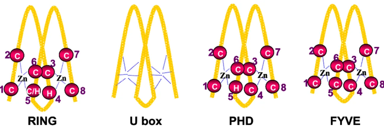
RING (really interesting new gene) finger domains are characterized by the presence of eight histidine and cysteine residues with a typical spacing in order to form a cross brace like structure (see Fig 2), which is stabilised by the presence of two zinc ions. The widely accepted consensus sequence for forming a RING domain is C-X2-C-X(9-39)-C-X-H -X(2-3)- C/H-X2-C-X(4-48)-C-X2-C28. Depending on the presence of C or H residues at the 5th position, the RING domains have been classified into two groups- C3HC4 (also known as HC-type RING), and C3H2C3 (also known as H2-type RING).
RING E3s were first discovered as integral part of several multi-subunit protein complexes- SCF (Skp1-cullin-F-box), APC (anaphase promoting complex) and CBC (cullin-elongin B and C) complexes. SCF complexes were initially identified as complexes consisting of at least three proteins Skp1, Cullin1 (Cdc53 in yeast), and a so called F-box protein. Later, it was shown that a RING finger protein ROC1 (or Rbx1) is also a part of the complex and it is essential for the E3 activity of the complex. Skp1, cullin1 and ROC1 are common to all SCF
Zn C
C
C
H C C
C/H C
Zn
1 2
8 3 5 4
6 7
Zn C
C
C
C C C
H C
Zn
1 2
8 3 5 4
6 7
Zn C
C
C
C C C
C C
Zn
1 2
8 3 5 4
6 7
RING U box PHD FYVE
Zn C
C
C C
C C
H C C C C
C/H CC Zn
1 2
8 3 5 4
6 7
Zn C
C
C
C C C
H C
Zn
1 2
8 3 5 4
6 7
Zn C
C
C C
C C
C C C C C
H CC Zn
1 2
8 3 5 4
6 7
Zn C
C
C
C C C
C C
Zn
1 2
8 3 5 4
6 7
Zn C
C
C C
C C
C C C C C
C CC Zn
1 2
8 3 5 4
6 7
RING U box PHD FYVE
Fig 2. Schematic representation of RING finger and RING finger related motifs.
A RING finger domain is stabilized by the presence of eight cysteine (C) or histidine (H) residues, which form a cross brace like structure in the presence of zinc ion. RING finger domains are usually classified into CH or H2- type RING fingers depending on whether the 5th conserved residue is cysteine or histidine. In contrast, a U box is stabilised by the formation of several salt bridges and hydrogen bonds with the conserved charged and polar residues. PHD and FYVE finger domains are very similar to the RING finger domain. The PHD finger domain contains cysteine as 4th conserved residue and an invariant tryptophan before the seventh residue, whereas the
YVE finger domain uses only cysteine residues to stabilise its structure.
F
complexes. The F-box protein, which mediates the substrate specificity of SCF complex, is the variable component of the complex. Depending on the presence of different F-box protein, the SCF complexes are designated accordingly (for example, SCFCdc4, SCFβ-TrCP).
The different SCF complexes perform different functions. For example, the SCFβ-TrCP complex targets phosphorylated IκBα29 and β-catenin30 for proteasomal degradation. Another RING E3 complex is the anaphase promoting complex (APC), which is best known for its function in timely degradation of mitotic substrates31. The APC complex contains the RING finger protein Apc11 as its core. The CBC complex is composed of substrate binding components (for example, Von Hippel-Lindau tumour suppressor protein, pVHL), the adaptors Elongins B and C, the regulatory protein cullin2, and the RING finger protein ROC1.
The most recently discovered RING E3s have their substrate binding site and the catalytic RING domain on the same polypeptide and have therefore been termed single-subunit RING E3 ligases. Mdm2, c-Cbl, Parkin, BRCA1 are a few examples of single-subunit RING E3s.
Mdm2 is well known for its ability to target p53 for proteasomal degradation in normal cells32; 33; c-Cbl targets activated receptor protein tyrosine kinases for ubiquitination and endocytosis34; and, mutations in the E3 activity of Parkin and BRCA1 have been implicated in Parkinson’s disease11 and familial breast carcinoma, respectively.
The mechanism underlying the substrate ubiquitination by RING finger E3s is different than HECT E3s. Unlike HECT E3s, RING E3s facilitate the direct transfer of ubiquitin from E2 to
the substrate lysine without forming an E3-Ub thioester intermediate. Hence, RING E3s function as a scaffold to bring the active site of E2 in close proximity to the lysine of the substrate. Structural data of RING E3s indicate that at least in some cases, RING fingers form dimers and the dimer formation is required for E3 activity. The dimer can be further stabilised by the presence of additional interacting residues adjacent to the N- and C terminus of the RING, as has been shown for the hetero-dimers of BRCA1/BARD135 and Ring1b/Bmi136, respectively. However, the structural details as to how RING E3s bring together E2s and their respective substrate proteins are not yet clear. One recent report indicates that binding of E2 to RING E3s causes allosteric changes in the E2, which may aid in bringing the catalytic cysteine of the E2 in close proximity to the substrate lysine residue for facilitating the catalysis of isopeptide bond formation37.
Although U box domains do not have conserved cysteine and histidine residues, the structure of the U box domain is very similar to the structure of the RING domain38. The U box domain is stabilised by the presence of several salt bridges and hydrogen bonds involving conserved charged and polar residues. Another RING finger like domain is the PHD (plant homeodomain) finger, which also has consensus cysteine and histidine residues. However, PHD finger contains a cysteine rather than histidine residue at the fourth zinc coordination position and a conserved tryptophan before the seventh zinc coordination residue. Finally, another yet poorly defined RING related structure known is the FYVE finger. The characteristic feature of the FYVE finger is the inclusion of the sequence (R/K)(R/K)HHCR encompassing the third zinc coordination residue. Another defining feature of FYVE finger is that it uses only cysteine residues to coordinate zinc atoms. Although U box and PHD domains have been implicated to have E3 ligase activity, no protein containing a FYVE finger has been shown so far to have E3 ligase function.
1.1.2 The Proteasome
The 26S proteasome is a large multi-subunit and multi-catalytic protease found in all eukaryotes and, unlike other proteases, requires the polyubiquitination of substrate proteins prior to degradation into small peptides2; 39. Given the importance of the proteasome in degradation of a vast array of proteins, it is not surprising that mutations in the proteasome subunits are lethal for cells. Both nucleus and cytoplasm have been shown to be the site of degradation of polyubiquitinated proteins by the proteasome. The 26S proteasome consists of three sub-complexes, a catalytic 20S core particle and two identical 19S regulatory lid particles each present on the two extremities of the 20S core particle. The 20S core particle is a barrel shaped structure consisting of four stacked ring like structures, two outer identical α
rings and two inner identical β rings. Each α and β ring is composed of seven distinct subunits. The catalytic activity of the 20S core particle is localised to some of the β subunits.
Several functions have been attributed to the 19S lid particle. One function is to recognise ubiquitinated proteins. The ATPase function of the 19S lid particle has been implicated in creating a gate in the α ring of the 20S core and also in unfolding of the ubiquitinated protein such that it can be inserted into the proteolytic chamber40.
1.2 p53 and its family members
The tumour suppressor protein p53 is the most extensively studied protein in cancer biology.
In 1979, p53 was identified as a binding partner of the SV40 large T antigen41. Subsequent studies revealed that p53 is a sequence specific DNA binding protein and acts as a transcriptional modulator and that the p53 gene is mutated in around 50% of all known human cancers42; 43. p53 is a short-lived protein that is very tightly maintained at threshold low levels within cells44. This low level of p53 is essential for the proper functioning of cells, as a decrease below the threshold level is oncogenic and increased levels cause cell cycle arrest or apoptosis or senescence. The low level of p53 is maintained by several RING finger E3 ligases including Mdm232; 33, PirH245, Cop15, Mule46, and Topors47. However, Mdm2 appears to be the main regulator of p53 levels in normal cells by targeting p53 for ubiquitination and degradation. In addition, Mdm2 also inhibits the transactivation activity of p53 by directly binding to the p53 transactivation domain and thus inhibiting the binding of transcriptional coactivators (for example, p300 and CBP)48.
Recently, two 53-related proteins, p7349 and p6350 (also known as p51, KET, p53CP, NBP, p40, p73L), have been identified. Structurally, p63 and p73 are more similar to each other than to p53. However, the three main functional domains of p53, the N-terminal transactivation domain (TA), the central DNA binding domain (DBD), and the C-terminal oligomerization domain (OD), are conserved in both p63 and p73 proteins (Fig 3). In addition, both p63 and p73 have C-terminal extensions. In contrast to p53, both the p63 and p73 gene produce multiple alternative splice variants with the respective proteins differing only in the length of C-terminal extension. At least 6 splice variants of p73 (α, β, γ, δ, ε, ζ) and 3 splice variants for p63 (α, β, γ) have been reported51. In addition, each splice variant can be produced in two forms (full-length and ∆N) by using two different promoters at the 5’- end of both p73 and p63 gene, which further adds to the complexity in the expression of p63 and p73. The ∆N form lacks the N-terminal transactivation domain and functions as a dominant-negative for the full-length protein, possibly by competing with the full-length protein for DNA binding52.
Nedd4 like HECT E3 ligase. Itch has been shown to bind to p73 and promotes its
TA TA PR
PR
PR
PR PR
PR DBD
DBD
DBD
OD
OD
OD
SAM
SAM
SAM
TA PR DBD OD SAM
PR DBD OD SAM
∆N
β α γ
δ ε
ζ α
α α γ β
∆N fl
fl
fl (c) p63 (b) p73 (a) p53
TA TA PR
PR
PR
PR PR
PR DBD
DBD
DBD
OD
OD
OD
SAM
SAM
SAM
TA PR DBD OD SAM
PR DBD OD SAM
∆N
β α γ
δ ε
ζ α
α α γ β
∆N fl
fl
fl (c) p63 (b) p73 (a) p53
Fig 3. Schematic representation of p53 and its family members.
The p53 family contains three proteins- p53, p73 and p63. All the domains of p53 - transactivation (TA), proline rich (PR), DNA binding (DBD), and oligomerization (OD), are conserved in each of the three members of the p53 family. Unlike p53, several splice variants of p73 and p63 are expressed within cells, which mainly differ at their C terminus following the oligomerization domain. p73 has at least six (α, β, γ, δ, ε, and ζ), whereas p63 has three (α, β, and γ) splice variants. The α splice variant is always the full-length protein. In addition, each splice variant of p73 and p63 can be expressed with two different promoters. Promoter 1 produces the full-length (fl) protein, while promoter 2 produces an N-terminal truncated protein (∆N) which lacks the transactivation domain.
Several functional properties of p63 and p73 are similar to p53 including homo-oligomer formation, DNA-binding, activation of the transcription of some of the p53-responsive genes, and induction of apoptosis50; 53; 54. However, unlike p53, both p63 and p73 are rarely mutated in cancers and in fact they are overexpressed in several cancers. Thus, p63 and p73 are unlikely to be classical tumour suppressor proteins. In addition, organ specific severe developmental abnormalities have been observed with p63 or p73 knockout mice, indicating tissue specific expression of p63 and p73 during development, whereas p53 is ubiquitously expressed in all tissues. p73 knockout mice show severe abnormalities mostly affecting brain functions, suggesting a role of p73 in the development of nervous system. Although p63 is very similar to p73, p63 knockout mice show very different characteristics. Unlike p73 knockout mice, which are viable, p63 null mice die shortly after birth due to significant skin, limb and craniofacial defects, suggesting a role of p63 in ectodermal development.
Similar to p53, Mdm2 also binds to p73 and inhibits its transcriptional activity. However, Mdm2 does not ubiquitinate and degrade p7355. In fact, p73 stability is regulated by Itch, a
1.3 Mdm2
proteasomal degradation26. In addition, p73 stability is also dependent on an intact Nedd8 (an ubiquitin like protein) attachment pathway56. The interaction of Mdm2 with p63 is still controversially discussed.
The mdm2 (murine double minute 2) gene was initially identified as one of three genes
became further evident with the
1.3.1 Mdm2 gene structure and protein domains
ns (Fig 4a). The first promoter (mdm1, mdm2, and mdm3) amplified in a spontaneously transformed BALB/c mouse cell line (NIH 3T3-DM)57. These three genes were located on short acentromeric extrachromosomal bodies (known as double minutes) and were present in >50 copies. Later, it was shown that overexpression of the mdm2 gene product (Mdm2) was responsible for the transformed phenotype of the 3T3-DM cell line58. In 1992, Mdm2 was shown to inhibit the transactivation activity of p53 by directly binding to the transactivation domain of p5359. This showed that overexpression of Mdm2 was yet another mechanism to inactivate p53 and hence transform a cell. At that time, one third of human sarcomas were observed to overexpress Mdm257, which further strengthened the notion that Mdm2 is an oncoprotein. In 1997, it was observed that Mdm2 not only inhibits the transactivation activity of p53, but it also controls the levels of p53 by ubiquitination and proteasomal degradation32; 33.
The importance of the mdm2 gene for cell viability
generation of Mdm2 knockout mice and Mdm2 hypomorphic allele containing mice, expressing ~30% of total Mdm2 level. Mdm2 knockout mice are embryonic lethal and die at 3.5dpc (days post coitum). Importantly, Mdm2 knockout mice are viable only in p53 null background60; 61. The mice with an Mdm2 hypomorphic allele have decreased body weight, defects in haematopoiesis and are more radiosensitive than normal mice62. These phenotypes are p53-dependent and further indicate that Mdm2 is the main regulator of p53 also in adult mice.
The mdm2 gene is composed of 2 known promoters and 12 exo
(P1) is a constitutive promoter and the second promoter (P2) is p53-dependent due to the presence of a p53-responsive element. These promoters produce two proteins- a full-length Mdm2 termed p90 and an N-terminal deleted Mdm2 termed p7663. The truncated p76-Mdm2 cannot bind p53 and functions as a dominant negative by binding to p90-Mdm263; 64. In addition, several splice variants of Mdm2 are known to be expressed in many human and mouse tumours, with Mdm2-A and Mdm2-B mRNAs representing the major splice variants observed in many human tumours65. Mdm2-A and Mdm2-B mRNAs have deletions of exons
. P1 is the constitutive promoter, whereas f p53 responsive elements (p53 RE). Both
rminal RING
finger domain are conserved across all species. The conserved cysteine and histidine
4-9 and exons 4-11, respectively, and the respective proteins lack the p53-binding domain and hence have been suggested to function as dominant negative interactors for p90-Mdm2.
100 200 300 400 489
(b) Mdm2 and MdmX protein
p53 binding Acidic Zn RING
(c) RING finger
NES NLS
I II III IV V VI VII VIII IX X XI XII
ATG ATG
P1 P2
Mdm2-A Mdm2-B
p53 RE
(a)Mdm2 gene
Mdm2
H.sapiens NAIEPCVICQGRPKNGCIVHGKTGHLMACFTCAKKLKKRNKPCPVCRQPIQMIVLTYFP M.musculus NAIEPCVICQGRPKNGCIVHGKTGHLMSCFTCAKKLKKRNKPCPVCRQPIQMIVLSYFN E.caballus NAIEPCVICQGRPKNGCIVHGKTGHLMACFTCAKKLKKRNKPCPVCRQPIQMIVLTYFP C.familiaris NAIEPCVICQGRPKNGCIVHGKTGHLMACFTCAKKLKKRNKPCPVCRQPIQMIVLTYFP F.catus NAIEPCVICQGRPKNGCIVHGKTGHLMACFTCAKKLKKRNKPCPVCRQPIQMIVLTYFP X.laevis TSIDPCVICQTRPKNGCIVHGRTGHLMACYTCAKKLKKRNKPCPVCREPIQMIVLTYFS D.rario TCLEPCVICQSRPKNGCIVHGRTGHLMACYTCAKKLKNRNKLCPVCREPIQSVVLTYMS
H.sapiens NLLKPCSLCEKRPRDGNIIHGRTGHLVTCFHCARRLKKAGASCPICKKEIQLVIKVFIA M.musculus NVLKPCSLCEKRPRDGNIIHGKTSHLTTCFHCARRLKKSGASCPVCKKEIQLVIKVFIA R.norvegicus NVLKPCSLCEKRPRDGNIIHGKTSHLTTCFHCARRLKKSGASCPACKKEIQLVIKVFIA P.troglodytes NLLKPCSLCEKRPRDGNIIHGRTGHLVTCFHCARRLKKAGASCPICKKEIQLVIKVFIA M.mulatta NLLKPCSLCEKRPRDGNIIHGRTGHLVTCFHCARRLKKAGASCPICKKEIQLVIKVFIA D.rario ALLEPCKLCRVRPRNGNIIHGRTAHLITCFPCARKLHKFHAPCPGCGQVIQKVIKTFIA
MdmX
440 450 460 470 480 489
100 200 300 400 489
p53 binding Acidic Zn RING
Mdm2
MdmX
440 450 460 470 480 489
100 200 300 400 489
(b) Mdm2 and MdmX protein
p53 binding Acidic Zn RING
(c) RING finger
NES NLS
I II III IV V VI VII VIII IX X XI XII
ATG ATG
P1 P2
Mdm2-A Mdm2-B
p53 RE
(a)Mdm2 gene
Mdm2
H.sapiens NAIEPCVICQGRPKNGCIVHGKTGHLMACFTCAKKLKKRNKPCPVCRQPIQMIVLTYFP M.musculus NAIEPCVICQGRPKNGCIVHGKTGHLMSCFTCAKKLKKRNKPCPVCRQPIQMIVLSYFN E.caballus NAIEPCVICQGRPKNGCIVHGKTGHLMACFTCAKKLKKRNKPCPVCRQPIQMIVLTYFP C.familiaris NAIEPCVICQGRPKNGCIVHGKTGHLMACFTCAKKLKKRNKPCPVCRQPIQMIVLTYFP F.catus NAIEPCVICQGRPKNGCIVHGKTGHLMACFTCAKKLKKRNKPCPVCRQPIQMIVLTYFP X.laevis TSIDPCVICQTRPKNGCIVHGRTGHLMACYTCAKKLKKRNKPCPVCREPIQMIVLTYFS D.rario TCLEPCVICQSRPKNGCIVHGRTGHLMACYTCAKKLKNRNKLCPVCREPIQSVVLTYMS
H.sapiens NLLKPCSLCEKRPRDGNIIHGRTGHLVTCFHCARRLKKAGASCPICKKEIQLVIKVFIA M.musculus NVLKPCSLCEKRPRDGNIIHGKTSHLTTCFHCARRLKKSGASCPVCKKEIQLVIKVFIA R.norvegicus NVLKPCSLCEKRPRDGNIIHGKTSHLTTCFHCARRLKKSGASCPACKKEIQLVIKVFIA P.troglodytes NLLKPCSLCEKRPRDGNIIHGRTGHLVTCFHCARRLKKAGASCPICKKEIQLVIKVFIA M.mulatta NLLKPCSLCEKRPRDGNIIHGRTGHLVTCFHCARRLKKAGASCPICKKEIQLVIKVFIA D.rario ALLEPCKLCRVRPRNGNIIHGRTAHLITCFPCARKLHKFHAPCPGCGQVIQKVIKTFIA
MdmX
440 450 460 470 480 489
100 200 300 400 489
p53 binding Acidic Zn RING
Mdm2
MdmX
440 450 460 470 480 489
Fig 4. Mdm2 gene structure and the protein domains.
(a) Mdm2 gene is composed of 12 exons and 2 promoters- P1 and P2 the P2 promoter is controlled by the p53 protein due to the presence o
exons I and II are present in the 5’ untranslated region. Exon III contains the first start codon which produces the full-length Mdm2 protein (p90), whereas exon IV contains the second start codon which is transcribed and translated to a N-terminal truncated Mdm2 protein lacking the p53 binding domain (p76). The splice variants Mdm2-A and Mdm2-B are produced by the deletion of exons IV to IX and IV to XI, respectively.
(b) A schematic representation of Mdm2 and MdmX polypeptides that illustrates the conserved N-terminal p53 binding domain, the central acidic domain followed by the zinc finger domain (Zn), and the C-te
finger domain.
(c) Multiple alignment of the RING finger domains of Mdm2 and MdmX from different species. Most of the residues in the RING
residues are shaded in red. Walker A motif sequences are shaded in yellow. The C-terminal 7 amino acid residues are shaded in green and the proposed dimerisation residues have been indicated by blue non-filled rectangles. Numbering of the amino acid residues is based on mouse Mdm2.
he full-length p90-Mdm2 protein is a 489 amino acid long polypeptide consisting of several
1.3.2 Structural requirements for p53 ubiquitination and degradation e C-
nd the RING finger domain, the acidic T
domains (Fig 4b). At the very N terminus of Mdm2, the p53 interaction domain is present within residues 29-128. This domain of Mdm2 binds to the transactivation domain of p53.
Hence, even if Mdm2 does not degrade p53, it interferes with the ability of p53 to interact with the transcriptional machinery. Downstream of the p53 binding domain, a nuclear localisation signal (NLS) and a nuclear export signal (NES) are present within residues 181- 185 and 191-199, respectively. These two motifs are responsible for Mdm2 to shuttle in and out of the nucleus and provide another means to control p53 level in the nucleus66. The central acidic domain (amino acid 222-272) is responsible for the interaction with a number of proteins including p300/CBP and ribosomal proteins L5, L11 and L23. Following the acidic domain, a well conserved structure is present between residues 300-332, identified as zinc finger. Recently, solution structure of this domain has been reported67. However, the physiological function of this domain is still unknown. At the C terminus, a RING finger domain is present within residues 438-478. Within the RING domain of Mdm2, a nucleolar localisation signal and Walker A motif have been reported.
In addition to the N-terminal Mdm2 binding domain, the oligomerization domain and th terminal regulatory domain of p53 are essential for Mdm2-mediated p53 ubiquitination and degradation68. A possible explanation for the requirement of the C-terminal regulatory domain could be the presence of six lysine residues at the very C terminus of p53, which could act as acceptors for ubiquitin. A p53 mutant, in which all the C-terminal six lysine residues were mutated to arginine (K370/372/373/381/382/386R; 6KR mutant), was reported to be impaired in Mdm2-mediated degradation69, supporting the importance of these lysine residues for ubiquitination. However, a recent report indicates that a 6KR p53 mutant is still ubiquitinated in vivo70. Mapping of the lysine residues ubiquitinated in the 6KR p53 mutant revealed several lysine residues in the DNA binding domain of p53. Furthermore, mutations of these lysine residues impair the ability of Mdm2 to ubiquitinate and degrade p53. This indicates that the six lysine residues at the C terminus of p53 are not required for ubiquitination. Thus, the exact role of the C-terminal regulatory sequence of p53 in its degradation is still unclear. The oligomerization domain of p53 is necessary for Mdm2 binding and Mdm2-mediated p53 degradation6871.
Along with the N-terminal p53 binding domain a
domain of Mdm2 has been found to be necessary for p53 ubiquitination and degradation as an acidic domain (residues 222-272) deletion mutant of Mdm2 is deficient in p53 degradation
1.3.3 The p53-Mdm2 feedback loop
s and degrades p53. On the other hand, p53
1.3.3.1 The p53-Mdm2 loop under non-stressed conditions
n as protein kinase B) activity72. Several possibilities have been suggested for the role of the acidic domain in p53 degradation. One possibility could be that Mdm2 directly interacts with the proteasome thereby facilitating p53 degradation73. Another possibility could be that the acidic domain is required for p300/CBP binding, which has been implicated in formation of polyubiquitin chain on p53. A recent report suggests that Mdm2 can only mono-ubiquitinate p53 and p300/CBP is required for polyubiquitin chain formation on mono-ubiquitinated p5374. Furthermore, the acidic domain of Mdm2 has been recently reported to be the another site for p53 interaction and has been suggested in stabilising the interaction of Mdm2 with p53 during p53 degradation75. However, the exact role of the acidic domain in p53 degradation requires further study.
It is well established that Mdm2 ubiquitinate
binds to the p53-responsive elements within the P2 promoter of the mdm2 gene and induces the expression of Mdm276. This forms a negative feedback loop, known as the p53-Mdm2 loop, where p53 controls its own level in the cell by regulating the expression of its antagonist Mdm2. Furthermore, Mdm2 not only ubiquitinates p53 but it can also ubiquitinate itself77 such that low levels of both p53 and Mdm2 are maintained in cells. Several post- translational modifications and the interaction with several regulator proteins have been reported to affect the p53-Mdm2 loop, both under non-stressed and stressed conditions of cells.
In the presence of growth factors and survival signals, Akt (also know
dependent phosphorylation of S166 and S186 of Mdm2 has been observed78. These amino acid residues lie within close proximity to the nuclear localisation signal and the nuclear export signal of Mdm2 and evidence indicates that phosphorylation of S166 and S186 promotes nuclear localisation of Mdm2 which promotes p53 degradation79. However, the mechanism of Akt-induced nuclear localisation of Mdm2 is still unclear. Moreover, several phosphorylated serine residues within the acidic domain of Mdm2 (S240, 242, 246, 253, 256, 260, and 262) have been recently observed80. Mutation of these residues, individually or in combination, impairs the ability of Mdm2 to degrade p53. However, these Mdm2 mutants still ubiquitinate p53, suggesting that phosphorylation of these residues may be required for p300/CBP binding to the acidic domain of Mdm2. The identity of the protein kinase(s) required to modify these residues in cells is not yet known.
also implicated in regulating the
1.3.3.2 The p53-Mdm2 loop upon stress
the negative effect of Mdm2 on p53 has been
s have been implicated to overcome the negative effect of Mdm2 on p53 The interaction with several regulatory proteins has been
p53-Mdm2 loop- Gankyrin, YY1, Daxx, HAUSP, and MdmX possibly the most important ones. Gankyrin, originally identified as a protein involved in phosphorylation and degradation of the tumour suppressor protein pRB, has recently been reported to be involved in p53 degradation as well81. It appears that Gankyrin enhances Mdm2-mediated p53 degradation by enhancing the interaction of p53 with Mdm2. YY1 (Yin Yang 1) protein has also been reported to enhance p53-Mdm2 binding and p53 degradation82, though, only in response to genotoxic stress. Both Daxx (death domain associated protein) and HAUSP (a ubiquitin specific protease, also known as USP7) have been shown to increase the Mdm2 stability by facilitating the deubiquitination of Mdm2, thereby enhancing p53 degradation.
Interestingly, Daxx-mediated recruitment of HAUSP has been recently reported to be the mechanism of action of stabilisation of Mdm2 by Daxx83. MdmX, a protein structurally related to Mdm2, has been reported to have both inhibiting and enhancing effects on Mdm2- mediated p53 degradation. However, the mechanism of action of MdmX is unclear (for more details see below).
In case of genotoxic stress (e.g. DNA damage),
observed to be inhibited in order to activate p53. The activated p53 induces the expression of its target genes, which causes either cell cycle arrest or apoptosis depending on the intensity of DNA damage.
Several mechanism
upon DNA damage. One major mechanism is to inhibit the complex formation between p53 and Mdm2. Several residues within the N-terminal interaction domain of both p53 and Mdm2 have been observed to be phosphorylated in response to a variety of DNA damaging agents including UV and ionizing radiation. DNA-PK (DNA-activated protein kinase) has been reported to phosphorylate S17 of Mdm2. Further studies indicate that indeed phosphorylation of S17 blocks the p53-Mdm2 interaction in vitro84. An NMR study of the N-terminal domain of Mdm285 revealed that residues 25-109 of Mdm2 form a hydrophobic cleft which accommodates the N-terminal region of p53. Residues 16-24 of Mdm2 form a “flexible lid”, which stabilises the N-terminal structure of Mdm2. Interestingly, S17 of Mdm2 has been observed close to T18 and S20 of human p5385, both of which have been implicated in DNA damage-induced phosphorylation of p53 and in weakening the p53-Mdm2 interaction.
However, a p53 mutant, in which all the N-terminal phosphorylation sites were substituted by non-phosphorylatable residues, has been observed to be stabilised by certain DNA-damaging