Lichtinghagen and Giaubitz: Competitive polymerase chain reaction for B. pertussis identification 87 Bur J Clin Chem Clin Biochem
1995; 33:87-93
© 1995 Walter de Gruyter & Co.
Berlin · New York
A Competitive Polymerase Chain Reaction Assay for Reliable Identification of Bordetella Pertussis in Nasopharyngeal Swabs
By Ralf Lichtinghagen and Ralf Giaubitz
Institut für Klinische Chemie I der Medizinischen Hochschule Hannover, Hannover, Germany
(Received August 12/November 25, 1994)Summary: In orderte optimize the identification of clinically relevant quantities of Bordetella pertussis in nasopha-
ryngeal swabs, an automated assay introducing competitive polymerase chain reaction was established. A 183 base pair DNA fragment from a repetitive region of the Bordetella pertussis genome was amplified in a polymerase chain reaction. An internal control DNA with nine base substitutions was coamplified in the same reaction. The differentiation between the amplified B. pertussis DNA and the internal control was based on hybridisation against two different probes using Enzymun Test® DNA Detection (Boehringer Mannheim). Nasopharyngeal swabs from serologically positive patients, clinically diagnosed with whooping cough, serologically negative patients after contact with B. pertussis and a negative group were compared. The advantages of competitive PCR are a reduced risk of false-positive and false-negative results and the possibility to differentiate between the different PCR posi- tive groups.
Introduction
The importance of the polymerase chain reaction for the identification of the Gra/w-negative bacterium Borde- tella pertussis in nasopharyngeal swabs or aspirates used for early diagnosis of whooping cough or epidemiologi- cal studies has increased in recent years. Standard meth- ods like culture of B. pertussis or ELISA serology are not sensitive enough or not useful for an early and fast identification of the infectious agent (1—5). For the diagnosis of whooping cough, various parts of the B.
pertussis genome have been used for amplification in PCR. Assays were described by different authors detect- ing a part of the pertussis toxin gene (4—7), the adenyl- ate cyclase gene (8) or a repetitive region of the B. per- tussis genome (2—4, 9—11). All regions are shown to be suitable for identification of B. pertussis with dif- ferent cross reactivities in detecting other Bordetellae like A parapertussis, B. bronchiseptica orB. avium. Our assay involves the amplification of the described repeti- tive element of the B. pertussis genome, because detec- tion of this region results in an increased sensitivity (2,4).
Frequently described problems in the routine use of the polymerase chain reaction include false-positive results due to contaminations, especially by carry over of DNA from other samples or positive controls. Secondly, false- negative results are obtained due to any disturbances in the entire assay, which involves all parts from DNA iso- lation to detection of the amplified DNA (11), such as inhibitory effects during PCR amplification. Another problem is the clinical relevance of any positive result in an assay with such a high sensitivity like a nested polymerase chain reaction. In order to investigate the questions we established a competitive polymerase chain reaction (12, 13) using a modified part of the re- petitive element of B. pertussis as internal control. We evaluated our assay by analyzing nasopharyngeal swabs from serologically confirmed children with whooping cough and serologically negative children with proven contact with B. pertussis.
Materials and Methods Samples
Samples were collected from patients from a pacdiatric practice and from the children's hospital of the Hannover Medical School
between March 1992 and June 1994. Nasopharyngeal swabs were collected with fine tip dacron swab sets (Transwab MD 173). The samples were placed in 100 μΐ physiological saline solution in a sterile reaction tube (Eppendorf, 0.5 ml), resuspended and stored at -20 °C until needed.
DNA i s o l a t i o n
An easy one tube DNA isolation method was performed as pre- viously described by Lichtinghagen et al. (11). A second method for DNA isolation involved proteinase-K treatment and subsequent adsorption of the DNA to a DNA-binding matrix. For this purpose we used QIAamp Blood Kit (Qiagen) according to the manufactur- er's instructions.
A third method was to incubate samples for 10 min at 99 °C, ex- tract the solution with an equal volume of phenol/chloroform (1 + 1) mixture and precipitate with a threefold volume ethanol.
The sedimented DNA was washed with 500 μΐ ethanol (75 g/l), dried and dissolved in 100 μΐ Η2Ο.
Implementation of the polymerase chain reaction PCR was performed as described by Saiki et al. with some modifi- cations (14). For a more specific annealing reaction, tetramethyl- ammonium chloride was used in the reaction mix (15). And we also preferred a hot start PCR in order to obtain a higher yield.
Samples (30 μΐ) treated as described above were placed in a 0.5 ml Eppendorf tube, overlaid with 50 μΐ of paraffin and incubated at 95 °C for 10 minutes (hot start PCR). PCR-mix (20 μΐ) and 2 units of Taq polymerase1) were then added at 80 °C (PCR-mix: see below). The PCR was carried out with the primer pair Perl/Per3- rev in a thermocycler (Landgraf) for 35 cycles. The cycles con- sisted of a denaturation step at 94 °C for 60 seconds, annealing at 59 °C for 60 seconds and elongation at 72 °C for 60 seconds.
The oligonucleotide sequences were:
PERI: 5'-GAT TCA ATA GOT TGT ATG CAT GGT T-31, PER3-rev: 5'-GCT TCA GGC ACA CAA ACT TGA TGG-31, PCR-mix (50 μΐ total volume): 0.2 mmol/1 dNTPs (Fa. Pharmacia, Boehringer Mannheim), 1 μπιοΐ/ΐ each of the oligonucleotides (Perl, Per3rev) HPLC grade (Biometra), 5 μΐ PCR buffer (Boeh- ringer Mannheim), 0.05 mmol/1 tetramethylammonium chloride, 2U Taq polymerase (Cetus, Boehringer Mannheim) ad 20 μΐ Η2Ο.
For an immunochemical detection, the DNA was labelled with di- goxigenin-ll-2'-desoxy-uridine-5l-triphosphate, which was added to the desoxynucleoside-triphosphate mixture in a ratio 8 : 1 (dTTP/Dig-11-dUTP). In case of a competitive polymerase chain reaction normally 1 pg internal control DNA was added to the PCR-mix.
Avoiding contamination in the PCR
In order to avoid false positive results from contaminated PCR samples, only sterile pipette tips with filters (Multy Technology Inc.) were used. The risk of contaminations was further reduced by using apportioned stock mixes to dimmish the number of pipet-
') Enzymes:
T&ypolymerase:
EcoRl:
Hindlll:
BamHl:
Ί4 DNA ligase:
DNA polymerase EC 2.7.7.7 from Thermits aquaticus
restriction endonuclease from E. coli restriction endonuclease from Haemophilus in uenzae Rd
restriction endonuclease from Bacillus amy- loliquefaciens H
DNA ligase from T4-infected E. coli
ting steps. The competitive polymerase chain reaction made it pos- sible to avoid a separate positive control, which would have been an additional source of contaminations. Working areas for sample preparation, PCR and detection of DNA were strictly separated.
Immunochemical detection of DN^r fragments Enzymun Test® DNA detection with an ES 300 analyser
PCR-specifically amplified and digoxigenin labelled sample were added to 360 μΐ denaturing reagent (50 mmol/1 NaOH) and pipetted into the ES 300 analyser. The reaction program was started accord- ing to the Enzymun Test® DNA Detection instructions (Boehringer Mannheim). The following reagents were also necessary: Biotiny- lated capture probes were added to different hybridisation solutions (phosphate buffer, pH 6.5) to a final concentration of 90 μg/l.
Capture probe (B. pertussis assay):
5' Biotin-CTG GAC CAT TTC GAG TCG AC 31
Capture probe (internal control):
5' Biotin-CTG CAC GTA GGA TCC TCG AC 3'
B. pertussis specific DNA and the internal control DNA were de- tected by binding of the biotin labelled nucleic acids to the strept- avidin solid phase (Enzymun Test® Streptavidin Tubes, Boehringer Mannheim) in two different reactions. A peroxidase conjugated anti-digoxigenin antibody was added to label the digoxigenin. The complex was then visualized by addition of the chromogen dir ammonium 2,2'-azmo-bis(3-ethylbenzothiazoline-6-sulphonate) (ABTS) giving rise to a green colour, the intensity of which y/as measured at 422 nm. Positive samples were determined by a cuT off value (cut-off = 2 X Eneg). Samples which had absorbances between —25% and + 50% of the cut-off value were seen as bor- derline values and were repeated.
Site directed mutagenesis using polymerase chain reaction (16)
The whole target DNA region was amplified in two different poly- merase chain reactions under standard conditions (14) with the primer pairs R1/R3 and R2/R4. The oligonucleotides Rl and R2 were the flanking primers of the target region with additional re- cognition sites for EcoRl1) (Rl) and Hindlll1) (R2). The oligonu- cleotide R3 corresponded to an internal part of this DNA region adjacent to the DNA sequence of oligonucleotide R4. Restriction endonuclease BamHl1) recognition sites at the 5'-end of the primers R3 and R4 for joining these fragments sites and three additional base substitutions in the sequence of oligonucleotide R4 were in- troduced.
The sequences were:
Rl: TAA GAA TTC GAT TCA ATA GGT TGT ATG CAT GGT R2: TAA AAG CTT CTC CAC GAT AGC CAG CGC CT R3: TAA GGA TCC TCG ACG TAG GAA GGT CAA TCG R4: TAA GGA TCC TAC GTC CAG CAA TTG ATC GCC The amplified products were gel purified using Sephaglas-Kit (Pharmacia) and cleaved with EccRl and BamHl (R1/R3) and with Hindlll and BamHl (R2/R4) to obtain DNA ready for ligation.
The enzymes were inactivated by phenol/chloroform extraction and ethanol precipitation. DNA fragments were subsequently dissolved in 10 μΐ Tris/EDTA buffer (10 mmol/1 Tris-HCl, pH 7.9, 1 mmol/1 EDTA) and ligated with EcoKUHindllV) digested vector pUCBM20 (Boehringer Mannheim) using T4-DNA4igase!) (Boeh- ringer Mannheim). Competent bacterial strain £. coli C600 was transformed with the recombinant plasmid DNA (17).
Plasmid preparation
LB medium (500 ml) containing 100 mg/l ampicillin was inocu- lated with 5 ml of a late log culture of a positive bacterial clone.
Lichtinghagen and Glaubitz: Competitive polymerase chain reaction for B. pertussis identification 89
The culture was incubated under vigorous shaking (180 min""1) at 37 °C for 6 hours (17). Plasmid preparation was performed accord- ing to instructions of Qiagen Plasmid Maxi Kit (Qiagen). The DNA was further extracted with phenol/chloroform and ethanol precipi- tated after RNase treatment of the plasmid DNA. The preparation was checked on an agarose gel. Agarose gel electrophoresis was performed on 20 g/1 agarose gels in Tris-borate buffer containing 89 mmol/1 Tris-borate, 2 mmol/1 EDTA, 400 μg/l ethidium bro- mide, pH 8.0 (17). Electrophoresis was carried out for 45 minutes with 10 V/cm in Tris-borate buffer. The DNA bands were detected with an UV Iransilluminator at 312 nm (17). The concentration of the DNA was determined by measuring the absorbance at 260 nm.
An absorbance of 1.0 corresponds to approximately 50 mg/1 double-stranded DNA (17).
Results
Cloning the internal control DNA in E. coli B. pertussis DNA isolated from a positive nasopharyn-
geal swab was amplified in two separate polymerase chain reactions using the primer pairs R1/R3 and R2/R4 (see fig. 1). All primers contained additional recognition sites for restriction endonucleases (see Material and Methods). The joining of the two resulting DNA frag- ments via the BamHl restriction site of R2 and R4 gen-
erated six substitutions in the original sequence. More bases were exchanged by introducing three mismatches next to the cloning site of oligonucleotide R4. After liga- tion with pUC BM20 and amplification in E. coli the recombinant plasmid was purified (see Material and Methods) and used as internal control DNA which was added to the PCR-mix.
Checking the assay conditions with internal control DNA
First the internal control DNA was tested for cross reac- tivity after hybridisation with the pertussis capture probe under the described assay conditions (see Material and Methods). For this purpose dilutions of the internal con- trol DNA were prepared from 10~
8g to 10~
13g per as- say and amplified in polymerase chain reactions with the primer pair Perl/Per3 rev. Internal control and pertussis capture probe were hybridised with the amplified pro- ducts in two different reactions. Table 1 shows absor- bances at 422 nm for these two detection steps from dilutions of the internal control DNA. Under standard assay conditions there was no cross reactivity between
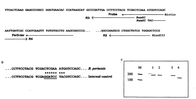
CTAGCTGTGA AGATTCAATA GGTTGTATGC ATGGTTCATC CGAACCGGAT TTGAGAAACT GGAAATCGCC AACCCCCCAG
> ΡθΠ EcoRJ > R1
TTCACTCAAG GAGCCCGGCC GGATGAACAC CCATAAGCAT GCCCGATTGA CCTTCCTACG TCGACTCGAA ATGGTCCAGC Probe < Biotin
R3 <
AATTGATCGC CCATCAAGTT TGTGTGCCTG AAGCGGCCCG. . .
Per3-rev <
GGCCAAGGCG CTGGCTATCG TGGAGCTCCG R2 < HindTIJ
. .CCTTCCTACG TCGACTCGAA ATGGTCCAGC. . . B. pertussis
****** ***
. . CCTTCCTACG TCGAGGATCC TACGTCCAGC. . . Internal control
Fig. 1 Construction of the internal control DNA
(a) The PCR amplified section from a repetitive element of B.
pertussis for detection of the infectious agent in nasopharyngeal aspirates or swabs is shown (2, 11). Oligonucleotide sequences used for amplification (see Materials and Methods) are labeled with arrows, additional restriction sites are shown in italics. The resulting DNA fragments have sizes of 183 base pairs (Perl/
PerSrev), 380 base pairs (R1/R2), 145 base pairs (R1/R3) and 241 base pairs (R2/R4).
M 200 — 100 —-
3 4
(b) The resulting nine sequence alterations in the internal control DNA after ligating the R1/R3 and R2/R4 DNA fragments with pUCBM20 are marked with asterisks in the sequence of the capture probe binding region.
(c) Electrophoretic separation of B. pertussis DNA (lane 1) and internal control DNA (lane 2) after PCR with primer pair Perl/
Per3rev results in 183 base pairs DNA fragments. After digestion with restriction endonuclease BamHl (lane 3: B. pertussis DNA, lane 4: internal control DNA) bands of 138 and 45 base pairs were only detected in lane 4.
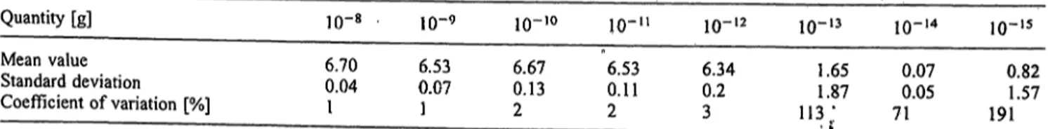
Tab. 1 Test for cross reactivity of the internal control DNA with the pertussis capture probe.
The cloned internal control DNA was diluted in several steps (10 - 10~l 3g per assay) and amplified in a PCR using the primer pair Perl/Per3rev (see Materials and Methods). Two capture probes (pertussis, internal control) were used for hybridisation. Absor- bances at 422 nm after detection of the PCR products with the ES300 analyser (Enzymun Test® DNA Detection) are shown.
Quantity [g] 1<T8 1(T9 ΙΟ"'0 ΚΓ" 1(T'2 1(T13
Pertussis 0.044 0.041 0.041 0.039 0.043 0.034 capture probe
Internal control 6.66 6.72 6.69 6.50 6.50 5.89 capture probe
internal control DNA and pertussis capture probe. Thus the introduced mismatches in the capture probe binding site are shown to be suitable for a differentiation of B.
pertussis and internal control in the assay.
The use of competitive PCR may result in a decreased amplification rate of the required DNA fragment. There- fore it was necessary to determine a minimum quantity of internal control DNA which could be coamplified in polymerase chain reaction. Table 2 shows the results of these investigations. Internal control DNA was diluted from 10~8g to 10~15g per assay. From each dilution four amplifications were performed and the products were detected with internal control capture probe using Enzymun Test® DNA Detection. Internal control DNA (1 pg per 50 μΐ total volume in the reaction mixture) was the lowest concentration which led to constant absor- bances at 422 nm. This fact is indicated by the increas- ing coefficients of variation at lower quantities of in- ternal control DNA. Furthermore we tested the competi- tive PCR with dilutions from 10~8 g to 10~14 g per as- say of the repetitive DNA fragment of B. pertussis cloned into pBluescript SK+ (11) and a constant amount of 1 pg internal control DNA per reaction (see fig. 2).
As expected we found a suppression of internal control DNA amplification at higher B. pertussis DNA concen- trations and vice versa. In ten different runs we investi- gated mean values for negative controls. We found for
internal control capture probe A422nm =s 5·83, SD = 0.6 (n = 20) and for pertussis capture probe A422nm
= 0.046, SD = 0.02 (n = 20). The cut-off value for negative samples was set at A^nm (pertussis capture probe) < 0.1 which is due to Enzymun Test® DNA De- tection specifications (negative sample < 2 X A422nm)·
Competitive polymerase chain reaction with patient samples
For reliable identification of Bordetella pertussis in na- sopharyngeal swabs it was necessary to check the qual- ity of the described assay conditions. Therefore we tested different pools of stored patient samples. The first group includes patients without any signs of infection which tested negative in a polymerase chain reaction (11). The absorbances after detection of the amplified DNA using pertussis capture probe were in the same range compared to the negative controls (A422nm
= 0.03, SD = 0.002 (n = 10)). Measuring negative pa- tient samples it was possible to study inhibitory effects of the DNA preparation techniques used in the assay.
We tested a commercial DNA preparation kit and two usual methods for DNA preparation (see materials afrd methods). The dependence of the quality of DNA prepa- ration methods on internal control DNA amplification is shown in table 3. Coamplification of 1 pg internal con- trol DNA in a competitive PCR with reproducible high absorbances in the detection step was only possible using phenol/chloroform purified DNA from nasopha- ryngeal swabs. In cases in which other DNA preparation methods were used it became necessary to increase the amount of internal control DNA.
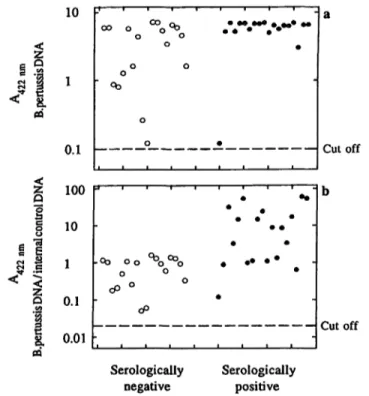
Finally we compared serologically confirmed patients with whooping cough and PCR-positive, serologically negative patients with proven contact to B. pertussis.
With these test results we wanted to show a possible detection of different amounts of the infectious agents in nasopharyngeal swabs between the point of infection and the paroxysmal stage. Results of these comparisons are shown in figure 3. Positive tests were confirmed in both groups using a competitive PCR assay. In addition, a ratio of absorbances at 422 nm of the B. pertussis
Tab. 2 Determining the minimum quantity of internal control
DNA in the assay. amplifications were performed. Hybridisation occured only with
the internal control capture probe. Mean values, standard devia- Intemal control DNA was diluted in several steps (1(T8 - I(r15g tions and coefficients of variation are shown for the measured ab- per assay) and PCR amplified using the primer pair Perl/Per3rev sorbances at 422 nm.
(see Materials and Methods). From each dilution four Quantity [g]
Mean value Standard deviation Coefficient of variation [%]
10~8
6.70 0.041
10~9
6.53 0.07 1
10-io 6.67 0.13 2
ΙΟ'11
6.53 0.11 2
io-
12 6.34 0.2 310~13
1.65 1.87 113 '
10-i4 0.07 0.05 71
io-
15 0.82 1.57 191Lichtinghagen and Glaubitz: Competitive polymerase chain reaction for B. pertussis identification 91
ΙΟ'14 ΙΟ'12 ΙΟ"10 ΙΟ'8' Positive control DNA [g]
Fig. 2 Competitive polymerase chain reaction of positive con- trol DNA
The B. pertussis DNA fragment (183 base pairs; Perl/Per3rev) cloned in pBluescript KS+ (11) was diluted from 10~8g to 10~15g per assay and coamplified with 10~l 2g internal control DNA. The primers Perl and Per3rev were used in the PCR. Result- ing DNA fragments were detected after hybridisation with pertussis (a) and internal control capture probe (b) using Enzymun Test®
DNA Detection (see Materials and Methods). Absorbances at 422 nm were plotted against concentrations of positive control plas- mid DNA.
DNA and the internal control DNA led to a better sepa- ration of different collectives. In case of the serologi- cally negative group we found maximum values only around 1.5 (values for negative samples were about 0.01). In the serologically positive group values between 10 and 100 could be obtained. Such a differentiation was not possible in a comparison only of the absorbances of the amplified B. pertussis DNA. To determine the influ- ence of the concentration of internal control DNA on the sensitivity of the assay, we investigated pooled sam- ples from serologically positive and negative patients with high absorbances in the detection assay (> 6.0), and samples with low absorbances (< 0.3).
From these pools four competitive PCR assays were performed with increasing concentrations (10~
12— 10~
9g per assay) of the internal control DNA. In
Tab. 3 Effect of DNA preparation technique on internal control DNA amplification.
Samples from patients without any signs of wooping cough were amplified in a competitive PCR with 1 pg internal control DNA after different routes of DNA isolation. Mean values for absor- bances at 422 nm after hybridisation with internal control capture probe, standard deviations and coefficients of variation are listed.
DNA preparation method ^internal control $D
Proteinase-K treatment 35 0.054 0.003 6 and boiling of the sample
QIAamp Blood Kit 25 0.71 0.91 128 Phenol/chloroform 10 4.47 0.37 8
case of the positive group a competition with about 10~~
9g internal control DNA and in case of the nega- tive group with about 10~
nto 10~~
12g leads to a ratio of 1 which indicates an equimolar amount of B.
pertussis and internal control DNA fragment. This shows that using an amount of 10~
l 2g of internal control plasmid DNA does not lead to a decreased diagnostic sensitivity of the assay.
Discussion
Usually the diagnosis of whooping cough can be done definitively with the help of polymerase chain reac- tion. But in some cases a bad correlation between laboratory results and the clinical picture leads to a decrease of diagnostic specificity and sensitivity. Diag- nostic specificity can be optimized by amplification of a proven germ specific DNA area (4—11), and by following hygienic precautions, like isolating the work areas and using filter tips for pipetting (11). In contrast to serology and culture, high sensitivity of PCR allows the detection of B. pertussis in early stages of disease
10
s ε
0.1 100 10
0.1 0.01
ο
ο Cut off
0 0 ·· ·
cP
Cut off Serologically
negative Serologically positive
Fig. 3 Competitive polymerase chain reaction with purified DNA from nasopharyngeal swabs
Bordetella pertussis DNA was isolated from nasopharyngeal swabs using phenol/chloroform extraction (see Materials and Methods).
Two groups of patients were tested in this comparison. Serologi- cally negative children without typical clinical presentations but proven contact to B. pertussis and serologically positive children with typical signs of whooping cough. Resulting DNA fragments were detected after hybridisation with pertussis and internal control capture probe using Enzymun Test® DNA Detection. Absorbances at 422 nm for pertussis capture probe are shown in (a). The listed values in (b) are the ratios A422nm (B. pertussis)/AA22 nm (internal control DNA).
and in case of infections of vaccinized patients (3, 11, 18, 19).
Competitive PCR is a useful tool in the identification of a diminished diagnostic sensitivity. Loss of sensitivity in PCR assays is mostly due to inhibitory effects (20);
e. g. we found an influence on the amplification of the target DNA due to insufficient inactivated proteinase K (see tab. 3), sometimes to haemoglobin and incomplete removal of phenol. During a competitive PCR an in- ternal control DNA is coamplified. A diminished ampli- fication of the internal control DNA discloses such disturbances in case of a negative detection of the target DNA. From our point of view one can omit a separate positive control like a cloned DNA fragment for quality control because it does not give any evidence of inhibi- tors in the individual sample and it presents a potential source of contamination.
Depending on the concentration of the internal control DNA, its coamplification leads to a reduction of the am- plification rate of the target DNA (see fig. 1). The sensi- tivity of the assay therefore has to be calibrated accord- ing to these demands (see fig. 4), so that the clinically
10
Q
< 1
0.1 100 10 l 0.1 0.01
a o
d ο— ·°^.—
'
Cutoff b
Cutoff '10
ΙΟ' ΙΟ'1 ΙΟ'12
CQ
Internal control DNA [g]
Fig. 4 Competition of different concentrations of B. pertussis DNA isolated from nasopharyngeal swabs
Pooled samples from serologically positive children with whooping cough (a, d), from serologically negative children with contact to B. pertussis and high absorbances after pertussis capture probe hy- bridisation (b, e) and samples with low absorbances after hybridisa- tion with pertussis capture probe (c, f) were tested in competitive PCR assays with increasing amounts of internal control DNA (10- l 2- lO'Vassay).
Absorbances at 422 nm after hybridisation with pertussis capture probe were plotted against internal control DNA quantities per as- say (a).
Ratios A422nm (B. pertussis DNA)/A422nm (internal control DNA) were plotted against internal control DNA quantities per assay (b).
relevant germ DNA concentration does not lie beneath the cut off value.
Today a quantitative interpretation of PCR results is partly possible in the diagnostics of virus diseases from homogeneous matrices like blood (13). For detection of B. pertussis nasopharyngeal swabs are suitable, but in this case they can be used only with limitations. B. per- tussis is an obligate pathogen, therefore each positive result, e. g. after cultivation of the infectious agent, is of clinical relevance. Quantitative results, however, lead to a different assessment of the clinical situation. Using the competitive PCR we could show that a significant differentiation is possible between patients with typical clinical symptoms and patients in prodromal stage. For example it can be of interest to estimate the infectious- ness during incubation stage. This is possible with the help of the target DNA/control DNA ratio.
A known disadvantage of a PCR assay is the detection of dead bacterial cells. Regarding the estimation of in- fectiousness the microbiological culture shows a better diagnostic specificity. Here positive results correlate well with the clinical condition. Therefore the PCR has priority in early diagnostics. With existing older infec- tions a possibly still existing infectiousness can riot be estimated because the bacteria die during the catarrhal stage. We obtained very weak positive results in some PCR positive patients who had proven contact with B.
pertusis infected persons (see fig. 4). A semiquantitative estimation with the help of the competitive PCR points to a number of bacterial cells that is clearly below nor- mally measured values (see fig. 4). After a few days a control test should give further information about the infectiousness under consideration of the target DNA/
control DNA ratio.
We find the competitive PCR to be a fast and depend- able procedure for the diagnosis of whooping cough.
Analytical sensitivity of single assays could be better controlled. The specificity of a result can be interpreted under consideration of the clinical condition and includ- ing the relation of target DNA to internal control DNA amplification. The clinical impact of the competitive PCR with weak positive results must be tested with a much bigger number of samples in correlation with the clinical findings.
Acknowledgements
The authors highly appreciate the skilful technical assistance of Bernadette Luens and Frank Dsiosa. We are grateful to Dr. M.
Scherf, Dr. U. Fahlbusch, Dr. D. Petersen and Dr. R. Zinck for helpful discussion. We thank Boehringer Mannheim for support, which rnade part of this work possible.
Lichtinghagen and Glaubitz: Competitive polymerase chain reaction for B. pertussis identification 93
References
1. Onorato IM, Wassilak SGF. Laboratory diagnosis of pertus- sis: the state of the art. Pediatr. Infect. Dis. J. 1987;
6:145-51.
2. Glare MG, Paton RP, Lawrence JL, Nisnet IN. Analysis of a repetitive DNA sequence from Bordetella pertussis and its application to the diagnosis of pertussis using the Polymerase Chain Reaction. J Clin Microbiol 1990; 28:1982-7.
3. He Q, Mertsola J, Soini H, Skurnik M, Ruuskanen O, Viljanen MK. Comparison of polymerase chain reaction with culture and enzyme immunoassay for diagnosis of pertussis. J Clin Microbiol 1993; 31:642-5.
4. He Q, Mertsola J, Soini H, Viljanen MK. Sensitive and specific polymerase chain reaction for detection of Bordetella pertussis in nasopharyngeal specimens. J Pediatr 1994; 124:421—6.
5. Grimprel E, Begue P, Anjak I, Betsou F, Guiso N. Comparison of polymerase chain reaction, culture and western immunoblot serology for diagnosis of Bordetella pertussis infection. J Clin Microbiol (United States) 1993; 31:2745-50.
6. Reizenstein E, Johansson B, Mardin L, Abens J, Mollby R, Hallander HO. Diagnostic evaluation of polymerase chain re- action discriminative for Bordetella pertussis, B. parapertussis and B. bronchiseptica. Diagn. Microbiol. Infect. Dis. (United States) 1993; 17:185-91.
7. Houard S, Hackel C, Herzog A, Bollen A. Specific identifica- tion of Bordetella pertussis by the polymerase chain reaction.
Res Microbiol. 1989; 140:477-87.
8. Douglas E, Coote JG, Parton R, McPheat W. Identification of Bordetella pertussis in nasopharyngeal swaps by PCR amplifi- cation of a region of the adenylate cyclase gene. J. Med.
Microbiol. 1993; 38:140-4.
9. McPheat, WL, McNally T. Isolation of a repeated DNA se- quence from Bordetella pertussis. J. Gen. Microbiol. 1987;
133:323-30.
10. Olcen P, Backman A, Johansson B, Esbjorner E, Tornqvist E, Bygraves J et al. Amplification of DNA by the polymerase chain reaction for the efficient diagnosis of pertussis. Scand.
J. Infect. Dis. 1992; 24:339-45.
11. Lichtinghagen R, Diedrich-Glaubitz R, von Horsten B. Identi- fication of Bordetella pertussis in nasopharyngeal swabs using the polymerase chain reaction: Evaluation of detection meth- ods. Eur. J. Clin. Chem. Clin. Biochem. 1994; 32:161-7.
12. Perre F. Quantitative or semi-quantitative PCR: reality versus myth. PCR Methods Appl. 1992; 2:1-9.
13. Clementi M, Menzo S, Bagnarelli P, Manzin A, Valenza A, Varaldo PE. Quantitative PCR and RT-PCR in virology. PCR Methods Appl. 1993; 2:191-6.
14. Saiki RK, Gelfand DH, Stoffel S, Scharf SJ, Higuchi R, Horn GT. Primer-directed enzymatic amplification of DNA with a thermostable DNA polymerase. Science 1988; 239:487-91.
15. Hung T, Mak K, Fong K. A specific enhancer for polymerase chain reaction. Nucl. Acids Res. 1990; 18:4953.
16. McPherson MJ. Directed mutagenesis - a practical approach.
Oxford University Press, New York, 1991.
17. Maniatis, T, Fritsch EF, Sambrook J. Molecular cloning: A lab- oratory manual. Cold Spring Harbor Laboratory, Cold Spring Harbor, N.Y., 1982.
18. Public Health Laboratory Service Epidemiological Research Laboratory and 21 Area Health Authorities. Efficacy of pertus- sis vaccination in England. Br. Med. J. 1982; 285:257-9.
19. Schlapfer G, Cherry JD, Heininger U, Just M. Use of the poly- merase chain reaction to increase the identification of B. per- tussis infections in vaccinees and family members in a pertus- sis vaccine efficacy trial. Pediatr. Res. 1994; 35 (4 Pt 20): 195 A.
20. Wadowsky RM, Laus S, Libert T, States SJ, Ehrlich GD. Inhi- bition of PCR-based assay for Bordetella pertussis by using calcium alginate fiber and aluminium shaft components of a nasopharyngeal swab. J. Clin. Microbiol. 1994; 32:1054-7.
Dr. Ralf Lichtinghagen
Medizinische Hochschule Hannover Institut für Klinische Chemie I Konstanty-Gutschow-Straße 8 D-30625 Hannover
Germany