Exploring the therapeutic potential of recombinant AAV vectors in stem cell and transplantation model systems
for the treatment of heart diseases
I n a u g u r a l - D i s s e r t a t i o n
zur
Erlangung des Doktorgrades
der Mathematisch-Naturwissenschaftlichen Fakultät der Universität zu Köln
vorgelegt von
Natascha Schuhmann
aus Weinheim
Köln 2008
Berichterstatter/in: Prof. Dr. Jens Brüning Prof. Dr. Herbert Pfister PD Dr. Hildegard Büning
Tag der mündlichen Prüfung: 24.6.2008
Universitätklinikums zu Köln angefertigt.
Im Verlauf dieser Arbeit entstanden folgende Publikationen:
Hacker, U. T., Wingenfeld, L., Kofler, D. M., Schuhmann, N. K., Lutz, S., Herold, T., King, S. B., Gerner, F. M., Perabo, L., Rabinowitz, J., McCarty, D. M., Samulski, R. J., Hallek, M., and Buning, H. (2005). Adeno-associated virus serotypes 1 to 5 mediated tumor cell directed gene transfer and improvement of transduction efficiency. J Gene Med 7(11), 1429-38.
Schuhmann, N.*, Burdorf, L.*, Postrach, J., Thein, E., Hallek, M., Reichart, B.
Buning, H.+, and Schmoeckel, M.+ (2007). AAV-mediated gene transfer to cardiac cells in a heterotopic rat heart transplantation model. Transplant Proc 39(2), 567-8. (* + equal contribution)
Zuber, C.*, Mitteregger, G.*, Schuhmann, N., Rey, C., Knackmuss, S., Rupprecht, W., Reusch, U., Pace, C., Little, M., Kretzschmar, H. A., Hallek, M., Buning, H., and Weiss, S. (2008). Delivery of single-chain antibodies scFvs directed against the 37 kDa/67 kDa laminin receptor into mice via recombinant Adeno-associated viral vectors for prion disease gene therapy. J Gen Virol, in press (* equal contribution)
Für meine Eltern
Table of contents
1 Zusammenfassung ...1
2 Abstract ...4
3 Introduction ...7
3.1 Adeno-associated virus...7
3.1.1 Classification of adeno-associated virus...7
3.1.2 Genome organization ...9
3.1.3 Infection biology of AAV...11
3.1.3.1 Virus-cell interaction ...13
3.1.3.2 Receptor-mediated endocytosis ...14
3.1.3.3 Endosomal processing and escape...14
3.1.3.4 Nuclear translocation...15
3.1.3.5 Latent and lytic life cycle of AAV ...16
3.1.4 Adenovirus-free AAV production and recombinant AAV vectors (rAAV)17 3.1.5 AAV as vector in gene therapy ...19
3.1.5.1 Gene therapy of ischemic cardiovascular diseases ...21
3.2 Stem cells ...25
3.2.1 Hematopoietic stem cells and endothelial progenitor cells ...26
3.2.2 Bone marrow stem cell niche, mobilization and homing ...29
3.2.3 Therapeutic potentials of EPC transplantation and gene therapy...32
3.2.4 Aim of the study ...35
4 Results...37
4.1 rAAV transfer into heart...37
4.1.1 rAAV transfer into rodent heart ...37
4.1.1.1 Heterogenic rat heart transplantation ...37
4.1.1.1.1 Delivery of vector genomes into transplanted hearts... 39
4.1.1.1.2 Transgene mRNA expression cannot be detected in ... transplanted hearts... 43
4.1.1.1.3 Beta-Galactosidase activity is not detectable in tissue sections ... of transplanted hearts... 45
4.1.1.2 In vitro analyses of rAAV-mediated rat cell transduction...48
4.1.1.2.1 AAV2 capsids are detectable in the cytoplasm and perinuclear ... area of RAECs... 49
4.1.1.2.2 CMV promoter induces transgene expression after transfection 50 4.1.1.2.3 RAECs are poorly transduceable with AAV serotypes 1 to 5, ... but show enhanced expression by administration of MG132... 51
4.1.1.2.4 Neonatal rat cardiomyocytes show highest transgene ... expression after transduction with rAAV1 and rAAV4... 54
4.1.2 rAAV-mediated gene transfer into porcine heart...56
4.1.2.1 Identification of the most suited serotype using PAECs as in vitro model ..56
4.1.2.1.1 PAECs are efficiently transduceable with rAAV2... 57
4.1.2.1.2 Visualization of rAAV2 capsids in the cytoplasm of PAECs... 59
4.1.2.2 Heterogenic pig heart transplantation...60
4.1.2.2.1 Successful vector delivery, but no transgene expression from ... single-stranded vector genomes in porcine heart... 63
4.1.2.2.2 Successful vector delivery and transgene expression from ... self-complementary vector genomes in porcine heart... 67
4.2 Establishment of rAAV-mediated gene transfer into CD34+ cells...74
4.2.1 Second-strand synthesis is a limiting step in CD34+ cell transduction ...74
4.2.2 Loss of transgene expression during prolonged cultivation times...78
4.2.3 Heparin inhibits transduction with rAAV2 approving HSPG as primary ...
receptor ...79
4.2.4 Transduction efficiency correlated with the availability of α5β1 integrins 81 4.2.5 Enhancement of transgene expression using retinoic acid and Trichostatin A ...84
4.2.6 CD34+ cells are able to take up Dil-AcLDL after endothelial differentiation assay ...87
4.3 Further applications of serotypes ...90
4.3.1 Serotype 2 is superior in transduction of primary melanoma cells ...90
4.3.2 Serotype 2 is superior in transduction of primary porcine fibroblasts and HeLa cells ...91
4.4 Vector genomes are detected in spleen following intracerebral injection ... of rAAV2...94
5 Discussion ...96
5.1 Heterotopic heart transplantations ...96
5.1.1 Approaches for endothelial transduction in rat model ...96
5.1.2 In vitro studies of RAECs ...99
5.1.2.1 Superiority of rAAV1 in rat cardiac endothelial and cardiomyocyte ... transduction ...99
5.1.2.2 Barriers in endothelial cells impair rAAV-mediated transgene expression100 5.1.3 Porcine endothelial cells are highly permissive for AAV ...103
5.1.4 AAV vector mediated transgene expression in porcine hearts ...103
5.1.5 Distribution of transgene DNA and product in the two animals...105
5.1.6 Potential of rAAV2 for animal cloning ...107
5.2 Investigations on CD34+ cells ...108
5.2.1 Most efficient serotype rAAV2 is limited by second-strand synthesis ..108
5.2.2 Transduction procedure does not interfere with endothelial differentiation ...110
5.2.3 Receptor and coreceptor studies ...111
5.2.4 Effects of the transcriptionally active drugs TSA and RA...113
5.2.5 Outlook ...115
6 Materials ...117
6.1 Chemicals and Solutions...117
6.2 Enzymes and Kits ...118
6.3 Plasmids ...118
6.4 Primers...120
6.5 Antibodies ...121
6.6 Bacteria Strains...121
6.7 Eukaryotic Cells ...121
6.7.1 Immortalized Cell Lines ...121
6.7.2 Primary cells ...122
6.8 Culture Media and Supplements...123
6.9 Laboratory Equipment and Disposables ...125
6.10 Data Treating Software ...126
7 Methods...127
7.1 Bacteria Culture ...127
7.1.1 Cultivation of Bacteria...127
7.1.2 Preparation of Competent Bacteria...127
7.1.3 Transformation of Bacteria ...128
7.2 Working with nucleic acids ...128
7.2.1 Plasmid amplification and extraction...128
7.2.2 DNA and RNA Quantification...129
7.2.3 Restriction Enzyme Digest...129
7.2.4 Agarose Gel Electrophoresis ...129
7.2.5 Tissue DNA extraction ...130
7.2.6 Tissue RNA extraction ...130
7.2.7 DNase I digest and cDNA synthesis ...131
7.2.8 Quantitative Polymerase-Chain-Reaction...132
7.3 Eukaryotic cell culture ...135
7.3.1 Cultivation of Cells ...135
7.3.2 Trypsinization...135
7.3.3 Counting ...135
7.3.4 Seeding / Passaging...135
7.3.5 Freezing and Thawing Cells ...136
7.3.6 Vector production and purification ...136
7.3.6.1 AAV-Vector Packaging...136
7.3.6.2 Iodixanol Gradient Purification...137
7.3.6.3 Heparin Affinity Chromatography ...137
7.3.6.4 Vector titration ...138
7.3.7 Working with CD34+ cells ...138
7.3.7.1 Isolation and culturing of CD34+ cells ...138
7.3.7.2 Thawing of CD34+ cells ...139
7.3.7.3 Transduction of CD34+ cells with AAV...139
7.3.7.4 Analysis of transduced CD34+ cells by FACS...139
7.3.8 Dil-AcLDL uptake...140
7.4 Determination of protein...140
7.4.1 Detection of beta-Galactosidase activity in tissue sections...140
7.4.2 Staining for beta-Galactosidase activity in cells ...141
7.4.3 Bradford Assay ...141
7.4.4 Luciferase Assay ...142
7.5 Heterotopic heart transplantation ...142
7.5.1 Rat heart transplantation ...142
7.5.2 Pig heart transplantation...143
8 Abbreviations ...146
9 References ...148
1 Zusammenfassung
Herzerkrankungen sind weltweit die Hauptursache für einen vorzeitigen Tod.
Sowohl Gentransfer als auch zelluläre Therapien werden derzeit als neue Behandlungsmöglichkeiten für diese Erkrankungen entwickelt.
Im ersten Teil dieser Arbeit sollte der AAV-vermittelte Gentransfer von Transgenen ins Herzgewebe von Donororganen, die anschließend transplantiert wurden, etabliert werden. Als Tiermodelle für die heterotopen Herztransplantationen wurden Sprague Dawley Ratten und Deutsche Landrasseschweine gewählt. rAAV Serotype 2, welcher für diesen Zweck als geeignet beschrieben wurde, wurde intracoronar entweder in das normotherme (n=3) oder hypotherme (n=3) Herz appliziert, welches anschließend in eine Empfängerratte transplantiert wurde. Der Gentransfer erfolgte mit einer besseren Effizienz in normotherme Herzen verglichen mit hypothermen Herzen. Trotz des erfolgreichen Transfers und der Nachweisbarkeit von Vektor-DNA in Gewebeproben 28 Tage nach Transplantation konnte jedoch weder Transgen-spezifische mRNA noch Proteinexpression detektiert werden. Zur Bestimmung potentieller Barrieren, welche die rAAV2- vermittelte Transgenexpression in unserem Rattenmodell beeinträchtigen, wurden in vitro Analysen in Rattenaortenendothelzellen (RAECs) durchgeführt. Wir konnten Zelleintritt und intrazelluläres trafficking der viralen Partikel ebenso als inhibierende Faktoren ausschließen wie die Expression vom gewählten CMV Promotor. Die Applikation des Proteasomeninhibitors MG132 erhöhte die Transgenexpression jedoch signifikant für die Serotypen 1 und 2 (6-fach und 7.3- fach). Ersterer war in unseren in vitro Experimenten der effizienteste Serotyp der analysierten 5 Serotypen (rAAV1 bis rAAV5). Da MG132 die Funktion der Proteasomen inhibitiert, könnte eine blockierte Vektordegradation die beobachteten Ergebnisse erklären. Darüberhinaus sind auch indirekte Effekte wie eine erhöhte Ubiquitinierung des Vektorkapsids denkbar, von der man annimmt, dass sie das sogenannte vector uncoating oder die Translokation in den Nucleus erleichtert (Duan et al., 2000; Yan et al., 2002). Des Weiteren konnte eine geringfügig eingeschränkte Fähigkeit zur Zweitstrangsynthese beobachtet werden.
Diese wird benötigt, um das einzelsträngige DNA-Genom herkömmlich verwendeter AAV-Vektoren in einen transkribierbaren Doppelstrang zu verwandeln. Zusammenfassend muss man feststellen, dass sich die analysierten rAAV Serotypen als ungeeignete Gentransfervektoren für Rattenendothelzellen
erwiesen haben. Im Gegensatz dazu wurde in primären Rattencardiomyocyten in vitro eine deutlich höhere Transduktionseffizienz erzielt. Dies veranlasste uns, die vasoaktive Substanz Histamin in unserem heterotopen porcinen Transplantations- modell anzuwenden, um in vivo sowohl Endothelzellen als auch Cardiomyocyten zu transduzieren. Interessanterweise zeigten in vitro Analysen porciner Aortenendothelzellen – im Gegensatz zu unseren erfolglosen Versuchen in RAECs – Transduktionseffizienzen von ca. 90 % mit rAAV2. Daher wurde dieser Serotyp zusammen mit Histamin in normotherme Herzen appliziert unter Verwendung des neuentwickelten in situ Langendorff Reperfusionssystems, welches eine verlängerte Rezirkulation des Vektors im Herzen erlaubte. In beiden Tieren konnte der Nachweis eines erfolgreichen Gentransfers anhand deutlich messbarer Transgen-DNA-Mengen erbracht werden. In einem der beiden Tiere wurde zudem funktionales Protein nachgewiesen. Dieses Schwein hatte zehnmal höhere Vektormengen erhalten. Dieser Vektor kodierte zudem für das Transgen Luciferase und wies eine self-complementary (Pseudo-Doppelstrang) Vektorgenom-Konformation auf. Im Gegensatz dazu wurde das Schwein, das keine Expression zeigte, mit einem Vektor behandelt, der für beta-Galaktosidase in einer Einzelstrang-Vektorgenom-Konformation kodierte. Obwohl weitere Experimente zur Bestimmung des Einflusses der drei Parameter (Vektormenge, Vektorgenom-Konformation und Wahl des Transgens), einzeln oder in Kombination, benötigt werden, konnten wir zeigen, dass ein rAAV2-vermittelter Gentransfer in porcines Herzgewebe im Rahmen eines (Xeno-) Transplantationsansatzes im Prinzip möglich ist.
Im zweiten Teil meiner Arbeit wurde ein Protokoll zur effizienten Transduktion humaner CD34+ Zellen aus Nabelschnurblut etabliert. Derartige Protokolle ermöglichen eine Kombination von Zell- und Gentherapie, welche für ein breites Spektrum an Anwendungen vorteilhaft ist. Vergleiche der Serotypen 2, 3 und 5 ermittelten rAAV2 als den effizientesten Serotyp in Zelleintritt und Transgenexpression. Der Zelleintritt von rAAV2 in CD34+ Zellen war abhängig von Heparansulfatproteoglycan, wie mithilfe von Kompetitionsexperimenten bestimmt wurde, und von α5β1 Integrin, was wir mittels einer Mutante für die Bindung des Rezeptors ermittelten. Interessanterweise waren nur auf vorexpandierten Zellen α5β1 Integrine nachweisbar. Dies erlaubt den Schluss, dass zumindest einige der kontroversen Berichte zur rAAV-vermittelten Transduktion von CD34+ Zellen auf
die verwendeten Kulturbedingungen zurückzuführen sind. Darüberhinaus ist die Synthese des Zweitstrangs in CD34+ Zellen beeinträchtigt, da nur die Zugabe von Vektoren mit Genomen in der self-complementary Konformation in erfolgreichen Transduktionen resultierte. Die ohnehin schon sehr effiziente Transduktion (61 %), die mit rAAV2 Vektoren mit self-complementary Genomen in vorexpandierten Zellen erzielt wurde, konnte signifikant (auf 86 %) durch die Zugabe von all-trans Retinsäure und dem Histon-Deacetylase-Inhibitor Trichostatin A erhöht werden.
Darüberhinaus weisen unsere Ergebnisse darauf hin, dass Transduktionen mit rAAV nicht mit der Fähigkeit der CD34+ Zellen zur endothelialen Differenzierung interferieren. Zusammenfassend kann festgestellt werden, dass unter Verwendung des hier etablierten Protokolls CD34+ Zellen effizient mit rAAV2 transduziert werden können und sich rAAV2 somit als ein geeignetes Vektorsystem zur transienten Modifikation dieser Zellen anbietet.
2 Abstract
Heart diseases are the main cause of premature death in the population world- wide. Gene transfer as well as cell-based therapies are currently developed as new treatment options.
In the first part of this thesis, AAV-mediated gene transfer to deliver transgenes into heart tissue before transplantation ought to be established. As model system, the Sprague Dawley heterotopic rat model and the German Landrace pig model were chosen. rAAV serotype 2, described to be suited for this purpose, was intracoronarily delivered either in the normothermic (n=3) or hypothermic (n=3) hearts which were subsequently transplanted into a recipient rat. Gene transfer into normothermic hearts occurred with a better efficiency compared to hypothermic hearts. However, despite successful delivery and detection of vector DNA in tissue samples 28 d post transplantation, neither transgene-specific mRNA nor protein expression could be detected. To identify potential barriers that impair rAAV2-mediated transgene expression in our rat model, in vitro analyses in rat aortic endothelial cells (RAECs) were performed. We could exclude cell entry, intracellular trafficking of viral particles as well as expression from the chosen CMV promoter as inhibiting factors. However, application of the proteasome inhibitor MG132 significantly enhanced rAAV1- and rAAV2-mediated transgene expression (6-fold and 7.3-fold, respectively). The latter was in our hand the most efficient serotype in RAEC transduction among the serotypes rAAV1 to rAAV5.
Since MG132 is a proteasome inhibitor blocking of vector degradation could be an explanation for the observed effect. Moreover, also indirect effects can be imagined like enhanced ubiquitination of the vector capsid, which is believed to facilitate vector uncoating or nuclear translocation of vector genomes (Duan et al., 2000; Yan et al., 2002). Furthermore, we observed a certain, albeit rather minor, limitation in second-strand synthesis. This step is necessary for the generation of a double-stranded DNA as template for transcription of the commonly used single- stranded DNA genome. In summary, the analyzed rAAV serotypes have been revealed as inappropriate gene transfer vectors in targeting of rat endothelial cells.
In contrast, in primary rat cardiomyocytes in vitro a higher transduction efficiency was observed. Therefore, we decided to administer the vasoactive substance histamine in our heterotopic pig heart transplantation model in order to target
porcine aortic endothelial cells revealed – in contrast to the unsuccessful attempts on RAECs – transduction efficiencies of about 90 % using rAAV2. Thus, this serotype was applied together with histamine into normothermic hearts using the newly developed in situ Langendorff reperfusion system which permitted prolonged recirculation of the vector in the heart. Transgene DNA detected in the graft of two transplanted animals displayed successful gene transfer. In one of the two animals functional protein was detected. This pig had received tenfold higher amounts of the vector which displayed a self-complementary vector genome conformation and encoded for the transgene luciferase. In contrast, the animal showing no expression was treated with a vector coding for beta-galactosidase in the single-stranded vector genome conformation. Although further experiments are needed to determine the influence of the three parameters (vector amount, vector genome conformation and choice of transgene) alone or in combination we could show that rAAV2-mediated gene delivery into the porcine heart tissue in a (xeno-) transplantation setting is in principle possible.
In the second part of my thesis, a protocol for efficient transduction of human cord blood-derived CD34+ cells was established. Such protocols enable a combination of cell and gene therapy which is advantageous for a wide range of applications.
Among the serotypes 2, 3 and 5, rAAV2 was identified as the most efficient serotype in cell entry and in transgene expression. Cell entry of rAAV2 into CD34+ cells was dependent on heparin sulfate proteoglycan as determined by competition experiments, and on α5β1 integrin as assessed by a receptor binding mutant. Interestingly, only pre-expanded cells displayed α5β1 integrin allowing to conclude that at least some of the contradictory reports on rAAV-mediated transduction of CD34+ cells are due to the applied cultivation conditions.
Furthermore, CD34+ cells are impaired in second-strand synthesis as only administration of vectors encoding the transgene in a self-complementary vector conformation resulted in successful transductions. The already high transduction level (61 %) achieved with rAAV2 using self-complementary vector genomes and pre-expanded cells could be significantly enhanced up to 86 % by addition of all- trans retinoic acid and the histone deacetylase inhibitor Trichostatin A.
Furthermore, our results provide strong evidence that transductions by rAAV2 vectors do not interfere with endothelial differentiation potential of CD34+ cells.
Thus, an efficient protocol for rAAV2-mediated transduction of CD34+ cells was
established revealing that rAAV2 is an appropriate vector system for transient modification of this cell type.
3 Introduction
3.1 Adeno-associated virus
3.1.1 Classification of adeno-associated virus
Adeno-associated viruses are classed into the Parvoviridae family. The Parvoviridae belong to the smallest known viruses (lat. parvus = small) consisting of a non-enveloped icosahedral capsid with a diameter of 18 to 26 nm and a linear single- stranded DNA genome (Figure 1). The Parvoviridae comprises two subfamilies: the vertebrate-infecting Parvovirinae and the Densovirinae which infect insects. The subfamily of Parvovirinae is further divided into the three genera Parvo-, Erythro- and Dependovirus. AAV belongs to the latter genus. Erythrovirinae infect erythroid precursor cells, whereas Parvovirus B19 is the only human pathogenic parvovirus and causes fifth disease (Erythema infectiosum) and complications during pregnancy (anemia, hydrops fetalis, abortions). Viruses belonging to the genus of Parvovirus are pathogenic for animals, examples are feline, canine and porcine parvovirus as well as minute virus of mice or aleutian mink disease virus. While erythro- and parvoviruses are autonomous viruses, dependoviruses require the presence of a helper virus like adenovirus (Ad), herpes simplex virus (HSV), vaccinia virus, human cytomegalovirus (HCMV) or papilloma virus (HPV) to undergo a productive life cycle (Atchison, Casto, and Hammon, 1965; McPherson, Rosenthal, and Rose, 1985; Richardson and Westphal, 1981; Schlehofer, Ehrbar, and zur Hausen, 1986). On the other hand, AAV seems to inhibit replication of helper viruses and also to interfere with malignant transformation induced by adenovirus, human or bovine papilloma virus (Heilbronn et al., 1990; Hermonat, 1992; Timpe, Verrill, and Trempe, 2006; You et al., 2006). Moreover, it was described that replication might be induced upon cellular genotoxic stress (Schlehofer, Ehrbar, and zur Hausen, 1986; Yakobson et al., 1989; Yakobson, Koch, and Winocour, 1987; Yalkinoglu et al., 1988).
Figure 1: Atomic structure of AAV serotype 2 determined by X-ray chrystallography. (Xie et al., 2003)
Until now, 12 serotypes have been described. In the 1960’s, AAV was discovered as contaminant of simian Adenovirus 15 preparations by several groups (Atchison, Casto, and Hammon, 1965; Mayor et al., 1965; Melnick et al., 1965). AAV serotype 2 has been isolated out of simian Adenovirus type 12 and AAV3 out of Adenovirus type 7 preparations (Hoggan, Blacklow, and Rowe, 1966). AAV4 has been found in African green monkeys infected with simian adenovirus 15 (Parks et al., 1967). In contrast, AAV5 was isolated out of a human clinical sample, a penile condylomatous lesion (Bantel-Schaal and zur Hausen, 1984). This virus is less related to the other serotypes considering sequence homology and serology (Chiorini et al., 1999). AAV6 has originally been identified as contaminant of an Adenovirus 5 stock. Its close relatedness to AAV1 with a variation of only 6 amino acids in the capsid sequence points either to a natural variant of serotype 1 or to a recombination between AAV1 and AAV2 as origin of AAV6 (Rutledge, Halbert, and Russell, 1998; Xiao et al., 1999). Wilson’s group discovered the serotypes AAV7 and AAV8 by PCR scanning for AAV sequence homologies in rhesus monkey tissues (Gao et al., 2002). The same group screened human tissues from various sources to detect latent AAV genomes and identified thereby a serologically different serotype, called AAV9 (Gao et al., 2004). Mori and colleagues isolated two new AAV variants out of cynomolgus monkey tissue designated AAV10 and AAV11 (Mori et al., 2004). Despite their isolation out of non-human primate tissues, these serotypes are also suited for transduction of human tissues (Gao et al., 2004). Recently, AAV serotype 12 has been identified in simian Adenovirus 18 contaminated vervet monkey cells from ATCC stocks (Schmidt et al., 2006).
Considering seroepidemiologic analyses, the serotypes AAV2, 3 and 5 are suggested to be endemic in humans (Gao et al., 2002). So far, AAV9 has only been found in human tissue. In contrast, monkeys are suggested to be the natural host for the serotypes 1, 4, 7 and 8 (Chiorini et al., 1997; Grimm and Kay, 2003;
Xiao et al., 1999). AAV serotypes differ in their tropism. For example, AAV1 is appropriate for transduction of skeletal muscle or retina, whereas AAV5 is better suited for applications in the central nervous system or the lung as determined in mouse models (Auricchio et al., 2001; Davidson et al., 2000; Xiao et al., 1999;
Zabner et al., 2000).
Moreover, AAV variants have also been isolated out of species other than primates including cow, bird, sheep, snake, lizard and goat (Bossis and Chiorini,
2003; Clarke et al., 1979; Farkas et al., 2004; Jacobson et al., 1996; Olson et al., 2004; Schmidt et al., 2004). So far, the best investigated AAV is the human serotype 2.
3.1.2 Genome organization
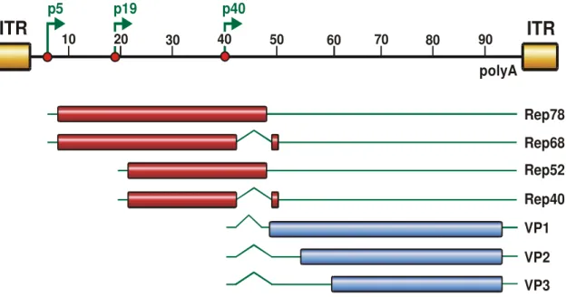
Wild type AAV contains a single-stranded DNA genome within an icosahedric capsid of 25 nm diameter. AAV serotype 2, the first serotype available as vector, has a genome size of 4680 nt with two open reading frames (ORFs) (Srivastava, Lusby, and Berns, 1983). These ORFs, encoding for the structural (cap; capsid) and non-structural proteins (rep; replication), are flanked by the inverted terminal repeats (ITRs) (Carter and Samulski, 2000) (Figure 2). This organization is conserved in all serotypes. The AAV2 genome contains three promoters (p5, p19, p40, describing their map position), but all transcripts share a common polyadenylation signal.
The 145 nt (for AAV2) long ITRs form the 3’- and 5’-end of the genome and hybridize to hairpin-like structures. Within the ITR region a Rep binding site (RBS) and a terminal resolution site (TRS) important for nicking of duplex DNA by the large Rep proteins are located (Im and Muzyczka, 1990; McCarty et al., 1994). In addition, this region is crucial for site-specific integration events, rescue of the provirus and serves as origin of replication (Berns, 1990; Feng et al., 2006;
Hauswirth and Berns, 1977; Labow and Berns, 1988; McLaughlin et al., 1988).
Moreover, the ITRs are required for packaging of the viral genomes into the preformed capsid.
The 5’-ORF encodes for four multifunctional, non-structural proteins named Rep78, Rep68, Rep52 and Rep40, according to their size (Lusby and Berns, 1982). While the p5 promoter controls the expression of the larger transcripts (Rep78 and Rep68) the expression of the smaller proteins Rep52 and Rep40 is under the control of the p19 promoter. Rep68 and Rep40 are splice variants of their larger counterparts (Figure 2). The Rep proteins have numerous functions.
The two larger Rep proteins which possess a nuclear localization signal at their C- terminus are essential for replication, transcription and site-specific integration (Cassell and Weitzman, 2004). The smaller Rep proteins mediate accumulation and packaging of the viral genome into the preformed capsid in a helicase- dependent manner (Dubielzig et al., 1999; King et al., 2001). While the Rep
proteins can act as transactivator of transcription of the three viral promoters in presence of helper virus, they can also repress transcription of the p5 and p19 promoters in absence of helper virus (Kyostio et al., 1994; Pereira, McCarty, and Muzyczka, 1997). Moreover, the large Rep proteins can regulate the processing of the cap transcripts (Qiu and Pintel, 2002).
Figure 2: Genome organization of AAV2. The AAV2 genome, flanked by the ITRs, spans 4680 nt divided into units of 100 nt. Shown are the three promoters p5, p19 and p40 at map position 5, 19 and 40 and the polyadenylation signal (polyA) at position 96. The open reading frames are indicated by rectangles, translated regions in red or blue, untranslated regions by thin solid lines while introns are marked as nicks. The p5 promoter controls expression of the large Rep proteins (Rep78, Rep68), while the p19 promoter is responsible for the expression of the small Rep proteins (Rep52, Rep40). Rep68 and Rep40 are spliced variants of Rep78 and Rep52, respectively. The gene encoding for the capsid proteins VP1, VP2 and VP3 is controlled by the p40 promoter. (Figure kindly provided by N. Huttner)
The three structural proteins VP1, VP2 and VP3 are situated in the 3’-ORF cap controlled by the p40 promoter. These three proteins form the 60 subunits of the viral capsid at a ratio of 1:1:8 (Kronenberg, Kleinschmidt, and Bottcher, 2001;
Rose et al., 1971). All capsid proteins share a common C-terminus, but differ in their N-terminus. The efficiency of translation for VP1 is regulated by alternative splicing while translation of VP2 is initiated from an unusual initiation codon (ACG) (Becerra et al., 1988; Becerra et al., 1985). This is the reason for the 10-fold lower
ITR
10 20polyA
40 50 60 70 80 90
ITR
p19 p40
p5
30
Rep78 Rep68 Rep52 Rep40 VP1 VP2 VP3
initiation codon (Becerra et al., 1985). The molecular weight of the three capsid proteins is 90 kDa (VP1), 72 kDa (VP2) and 60 kDa (VP3). Considering the functions of the capsid proteins, VP1 seems to be essential for infectivity whereas VP3 is sufficient for capsid formation (Warrington et al., 2004). VP2 is proposed to be neither necessary for capsid formation nor for production of infectious particles (Lux et al., 2005; Warrington et al., 2004).
Regarding phylogenetic relations between AAV2 and the other serotypes, a homology of 80 to 90 % was
observed for AAV1, 3, 6 to 8 and 10 in the amino acid sequence of VP1 (Gao et al., 2002; Mori et al., 2004) (Figure 3). AAV4 and AAV11 showed a 60 % and 65 % homology to AAV2 VP1, respectively (Gao et al., 2004; Mori et al., 2004).
AAV12’s closest relatives within the AAV family are AAV11 (84
%) and AAV4 (78 %) (Schmidt
et al., 2007). AAV5 is the most divergent serotype with only 58 % similarity compared to AAV2 VP1 (Bantel-Schaal et al., 1999). Additionally to the divergence of the capsid protein to the other serotypes, AAV5 contains an extra polyadenylation signal located within the intron thus producing mainly the unspliced Rep proteins Rep78 and Rep52 (Qiu et al., 2002). In general, homologies in the VP1 proteins are comparable to the phylogenetic results obtained by the nucleotide sequence (Grimm and Kay, 2003). About the recently described serotypes AAV9 to 12 only little is known so far.
3.1.3 Infection biology of AAV
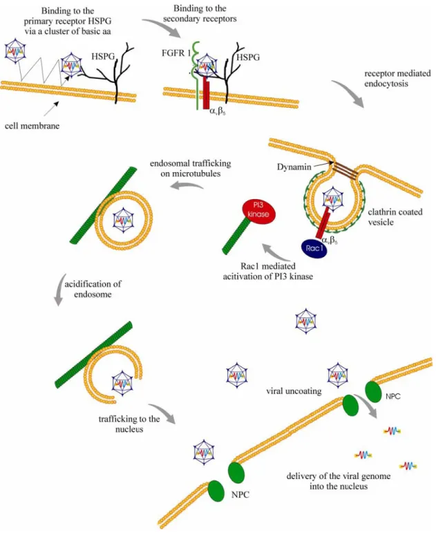
A successful infection of cells by AAV is a multistep process including attachment, uptake, intracellular trafficking, nuclear translocation and replication of the virus (Figure 4). Its understanding is crucial to identify potential barriers in AAV infection that have to be overcome for its use as gene therapy vector. The current knowledge of AAV2 infection is described in detail in the following chapters.
Figure 3: Phylogenetic analysis of the amino acid sequences of the capsid protein VP1. Modified scheme (grey) (Mori et al., 2004).
Figure 4: Infection pathway of AAV2 in HeLa cells. AAV2 touches the host cell several times and attaches to its primary receptor heparan sulfate proteoglycan (HSPG) and to the coreceptors fibroblast growth factor receptor 1 (FGFR-1) and the integrin αvβ5. The virus is internalized by receptor-mediated endocytosis into clathrin-coated vesicles in a dynamin-dependent way. The GTP-binding protein Rac1 is believed to be activated by integrin-binding and rearranges the cytoskeleton thus facilitating endosomal trafficking. Acidification of the endosomes leads to an escape of the AAV particles, maybe due to conformational changes. Viral uncoating takes place before or during nuclear entry. Also the exact mechanism of viral DNA import into the nucleus is yet
3.1.3.1 Virus-cell interaction
As Single Virus Tracing studies revealed, AAV2 contacts the cell membrane several times before it enters the cell (Seisenberger et al., 2001). For AAV2, the widely expressed cell surface receptor heparan sulfate proteoglycan (HSPG) has been identified as primary receptor (Summerford and Samulski, 1998). This contact is mediated by surface structures on the AAV capsid, namely the residues R484, R487, K532, R585 and R588 in the common VP3 region (Kern et al., 2003;
Wu et al., 2000). Attachment to HSPG seems to induce a reversible structural change thus facilitating coreceptor binding and cell entry (Asokan et al., 2006).
Surprisingly, some cell lines have been shown to take up virions even in the absence of HSPG (Duan et al., 1998b; Duan et al., 2000). Also AAV3 is suggested to use HSPG as primary receptor whereas the serotypes 1, 4, 5 and 6 bind to sialic acid (Rabinowitz et al., 2002). In 2006, Wu identified α2,3 and α2,6 sialic acids present on N-linked glycoproteins as primary receptors for AAV1 and AAV6 (Wu et al., 2006). AAV4 and AAV5 both bind to α2,3 sialic acid, but differ in their linkage specificity. While AAV4 requires O-linked, AAV5 prefers N-linked α2,3 sialic acids (Kaludov et al., 2001; Walters et al., 2001). Recently, the 37/67 kDa laminin receptor was proposed as a receptor for AAV8 (Akache et al., 2006).
Interestingly, overexpression of this receptor rendered cells also more susceptible for transduction with AAV2, 3 and 9 proposing a role for laminin receptor for cell infection of these serotypes. For AAV12, recently published data point towards a HSPG and sialic acid independent entry mechanism (Schmidt et al., 2007). The primary receptors for AAV7 and 9 to 12 have yet to be determined.
For efficient internalization, the additional binding to coreceptors is required. For AAV2, five secondary receptors have been proposed so far. Human fibroblast growth factor receptor 1 (FGFR-1), hepatocyte growth factor receptor (HGFR or c- met) and laminin receptor seem to support virus:cell interaction (Akache et al., 2006; Kashiwakura et al., 2005; Qing et al., 1999). On the other hand, the integrins αvβ5 and α5β1 are proposed as further coreceptors (Asokan et al., 2006; Sanlioglu et al., 2000; Summerford, Bartlett, and Samulski, 1999).
For AAV3, FGFR-1 has been described as potential coreceptor (Blackburn, Steadman, and Johnson, 2006). Concerning AAV5, the platelet-derived growth factor receptor (PDGFR) was identified as secondary receptor (Di Pasquale et al., 2003). It is conceivable that PDGFR might act alone as AAV5 receptor as it is a
sialo-proteoglycan containing oligosaccharides chains with sialic acids (Daniel et al., 1987; Hosang, 1988).
3.1.3.2 Receptor-mediated endocytosis
Following receptor binding and structural rearrangement, the virion enters the cell predominantly by receptor-mediated endocytosis in a dynamin-dependent manner (Bartlett, Wilcher, and Samulski, 2000; Duan et al., 1999; Hinshaw and Schmid, 1995). Single Virus Tracing studies revealed that the uptake of virions occurs within 100 ms (Seisenberger et al., 2001). Clustering of αvβ5 integrins seems to facilitate localization of the virion into clathrin-coated pits (Bartlett, Wilcher, and Samulski, 2000). Also for AAV5, localization in clathrin-coated pits has been claimed despite usage of alternate receptors (sialic acid and/or PDGFR) (Bantel- Schaal, Hub, and Kartenbeck, 2002). In addition, integrins interact with intracellular signalling molecules, e.g. Rac1, which support the internalization processes (Sanlioglu et al., 2000). Moreover, the activation of this small GTP- binding molecule leads to a subsequent activation of the phosphatidylinositol-3 kinase (PI3K) pathway which is involved in vesicular trafficking and rearrangement of microtubules and microfilaments (Kapeller and Cantley, 1994; Sanlioglu et al., 2000). Interestingly, Rac1 and PI3K pathways are also crucial for internalization of adenovirus, a helper virus of AAV which is also located in clathrin-coated vesicles shortly after cell entry (Li et al., 1998).
3.1.3.3 Endosomal processing and escape
Even though details about the endosomal pathway used by AAV remain to be elucidated, it seems to be assured that trafficking of the virion-containing endosomes along the microtubules and microfilaments towards the nuclear area is essential for successful transduction (Bartlett, Wilcher, and Samulski, 2000; Douar et al., 2001). As Sanlioglu and colleagues described, application of nocodazole to depolymerize microtubules reduces perinuclear accumulation of AAV2 (Sanlioglu et al., 2000). Moreover, investigations on certain non-transduceable cell types revealed inefficient endosomal processing and nuclear trafficking as critical steps (Duan et al., 2000; Hansen, Qing, and Srivastava, 2001a). However, publications about intracellular processes remain controversial. While some groups postulate an escape from the early endosome, others observe a trafficking into the late endosomal compartment (Bartlett, Wilcher, and Samulski, 2000; Douar et al.,
2001; Hansen, Qing, and Srivastava, 2001a; Xiao et al., 2002). For AAV2 and AAV5, also an accumulation in the Golgi compartment was stated (Bantel-Schaal, Hub, and Kartenbeck, 2002; Pajusola et al., 2002).
To escape from the endosomes for trafficking to the nucleus, AAV requires endosomal acidification. This assumption is based on the observation that inhibition of acidification by bafilomycin A or ammonium chloride blocks transduction (Bartlett, Wilcher, and Samulski, 2000). It has been postulated that the progressively decreasing pH inside the endosomes induces a conformational change in the capsid leading to the exposure of a phospholipase A2 (PLA2) homology domain within the N-terminus of VP1 (Kronenberg et al., 2005; Sonntag et al., 2006). This domain is conserved among parvoviruses and required for infectivity (Girod et al., 2002). It is discussed to be involved in nuclear entry or, most likely, in endosomal escape (Girod et al., 2002; Sonntag et al., 2006). The importance of endosomal acidification is also known for other viruses, e.g. for rhabdovirus which exposes domains to facilitate membrane fusion or for adenovirus to disrupt the endosome (Marsh and Helenius, 1989).
When released from the endosomes, as shown for AAV2 and AAV5, the capsids are a target for ubiquitination which usually serves as a signal for proteasomal degradation (Yan et al., 2002). Ubiquitin, however, also mediates proteasome- independent functions (Mukhopadhyay and Riezman, 2007). Interestingly, addition of proteasome inhibitors, e.g. MG132, resulted in an enhancement of transgene expression in some cell lines transduced by the serotypes 1 to 5 and by AAV2 in mouse lungs in vivo (Douar et al., 2001; Duan et al., 2000; Hacker et al., 2005;
Jennings et al., 2005; Yan et al., 2004). Though the mechanisms remain unclear, it has been suggested that proteasome inhibitors block capsid degradation, facilitate vector uncoating, lead to an increased perinuclear accumulation or translocation into the nucleus (Duan et al., 2000; Yan et al., 2002).
3.1.3.4 Nuclear translocation
Viral particles start to accumulate in the perinuclear area between 15 and 30 min post infection (p.i.) (Bartlett, Wilcher, and Samulski, 2000; Seisenberger et al., 2001). Moreover, viral capsids can be detected in nuclear invaginations (Lux et al., 2005; Seisenberger et al., 2001). In comparison to entry and intracellular trafficking, translocation of the virus into the nucleus is a comparably slow and
inefficient step (Lux et al., 2005). However, reports on intact viral particles within the nucleus have been published (Sanlioglu et al., 2000).
If viral uncoating occurs before or after entering the nucleus is still a controversially discussed question. Lux and colleagues reported that uncoating occurs before or during entry into the nucleus independently of the helper virus since at viral-to-cell ratios at which viral genomes could be detected within the nucleus. Signals for intact viral capsid were exclusively detected outside the nucleus in the perinuclear area or in nuclear invaginations (Lux et al., 2005). In presence of helper virus, however, the rare event of intranuclear localization of intact virals capsids is increased (Xiao et al., 2002).
Moreover, it is still discussed whether AAV and/or AAV genomes enter the nucleus through the nuclear pore complex (NPC) or in a NPC-independent way (Hansen, Qing, and Srivastava, 2001b). However, several agents have been shown to enhance nuclear accumulation and gene expression of AAV including adenovirus as well as hydroxyurea and the previously mentioned proteasome inhibitors (Hansen, Qing, and Srivastava, 2001a; Jennings et al., 2005; Xiao et al., 2002).
3.1.3.5 Latent and lytic life cycle of AAV
The presence or absence of helper virus determines if AAV enters a lytic or latent life cycle. Lacking the helper viral functions, the virus latently infects cells by integrating into the genome. Integration occurs in dividing and, to a lesser extent, in non-dividing cells (Podsakoff, Wong, and Chatterjee, 1994; Russell, Miller, and Alexander, 1994). First, second-strand synthesis of the single-stranded virus genome and a basal expression of the Rep proteins are activated (Brister and Muzyczka, 2000; Redemann, Mendelson, and Carter, 1989). In presence of the large Rep proteins (Rep68, Rep78) and intact ITRs, integration occurs, although not exclusively, at the so-called AAVS1 site on the human chromosome 19 (19q13.3-qter) (Kotin, Linden, and Berns, 1992; Kotin et al., 1990). The AAVS1 locus resides a Rep binding element (RBS) and a terminal resolution site (TRS) equivalent to the AAV genome (Linden et al., 1996; Linden, Winocour, and Berns, 1996; Weitzman et al., 1994). Usually, proviral sequences are integrated as viral concatemers in a head-to-tail conformation (Linden et al., 1996). The ability to integrate site-specifically into the human genome is unique among eukaryotic viruses and explains the attractivity of AAV as vector for gene therapy. Helper viral
superinfection can rescue the integrated provirus initiating a lytic, productive life cycle (Berns and Giraud, 1996).
During virus replication, the 3’-OH end of the hairpin-like ITR may serve as primer for second-strand synthesis (Berns, 1990). The large Rep proteins unwind the ITR by their helicase activity which leads to exposure of the TRS which is nicked by the Rep endonuclease and enables complete synthesis of the second-strand by switching templates (Brister and Muzyczka, 2000; Im and Muzyczka, 1990; Ni et al., 1994). The single-stranded DNA is then converted into a parental duplex replicative form and production of viral progeny can proceed.
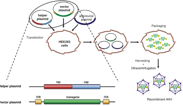
3.1.4 Adenovirus-free AAV production and recombinant AAV vectors (rAAV)
For the production of recombinant AAV (rAAV), the only viral elements required in cis are the ITRs while the two ORFs rep and cap are sufficient when provided in trans (helper plasmid) (Collaco, Cao, and Trempe, 1999). The deleted rep/cap sequences of the parental virus can then be replaced by marker or therapeutic genes resulting in the production of vectors which are unable to replicate even in presence of helper virus (Collaco, Cao, and Trempe, 1999). The flanking ITRs are necessary for packaging into the newly formed capsids. In general, rAAV is produced in a helper virus-free manner to avoid helper virus contaminations of vector preparations. The essential adenoviral genes VA, E2A and E4 have been cloned into an adenoviral helper plasmid and are provided in trans (Collaco, Cao, and Trempe, 1999; Grimm and Kleinschmidt, 1999; Xiao, Li, and Samulski, 1998).
HEK293 cells which are commonly used for the production of viral particles are transgenic for the adenoviral genes E1a and E1b.
The helper, vector and adenoviral plasmids are brought in HEK293 cells by triple transfection (Figure 5). 48h later, viral progeny can be isolated out of the cell lysates and purified by either CsCl or Iodixanol gradient ultracentrifugation (Hermens et al., 1999; Zolotukhin et al., 1999). AAV2 is appropiate for purification directly from crude lysates or from gradient purified fractions by heparin affinity chromatography (Zolotukhin et al., 1999).
All serotypes can be produced as recombinant vectors as described above.
Therefore, only the cap sequence of AAV2 has to be replaced by the serotype-
specific cap. The ITRs as well as the rep ORF are typically derived from AAV2.
This method is called pseudotyping or cross-packaging (Rabinowitz et al., 2002).
Figure 5: Packaging of recombinant AAV vectors. HEK293 cells are transfected by 3 plasmids: A helper plasmid encoding for the rep and cap ORFs, a vector plasmid carrying the desired transgene flanked by the packaging sequences (ITRs) and an adenoviral helper plasmid to provide helper virus functions. After vector assembly, the cells are lysed and rAAV is purified, e.g. by ultracentrifugation. (Figure was kindly provided by H. Büning)
Transduction efficiency in numerous cell lines has been reported to be limited by insufficient second-strand synthesis of the single-stranded (ss) DNA genome (Ferrari et al., 1996; Fisher et al., 1996). This step is necessary to obtain a double- stranded DNA template for initiation of gene expression. Hence, McCarty and colleagues developed a pseudo double-stranded, self-complementary (sc) genome in order to overcome this limitation (McCarty, Monahan, and Samulski, 2001). Their construct contains an extra copy of the palindromic terminal repeat thus enabling the DNA to re-fold and form a duplex DNA (Figure 6). Thereby, the requirement for host cell-mediated second-strand DNA synthesis can be circumvented and high transduction efficiencies are obtained in vitro and in vivo (Hacker et al., 2005; McCarty et al., 2003; Wang et al., 2003). Due to the duplex structure of the self-complementary genome conformation, its packaging capacity
HEK293 cells helper
plasmid
vector plasmid
Transfection
Harvesting + Ultrazentrifugation
Packaging
transgene
rep cap
ITR ITR
Helper plasmid
Vector plasmid Recombinant AAV
adenoviral plasmid
4.6 kb including ITRs. Therefore, self-complementary vectors are not suitable for larger transgenes.
Figure 6: Single-stranded and self-complementary vector genome conformation. On the left side, the natural, single-stranded conformation of AAV is shown. On the right side, the same transgene cassette consisting of a CMV promoter-driven eGFP gene is depicted as a self-complementary DNA. An additional terminal repeat allows folding into a duplex DNA. (McCarty, Monahan, and Samulski, 2001)
3.1.5 AAV as vector in gene therapy
Gene therapy bases on the idea of introducing genetic material into an organism in order to cure or improve the status of a disease. In general, two different systems are applied, the viral and non-viral vectors. Whereas the viral systems include adeno-, retro-, vaccinia-, pox-, herpes simplex- and adeno-associated-viral vectors, the non-viral vector strategy uses naked DNA and lipid- or polyethylenglycol- (PEG) covered DNA (Gould and Favorov, 2003; Minato et al., 2003; Omori et al., 2003).
Ideally, gene therapeutical vectors should combine efficiency and safety. Unique for AAV is that no disease could be related to this virus despite its broad tissue tropism (Berns and Linden, 1995). The transduceability of various cell types including dividing as well as post-mitotic or quiescent cells and differentiated tissues such as brain, muscle, lung and liver, qualifies AAV for a wide range of applications (Alexander et al., 1996; Fisher et al., 1997; Flotte et al., 1993; Kaplitt et al., 1994; Kaplitt et al., 1996; Manno et al., 2006; Podsakoff, Wong, and Chatterjee, 1994). Moreover, AAV has also been shown to mediate long-term expression, e.g. in a muscle-directed trial where transgene expression sustained for more than four years in a canine hemophilia B model (Fisher et al., 1997;
Herzog et al., 1999). As another important aspect, AAV does not need to integrate into the host genome in contrast to lenti- or retroviral vectors. Actually, vector genomes seem more likely to exist as episomes (Duan et al., 1998a; Nakai et al.,
2001). Moreover, in presence of the large Rep proteins, AAV is able to integrate site-specifically into the human chromosome 19 thus minimizing the risk of insertional mutagenesis (Huttner et al., 2003; Kotin et al., 1990). By the development of high titer-reaching helpervirus-free production methods as well as improvements in purification this vector system has become even more attractive.
As recombinant AAV vectors are gutless vectors, they are unable to replicate even in presence of helper virus (Samulski, Chang, and Shenk, 1987).
In general, immunologic reactions to AAV are low. The importance of that aspect becomes evident when AAV is compared to adenovirus which elicits high immune responses (Raper et al., 2003; Zaiss et al., 2002). Apparently, AAV has only a minimal inflammatory potential and seems not to engage pattern recognition receptors like toll-like receptors (TLRs) mediating innate immune responses (Hensley and Amalfitano, 2007; Zaiss et al., 2002). The prevalence of antibodies against AAV2 due to natural infections is as high as 50 to 96 % in the human population. The amount of neutralizing antibodies varies from 18 to 68 % depending on age and ethnic group (Chirmule et al., 1999; Erles, Sebokova, and Schlehofer, 1999; Moskalenko et al., 2000). Animal experiments have confirmed that neutralizing antibodies have strong negative implications on transduction efficiency if the same serotype is reapplied (Scallan et al., 2006). Although human data are limited, at least for one patient in a clinical hemophilia B trial, neutralizing antibodies seem to account for the absence of transgene expression (Manno et al., 2006). Additionally, an anti-capsid response was observed.
Disadvantages of the AAV vector system are its small genome size limiting the coding capacity for transgenes including ITRs to a maximum of 4.1 to 4.9 kb and its broad tissue tropism interfering with a cell-specific in in vivo gene transfer (Dong, Fan, and Frizzell, 1996).
For overcoming pre-existing immune reactions and off-target gene expression, several options are available. The use of serotypes other than AAV2 which show different tropisms and immune responses can be considered and technology, respectively, are likely to cope with these limitations (Buning et al., 2003b; Grimm and Kay, 2003; Limberis and Wilson, 2006; Wu, Asokan, and Samulski, 2006).
Furthermore, several strategies have been developed to overcome the size
limitation (Duan et al., 2001). Overall, AAV is a promising vector for gene therapy as assessed in clinical trials.
Since the first gene therapy clinical trial in 1989, 1308 more studies have been initiated worldwide (http://www.wiley.co.uk/genmed/clinical/). The main focus for gene therapeutical applications is the treatment of cancer with 66.5 % clinical trials followed by cardiovascular (9.1 %) and monogenic (8.3 %) diseases. In most cases, adeno- (24.7 %) or retroviral (22.8 %) vectors find application, while AAV vectors are used only in 3.5 % of the approaches. In complete, 32 clinical trials involving AAV vectors are still open, whereas 14 are already closed. Currently, evaluation of safety of AAV as a vector system is of main interest in clinical trials.
First published data dealt with the monogenic diseases cystic fibrosis and hemophilia B in gene therapy trials. Administration of the cystic fibrosis transmembrane conductance regulator (CFTR) as transgene on the nasal sinus and bronchial epithelium resulted in an improvement of pulmonary function and partial correction of hyperinflammatory responses and electrophysiological defects (Moss et al., 2004; Wagner et al., 1999; Wagner et al., 1998). AAV was approved to be safe in these clinical settings as well as in the treatment of hemophilia B by intramuscular or intrahepatic vector administration (Kay et al., 2000; Manno et al., 2003; Manno et al., 2006). Evidences for transduction were found in all patients of the muscle-directed study and long-term expression of the therapeutic gene, coagulation factor IX (FIX), could be detected albeit at low levels. Highest vector amount administered into the hepatic artery resulted in therapeutic, but transient (<8 weeks) transgene expression levels.
3.1.5.1 Gene therapy of ischemic cardiovascular diseases
As mentioned above, cardiovascular diseases are a main target for human gene therapy. Despite considerable advances in conventional treatment strategies, heart diseases remain the prevalent cause of disability and premature death in the human population (17 million deaths per year) world-wide (World Health Organization 2008). Since organ regeneration, pharmacotherapy and invasive interventions are limited, alternative therapies are urgently needed. Currently, xenotransplantation, gene- and cell-based therapies are the focus of intense investigations. Most efforts in the latter two fields are made on the development of strategies to induce angiogenesis (vessel formation from pre-existing ones) and vasculogenesis (de novo vessel formation) (Khan, Sellke, and Laham, 2003; Melo
et al., 2004). Such therapies could be administered either for protection of myocardium at risk or for rescue after infarction (Khan, Sellke, and Laham, 2003).
Investigations on the molecular and cellular basis of cardiac diseases have identified potential therapeutic genes. The promising therapeutic potential of angiogenic factors such as vascular endothelial growth factor (VEGF), fibroblast growth factor (FGF) or hepatocyte growth factor (HGF) has already been successfully demonstrated in clinical trials (Baumgartner et al., 1998; Grines et al., 2003; Losordo et al., 2002; Morishita et al., 2004). Overexpression of cytoprotective genes like antioxidant genes (e.g. heme oxygenase 1 (HO-1)), survival genes (e.g. Bcl-2, HGF), genes encoding for heat shock proteins and anti- inflammatory cytokines (e.g. IL-4, IL-10, IL-13, TGF-β) as well as inhibition of proapoptotic genes (e.g. Bad) might be useful in myocardial protection as described in various publications and reviewed by Melo and colleagues (Melo et al., 2005).
Generally, gene transfer vectors are administered into the heart tissue by either of the two routes, intracoronary or intramyocardial. Injection into the myocard resulted in local transgene expression in a patchy pattern (French et al., 1994). In order to reach homogenous vector distribution, vector can be applied intracoronary. However, this procedure is limited by the short exposure time of the vector to the endothelium and fast systemic distribution. If transduction of the myocardium is desired, the vector has to overcome the endothelial barrier.
Therefore, novel techniques have been developed to increase endothelial permeability and to prolong exposure time. Capillary-modulating substances like histamine, serotonin or VEGF as well as high intravascular pressure or ultrasound have been described to permeabilize the endothelial barrier (Beeri et al., 2002;
Bekeredjian et al., 2003; Donahue et al., 1998; Logeart et al., 2001). As demonstrated in a rat model, simultaneous clamping of the pulmonary artery and the aorta allowed the adenoviral vector to recirculate in the coronaries over a short period of time and resulted in successful transgene expression (Hajjar et al., 1998). Prolongation can also be reached by hypothermia or cardiac arrest (Ding et al., 2004; Iwanaga et al., 2004).
On the other hand, direct targeting of the endothelium might be favourable in endothelial dysfunction which plays a pivotal role in atherosclerosis, coronary
artery disease and hypercholesterolemia. Under physiological conditions, the endothelium retains numerous prominent roles in maintaining vessel wall homeostasis such as regulation of angiogenesis, thrombolysis, leukocyte adhesion, platelet adhesion and aggregation (Cooke, 2000). Therefore, anti- thrombotic, anti-adhesion (e.g. inhibition of intercellular adhesion molecule 1 (ICAM-1)) or anti-inflammatory genes are considered as therapeutic targets in endothelial dysfunction and should preferentially be expressed by endothelial cells (Vassalli et al., 2003). In diseases associated with high oxidative stress, the overexpression of enzymes that act as anti-oxidants could be beneficial as shown in a rat postmyocardial infarction model using rAAV to deliver heme oxygenase 1 (HO-1) delivery (Liu et al., 2006).
AAV2 was reported to transduce endothelial cells in vitro and in vivo with low efficiency (Nicklin et al., 2001a; Pajusola et al., 2002). Besides AAV, also adeno-, retro- and lentiviruses have been used as gene therapy vectors for cardiac diseases. Additionally to the disadvantages discussed above, the potential of retroviruses is limited as they require dividing cells for efficient transduction, whereas lentiviruses are appropriate vectors for endothelial transduction (Byun et al., 2000; Sakoda et al., 2007). Adenovirus might provoque myocarditis in response to immune reactions and shows only short-term transgene expression (Calabrese and Thiene, 2003; Guzman et al., 1993). In targeting of the myocardium, comparative analyses of the AAV serotypes in mouse and non- human primate models revealed the recently discovered AAV9 as the most efficient serotype (Pacak et al., 2006; Palomeque et al., 2007).
Recently, the identification of stem cells capable of contributing to tissue regeneration has ignited significant interest in the possibility that cell therapy might be used therapeutically for repair of damaged myocardium. Thereby, the combination of cell- and gene-based therapies could result in even higher beneficial effects. This interesting field is discussed separately in chapter 3.2.3.
Transplantations as potential therapy are limited primarily by donor organ shortage and the need for optimal tissue matching to minimize the risk of organ rejection.
Nevertheless, organ recipients require life-long immunosuppression. Currently, researchers are determining the potential and limiting factors of porcine grafts which are functionally and physically closely related to human hearts. In
xenotransplantation and transplantation, gene therapy could allow the production of immunomodulatory proteins locally within the donor graft or the induction of donor-specific tolerance and other mechanisms preventing graft rejection (Chen, Sung, and Bromberg, 2002). Moreover, the identification of beneficial effectors could account for the generation of transgenic animals for xenotransplantation.
Both, innate and adaptive immune reactions are responsible for organ rejection.
The hyperacute rejection occurring within minutes is due to complement- dependent reactions of pre-existing alloantibodies to blood group or major histocompatibility complex (MHC) antigens. As complement-regulatory molecules are working less efficient across species-barriers and recognize directly certain porcine oligosaccharides (e.g. αGal), this step is very problematic for xenografts.
Nevertheless, the main mediators for acute organ rejection are T-cells. They can be either activated directly by donor antigen-presenting cells (APC) or indirectly by the recipient’s APCs which present phagozytosed non-self molecules to T-cells.
Additionally, T-cells play an important role in chronic rejection processes caused by inflammatory vascular injury. Briefly, alloreactive T-cells infiltrate the graft and recruit inflammatory cells by cytokine release and stimulation of endothelial adhesion molecules. Gene therapeutical approaches encompass the inhibition of anti-graft responses and induction of graft protective mechanisms (Chen, Sung, and Bromberg, 2002). As already discussed above, delivery of genes encoding for anti-adhesive, anti-apoptotic, anti-inflammatory and antioxidant proteins might be useful in this regard as well. Moreover, blockage of specific functions of the adaptive immune system showed promising effects. For example, inhibition of the costimulatory signal (CD28-CD80/86) between APC and T-cell by expression of CTLA-4Ig prolongs cardiac graft survival using AAV as vector system (Chen et al., 2003). Expression of immunomodulatory cytokines such as IL-4, IL-10, IL-13 and TGF-β resulted in prolonged allograft survival in various models (Chan et al., 2000;
David et al., 2000; Ke et al., 2000; Ke et al., 2002). Another example is viral IL-10 (vIL-10) which is encoded by the Epstein-Barr virus. It has the same properties like IL-10, but lacks T-cell immunostimulatory functions and has been shown to prolong heart survival time in an adenovirus-mediated vIL-10 rat transplantation model (Zuo et al., 2001).
3.2 Stem cells
By definition, stem cells (SC) are undifferentiated cells able to generate new stem cells of identical differentiation potency or to produce cells that differentiate along a lineage pathway. The first of these two activities of stem cells is described as symmetric, the second as asymmetric division. Both types of cell divisions contribute to the homeostasis of the stem cell population within the stem cell niche, whereas asymmetric divisions are responsible for renewal of the respective, differentiated tissue. In the adult tissue, quiescent stem or progenitor cells are normally mobilized upon stimuli for physiological and pathological tissue regeneration.
Stem cells can be divided into three different types depending on their developmental potential. Embryonic totipotent stem cells are able to differentiate into all embryonic and extra-embryonic cell types (e.g. placenta, umbilical cord) while pluripotent – the so-called embryonic stem cells – possess the ability to generate all tissues of an adult organism. Pluripotent cells give rise to the three types of germ layer stem cells for ecto-, endo- and mesoderm (Figure 7). As development proceeds the differentiation properties get more restricted. The multipotent stem cell gives rise to only a limited number of cell types of fully developed organs maintaining a steady-state homeostasis in the tissue. If only one terminally differentiated cell type can be generated, the cell is referred to as unipotent.
Multipotent and unipotent cells are designated as somatic or adult stem cells and are present in tissues where terminally differentiated cells do not divide or have only a short life span. Indeed, long time it was believed that organs responding poorly to regenerative pressure (e.g. heart, brain) would not reside any stem cells.
Then it turned out that even organs considered as post-mitotic were able to regenerate although at lower levels. Anyway, multipotent stem cells are more abundant in tissues with a high cell turnover rate such as epithelia, vasculature or blood and to a lesser extent in organs or tissues undergoing little self-renewal like the central nervous system or the myocard (Beltrami et al., 2003; Lemoli, 2005;
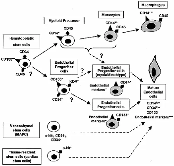
Lois and Alvarez-Buylla, 1993; Oh et al., 2003). The best characterized multipotent cells are hematopoietic stem cells (HSC) which give rise to the entire blood lineages.
Figure 7: Postnatal stem and progenitor cells. SC, stem cell. (Asahara and Kawamoto, 2004)
3.2.1 Hematopoietic stem cells and endothelial progenitor cells
Since blood cells have only a limited life span and essential functions like gas transport, immunity and other vital functions have to be maintained, a continuous production of new cells is needed. The hematopoietic stem cell (HSC) provides all the blood lineage cells and is well described. In the 1960’s first evidence for the existence of clonogenic cells able to generate myeloerythroid cells after bone marrow transplantation in lethally irradiated mice was given (Becker, Mc, and Till, 1963; Wu et al., 1968).
Regarding embryonic development hematopoietic and endothelial progenitor cells share a common mesodermal precursor called hemangioblast. These cells can be found in the extra embryonic yolk sac where they are accumulating in the so-called blood islands. In the inner part of these aggregates cells possess hematopoietic properties whereas the outer cells begin to form endothelial cells (EC) (Asahara and Kawamoto, 2004). These first developing vessels are marking the onset of vasculogenesis. Long time it was believed that this process only takes place during embryogenesis in contrast to angiogenesis, meaning the sprouting of new vessels out of pre-existing ones. Then, in 1997, Asahara and colleagues published the intriguing observation that CD34+ hematopoietic progenitor cells purified from