Improving safety and
establishing episomal maintenance of Adeno-associated viral vectors
I n a u g u r a l - D i s s e r t a t i o n zur
Erlangung des Doktorgrades
der Mathematisch-Naturwissenschaftlichen Fakultät der Universität zu Köln
vorgelegt von Maria Angelika Schnödt
aus Auerbach i.d.OPf.
Köln, 2016
I Berichterstatterin: Prof. Dr. Dagmar Mörsdorf
Prof. Dr. Mirka Uhlirova Prof. Dr. Hildegard Büning
Tag der mündlichen Prüfung: 24.10.2016
II
Kurzzusammenfassung
Adeno-assoziierte virale (AAV) Vektoren sind eine der am häufigsten verwendeten Gentransfersys- teme in der Grundlagen- und der präklinischen Forschung und wurden bereits in mehr als 160 klini- schen Studien angewendet. Üblicherweise werden sie durch Kotransfektion eines sogenannten Vek- torplasmids und zweier, oder wie in meiner Arbeit, eines Helferplasmids in einer Produktionszelllinie hergestellt. Das Vektorplasmid enthält die Transgenexpressionskassette („transgene cassette of inte- rest“ (TEC)) flankiert von den viralen „inverted terminal repeats“ (ITRs), palindromischen Sequenzen, die als Verpackungssignale fungieren, während das Helferplasmid die notwendigen AAV- und Hel- fervirusgene in trans bereitstellt. Ein entscheidender Aspekt der AAV-Vektorologie ist die Herstellung von AAV-Vektoren frei von durch den Produktionsprozess anfallenden Unreinheiten. Zu diesen Un- reinheiten gehören AAV-Kapside, die prokaryotische Sequenzen wie z.B. antibiotische Resistenzge- ne enthalten, die von den Ausgangsplasmiden stammen.
Das Ziel des ersten Teils dieser Arbeit war die Verbesserung der Sicherheit von AAV-Vektoren. Da es nicht möglich ist, in AAV-Kapside verpackte prokaryotische Sequenzen durch Standard- Reinigungsprotokolle zu entfernen, wurde untersucht, ob die Ausgansplasmide für die Vektorproduk- tion durch „Minicircles“ (MCs) ersetzt werden können. MCs sind zirkuläre DNS-Konstrukte die keine funktionalen oder kodierenden prokaryotischen Sequenzen beinhalten; sie bestehen nur aus der TEC und einem kurzen Abschnitt der für ihre Herstellung und Aufreinigung nötig ist. Ein Vektor-MC als Gegenstück für ein Vektorplasmid das für das enhanced green fluorescent (eGFP) Protein kodiert und ein Helfer-MC als Gegenstück für das Helferplasmid welches für die Gene von AAV Serotyp 2 (AAV2) und die Gene des Helfervirus Adenovirus Typ 5 kodiert, wurden von PlasmidFactory (Biele- feld, Germany) entwickelt und produziert. Die vier möglichen Kombinationen von MCs und Plasmiden wurden anschließend verwendet, um einzelsträngige („single-stranded“) AAV2-Vektoren (ssAAV) und self-complementary” AAV-Vektoren (scAAV) herzustellen. Die Vektorpräparationen wurden ge- mäß Vektorquantität, –qualität und –funktionalität charakterisiert. Diese Analysen zeigten, dass die Vektor- und Helferplasmide durch MCs ersetzt werden können, ohne die Effizienz der Vektorproduk- tion oder die Vektorqualität zu verringern. MC-basierte scAAV-Vektorpräparationen wiesen im Ver- gleich zu Plasmid-basierten Präparationen sogar eine bis zu 30-fach verbesserte Transduktionseffi- zienz auf. Durch Verwendung der verschiedenen Kombinationen von Plasmiden und MCs konnte das Vektorplasmid als Hauptquelle der falsch in Kapside verpackten prokaryotischen Sequenzen identifiziert werden. Bemerkenswerterweise beinhalteten die Plasmid-basierten scAAV- Vektorpräparationen eine beträchtlich höhere Menge prokaryotischer Sequenzen (bis zu 26,1 %, im Verhältnis zur TEC) als ssAAV-Vektorpräparationen (bis zu 2,9 %). Durch Ersetzen beider Plasmide durch MC wurde die Menge an kodierenden prokaryotischen Sequenzen unter die Nachweisgrenze reduziert. Weitere Analysen zeigten, dass scAAV-Vektoren im Allgemeinen einen höheren Anteil weiterer DNS-Unreinheiten (wie z.B. adenovirale Sequenzen) als ssAAV-Vektoren aufwiesen. So-
III wohl ssAAV- als auch scAAV-Vektorpräparationen die mit MCs hergestellt wurden tendierten dazu, kleinere Mengen Fremd-DNS zu beinhalten als Vektorpräparationen die mit Plasmiden hergestellt wurden. Keine der getesteten Vektorpräparationen induzierte Immunogenität. Somit lässt sich zu- sammenfassend sagen, dass die Qualität von AAV-Vektorpräparationen signifikant verbessert wird, wenn statt Plasmiden MCs zur Herstellung verwendet werden.
Nach erfolgreicher Zelltransduktion bilden die AAV-Vektorgenome überwiegend doppelsträngige DNS-Ringe oder DNS-Konkatemere aus. Diese episomalen Moleküle persistieren in post-mitotischen Zellen und vermitteln so langfristige Transgenexpression, gehen jedoch in proliferierenden Zellen mit fortschreitender Zellteilung verloren. Für den zweiten Teil dieser Arbeit wurde, in Kooperation mit Claudia Hagedorn und Hans J. Lipps (Universität Witten/Herdecke), ein AAV-Vektor mit einem auto- nom replizierenden Element (Scaffold/matrix attachment region (S/MAR)) ausgestattet. Vektor AAV- S/MAR, kodierend für eGFP und ein Blasticidin-Resistenzgen und ein Kontrollvektor mit der gleichen TEC, aber ohne das S/MAR-Element wurden produziert und in schnell proliferierende HeLa-Zellen transduziert. Durch Inkubation mit dem Antibiotikum Blasticidin wurden die Zellen selektioniert die das Vektorgenom stabil bewahrten. AAV-S/MAR-transduzierte Zellen wiesen eine höhere Anzahl an überlebenden Zellkolonien auf als AAV-ΔS/MAR-transduzierte Zellen. Zellkolonien beider Vektoren wurden isoliert und kultiviert. Sie blieben bis zu 70 Tage (maximaler Kultivierungszeitraum) eGFP- positiv – ohne Selektionsdruck durch Antibiotikagabe. Erstaunlicherweise war die mitotische Stabilität sowohl von AAV-S/MAR als auch des Kontrollvektors AAV-ΔS/MAR ein Resultat episomaler Persis- tenz des jeweiligen Vektorgenoms. Diese Ergebnisse lassen die Annahme zu, dass „gewöhnliche“
AAV-Vektorgenome unter spezifischen Bedingungen, wie durch den verwendeten milden Selektions- druck, episomal persistieren können. Unter diesen Umständen erhöht das S/MAR-Element die Häu- figkeit der Etablierung des stabilen Episoms, ist aber keine Grundvoraussetzung.
IV
Abstract
Adeno-associated viral (AAV) vectors are among the most widely used gene transfer systems in basic and pre-clinical research and have been employed in more than 160 clinical trials. AAV vectors are commonly produced in producer cell lines like HEK293 by co-transfection with a so-called vector plasmid and one (in this work) or two so-called helper plasmids. The vector plasmid contains the transgene cassette of interest (TEC) flanked by AAV’s inverted terminal repeats (ITRs) which serve as packaging signals, whereas the helper plasmid provides the required AAV and helper virus func- tions in trans. A pivotal aspect of AAV vectorology is the manufacturing of AAV vectors free from im- purities arising during the production process. These impurities include AAV vector preparations that contain capsids containing prokaryotic sequences, e.g. antibiotic resistance genes originating from the producer plasmids.
In the first part of the thesis we aimed at improving the safety of AAV vectors. As we found that en- capsidated prokaryotic sequences (using the ampicillin resistance gene as indicator) cannot be re- moved by standard purification methods we investigated whether the producer plasmids could be replaced by Minicircles (MCs). MCs are circular DNA constructs which contain no functional or cod- ing prokaryotic sequences; they only consist of the TEC and a short sequence required for produc- tion and purification. MC counterparts of a vector plasmid encoding for enhanced green fluorescent (eGFP) protein and a helper plasmid encoding for AAV serotype 2 (AAV2) and helper Adenovirus (Ad) genes were designed and produced by PlasmidFactory (Bielefeld, Germany). Using all four pos- sible combinations of plasmid and MCs, single-stranded AAV2 vectors (ssAAV) and self- complementary AAV vectors (scAAV) were produced and characterized for vector quantity, quality and functionality. The analyses showed that plasmids can be replaced by MCs without decreasing the efficiency of vector production and vector quality. MC-derived scAAV vector preparations even exceeded plasmid-derived preparations, as they displayed up to 30-fold improved transduction effi- ciencies. Using MCs as tools, we found that the vector plasmid is the main source of encapsidated prokaryotic sequences. Remarkably, we found that plasmid-derived scAAV vector preparations con- tained a much higher relative amount of prokaryotic sequences (up to 26.1 %, relative to TEC) com- pared to ssAAV vector preparations (up to 2.9 %). By replacing both plasmids by MCs the amount of functional prokaryotic sequences could be decreased to below the limit of quantification. Additional analyses for DNA impurities other than prokaryotic sequences showed that scAAV vectors generally contained a higher amount of non-vector DNA (e.g. adenoviral sequences) than ssAAV vectors. For both, ssAAV and scAAV vector preparations, MC-derived vectors tended to contain lower amounts of foreign DNA. None of the vectors tested could be shown to induce immunogenicity. In summary we could demonstrate that the quality of AAV vector preparations could be significantly improved by re- placing producer plasmids by MCs.
V Upon transduction of a target tissue, AAV vector genomes predominantly remain in an episomal state, as duplex DNA circles or concatemers. These episomal forms mediate long-term transgene expression in terminally differentiated cells, but are lost in proliferating cells due to cell division.
Therefore, in the second part of the thesis, in cooperation with Claudia Hagedorn and Hans J. Lipps (University Witten/Herdecke) an AAV vector genome was equipped with an autonomous replication element (Scaffold/matrix attachment region (S/MAR)). AAV-S/MAR encoding for eGFP and a blasti- cidin resistance gene and a control vector with the same TEC but lacking the S/MAR element (AAV- ΔS/MAR) were produced and transduced into highly proliferative HeLa cells. Antibiotic pressure was employed to select for cells stably maintaining the vector genome. AAV-S/MAR transduced cells yielded a higher number of colonies than AAV-ΔS/MAR-transduced cells. Colonies derived from each vector transduction were picked and cultured further. They remained eGFP-positive (up to 70 days, maximum cultivation period) even in the absence of antibiotic selection pressure. Interestingly, the mitotic stability of both AAV-S/MAR and control vector AAV-ΔS/MAR was found to be a result of epi- somal maintenance of the vector genome. This finding indicates that, under specific conditions such as the mild selection pressure we employed, “common” AAV vectors persist episomally. Thus, the S/MAR element increases the establishment frequency of stable episomes, but is not a prerequisite.
.
VI
Table of Contents
List of figures ... IX List of tables ... X List of abbreviations ... XI
1 Introduction ... 1
1.1 Adeno-associated virus ... 1
1.1.1 Classification ... 1
1.1.2 Genome and proteins ... 2
1.1.3 Life cycle ... 4
1.2 AAV vectorology ... 7
1.2.1 Production ... 8
1.2.1.1 Upstream processing ... 8
1.2.1.2 Downstream processing ... 9
1.2.1.3 Challenges ... 10
1.3 AAV vector transduction and optimization ... 12
1.3.1 Vector tropism and immunogenicity ... 12
1.3.2 Intracellular trafficking ... 13
1.3.3 Vector genome fate in the nucleus ... 13
1.3.3.1 Interaction with DNA damage response proteins ... 13
1.3.3.2 Double strand synthesis ... 14
1.3.3.3 Long-term persistence ... 16
2 Aim of the study... 17
2.1 Aim of study I: Improving safety of AAV vectors ... 17
2.2 Aim of study II: Establishment of episomal maintenance ... 18
3 Materials ... 19
3.1 Chemicals, reagents and enzymes ... 19
3.2 Commercial kits ... 19
3.3 Cell culture media and supplements ... 19
3.4 Plasmids and MC ... 20
3.5 Primers for qPCR ... 21
3.6 Eukaryotic cell lines ... 21
3.7 Bacteria strain... 21
3.8 Laboratory equipment ... 22
3.9 Data treating software ... 23
VII
4 Methods ... 24
4.1 Bacteria Culture ... 24
4.1.1 Cultivation ... 24
4.1.2 Preparation of competent bacteria ... 24
4.1.3 Transformation ... 24
4.2 Nucleic acid techniques ... 25
4.2.1 pDNA amplification and extraction ... 25
4.2.2 DNA extraction from eukaryotic cells ... 25
4.2.3 RNA extraction from eukaryotic cells ... 25
4.2.4 Determination of pDNA and RNA concentration ... 25
4.2.5 Restriction enzyme digest ... 25
4.2.6 Agarose gel electrophoresis ... 25
4.2.6.1 Neutral ... 25
4.2.6.2 Denaturing ... 26
4.2.7 cDNA synthesis ... 26
4.2.8 Quantitative real-time PCR ... 26
4.3 Protein techniques ... 27
4.3.1 ELISA ... 27
4.3.2 Western Blot... 27
4.4 Eucaryotic cell culture ... 29
4.4.1 Cultivation conditions ... 29
4.4.2 Passaging, counting and seeding ... 29
4.4.3 Freezing and thawing ... 29
4.5 AAV vector production ... 30
4.6 AAV vector purification ... 31
4.6.1 Iodixanol density gradient ... 31
4.6.2 Affinity chromatography ... 31
4.6.3 Centrifugal filtration ... 32
4.6.4 Gel filtration ... 32
4.7 AAV vector characterization ... 32
4.7.1 Genomic titer ... 32
4.7.2 Transducing titer ... 32
4.7.3 Capsid titer and composition ... 33
4.7.4 DNA impurity content ... 33
4.8 Cell transduction by AAV vectors ... 33
4.8.1 TLR9 activation assays ... 33
4.8.1.1 Quantification of SEAP protein levels ... 33
4.8.1.2 Quantification of IL8 transcription levels ... 33
4.8.2 Colony forming assay ... 33
VIII
4.9 Statistical Analysis ... 34
5 Results ... 35
5.1 Improving the purity of AAV vector preparations using DNA Minicircle (MC) technology ... 35
5.1.1 Encapsidated prokaryotic sequences in AAV vector preparations cannot be removed by standard purification methods ... 35
5.1.2 Evaluation of MC constructs for ssAAV vector production ... 37
5.1.3 Vector plasmids are the main source of encapsidated prokaryotic sequences ... 41
5.1.4 Proximity to ITRs rather than a specific sequence element is decisive for packaging of backbone sequences ... 41
5.1.5 Self-complementary AAV (scAAV) vector preparations produced by MC constructs show improved transduction efficiencies and contain no ampR DNA impurities ... 43
5.1.6 Further characterization of plasmid and MC-derived ssAAV and scAAV vectors ... 46
5.1.7 AAV vectors do not activate Toll-like receptor 9 in vitro ... 48
5.2 Establishment of episomal maintenance of AAV vectors ... 51
5.2.1 Colony forming assay ... 51
5.2.2 Mitotic stability of AAV-S/MAR vector genomes in HeLa cells ... 53
5.2.3 Mitotic stability of AAV-∆S/MAR vector genomes in HeLa cells ... 57
6 Discussion ... 60
6.1 MC technology for AAV vector production ... 60
6.1.1 Prokaroytic DNA is encapsidated during AAV vector production ... 60
6.1.2 Comparison of plasmid and MC-derived AAV vector preparations ... 60
6.1.3 Model for packaging of prokaryotic sequences ... 61
6.1.4 DNA impurities in AAV vector preparations ... 64
6.1.5 Interaction of AAV vectors with the innate immune system ... 65
6.1.6 Challenges and chances of using MC for AAV vector production ... 66
6.2 Episomal maintenance of AAV vectors ... 67
6.2.1 S/MAR-based vectors for gene therapy ... 67
6.2.2 Episomal AAV-S/MAR and AAV-ΔS/MAR vector genomes ... 68
6.2.3 Conditions for establishment of stable AAV vector episomes ... 69
6.2.4 Outlook ... 70
7 References ... 72
Appendix ... 94
Danksagung ... 98
Erklärung ... 99
IX
List of figures
Figure 1 Genome organization of AAV2. ... 2
Figure 2 Capsid of AAV2. ... 4
Figure 3 Schematic model of AAV2 infection. ... 6
Figure 4 AAV vector production by transient transfection. ... 8
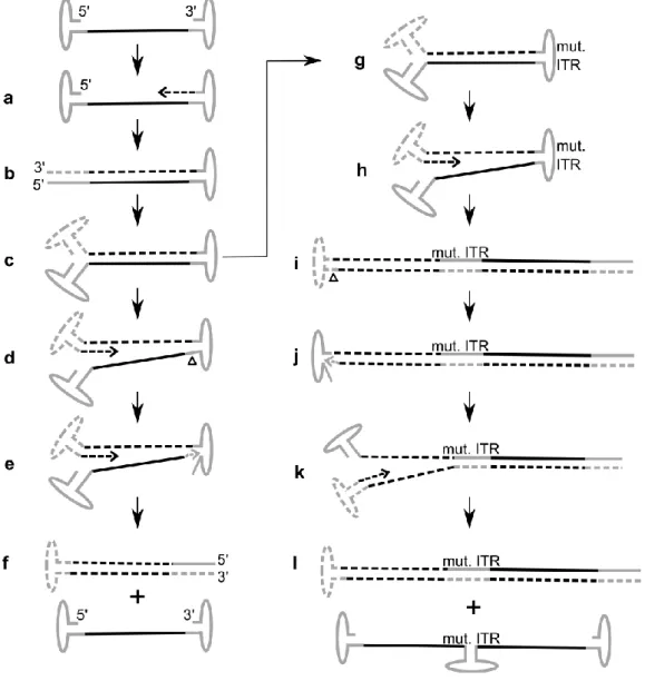
Figure 5 AAV genome replication and generation of self-complementary AAV vector genomes. ... 15
Figure 6 Antibiotic resistance gene (ampR) sequences in standard AAV vector preparations. ... 36
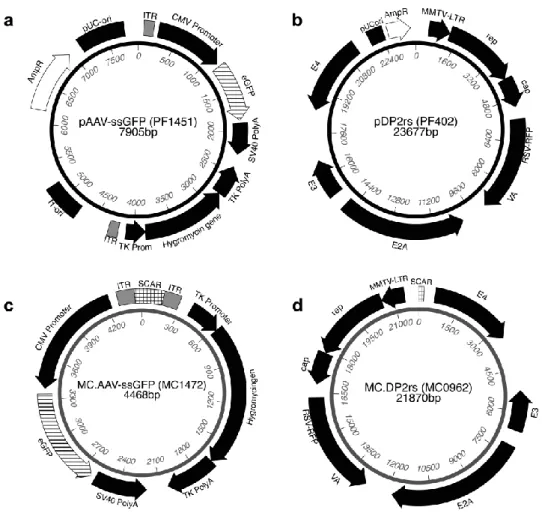
Figure 7 ssAAV vector and AAV/Ad helper plasmids and thereof derived MC constructs. ... 37
Figure 8 Western Blot analysis of ssAAV vector preparations. ... 39
Figure 9 ampR sequences in ssAAV preparations. ... 41
Figure 10 scAAV vector plasmid and thereof derived MC construct. ... 43
Figure 11 ampR sequences in scAAV preparations. ... 46
Figure 12 Analysis of ssAAV and scAAV vector genomes by denaturing gel electrophoresis. ... 47
Figure 13 SEAP production of HEK blue hTLR9 cells upon transduction. ... 49
Figure 14 IL8 gene expression of HEK blue hTLR9 cells upon transduction. ... 50
Figure 15 Plasmid maps of pAAV-S/MAR and pAAV-ΔS/MAR. ... 51
Figure 16 Cultivation of AAV-S/MAR and AAV-ΔS/MAR-transduced cells in absence of selection. .. 52
Figure 17 Colony count of blasticidin-resistant cells. ... 53
Figure 18 Transgene expression of AAV-S/MAR-derived colonies. ... 53
Figure 19 Long-term transgene expression of AAV-S/MAR-derived colonies in absence of selection. ... 54
Figure 20 Episomal maintenance of AAV-S/MAR vector genomes in HeLa cells (I). ... 55
Figure 21 Episomal maintenance of AAV-S/MAR vector genomes in HeLa cells (II). ... 56
Figure 22 Transgene expression of AAV-ΔS/MAR-derived colonies. ... 57
Figure 23 Long-term transgene expression of AAV-∆S/MAR-derived colonies in absence of selection. ... 57
Figure 24 Episomal maintenance of AAV-∆S/MAR vector genomes in HeLa cells (I). ... 58
Figure 25 Episomal maintenance of AAV-∆S/MAR vector genomes in HeLa cells (II). ... 59
Figure 26 Model of proposed rescue mechanism. ... 63
X
List of tables
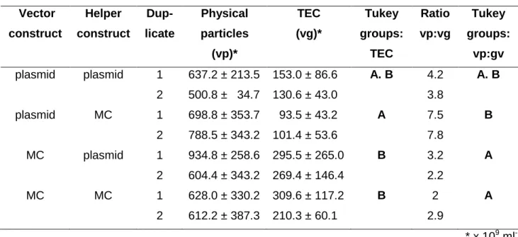
Table 1 Particle titer and packaging efficiency of ssAAV vector preparations. ... 38
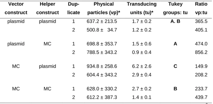
Table 2 Transducing titer and transduction efficiency of ssAAV preparations. ... 40
Table 3 Quantification of prokaryotic DNA in the dual plasmid preparations. ... 42
Table 4 Quantification of SCAR sequences in ssAAV vector preparations. ... 42
Table 5 Particle titer and packaging efficiency of scAAV vector preparations. ... 44
Table 6 Transducing titer and transduction efficiency of scAAV vector preparations. ... 45
Table 7 Further DNA impurities in ssAAV and scAAV vector preparations. ... 48
XI
List of abbreviations
°C Degree celsius HEPES (4-(2-hydroxyethyl)-1-
piperazineethanesulfonic acid ) µ (g,l,M) micro (gram, liter, mole/molar) HP Hairpin
A Ampere HRP Horseradish peroxidase
Å Ångström HSPG Heparan sulfate proteoglycan
AAP Assembly-activating protein HSV Herpes simplex virus AAV Adeno-associated virus i.e. lat. Id est, engl. That is AAVS1 AAV integration site 1 IDGC Iodixanol density gradient cen-
trifugation
abs. Absorption IL Interleukin
Ad Adenovirus IRES Internal ribosome entry site
ampR Ampicillin resistance gene ITR Inverted terminal repeat
ANOVA Analysis of variance IU International units
AP-1 Activator protein 1 kanR Kanamycin/neomycin re-
sistance gene
APS Ammonium persulfate kb Kilo bases
bp Base pair L Liter
cDNA complementary DNA LB Lysogeny broth
CMV Cytomegalovirus m (g, l, M) milli-(gram , liter, mole) DMEM Dulbecco's modified eagle medium MC Minicircle
DMSO Dimethyl sulfoxide MFI Mean fluorescence intensity
DNA Deoxyribonucleic acid min Minutes
DNase Deoxyribonuclease n.s. Not significant
ds Double-strand NF-κB Nuclear factor kappa B
DTT Dithiothreitol NPC Nuclear pore complex
E.coli Escherichia coli OD Optical density
e.g. lat. Exempli gratia, engl. For example ORF Open reading frame EDTA Ethylenediaminetetraacetic acid ori Origin of replication eGFP enhanced green fluorescent protein p.t. Post-transduction
ELISA Enzyme-linked immunosorbent assay PBS Phosphate-buffered saline FACS fluorescence-activated cell sorting pDNA plasmid DNA
FCS Fetal calf serum PLA2 Phospholipase A2
FISH Fluorescence in situ hybridization PLAT Tissue plasminogen activator GAPDH Glycerinaldehyd-3-phosphat-
Dehydrogenase qPCR quantitative polymerase chain
reaction
GOI Genomic particles per cell rAAV recombinant adeno-associated virus
h, hrs Hour, hours RFP Red fluorescent protein
HBS HEPES-buffered saline RNA Ribonucleic acid
XII
rpm Rounds per minute Tris Tris(hydroxymethyl)aminomethane
RT Room temperature trs Terminal resolution site
RT-PCR
Reverse transcriptase polymerase chain
reaction U/µl Units per µl
sc Self-complementary UV Ultraviolet light
SCAR
Sequence for chromatography, affinity and
recombination V Volt
SDS Sodium dodecyl sulfate vg Vector genomes
SEAP Secreted embryonic alkaline phosphatase VP Viral protein
ss Single-stranded wt Wild type
TAE Tris-acetate-EDTA x g Gravitational force
TEMED Tetramethylethylenediamine Δ, delta Deletion TLR Toll-like receptor
XIII
Introduction
1
1 Introduction
1.1 Adeno-associated virus
1.1.1 Classification
Adeno-associated virus (AAV) belongs to the parvovirus family (Parvoviridae), which comprises all small, isometric, non-enveloped DNA viruses with a linear single-stranded genome. Parvoviruses are divided into the subfamilies Parvovirinae, infecting vertebrates, and Densovirinae, infecting arthro- pods.1 With capsid diameters of just around 25 nm, Parvoviridae contain only a short DNA sequence of about 5 kb. Because of the resulting genetic simplicity Parvoviridae are greatly dependent on their host cell to support their life cycle. AAV additionally relies on functions provided by more intricate helper viruses, such as adenovirus (Ad), human papillomavirus (HPV), and members of the herpes virus family, including herpes simplex virus (HSV) -1 and -2, human cytomegalovirus, Epstein-Barr virus and varicella virus to foster its propagation.2–6 Thus, AAV is classified in the genus Dependo- virus within the Parvovirinae.
Since its discovery in 1965 2, 7 at least twelve AAV serotypes differing in tissue tropism and originat- ing from humans and non-human primates have been described.8 Moreover, more than 100 AAV genomic variants have been found in human and non-human primate tissues.9 This reflects the wide- spread dissemination of AAV: up to 80% of humans (population-to-population variation, region- dependent) possess anti-AAV antibodies to serotypes 1, 2, 3 and 5, with up to 60 % harboring neu- tralizing antibodies.10, 11 Given the high prevalence of AAV, reports of AAV-induced pathology are scarce. Except for reports associating early AAV infection in pregnancy with spontaneous abortion,12 AAV is considered as non-pathogenic.13 Recently, Nault and colleagues found integrated AAV2 se- quences in hepatocellular carcinoma driver genes, which led them to propose that insertional muta- genesis of AAV2 may cause malignant transformation in the liver.14 Yet, in the meantime, this report has been challenged regarding methodology of the analyses, the conclusions drawn from the data, and the implications of the findings.15–17 On the other side, researchers have put forward that AAV may actually be beneficial for its host. This hypothesis is based on the finding that AAV inhibits repli- cation of its pathogenic helper viruses.18, 19 Indeed, possibly due to AAV impeding replication of HPV, serological studies reported a negative correlation of AAV with cervical carcinoma.13, 20
Introduction
2 1.1.2 Genome and proteins
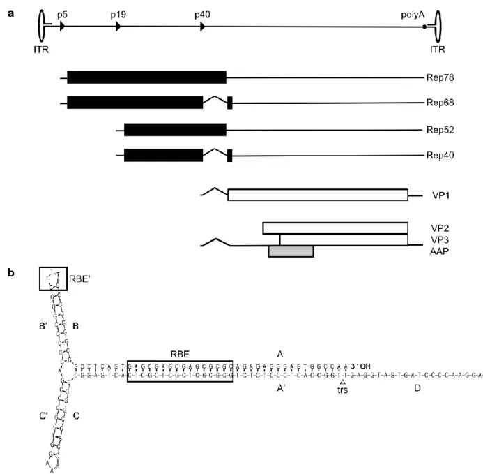
Limited to a genome size of a mere 4.7 kb, AAV2 evolved alternative splicing and alternative open reading frames (ORF) to increase its coding capacity. Structurally, the AAV2 genome contains ORFs rep, cap and AAP in sense orientation, flanked by inverted terminal repeats (ITRs) (Figure 1).
Figure 1 Genome organization of AAV2. (a) The single stranded DNA genome of AAV2 has a length of 4.7kb, divided into 100 map units. The positions of promoters p5, p19 and p40 (named according to their map position) are indicated as solid triangles, the polyadenylation signal (polyA) at position 96 is indicated as solid circle. The inverted terminal repeats (ITRs) are represented by ellipses (not to scale). The transcripts from the three promoters are shown below the genome map. Open reading frames (ORF) are depicted by rectangles (in black for rep ORF, white for cap ORF 21 and light grey for AAP ORF 22), untranslated regions by thin solid lines and introns as nicks. (b) Secondary structure of an AAV2 ITR. The AAV2 ITR is composed of two palindromic regions (B-B’ and C-C’) within a stem palindrome (A-A’), and a single-stranded D region. The ITR can fold in a flip (depicted here) or flop conformation, with the B-B’ or C-C’ palindrome closest to the 3’ end, respectively. The rectangles mark the binding motif for Rep proteins at the A-A’ stem (Rep-binding element, RBE) and the apex of the ITR hairpin (HP) structure (RBE’).23–25 The triangle indicates the terminal resolution site (trs), the specific nicking site of Rep protein. Depiction of the ITR structure is based on Mfold analysis.26
Introduction
3 The ITRs consist of two 125 nucleotide (nt) palindromes with six segments (A-A’, B-B’, C-C’) forming a T-shaped hairpin (HP) structure, and a 20 nt D sequence (Figure 1b). AAV’s origin of replication, the terminal resolution site (trs), is located between the A and D sequences.27, 28 The ITRs act as self- priming HP during genome replication 29 (see Figure 5) and as signals for packaging and integra- tion.30–32
The rep ORF codes for four non-structural proteins named Rep78, Rep68, Rep52 and Rep40, ac- cording to their molecular mass (Figure 1a). Transcription of Rep78 and Rep68 is initiated at promot- er site p5, while transcription of Rep52 and Rep40 starts at promoter p19. The smaller Rep proteins of each transcript, Rep68 and Rep40 are generated by splicing of the same intron sequence.21 The large Rep proteins, Rep78 and Rep68, which possess DNA binding, helicase and site-specific endo- nuclease activity, are the main protagonists of transcriptional regulation, AAV DNA replication and site-specific integration.33–38 Rep52 and Rep40 do not seem to be required for DNA replication,39 but are necessary for generation or accumulation of replicated single-stranded viral DNA in the host cell.40 Ultimately, Rep52 and Rep40 mediate the packaging of viral DNA into the preformed cap- sids.32, 41 The cap ORF encodes the capsid structural proteins VP1, VP2 and VP3. They all share a common C-terminus and the complete VP3 sequence of 62kDa. VP1 (87kDa) and VP2 (73kDa) pos- sess additional N-terminal sequences. The N-terminus of VP1 contains motifs which are required for infection (see below); whereas to date, no function of VP2’s N-terminus has been determined.42 Since VP1 is the product of a minor splice transcript and the initiation codon of VP2 is a non- traditional ACG which is frequently skipped, both VP1 and VP2 are translated at lower levels than VP3. Therefore, the AAV capsid consists of 90% VP3, with VP1 and VP2 contributing the remaining 10%.43, 44 Nested within the cap ORF lies an alternative ORF which codes for the assembly activating protein (AAP).22 AAP targets capsid proteins to the nucleolus and interacts with capsid proteins to provide an assembly scaffold.45
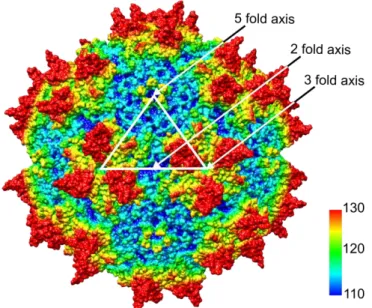
The AAV capsid is formed by 60 subunits as an icosahedron,46 which is characterized by three levels of rotational symmetry, namely fifteen 2-fold axes, ten 3-fold axes and six 5-fold axes (Figure 2). The outward protrusions (red in Figure 2) and adjacent plateau regions at the 3-fold symmetry axes de- fine the interaction of the AAV capsid with cell receptors and represent the capsid’s epitope struc- tures.10, 47–52 At the 5-fold axes, pentameric pores are located (surrounded by elevated rim structures, depicted in yellow in Figure 2). These pores function as channels connecting the capsid inside to the environment and act as portals for externalization of the N-terminal sequences of VP1 and packaging of progeny AAV genomes into the pre-formed capsids.32, 53
Introduction
4
Figure 2 Capsid of AAV2. The capsid is shown as space-filling surface representation model with coloring of the amino acids according to the relative distance from the center of the capsid (blue-cyan-green-yellow-red: ~ 110-130 Å). The white triangle indicates the approximate boundary of one viral asymmetric subunit (60 of which compose the capsid).The apex of the triangle touches a pentameric pore at a 5-fold symmetry axis. The outward vertices touch the protrusions (at the right) and plateau regions (at the left) of a 3-fold symmetry axis. The midpoint of the base side touches a 2-fold symmetry axis.
This model was generated from the crystal structure of AAV2 (RCSB PDB #1LP3)46 using Chimera software.54
Research into the transcriptome and proteome of AAV is ongoing. Recently, further gene products involved in different stages of AAV’s life cycle have been proposed, such as an additional ORF termed X under the control of a promoter at map position 81, as well as novel transcripts and splice variants, which are involved in specific stages of viral replication.19, 55
1.1.3 Life cycle
AAV harnesses different types of glycans as primary receptors for initial attachment to the host cell, followed by interaction with a secondary receptor which facilitates cell entry. AAV2 uses heparan sulfate proteoglycan (HSPG) as attachment receptor.56 HSPG binding possibly triggers changes in capsid conformation, thus increasing the affinity to a receptor which mediates uptake.57 As secondary receptors, fibroblast growth factor receptor 1 (FGFR-1),58 hepatocyte growth factor receptor (HGFR)59 and laminin receptor,60 and integrins αvβ5 61 and α5β1 57 have been proposed, although at least the role of αvβ5 integrin, FGFR-1 and HGFR in AAV2 cell entry has been challenged.62, 63 For other AAV serotypes different sets of primary and secondary receptors were described, thus accounting for the differences in tissue tropisms.8
Very recently, Pillay et al. identified a previously unknown receptor which is essential for AAV infec- tion in a range of mammalian cell types: transmembrane protein KIAA0319L, which was termed AAV
Introduction
5 receptor (AAVR). AAVR is assumed to function as universal receptor for AAV, as infection by sero- types 1-9 is dependent on its presence.63 It is not clear yet which ubiquitous viral structures partake in the AAV-AAVR interaction or at which stage of infection this interaction takes place. AAVR could act as cell entry receptor, either as monomer, dimer or in a multimeric complex with other receptors, or promote later steps in the virus life cycle.64
The major pathway for AAV2 internalization is believed to be clathrin-mediated dynamin-dependent endocytosis.65, 66 In addition, AAV is capable of exploiting several other entry strategies, such as the clathrin-independent carrier mediated/glycosylphosphatidylinositol-anchored protein- enriched endo- somal compartment-associated (CLIC/GEEC) endocytic pathway and macropinocytosis.67–70 For ef- fective infection AAV processing to an acidified compartment is essential, irrespective of the route of cell entry.65 In the course of a productive pathway, AAV travels in endosomes toward the nucleus via the cytoskeletal network.67, 68, 71–73 AAV trafficking to the trans-Golgi network (TGN) and the Golgi apparatus has been proposed,63, 74, 75 although this also may not be an absolute requirement.64 Grad- ual acidification of the endosomal compartment, possibly with assistance of cellular proteases such as cathepsins B and L,76 induces conformational changes of the AAV capsid. Thus, the N-terminus of VP1 (VP1 unique region, VP1u) is exposed to the capsid outside.77 VP1u contains a phospholipase A2 (PLA2) domain78 and nuclear localization signals.79, 80 The PLA2 domain then mediates virion es- cape from the endosome into the cytoplasm.81 Translocation into the nucleus likely occurs through nuclear pore complexes (NPC)65,82 in an active transport process supported by cellular karyopher- ins,83 although NPC-independent entry mechanisms have been proposed as well.84 Several studies suggest that AAV2 enters the nucleus as intact particle,65, 77, 85 while other researchers proposed that uncoating and release of the viral genome already occurs before or during nuclear entry.86
Introduction
6
Figure 3 Schematic model of AAV2 infection. (a) AAV2 binds to heparan sulfate, a glycan chain of heparan sulfate pro- teoglycan (HSPG) which serve as AAV2’s primary receptor. This binding possibly induces conformational changes to the capsid. Binding to an uptake receptor (see text) initiates endocytosis. The major pathway for AAV2 endocytosis is believed to be clathrin-mediated and dynamin-dependent; but additional other entry mechanisms have been reported. (b) AAV2 traffics toward the nucleus through the endosomal system. Sorting toward the trans-Golgi network (TGN) has been pro- posed, but may not be an absolute requirement and/or cell-type specific. (c) Acidification of the endosome triggers the ex- ternalization of VP1u (VP1 unique region) which contains a phospholipase domain (PLA2) and nuclear localization signals.
PLA2 mediates endosomal escape into the cytoplasm. (d) Nuclear localization signals target the virion to the nucleus where entry likely occurs via the nuclear pore complex (NPC). It is not yet clear whether AAV2 uncoats in the nucleus or before or during nuclear entry.
Upon arrival in the nucleus the further fate of AAV depends on whether a helper virus co-infects. In the absence of helper viral functions, only minimal gene expression takes place and Rep gene prod- ucts negatively regulate further gene expression and viral DNA replication.37 AAV establishes a latent
Introduction
7 infection by persisting episomally 87 or by integration into the host genome. Thereby, AAV2 is unique among eukaryotic viruses as it exhibits a preference for integration in a specific locus, on chromo- some 19 (19q13.4)88 at a site termed AAVS189 possessing homology to sequences within the ITR.
This process of targeted integration is mediated by Rep78 and Rep68.31, 90 Super-infection with a helper virus creates a cellular environment which favors AAV replication and propagation. Subse- quently AAV enters the productive phase of infection: the suppression of AAV gene expression is relieved, the viral genome is rescued from the integrated state, DNA replication ensues (Figure 5a-f) and progeny viruses are produced.37, 38, 91
Even in absence of helper virus infection, however, the factors required for AAV replication can be provided by specific cellular conditions. Such conditions include cellular stress induced by genotoxic treatments, such as UV irradiation 92 or carcinogenic agents.93, 94 Furthermore, autonomous replica- tion of AAV has been observed in differentiating keratinocytes.95
1.2 AAV vectorology
AAV’s hallmark features of non-pathogenicity and ability to persist in a host cell, in addition to its abil- ity to produce high yields of progeny, prompted interest in exploring AAV as gene delivery platform.96 By 1982 the complete AAV2 genome had been cloned into a plasmid vector. This enabled production of AAV in Ad-infected cells upon transfection.97 First experiments with a recombinant AAV (rAAV) in which the cap/AAP gene was replaced by a neomycin resistance cassette showed that rAAV could be used to transduce non-AAV DNA into mammalian cells.98 Subsequently, it was demonstrated that the ITRs are the only viral sequences which are required in cis to generate rAAV particles, while rep and cap/AAP functions can be provided in trans.30 This finding paved the way for the development of
“gut-less” AAV vectors containing no coding viral sequences. Instead, a transgene cassette of inter- est (TEC) of up to 4.9kb could be placed between the ITR sequences and be packaged as genetic payload into the viral vector particle.99 Today, AAV vectors are considered as one of the most promis- ing delivery systems in human gene therapy, especially for in vivo administration into post-mitotic tissues, such as liver, muscle, eye and brain.100–103 More than 160 clinical trials using AAV vectors have been conducted.104 In 2012 Glybera® (alipogene tiparvovec) an rAAV1-based therapeutic en- coding a hyperactive variant of lipoprotein lipase (LPLS447X)105 for treatment for lipoprotein lipase defi- ciency was granted marketing authorization by the European Medicines Agency as first gene thera- peutic medicine.106
Introduction
8 1.2.1 Production
The production process is a critical aspect of AAV vectorology. To obtain AAV vectors suitable in quality and quantity for gene therapy applications different strategies are employed for production und purification.
1.2.1.1 Upstream processing
The upstream process of AAV vector production encompasses the bioprocess, that is, all steps of cell culture work up to harvesting of producer cells. At laboratory scale, transient transfection is the most commonly used method, as it is easily set up and flexible to changes of vector genome and capsid (Figure 4). The vector plasmid containing the TEC flanked by the ITRs serves as template for vector genome replication. AAV genes rep and cap/AAP are provided in trans by co-transfection of an AAV helper plasmid. Earlier in development of AAV vector production, host cells were infected with Ad helper virus to initiate vector replication.107 This created the need to remove infectious Ad contaminants in AAV vector stocks, in addition to Ad infection diminishing host cell viability. To obvi- ate helper virus infection, plasmids have been constructed, which encode the Ad genes E2A, E4 and VA RNA,108 or which contain both Ad and AAV helper genes on a single AAV/Ad helper plasmid.109 For DNA transfer, the most commonly used methods are DNA co-precipitation with calcium phos- phate, polycations such as polyethylenimine or cationic lipids.110 As producer cells, HEK293 are usu- ally used. They have been transformed with Ad5 DNA and stably express E1A and E1B,111 and thus complete the essential set of Ad functions. Inherently, transfection methods based on adherent HEK293 cells are challenging to scale-up. To generate a more readily scalable production platform Grieger et al. recently adapted HEK293 to grow in suspension.112
Figure 4 AAV vector production by transient transfection. (a) HEK293 cells are transfected with the vector plasmid containing the TEC flanked by ITRs and plasmids providing AAV and Ad helper functions. (b) Upon replication, the AAV vector genome is rescued from the plasmid, and the capsids are assembled. (c) The vector genomes are packaged into the capsids. Cells are harvested and lysed 48 hrs after transfection. Figure shows schematic depiction of a cell with no distinc- tion between cytoplasm and nucleus.
Introduction
9 Other production strategies include generation of stable cell lines. So-called packaging cell lines con- tain rep and cap/AAP as integrated copies, while so-called producer cell lines further contain the vec- tor genome to be packaged. AAV vector production is initiated by infection with a helper virus, usually Ad, and, in case of packaging cell lines, by co-infection with an additional virus providing the vector genome.113 Further, HSV’s ability to provide helper functions is harnessed for manufacturing of AAV vectors. Commonly, mammalian cells are simultaneous infected with two replication-deficient recom- binant HSV, one carrying AAV genes and one transporting the vector genome.114 As yet another concept, a baculovirus (BV)-based platform in insect cells was implemented. For AAV vector produc- tion, one or two BVs encoding the AAV genes under the control of BV-specific promoters and a BV containing the TEC are co-infected, commonly in Sf9 (from Sodoptera frugiperda).113 Alternatively, stable Sf9 cell lines have been developed. They contain integrated copies of AAV genes which are activated upon infection with the BV containing the TEC.115 A BV-expression vector system has been used for the production of Glybera. All methods relying on helper virus infections, albeit scalable, maintain the risk of contaminating helper virus in the AAV vector preparations.
1.2.1.2 Downstream processing
The downstream process encompasses the recovery of AAV vectors from the producer cells and subsequent purification protocols. The specific goal is the removal of process- and product-related impurities. Process-related impurities stem from the materials and components used for manufacture, including host cell nucleic acids and proteins, residue plasmid DNA, components of the cell culture medium and buffers. Product-related impurities are structurally similar to the vector product, but do not meet the specifications for safety and efficacy. Such impurities are empty AAV capsids, AAV capsids containing DNA other than the TEC or replication-competent AAV.116
In the first step, upon harvest of producer cells, host cell membranes are disrupted to release the produced AAV vectors into a cell lysate. At laboratory scale, cells are commonly subjected to repeti- tive freeze thaw cycles or ultrasonication. Next, the vector-containing lysate is incubated with a nu- clease to remove contaminating nucleic acids from the production process, followed by removal of cellular debris by centrifugation. In some protocols a precipitation step using polyethylene glycol be- fore or after nuclease treatment is included.117, 118 Large scale approaches employ chemical agents such as surfactants or mechanical protocols such as microfluidizers or homogenizers and clarify the lysate by microfiltration.113 Subsequent purification methods harness specific characteristics of AAV particles for their isolation. As the AAV capsid provides high particle stability in temperatures up to 65°C 119 and pH between 2.5 and 8,120 vector preparations can sustain sophisticated purification pro- tocols.
Introduction
10 At laboratory scale, vector-containing cell lysates are commonly purified by density gradient ultracen- trifugation: The cleared lysate in a centrifuge tube is mixed with cesium chloride (CsCl) or sub- layered with solutions containing increasing concentrations of iodixanol. While CsCl is a cytotoxic agent and must be removed carefully, iodixanol is a non-toxic contrast agent,121 which is tolerated by some cell types. After applying high centrifugal force, TEC-containing AAV particles accumulate in specific phases of density and can be harvested. Density gradient ultracentrifugation is applicable to any AAV serotype. Importantly, this method removes a large portion of empty capsids from the vector lysate, as these accumulate in phases of lower density.118, 121 For further purification, or as alternative approaches, column-based protocols have been implemented, many of which are commercially available. Affinity chromatography utilizes the chemical properties of the capsid surface, such as the heparin binding of AAV2, to enrich vector particles.121 Further, an immunoaffinity column (AVB col- umn) carrying an anti-AAV single domain antibody specific for a surface-exposed epitope region on the capsid which is common to several serotypes122 has been developed. Ion-exchange chromatog- raphy is based on the isoelectric point (IEP), i.e. the pH at which a particular molecule has a neutral net charge. When the pH is lower than the IEP of a target molecule, it is protonated and binds to a cation exchanger (a negatively charged ion exchange resin, such as sulfonic acid groups). With the buffer pH value approaching the IEP, the target molecule’s net change becomes charge-neutral which allows it to dissociate from the resin and elute from the column. Vice versa, the target molecule carries a negative charge when the pH is higher as it its IEP and thus binds to a positively charged anion exchange resin, such as quaternary ammonium groups. By lowering the pH to the specific val- ue of IEP, elution takes place. As empty AAV capsids have an IEP of 6.3, as opposed to the IEP of 5.9 of capsids containing DNA,123 ion-exchange chromatography is broadly used to specifically enrich the latter. Different protocols using cation exchanger resin, anion exchanger resin, two-step and dual exchange membrane chromatography have been described.120, 124–128 Another advantage of this method is the broad applicability to different serotypes. Other column-based methods rely on capsid modifications, such as genetic insertion of histidine-tags (his-tags). Capsids with inserted his-tag are purified using immobilized metal affinity chromatography (IMAC) with nickel columns.129, 130
As final polishing methods, size-exclusion chromatography (e.g. gel filtration), ultrafiltration or tangen- tial flow filtration are commonly used for buffer exchange or to concentrate the vector suspension.
The final AAV vector product can be stored frozen or even lyophilized without losing infectivity.131
1.2.1.3 Challenges
As mentioned above, vector preparations may contain process- and product-related impurities. In an assessment of type and significance of process-related impurities, J. Fraser Wright concludes that these are similar to those arising in the production of established biologics, and can therefore be re-
Introduction
11 solved by appropriate established procedures. Product-related impurities, in contrast, are unique to AAV vector production, pose specific risks to safety and efficacy and so closely resemble the vector product as such, that it becomes challenging to remove them.116
Regardless of production method, empty capsids are present in abundance in AAV vector prepara- tions, constituting <50 to > 98 % of total AAV particles.128, 132, 133 The variation in relative empty capsid content depends on size and sequence of vector genome, cell culture system or transfection efficien- cy; additionally, since lot-to-lot variability has been observed, other, yet unidentified factors are in involved.126, 132 For most applications, the presence of large numbers of empty capsids is highly un- desirable, as they form a source of antigenic material which may induce or contribute to a capsid- triggered anti-AAV immune response.134, 135 Moreover, they may competitively inhibit transduction and induce capsid particle aggregation.126 As empty capsids differ from DNA-containing capsids in density and IEP, they can be largely removed by density centrifugation and ion exchange chromatog- raphy.
Additionally, capsids containing non-vector DNA are present in vector preparations. Replication- competent AAVs (rcAAV) are capsids containing the wild type (wt) AAV rep and cap/AAP genes flanked by ITRs. They are generated by recombination events between the ITRs of the vector ge- nome cassette with rep and cap/AAP sequences provided on helper constructs. Indeed, wtAAV is largely regarded as non-pathogenic (see 1.1.1). Yet, unintended transfer of viral DNA may result in production of AAV proteins, which possess helicase or DNA nickase activity,33, 34 or induce cytotoxic T lymphocyte reactions.136 Several strategies to prevent the generation of rcAAV have been reported, including the elimination of sequence homologues within vector and helper plasmids,137 replacing the p5 promoter which is implicated in recombination events,109 placing rep and cap/AAP genes in op- posing transcriptional orientations138 or generating an oversized rep-cap helper plasmid.139 Further, fragments of host cellular DNA and helper sequences may get packaged into capsids. In Glybera, for example, encapsidated baculovirus DNA stemming from the production system was specified as im- purity and as of major concern in the EMEA assessment report.140
In vectors derived from plasmid transfection, encapsidated prokaryotic backbone sequences, e.g.
antibiotic resistance genes have been reported as substantial impurity, ranging from 1-8 % of pack- aged genomic particles.116, 141–143 Although prokaryotic promoter sequences are not functional in mammals, transfer of plasmid backbone sequences should nevertheless be avoided, since they con- tain motifs that are recognized by the cell-autonomous immune system and are thus prone to induce inflammatory responses or gene silencing.144, 145 In addition, antibiotic resistance genes have been found to get integrated into the target genome,141, 146 thus bearing the risk to come under the control of eukaryotic promoters. In an approach to reduce the packaging of prokaryotic sequences Hauck and colleagues inserted a stuffer into the vector plasmid backbone to render it too large to be pack-
Introduction
12 aged. This reduced the packaging of plasmid backbone sequences 7.6-fold, but did not completely avoid it.143
1.3 AAV vector transduction and optimization
The success of AAV vector transduction depends on the target cell type; in some, AAV vector trans- duction is rather inefficient and requires high particle numbers, while others are altogether refractory.
Researchers aim to identify and overcome bottlenecks by modifying both the capsid and the vector genome.
1.3.1 Vector tropism and immunogenicity
AAV2 is the most extensively studied serotype and is widely used for gene transfer applications, but increasingly vectors based on other AAV serotypes are harnessed for their differential tissue tropism and partially lower immunogenicity.8 Conveniently, it is possible to package a vector genome with ITRs stemming from one serotype into a capsid of another serotype, a process referred to as pseudotyping.147 For treatment of hemophilia B in a human clinical phase I/II trial148 for example, a codon-optimized human factor IX (FIX) transgene flanked by AAV2 ITRs was packaged in capsids of AAV8, since the capsid of the latter has a strong tropism for the liver and a lower seroprevalence in humans than the capsid of AAV2.11 This pseudoytped vector achieved long-term expression of the FIX transgene at therapeutic levels.148 Moreover, capsid-engineering approaches using rational de- sign or directed evolution aim to increase the efficiency and specificity of AAV vector transduction.
Non-genetic approaches target the capsid to a specific receptor by equipping it with a suitable ligand by chemical binding or via bi-specific linkers.149 In genetic targeting, the ligands are directly incorpo- rated into the capsid structure. Hereby, the ligand DNA sequence is cloned within the cap ORF at specific sites where the ligand does not interfere with capsid assembly and vector genome packag- ing. Insertion of short peptides (up to 34 amino acids) into amino acid position 587 of the VP3 both interrupts the binding motif for HSPG,49, 51 thereby abolishing the natural tropism of AAV2, and estab- lishes novel ligand-receptor interactions.150 Larger peptides can be inserted into the N-terminus of VP2.42, 86 Using this position, Münch and colleagues incorporated designed ankyrin repeat proteins (DARPins) specific for Her2/neu or CD4, thus targeting human Her2/neu-positive tumors or CD4- positive lymphocytes in vivo, without off-targeting activity.151
Directed evolution strategies were developed to generate targeting vectors, even if a receptor to be targeted or the cell-specific factors hampering transduction are not known. In these approaches, tar- get cells are infected with libraries of capsid mutants carrying random peptide insertions. The mu- tants achieving successful infection are used for subsequent selection rounds to finally obtain the optimal transducing capsid variant.149, 152, 153 Employing the directed evolution strategy, Sallach et al.