Exploiting high-throughput screens to optimize Adeno-
Associated Viral Vectors for gene transfer into primary human
keratinocytes
Inaugural-Dissertation
zur
Erlangung des Doktorgrades Dr.nat.med.
der Medizinischen Fakultät und
der Mathematisch-Naturwissenschaftlichen Fakultät der Universität zu Köln
vorgelegt von Jessica Sallach
Köln 2013
i Berichterstatter/-in: Prof. Dr. Dagmar Knebel-Mörsdorf PD Dr. Bent Brachvogel
Tag der letzten mündlichen Prüfung: 26.09.2013
ii Die vorliegende Arbeit wurde in der Zeit von August 2008 bis Mai 2013 unter der Anleitung von PD Dr. Hildegard Büning in der Medizinischen Klinik I des Universitätsklinikums Köln angefertigt.
Im Verlauf dieser Arbeit entstanden folgende Publikationen:
Schuhmann, N. K.; Pozzoli, O.; Sallach, J.; Huber, A.; Avitabile, D.; Perabo, L.;
Rappl, G.; Capogrossi, M. C.; Hallek, M.; Pesce, M. & Büning, H. Gene transfer into human cord blood-derived CD34(+) cells by adeno-associated viral vectors.
Exp Hematol. 2010, 38, 707-717
Sallach, J; DiPasquale, G; Larcher, F; Niehoff, N; Rübsam, M; Huber, A; Chiorini, J; Almarza, D; Eming, S; Ulus, H; Hacker, U; Nishimura, S; Hallek, M; Niessen, C and Büning, H. Tropism-modified AAV vectors overcome barriers to successful cutaneous therapy. Mol Ther., 2014 Jan 28. doi: 10.1038/mt.2014.14.
Teile dieser Arbeit wurden auf folgenden Kongressen vorgestellt Poster presentations
18th Annual Meeting of the German Society of Gene Therapy (DG-GT), 2012.
15th Annual Meeting of the American Society of Gene Therapy (ASGT), 2012.
19 th Annual Meeting of the German Society of Gene Therapy (DG-GT), 2013.
Oral presentation:
17th Annual Meeting of the German Society for Gene Therapy (DG-GT) 2010
iii
Erklärung
Ich versichere, dass ich die von mir vorgelegte Dissertation selbständig angefertigt, die benutzten Quellen und Hilfsmittel vollständig angegeben und die Stellen der Arbeit − einschließlich Tabellen, Karten und Abbildungen −, die anderen Werken im Wortlaut oder dem Sinn nach entnommen sind, in jedem Einzelfall als Entlehnung kenntlich gemacht habe; dass diese Dissertation noch keiner anderen Fakultät oder Universität zur Prüfung vorgelegen hat; dass sie − abgesehen von unten angegebenen Teilpublikationen − noch nicht veröffentlicht worden ist sowie, dass ich eine solche Veröffentlichung vor Abschluss des Promotionsverfahrens nicht vornehmen werde. Die Bestimmungen der Promotionsordnung sind mir bekannt. Die von mir vorgelegte Dissertation ist von Frau PD Dr. Hildegard Büning betreut worden.
____________________ ____________________
Datum, Ort Unterschrift
iv
Danksagung
Ich möchte mich herzlich bei Frau PD Dr. Hildegard Büning bedanken, die mir die Möglichkeit gab in ihrer Arbeitsgruppe an interessanten Fragestellungen zu arbeiten. Außerdem Danke ich ihr für die ständige und umfassende Betreuung zu allen Uhr- und Tageszeiten, für die ständige Diskussions- und Erklärungsbereitschaft, die geduldige Führung und für die Korrektur dieser Arbeit.
Sie stand mir mit wissenschaftlicher Anleitung und freundlichen Worten immer zur Seite.
Ich danke Prof. Dr. M. Hallek für die Möglichkeit meine Promotionsarbeit in der Medizinischen Klinik I des Universitätsklinikum zu Köln anzufertigen.
Bei Prof. Dr. Dagmar Knebel-Mörsdorf und Dr. Bent Brachvogel möchte ich mich für die hervorragende Betreuung als meine Tutoren im Rahmen des IPMM- Programms, sowie für die Begutachtung dieser Arbeit, bedanken.
Ganz besonders möchte ich mich bei Giovanni DiPasquale für die Durchführung des Rezeptor-Screening bedanken.
Bei Prof. Dr. Carien Niessen und Matthias Rübsam möchte ich mich für die 3D Kulturen bedanken sowie für die inspirierenden Ideen bezüglich dieser Technik.
Ganz besonders möchte ich mich bei Nadine Niehoff für ihre engagierte Anleitung und kontinuierliche Hilfe und Unterstützung bei der Herstellung der 3D Kulturen bedanken.
Weiterhin möchte ich mich ganz herzlich bei Dr. Linus Völker und dem Nephro- Labor für die Unterstützung bei den histologischen Färbungen bedanken.
Ein ganz herzlicher Dank gilt den früheren und aktuellen Kollegen aus meiner Arbeitsgruppe und den Kollegen aus den anderen Arbeitsgruppen aus dem LFI Ebene 4 und dem ZMMK Ebene 5. Ganz lieben Dank für die Hilfsbereitschaft und die guten Diskussionen, aber auch für gemeinsames Lachen, Feiern und die Aufmunterungen. Besonders möchte ich Hanna für alles Danken: Hilfe im Labor, fürs Zuhören, für super leckeres Essen, tanzen, tiefgreifende Gespräche, einen gelegentlichen Schubs in die richtige Richtung……
Bei meinem Bruder Tobias und seiner Frau Mary möchte ich mich herzlichst für die schnellen, inhaltlichen und grammatikalischen Korrekturen bedanken.
Außerdem möchte ich mich bei Christoph Helbig nicht nur für die Hilfe bei Computerfragen ganz besonders bedanken, sondern auch für seine Geduld und für liebe Worte zwischendurch. Ich möchte mich auch bei allen meinen Freunden und meinen Eltern für die liebevolle Unterstützung während meiner Docktorarbeit bedanken.
v
Für meine Familie
Der Ursprung der Wissenschaft liegt im Wissen, dass wir nichts wissen.
Fernando Pessoa (13.06.1888 - 30.11.1935 )
Table of Content vi
Table of Content
Erklärung... iii
Danksagung ... iv
List of tables and figures ... x
Zusammenfassung ... 1
Abstract ... 3
1 Introduction ... 5
1.1 Adeno Associated Virus (AAV) ... 5
1.1.1 Viral genome and AAV proteins ... 6
1.1.2 AAV infectious biology ... 8
1.1.3 Adenovirus-free AAV production and recombinant AAV vectors (rAAV) . 12 1.2 AAV in Gene Therapy ... 14
1.2.1 Improvements of naturally occurring AAVs ... 17
1.2.1.1 Mosaic rAAV vectors ... 17
1.2.1.2 Chimeric rAAV vectors ... 18
1.2.1.3 Pseudotyped rAAV vectors ... 18
1.2.2 Generation of rAAV targeting vectors with increased transduction efficiencies... 18
1.2.2.1 Non-genetic vector targeting using adaptors ... 19
1.2.2.2 Genetic vector targeting ... 20
1.3 AAV peptide display ... 21
1.4 Skin ... 23
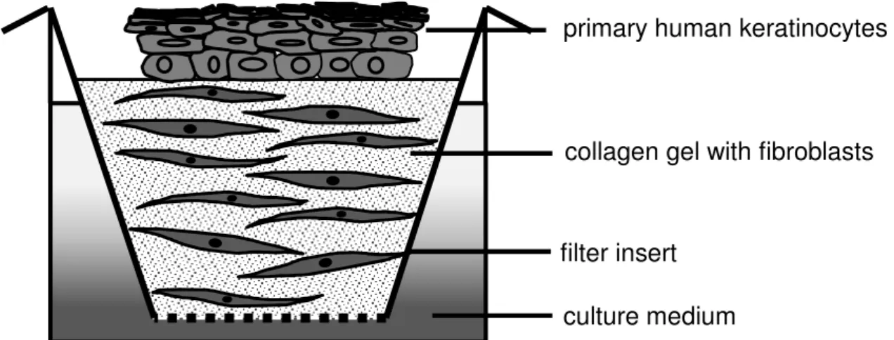
1.4.1 Organotypic skin co cultures ... 25
1.4.2 Wounds and wound healing therapies ... 27
1.5 Objective ... 28
2 Materials and Methods ... 30
2.1 Materials ... 30
2.1.1 Chemicals, solutions and enzymes ... 30
2.1.2 Standard kits ... 32
2.1.3 Plasmids ... 32
2.1.4 Enzymes ... 33
2.1.5 Primers ... 33
2.1.6 Antibodies ... 34
2.1.6.1 Direct labeled antibodies ... 34
2.1.6.2 Primary antibodies ... 34
2.1.6.3 Secondary antibodies ... 34
Table of Content vii
2.1.7 Peptides ... 34
2.1.8 Bacteria strain ... 35
2.1.9 Eukaryotic cells ... 35
2.1.9.1 Immortalized cell lines ... 35
2.1.9.2 Primary human keratinocytes ... 36
2.1.9.3 Primary murine keratinocytes ... 36
2.1.10 Culture Media and Supplements ... 36
2.1.11 Laboratory equipment and disposables... 38
2.1.12 Data treating Software ... 41
2.2 Methods ... 42
2.2.1 Bacteria culture ... 42
2.2.1.1 Cultivation of bacteria ... 42
2.2.1.2 Preparation of chemically competent bacteria ... 42
2.2.1.3 Transformation of bacteria ... 43
2.2.2 Working with nucleic acid ... 43
2.2.2.1 Plasmid amplification and extraction ... 43
2.2.2.2 DNA quantification ... 43
2.2.2.3 Restriction Digest of DNA ... 43
2.2.2.4 Gel Electrophoresis ... 43
2.2.2.5 DNA extraction from eukaryotic cells ... 44
2.2.2.6 Polymerase chain reaction ... 44
2.2.2.7 Quantitative real-time PCR (qPCR) ... 45
2.2.2.8 Sequencing ... 46
2.2.2.9 Molecular cloning ... 47
2.2.3 Capsid ELISA ... 47
2.2.4 Eukaryotic cell culture ... 48
2.2.4.1 Cultivation of cells ... 48
2.2.4.2 Counting ... 48
2.2.4.3 Seeding and culturing ... 48
2.2.4.4 Seeding of primary human keratinocytes as mixed culture with mouse embryonic fibroblast cells (NIH3T3) ... 48
2.2.4.5 Freezing and thawing of cells ... 48
2.2.4.6 Isolation of primary human keratinocytes (monolayer) ... 49
2.2.4.7 Preparation of organotypic human skin co-cultures ... 49
2.2.5 Vector production and purification ... 50
2.2.5.1 AAV library and vector packaging ... 50
2.2.5.2 Iodixanol gradient purification ... 51
2.2.5.3 Vector titration ... 51
2.2.5.4 Coupling of pheno- and geno-type of mutants ... 52
2.2.5.5 Transducing titer of viral vectors encoding for GFP ... 52
2.2.5.6 Heparin affinity chromatography ... 52
2.2.5.7 AAV peptide display on primary HK ... 52
Table of Content viii
2.2.6 Cell transduction by rAAV vectors ... 53
2.2.6.1 Quantification of vector entry efficiency ... 53
2.2.6.2 Drug treatment ... 53
2.2.6.3 Cell transduction assays ... 54
2.2.6.4 Heparin competition assay ... 54
2.2.6.5 Peptide and αV blocking-antibody competition assay ... 54
2.2.6.6 αVβ8 antibody competition assay ... 54
2.2.6.7 Transduction of mixed cultures ... 55
2.2.6.8 Transduction of organotypic human skin co-cultures ... 55
2.2.7 Immunohistochemistry ... 55
2.2.7.1 Immunofluorescence staining of cryosections of organotypic human skin co-cultures ... 55
3 Results ... 56
3.1 Characterization of cell surface receptors of primary human keratinocytes 56 3.1.1 Selection of rAAV targeting vectors from a library enriched for non-HSPG binding mutants ... 59
3.2 Characterization of rAAV peptide insertion variants regarding cell entry and transduction efficiency on primary HK ... 62
3.2.1 Infectivity of rAAV2 and rAAV2 selected peptide insertion variants on primary human keratinocytes ... 66
3.3 Transduction efficiencies of rAAV2 and rAAV2 peptide insertion variants in presence or absence of Heparin ... 66
3.4 Peptide competition of selected rAAV2 peptide insertion variants on human primary keratino-cytes ... 67
3.5 Selected rAAV2 peptide insertion variants enter via clathrin-mediated endocytosis ... 69
3.6 rAAV2 peptide insertion variants show altered tropism ... 71
3.6.1 Cell transduction of rAAV2 peptide insertion variants on feeder cultivated primary human keratinocytes ... 73
3.7 Efficient and specific transduction of differen-tiated keratinocytes in human organotypic skin cultures ... 76
3.7.1 Efficient transduction of primary murine keratinocytes ... 78
3.8 Identification of candidate receptor for Kera2 ... 79
3.8.1 αVβ8 integrin inhibition blocks Kera2 transduction ... 81
4 Discussion ... 86
4.1 Selection of AAV capsid variants ... 86
4.2 Kera1, Kera2 and Kera3 transducing target cells peptide-dependent through the clathrin entry route ... 88
4.3 Kera2 possesses the highest receptor specificity ... 90
4.4 Kera1, Kera2 and Kera3 are capable of transducing differentiated keratinocytes in human organotypic skin co-cultures ... 91
4.5 αVβ8 integrinserves as receptor for Kera2 ... 91
Table of Content ix
4.6 Summary and outlook ... 92 List of Abbreviations ... 95 References ... 97 Lebenslauf ... Fehler! Textmarke nicht definiert.
List of tables and figures x
List of tables and figures
Table 1: Examples of clinical trials using AAV gene transfer [97] ... 15
Table 2: Sequences identified after the fifth selection round ... 61
Table 3: Characterization of selected rAAV peptide insertion variants ... 62
Table 4: Transducing titer and infectivity of rAAV2 and rAAV peptide insertion variants determined on primary HK ... 66
Table 5:The output of the COMPARE analysis. ... 80
Table 6: Keratinocyte integrins [174], RGD recognition sequence [238] ... 89
Table of Figures Figure 1: Atomic structure of AAV serotype 2. Figure was kindly provided by J. Boucas. ... 5
Figure 2: Genome organization of AAV2 ... 6
Figure 3: Infectious pathway of AAV2 in HeLa cells ... 9
Figure 4: Schematic representation of latent and lytic life cycle of AAV2 ... 11
Figure 5: Packaging of recombinant AAV (rAAV) vectors ... 13
Figure 6: Overview for modifications of viral capsids ... 17
Figure 7: Principle of cell surface targeting using the example of AAV2 ... 19
Figure 8: AAV peptide display ... 22
Figure 9: Structure of the human skin (www.physioweb.org) ... 24
Figure 10: Schematic drawing of an organotypic skin co-culture ... 26
Figure 11: Transduction efficiencies of rAAV2 with wild-type capsid on primary HK of different donors. ... 57
Figure 12: Characterization of cell surface receptors on primary HK and HeLa cells ... 58
Figure 13: Schematic representation of AAV peptide display selection on primary HK ... 60
Figure 14: Cell entry efficiencies of indicated vectors ... 63
Figure 15: Microscopic images of primary HK transduced with rAAV2, Kera1, Kera2 and Kera3 ... 64
Figure 16: FACS analysis of rAAV2 and rAAV peptide insertion variants on primary HK. ... 65
Figure 17: Heparin competition assay on primary HK ... 67
Figure 18: Peptide competition on primary HK ... 68
Figure 19: Cell transduction in presence and absence of Genistein ... 69
List of tables and figures xi
Figure 20: Cell transduction in presence and absence of CPZ... 70 Figure 21: Transduction experiments of indicated vectors on non-target cells ... 72 Figure 22: Transduction efficiencies of indicated vectors on DU-145 cells (dark
grey) and A375 cells (light grey) ... 73 Figure 23: Target cell specificity of indicated vectors in mixed cultures ... 74 Figure 24: Target to-noise ratio of indicated vectors ... 75 Figure 25: Histological examination of cryosections of human organotypic skin co-
cultures ... 77 Figure 26: Flow cytometric measurements of primary murine keratinocytes
incubated with indicated vector preparations ... 78 Figure 27: Transduction profiles of rAAV2 and Kera2 on NCI60 cell panel ... 79 Figure 28: αVβ8 integrin expression on primary HK (A) and non-target cells (B) ... 81 Figure 29: Characterization of RGD-binding integrins expressed on SW480 αVβ8
cells and parental SW480 cells ... 82 Figure 30: Blocking experiment using MAB specific for the αV chain... 83 Figure 31: Blocking experiment using an αVβ8 integrin antibody ... 84
Zusammenfassung
1
Zusammenfassung
Die Heilung chronischer Wunden, wie z.B. diabetischer Ulzera oder großflächiger Verbrennungswunden, stellt ein nicht unerhebliches medizinisches Problem dar.
Der Heilungsprozess kann sehr langwierig und schmerzvoll sein und schränkt dadurch die Lebensqualität der Patienten massiv ein. Mit den traditionellen Vorgehensweisen und Maßnahmen zur Behandlung akuter Erkrankungen allein kann auf Grund der Vielzahl negativer Einflussmöglichkeiten kein optimales Ergebnis erzielt werden. Daher ist die Einführung neuer, innovativer, therapeutischer Strategien von Nöten, wie zum Beispiel die Verwendung von primären humanen Keratinozyten für die Herstellung autologer Hauttransplantate.
Gentherapeutische Vektorsysteme könnten das Anwachsen von Hauttransplantaten durch z.B. gezielte, aber transiente Bereitstellung von Wachstumsfaktoren mittels Gentransfer verbessern. Rekombinante adeno- assoziierte Virus Vektoren (rAAV) wären hierfür ein potentiell geeignetes System.
Sie sind wenig immunogen und stabil, lassen sich mit hohen Titern herstellen und sind als nicht-integrierende Vektoren in proliferierenden Zellen nur transient vorhanden. Allerdings scheint die Haut ein schlechtes Zielorgan für AAV Vektoren des Serotypes 2, sowie pseudotypisierte AAV Vektoren mit Kapsiden anderer AAV Serotypen zu sein, da sich primäre humane Keratinozyten nur unzureichend von AAV transduzieren lassen. Ein Grund hierfür wurde im Rahmen dieser Arbeit gefunden. Es konnte gezeigt werden, dass primäre humane Keratinozyten den AAV2-Primärrezeptor Heparansulfat-Proteoglykan (HSPG) nur unzureichend oder gar nicht exprimieren.
Kürzlich wurde demonstriert, dass die genetische Modifizierung des AAV-Kapsids durch Insertion rezeptorspezifischer Liganden („AAV targeting“) die Transduktion von Zellen unabhängig vom Vorhandensein der natürlichen AAV-Rezeptoren ermöglicht. Die „AAV-targeting“-Technologie bietet einen möglichen Lösungsansatz um spezifische rAAV2-Targeting-Vektoren für primäre humane Keratinozyten zu generieren.
Im Rahmen dieser Arbeit wurden neue, vielversprechende rAAV Vektoren für die Modifikation primärer humaner Keratinozyten generiert. Mit Hilfe einer „AAV
Zusammenfassung
2 peptide display“ Bibliothek wurden drei rAAV Peptidinsertionsmutanten (Kera1, Kera2 und Kera3), die sich in der inserierten Sequenz unterscheiden, selektioniert.
Die AAV2-Bibliothek besteht aus Mutanten, die 7-mer Peptide mit zufälliger Sequenz im Kapsid in der Position 587 präsentieren. Um „targeting“-Vektoren mit einem veränderten Tropismus zu generieren, wurde die AAV-Bibliothek optimiert, indem Mutanten die an HSPG binden können vor der Selektion durch Heparinaffinitätschromatographie abgereichert wurden. Eine weitere Optimierung des Selektionsschemas wurde durch die Verwendung von verschiedenen Keratinozyten-Spendern in jeder Selektionsrunde erzielt. Das erhöhte die Wahrscheinlichkeit Mutanten mit Spezifität für einen allgemeingültigen Rezeptor für primäre humane Keratinozyten zu selektionieren. Die auf diese Weise selektionierten Mutanten Kera1 (RGDTATL), Kera2 (PRGDLAP) und Kera3 (RGDQQSL) weisen eine außergewöhnliche Änderung des Tropismus auf. Sie transduzieren primäre humane Keratinozyten mit einer hohen Effizienz und Spezifität, was selbst in Mischkultur-Experimenten mit Nicht-Ziel-Zellen zu einer präferentiellen Transduktion von Keratinozyten führte. In dieser Arbeit wurde zudem erstmalig die neue bioinformatische Methode der komparativen Genanalyse (CGA) zur Identifizierung des Ziel-Rezeptors eines rAAV-targeting Vektors angewandt. In Kooperation mit Giovanni Di Pasquale (NCI/NIH, Bethesda, USA) wurde zu diesem Zweck ein Zellscreening auf der NIH Zellliniensammlung durchgeführt. Für die Mutante Kera2 konnte mit Hilfe dieses Verfahren eine hohe Affinität zu dem Integrin-Rezeptor beta8 festgestellt werden. Die Integrin beta8 Untereinheit bildet mit der Integrin alpha V Untereinheit ein Heterodimer. Das Intergin αVβ8 wird tatsächlich auf der Oberfläche von primären Keratinozyten expressioniert. Durch Experimente mit blockierenden αV- oder αVβ8-Antikörpern konnte nachgewiesen werden, dass das Integrin αVβ8 als Rezeptor für Kera2 fungiert.
Außerdem war es möglich differenzierte Keratinozyten einer 3D Kultur nach topischer Anwendung der „targeting“-Vektoren Kera1, Kera2 und Kera3 zu transduzieren. Zusammenfassend lässt sich sagen, dass die drei in dieser Arbeit entwickelten und charakterisierten „targeting“-Vektoren Kera1, Kera2 und Kera3 Schlüsselfunktionen für die klinische Anwendung erfüllen.
Abstract
3
Abstract
Chronic non-healing wounds such as diabetic ulcers or burns represent a devastating health problem with significant clinical, physical and social implications. The healing can be frustrating and painful for patients. The difficult healing process requires advanced therapeutic strategies such as the use of primary human keratinocytes (HK) as autologous transplants, which may be considered for clinical use. To improve engraftment or to introduce therapeutic genes into primary HK, efficient and safe vectors are required. One of the most promising vector systems today is based on the adeno-associated virus (AAV), a member of the parvovirus family. Recombinant AAV (rAAV) vectors possess a number of attractive properties including low immunogenicity, high stability and the potential to integrate site-specifically without known side-effects. Unfortunately, cell entry into primary HK of rAAV2 is barely detectable and consequentially, HK are poor targets of rAAV2-mediated transductions. As demonstrated in this thesis, primary HK do not express AAV2´s primary receptor heparan sulphate proteoglycan (HSPG), the presence of which, however, is required for binding to AAV2´s internalization receptors. Cell surface targeting allows re-directing the viral vector tropism towards a novel receptor mediating thereby transduction of cells in absence of AAV’s natural receptors. These AAV capsid mutants have displayed improved transduction efficiency in wild-type-AAV non-permissive cells and have provided the opportunity of rAAV-mediated, cell-type-specific gene transfer.
As documented in this study, new rAAV vectors were developed as promising tools for modifying primary HK. Using an AAV peptide display library that displayed 7mer peptides of random sequence at capsid position 587; three AAV peptide insertion mutants differing in sequence of inserted ligand (Kera1, Kera2 and Kera3) were selected and subsequently analyzed. To select rAAV targeting vectors with a re-directed tropism, the library was optimized by depleting mutants capable of binding to HSPG prior to selection by heparin affinity chromatography.
Furthermore, the selection was performed on primary HK obtained from different donors to target a common receptor and the selection pressure was continuously increased by decreasing the vector genomes per cell ratio to select for the fittest variant. The thereby developed rAAV targeting vectors Kera1 (RGDTATL), Kera2
Abstract
4 (PRGDLAP) and Kera3 (RGDQQSL) showed a remarkable change in tropism, transducing primary HK with high efficiency and specificity even in mixed cultures of target and non-target cells. In this study, a novel microarray based bioinformatic approach (comparative gene analysis (CGA)), was used for the identification of the receptor that targeted the mutant that showed the most striking change in tropism, Kera2. Briefly, in cooperation with Giovanni Di Pasquale (NCI/NIH, Bethesda, USA), a screening of the NIH cell line panel was performed, pointing towards the involvement of beta8 integrin subunit for cell transduction by Kera2. Beta8 is unique as it is solely described as heterodimer with alpha V and the integrin αVβ8 could be detected on cell surface of primary human keratinocytes. By blocking experiments with blocking αV- or αVβ8-antibodies experimental evidence was provided that the integrin αVβ8 serves as receptor for Kera2. Finally, this study has shown that the targeting vectors Kera1, Kera2 and Kera3 transduced airlifted differentiated keratinocytes in organotypic 3D cultures. In summary, the three rAAV targeting vectors Kera1, Kera2 and Kera3, selected from an optimized library and using a novel selection strategy, are excellent candidates for successful application in clinical use.
1 Introduction
5
1 Introduction
1.1 Adeno Associated Virus (AAV)
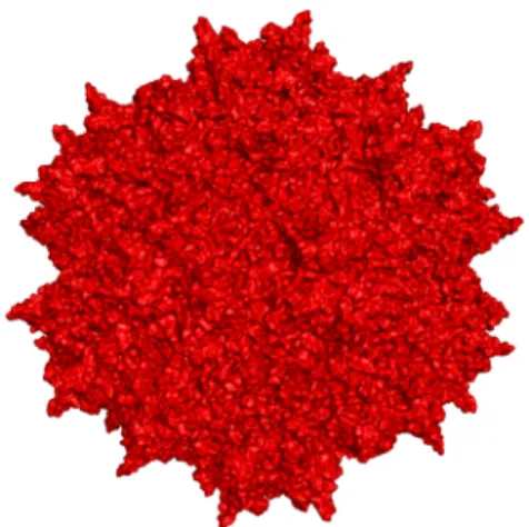
Adeno-Associated Viruses (AAVs) belong to the genera of Dependovirus and the subfamily Parvovirinae that infects vertebrates. Parvovirinae together with the insect-infecting Densovirinae form the family of Parvoviridae. These include viruses with a linear, single-stranded DNA genome of about 4.7 kb and a non- enveloped icosahedric capsid of 18-30 nm in diameter [1].
Figure 1: Atomic structure of AAV serotype 2. Figure was kindly provided by J. Boucas.
AAV were first described in 1965 as DNA-containing particles in preparations of a simian adenovirus [2]. Later, AAV was defined as a unique virus family. For replication and initiation of a productive infection cycle, AAV is, as the name implicates, dependent on co-infection by a helper virus. Known helper viruses are Adenoviruses, Herpes Simplex Viruses, human Cytomegaloviruses (CMV) and Papillomaviruses [3], [4]. In absence of co-infection with a helper virus, AAV establishes a latent infection where the viral DNA is either maintained as episomes or integrated into the host genome. Integration occurs in 70% of cases, site- specific into the human chromosome 19 at position 13.4-qter (AAVS1) [5], [6], [7].
After super-infection with a helper virus, the provirus enters the lytic cycle, leading to viral gene expression, rescue and replication of the AAV genome with subsequent production of viral progeny (see 1.1.2), [8]. Thus far, 12 different serotypes (AAV1-12) and over 100 variants of AAV have been isolated from
1 Introduction
6
ITR ITR
p5 p19 p40
10 20 30 40 50 60 70 80 90
polyA
Rep78 Rep68 Rep52 Rep40 VP1 VP2 VP3 AAP
adenoviral isolates and tissue samples [4]. They differ in the amino acid (aa) composition of their capsids, but show similar capsid morphology, genome length and genome organization. AAV serotype 2 (AAV2), the best-characterized serotype, is frequently applied in human gene therapy [9].
1.1.1 Viral genome and AAV proteins
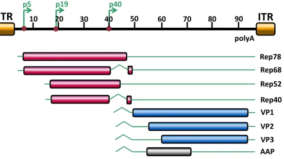
The single stranded DNA genome of AAV2 contains four functional units, the open reading frame (ORF) for the Rep proteins (rep), the cap ORF (ORF1), the alternative cap ORF (ORF2) and the inverted terminal repeats (ITR) flanking these ORFs (Figure 2). The alternative cap ORF was just recently discovered and encodes a 23 kDa protein, which was named assembly-activating protein (AAP), required for initiation of capsid formation [34].
Figure 2: Genome organization of AAV2
The AAV2 genome is flanked by the ITRs, spans 4680 nt divided into 100 map units. Shown are the three promoters p5, p19 and p40 at map position 5, 19 and 40 and the polyadenylation signal (polyA) at position 96. The open reading frames are indicated by rectangles, translated regions in red, blue or grey, untranslated regions by thin solid lines, while introns are marked as nicks. The p5 promoter controls expression of the large Rep proteins (Rep78, Rep68), while the p19 promoter is responsible for expression of the small Rep proteins (Rep52, Rep40). Rep68 and Rep40 are splice variants of Rep78 and Rep52, respectively. The expression of capsid proteins VP1, VP2, VP3 and AAP is controlled by the p40 promoter. Figure was kindly provided by N. Huttner [10] and modified according to F. Sonntag [11].
The genome contains three promoters (p5, p19 and p40) and a single polyadenylation signal (poly A). The 5´-ORF rep encodes four Rep proteins. These are multifunctional, non-structural proteins that are termed by their molecular
1 Introduction
7 weights (Rep78, Rep68, Rep52, and Rep40). Transcription of the larger Rep proteins (Rep78, Rep68) is controlled by the p5 promotor, while the smaller ones (Rep52, Rep40) are transcribed by the p19 promotor [12]. Rep68 is a splice variant of Rep78 and Rep40 is a splice variant of Rep52. The larger Rep proteins are necessary for site-specific integration into AAV2, thus they possess site- and strand-specific endonuclease activity. In addition, they are required for transcription of the viral ORFs, control of viral replication (see below) and packaging of the viral genome. Specifically, Rep78 and 68 possess DNA binding, ATPase, DNA helicase, and endonuclease activities [13], [14], [15], [16], [17], while the Rep proteins are involved in accumulation and packaging of the single- stranded DNA genome into the preformed capsid [18], [19]. Furthermore, all Rep proteins contain in the common C-terminal part a nuclear localization signal (NLS) [15], [20]. The smaller Rep proteins seem to be involved in accumulation and packaging of single-stranded DNA into the preformed capsid [18], [19].
The 3´-located ORFs encode the capsid proteins, VP1, VP2, VP3 and AAP, the latter of which is required for viral capsid assembly. The VP proteins are expressed from ORF1, while ORF2 encodes for AAP [11]. Expression of the AAV2 capsid proteins is controlled by the p40 promoter. The three VP proteins assemble the viral capsid in a 1:1:10 ratio [21]. The capsid proteins VP1 and VP2 share identical sequences at the C-terminus but differ in their N-terminal sequences. The translation of VP1 is regulated by alternative splicing of the p40 -transcripts [22].
Translation of VP2 is initiated from an alternative start codon (ACG) [23]. All three VP proteins use the same stop codon. The molecular weights of the capsid proteins are 90 kDa (VP1), 72 kDa (VP2) and 60 kDa (VP3). All capsid proteins are required for the formation of infectious particles, while intact non-infectious capsids assemble in absence of VP1 or in absence of VP1 and VP2, when AAP is present to mediate nuclear transport of VP3 [11]. VP2 seems to be dispensable for the formation of infectious particles, at least in an in vitro application [24], [25]. The capsid formation takes place in the cell nucleus [26], [27].
The 5´- and the 3´-end of the viral genome are formed by the ITRs consisting of 145 nt. Due to their palindromic sequence, a hairpin structure is formed by the first 125 bp [28], [29]. The ITRs serve as signal sequence – recognized by the viral Rep proteins - for packaging of the viral genomes into the capsid. In addition, the ITRs serve as origin of replication (ori). For this function, a Rep binding site (RBS),
1 Introduction
8 a specific cleavage site for Rep proteins (terminal resolution site, TRS) and a certain distance between the former two sites are required [14], [30], [31]. The ITRs play a key role in the site-specific integration into AAVS1, as well as in the subsequent rescue of viral DNA from the integrated state in the presence of helper viruses [32], [33], [34], [35].
1.1.2 AAV infectious biology
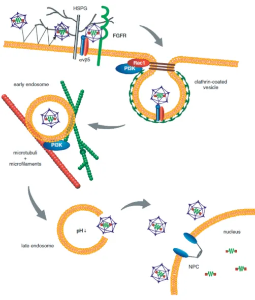
A successful infection of cells by AAV is a multistep process including attachment, uptake, intracellular trafficking, nuclear translocation and replication of the virus (Figure 3). Many steps of the AAV-cell interaction are still unknown. As single virus tracing studies have revealed, AAV2 contacts the cell membrane several times before entering the cell. On average, AAV contacts the cell 4.4 times [36]. For AAV2, the widely expressed cell surface receptor heparan sulfate proteoglycan (HSPG) has been identified as primary receptor [37]. This contact is mediated by binding motives present on the AAV capsid, that are formed by residues R484, R487, K532, R585 and R588 in the common VP3 region [38]. Binding to HSPG is believed to induce a conformational change in the capsid, which is required for internalization into the cell [39]. For efficient internalization, co-receptors are required. So far, five co-receptors have been described for AAV2. Human fibroblast growth factor receptor 1 (FGFR-1), hepatocyte growth factor receptor (HGFR) and laminin receptor seem to support virus-cell interaction, facilitating the HSPG-induced structural rearrangement of the capsid [40], [41], [42]. The integrins αVβ5 and α5β1 are thought to mediate endocytosis of AAV2 [39], [43]. In addition, integrin binding subsequently leads to the activation of the small GTPase Rac1 and phosphatidylinositol-3 kinase (PI3K), resulting in cytoskeletal rearrangements that promote clathrin-dependent internalization of AAV2 as well as trafficking of AAV2 from the cell periphery towards the nucleus [43], [44], [45], [46].
1 Introduction
9
Figure 3: Infectious pathway of AAV2 in HeLa cells
Following multiple contacts with the cell, AAV binds to HSPG on the cell membrane. The attachment is likely enhanced by co-receptors such as FGFR1 and/or HGFR. Subsequent binding to integrins lead to endocytosis via clathrin-coated pits. Integrin binding activates the small GTP binding protein Rac1, which stimulates the PI3K pathway. The resulting rearrangement of the cytoskeleton allows for trafficking of AAV2-containing endosomes. Acidification of the endosome may lead to conformational changes in the AAV2 capsid and its release. Once inside the nucleus the AAV genome is replicated (lytic phase; requires the presence of helper virus), stays episomally or is integrated into the host genome (latent phase) [47]. NPC: nuclear pore complex. Picture was kindly provided by H. Büning © 2008
Once internalized, AAV is trafficked mainly inside endosomes [36], [44], [45], [48], [49], [50]. The transport of the endosomal vesicle takes place via motor proteins along microtubules and microfilaments [43], [44], [45]. AAV particles remain in the endosomal compartment until late stages. When and how AAV escapes from the endosome is still subject of debate and may be cell type specific [45], [50].
Acidification inside the endosomes appears to be essential for priming AAV for nuclear entry. This assumption is based on the observation that microinjection of AAV2 particles directly into the cytoplasm (instead of natural infection) did not
1 Introduction
10 result in gene expression [51]. The same effect can be reached by the addition of inhibitors of acidification like bafilomycin A1 or ammonium chloride [45]. The acidification of endosomes during maturation may lead to a conformational change of the viral capsid, leading to exposure of a phospholipase A2 (PLA2) homology domain, present within the N-terminus of VP1 [52], [53]. The PLA2 domain is conserved among parvoviruses [54] and AAV2 requires this domain for endosomal escape through lipolytic pore formation [53], [55]. When AAV2 is released from the endosome the capsids are target for ubiquitination, which is a general signal for proteasomal degradation [56]. Several groups have shown that the addition of proteasome inhibitors results in an enhancement of transgene expression at least in some cell lines [44], [57] [58], [59], [60]. Though, the mechanism remains unclear, studies suggested that, conceptually proteasome inhibitors block capsid degradation, facilitate vector uncoating and lead to an increased perinuclear accumulation or translocation into the nucleus [57], [56].
It is still unknown how the virus enters the nucleus and where viral uncoating occurs. Viral particles start to accumulate in the perinuclear area between 15 and 30 min post infection (p.i.) [45], [36]. The majority of these virions still have intact viral capsids containing viral genomes [25]. Several studies have reported of intact AAV particles in the nucleus. But there are controversial reports concerning the mechanism and efficiency of capsid import as well as their role in viral infection [25], [43], [45], [50], [61]. Lux et al. showed that when using a low number of virions for infection, viral genomes, but no intact capsids, are found within the nucleus, whereas intact full and empty capsids were still evident in the perinuclear area [25]. This study suggested that viral genomes rather than intact capsids are transported into the nucleus. In contrast, Sonntag and colleagues blocked AAV infection completely by injection of capsid specific antibodies into the nucleus.
These results suggest that viral genomes are transferred into the nucleus by intact viral capsids and that the uncoating event takes place there [55]. Moreover, whether AAV and/or AAV genomes enter the nucleus through the nuclear pore complex (NPC) or in a NPC-independent way is still discussed [62].
1 Introduction
11
Figure 4: Schematic representation of latent and lytic life cycle of AAV2
Infection of cells with AAV, in the absence of a helper virus results in the establishment of a latent infection that is characterized by the persistence of viral DNA - frequently integrated within the host genome - and by absence of viral gene expression. In the presence of a helper virus, wild type AAV2 enters a productive cycle leading to the replication of viral DNA, expression of viral genes and packaging of viral DNA into pre-assembled capsids. AD = adenovirus, HSV herpes simplex virus, HPV = human papillomavirus
Inside the nucleus, the presence or absence of a helper virus determines whether AAV enters a lytic or latent life cycle. In the absence of helper viral functions second-strand synthesis of the single-stranded virus genome and the basal expression of the Rep proteins are activated [63], [64]. First, second-strand synthesis of the single-stranded virus genome and a basal expression of the Rep proteins are activated [63]. In presence of the large Rep proteins (Rep78, Rep68) and intact ITRs, integration occurs, although not exclusively, at the so-called AAVS1 site on the human chromosome 19 (19q13.3-qter) [65], [66]. The AAVS1 locus resides a Rep binding element (RBS) and a terminal resolution site (TRS) equivalent to the AAV genome [67], [68], [69]. Usually, proviral sequences are integrated as viral concatemers in a head-to-tail conformation [67]. Helper viral superinfection can rescue the integrated provirus initiating a lytic, productive life cycle (Figure 4), [8]. Alternatively, AAV genomes can form episomes, which at least in non-dividing cells, also results in a latent life cycle.
In the presence of a helper virus, AAV can undergo a productive infection. During viral replication, the 3’-OH end of the ITR serve as the primer for second-strand
Latency Replication
co-infection with helper virus (AD, HSV, HPV) AAV
+
helper virus (AD, HSV, HPV) co-infection
1 Introduction
12 synthesis [3]. The large Rep proteins unwind the ITR by their helicase activity, leading to exposure of the TRS, which is nicked by the Rep endonuclease enabling complete synthesis of the second-strand by switching templates [13], [63]. The single-stranded DNA is then converted into a parental duplex replicative form where production of viral progeny can proceed.
1.1.3 Adenovirus-free AAV production and recombinant AAV vectors (rAAV)
The structural properties of the AAV capsid allow for the production of recombinant viral particles that package a DNA genome of approximately 5 kb [70]. For the generation of rAAV vectors, all ORFs are deleted leaving only the ITR sequences of the parental virus. The ITRs are the solely required cis elements necessary for the production of viral particles (replication and packaging). The deleted ORF sequences are replaced by an exogenous DNA sequence (transgene expression cassette).
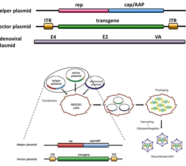
A successful approach to produce rAAV vectors at high titers for laboratory scale uses triple transfection of AAV vector, AAV helper and adenoviral helper plasmids (Figure 5A). The vector plasmid contains the transgene flanked by the ITRs. The AAV-specific ORF required in trans, rep and cap/AAP, are cloned onto the helper plasmid, which lacks the ITR sequences [34], [71]. These plasmids are co- transfected with the third plasmid carrying the essential adenoviral genes VA, E2A and E4, necessary for AAV replication (Figure 5B) [71], [72], [73]. For viral particle production, HEK293 cells, which are transgenic for the adenoviral genes E1a and E1b (also required for AAV progeny production), are commonly used. After transcription and translation of rep and cap/AAP proteins and replication of the vector genome, the vector genome is shuttled into preformed AAV capsids.
Finally, vector particles are harvested and purified by density gradient centrifugation (CsCl or Iodixonal) and/or column chromatography [74], [75].
1 Introduction
13
Figure 5: Packaging of recombinant AAV (rAAV) vectors
(A) Plasmid constructs used for packaging of rAAV vectors. The vector plasmid is devoid of all viral genes, only the ITRs are left, which flank the transgene expression cassette (“transgene”) and serve as packaging signal. The helper plasmid encodes for the non-structural, multifunctional Rep proteins (rep) and proteins required for capsid production (cap/AAP). These proteins are necessary for replication of the vector genome, production and assembly of the capsid and the subsequent packaging of the vector genome into preformed capsids. The adenoviral plasmid carries the essential adenoviral genes for rAAV production (VA, E2A and E4).
(B) Packaging of rAAV vectors. AAV vector plasmids, AAV helper plasmids, and adenoviral helper plasmids are transfected into HEK293 cells. After replication and assembly of viral vector particles, cells are lysed and vector particles are harvested and purified by e.g. iodixanol gradient centrifugation[47]. Figure A was kindly provided by N. Huttner and Figure B by H. Büning.
ITR ITR
rep cap/AAP
transgene adenoviral
plasmid
helper plasmid vector plasmid
E4 E2 VA
B A
cap/AAP
1 Introduction
14
1.2 AAV in Gene Therapy
Gene therapy is based on the idea of introducing genetic material into an organism in order to cure or improve the status of a disease [76]. A key factor for the success of gene therapy is the development of gene delivery systems that combine efficiency and safety. Currently, viral as well as non-viral vectors have been developed for this purpose. Whereas the viral systems include adeno-, retro-, vaccinia-, pox-, herpes simplex- and adeno-associated-viral vectors, the non-viral vector strategy uses naked DNA within lipoplexe or polyplexe [77], [78].
However, each vector has its own advantages and disadvantages. The simplest way of gene delivery is injecting naked DNA encoding the transgene expression cassette. But this strategy lacks efficiency [79]. Viral vector systems are very efficient at transferring DNA into host cells but are in general more immunogenic, more sophisticated to produce and are limited in the size of foreign DNA that can be delivered. AAV has many features that make it attractive for use as a gene therapy vector. Briefly, rAAV vectors are based on a non-pathogenic virus [80], [81] and transduce dividing as well as post-mitotic or quiescent cells [82], [83].
Furthermore, they show a broad tissue tropism infecting diverse organs such as brain, liver, muscle, lung, retina and heart [84], [85], [86], [87], [88]. Moreover, in non-dividing cells or tissues AAV mediates long-term expression without the need for integration. Examples of such tissues are muscle or liver where e.g. in a muscle-directed trial transgene expression was sustained for at least four years in a canine hemophilia B model [85]. Another important aspect that – as already mentioned – AAV in contrast to lenti- or retroviral vectors stays as episomes [89], [90], [91], reducing thereby the risk for insertional mutagenesis. Moreover, if integration is required, expression of Rep proteins can be exploited to direct AAV towards integration at AAVS1 [5], [10]. The immunological reactions to AAV are low comparing to adenovirus [92], [93]. As such, AAV have only a minimal inflammatory potential. Nevertheless, in a clinical trial of liver-directed gene transfer, re-direction of memory T cells caused failure of long-term gene expression [94]. Recently, our group demonstrated that primary human liver cells, like Kupffer cells (KC) and liver sinusoidal endothelial cells (LSEC) are capable of sensing AAV. The AAV capsid represents pathogen-associated molecular patterns (PAMPs) that are detected by the pattern recognition receptors (PPR) Toll-like
1 Introduction
15 receptor-2 (TLR-2) [95] known to activate innate immune response. Minimizing this recognition will be a key to improving rAAV-mediated gene transfer and reducing side effects in clinical trials due to immune responses against rAAV [95].
Disadvantages of the AAV vector system include the small genome size limiting the coding capacity for transgenes including ITRs to approximately 5 kb [96] and the broad tissue tropism interfering with a cell-specific in in vivo gene transfer. To date, AAV vectors have been applied in over 80 clinical trials (Table 1).
Table 1: Examples of clinical trials using AAV gene transfer [97]
Disease Transgene
product
Serotype Route
administration
Clinical trial
Clinical Trials.
gov identifier
Refs AAV clinical trials for inherited disease
α1antitrypsin deficiency
α1 antitrypsin AAV2
Intramuscular Phase I/II
NCT00377416 [98], [99]
AAV1 NCT00430768
Batten’s disease CLN2 AAV2 Direct intracranial
administration Phase I/II
NCT00151216
[100]
AAVrh10 NCT01161576
Canavan’s disease
Aspartoacylase AAV2 Direct intracranial administration
Phase I NA [101]
Cystic fibrosis CFTR
AAV2
Direct instillation to maxillary sinus,
bronchoscopy to right lower lobe, aerosol to whole lung
Phase
I/II NCT00004533
[102], [103], [104], [105]
Haemophilia B FactorIX AAV2 Intramuscular
Phase I/II
NCT00076557
[106], [107]
Hepatic NCT00515710
AAV8 Intravenous Phase I/II
NCT00979238 Muscular
dystrophy:
Duchenne
Microdystrophin AAV1- AAV2
hybrid Intramuscular Phase I NCT00428935 [108]
AAV clinical trials for acquired diseases Severe heart failure SERCA2a AAV1 Antegrade
epicardial coronary artery infusion
Phase I/II
NCT00454818
[109]
AAV6 NCT00534703
Parkinson’sdisease AADC
AAV2 Intracranial Phase I/II
NCT00229736 [110], [111]
GAD
NCT00643890, NCT00195143, NCT01301573
[112], [113]
Neutrophin NCT00252850,
NCT00985517, NCT00400634
[114]
AADC, aromatic-L‑amino-acid decarboxylase; AAV, adeno-associated virus; CFTR, cystic fibrosis transmembrane regulator; CLN2, also known as tripeptidyl peptidase 1 (TPP1); GAD, glutamic acid decarboxylase; SERCA2a, sarcoplasmic reticulum calcium ATPase 2a
1 Introduction
16 Early published data dealt with the monogenic diseases cystic fibrosis and hemophilia B in gene therapy trials. Administration of the cystic fibrosis transmembrane conductance regulator (CFTR) as a transgene on the nasal sinus and bronchial epithelium resulted in an improvement of pulmonary function and partial correction of hyperinflammatory responses and electrophysiological defects [104], [105], [103]. AAV was approved for safe usage in these clinical settings as well as in the treatment of hemophilia B by intramuscular, intrahepatic or intravenous vector administration [115], [107], [106], [116]. Evidences for transduction were found in all patients of the muscle-directed study as well as the intravenous study and long-term expression of the therapeutic gene, coagulation factor IX (FIX), could be detected albeit at low levels.
Further success was achieved by Bainbridge et al., Cideciyan et al. and Hauswirth et al.. They used AAV2-based RPE65 gene replacement therapy to treat patients, afflicted with RPE65 Leber congenital amaurosis. All three groups observed an increase in visual sensitivity [32], [117], [118].
In November 2012, the first AAV based gene therapy drug (Glybera®) was approved by regulatory authorities in Europe. This drug was developed by uniQure (former Amsterdam Medical Therapeutics) for treating patients suffering from lipoprotein lipase deficiency (LPLD). In 2004, Rip and colleagues reported on the rAAV1-lipoprotein lipase (LPL)S447X vector, which aims to introduce episomal copies of a functional LPL gene variant into muscle tissue of patients with LPLD [119], [120], [121]. After several interventional clinical studies, conducted in the Netherlands and in Canada, the therapy was judged to be successful, based on tolerance, safety and efficiency, and Glybera® was authorized for patients suffering from LPLD.
Despite these successes, AAV´s broad host range remains a challenge as higher vector doses have to be applied and only those transgenes that do not harm the patient when expresses off-target are applied. In case of cancer therapy with suicide genes e.g., unspecific transduction of neighboring tissue would cause severe damage [122]. The specificity is not only important because of safety aspects but also helpful in reducing the number of particles required to be delivered [122], [123].
1 Introduction
17
1.2.1 Improvements of naturally occurring AAVs
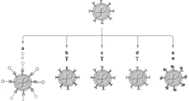
Increasing the efficiency of vectors is possible by modifying the viral tropism through capsid engineering, improving thereby gene delivery properties. There are different methods for modification of the viral capsid (Figure 6).
Figure 6: Overview for modifications of viral capsids
Rational design methods include (a) the use of a bispecific adaptor, (b) pseudotyping with an alternate serotype capsid (c, d) the generation of mosaic or chimeric particles, and (e) genetic engineering of the capsid sequence by peptide insertion or point mutations. Adapted by permission of Annual Reviews, Inc: Annual review of biomedical engineering [124] © 1999.
1.2.1.1 Mosaic rAAV vectors
A possible method to change the feature and to expand the tropism of rAAV vectors is the combination of capsid proteins from different serotypes resulting in viral capsids that accumulates the attributes of the respective serotypes [47], [125]. For example, an AAV1/AAV2 mosaic vector achieved gene expression levels similar to those of AAV1 in muscle and AAV2 in liver and could be purified by Heparin affinity chromatography like wild-type AAV2 [126]. However, since these vectors are produced by transfection of plasmids encoding the capsid proteins of the different serotypes, such viral preparations consist of virions with non-uniform capsid compositions, which in turn make standardization of this technology difficult [47], [125].
1 Introduction
18 1.2.1.2 Chimeric rAAV vectors
Chimeric rAAV vectors contain capsid proteins that have been modified by domain or aa swapping between different serotypes [125]. Bowels and colleagues generated isolated virions, co-transfected by a non-functional, HSPG-deficient AAV2 capsid mutant and an AAV3 capsid sequence in AAV replication supporting cells. This allowed for the rescue of chimeric functional viruses from these cells, which showed HSPG binding ability (the parental AAV variant was deficient in HSPG binding) and transduced the target cells [127].
1.2.1.3 Pseudotyped rAAV vectors
Pseudotyping is the process of producing viral particles that incorporate foreign viral proteins. A pseudotyped AAV vector containing the ITRs of serotype X encapsulated with the proteins of serotype Y and will be designated as AAVX/Y.
For example, a vector plasmid carrying a transgene flanked by AAV2 ITRs is co- transfected with an AAV helper plasmid coding simultaneously for Rep proteins derived from AAV2 and for capsid proteins and AAP from the serotype of choice [47]. Initial studies testing these vectors for gene delivery demonstrated far superior transduction efficiency for retina with AAV4 and AAV5 in comparison to AAV2 [128], [129], [130]. This method leads to broadening the viral tropism and may circumvent pre-existing immunity to one serotype by using a different capsid [131]
1.2.2 Generation of rAAV targeting vectors with increased transduction efficiencies
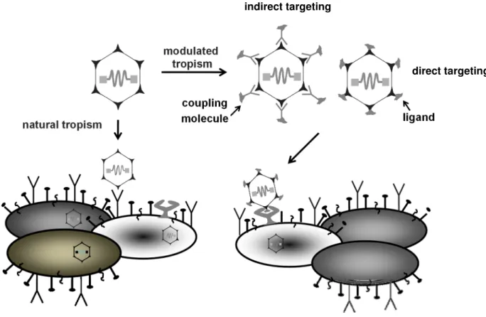
The possibility to engineer viral particles displaying selective binding domains that enable stringent interaction with target cell specific receptors (vector targeting) is desirable. Vector targeting allows the transduction of cell types that are refractory to infection with natural occurring AAVs [47]. Two main strategies have been used to achieve an altered tropism of AAV in the past; non-genetic (indirect) targeting and genetic (direct) targeting (Figure 7).
1 Introduction
19
Figure 7: Principle of cell surface targeting using the example of AAV2
By natural tropism AAV2 binds to the cell surface molecules HSPG for cell attachment and integrins αVβ5 orα5β1 for internalization. These receptors are very common and hence AAV2 shows a broad tropism, which may result in off-target transduction. It is possible to redirect the natural tropism of AAV to a more specific receptor. Furthermore, certain cell types do not express AAV receptors and therefore it would be beneficial to expand tropism to a receptor present on these cells. This constraint can be circumvented by modulating the tropism using adaptor molecules (indirect targeting) or by the insertion of peptide ligands (direct targeting) into the capsid. Figure was kindly provided by H. Büning.
1.2.2.1 Non-genetic vector targeting using adaptors
The non-genetic (indirect) targeting approach uses an adaptor molecule, which acts as a bridge between the viral capsid surface and a specific cell surface molecule (Figure 7), [47]. This technique is applicable even with limited knowledge of the viral structure [132]. This method allows for high flexibility as different adaptors can readily be coupled to the same vector and do not induce changes in capsid structure that may negatively effects vector gene transfer efficiency and packaging efficiency. Most adaptors can achieve the two main goals of targeted delivery: ablating native tropism and conferring novel tropism towards the desired target [132].
indirect targeting
direct targeting
1 Introduction
20 Barlett et al. used a bi-specific F(ab´Y)2 antibody that was subsequently linked to the capsid of AAV2. The capsid-antibody linked rAAV vectors were retargeted successfully to αIIbβ3-expressing cell lines. Results showed an increased transduction by up to 70-fold in receptor-positive cell lines [122]. Another approach used avidin-linked epidermal growth factor (EGF) or fibroblast growth factor (FGF) fusion proteins conjugated to biotinylated AAV capsids to transduce human ovarian cancer and megakaryocytic cell lines [133]. Despite the promising and successful studies of diverse adaptor systems in vitro, their usability in an in vivo setting remains to be demonstrated. Obstacles in this regard are maybe the stability of the vector-adaptor complex, in particular when host factors compete with adaptor binding [132].
1.2.2.2 Genetic vector targeting
By using the genetic vector targeting approach, cell specific targeting of the vector is mediated by genetically incorporating ligands into viral capsid proteins by simultaneously shielding the natural binding receptor (Figure 7), [134].
A first attempt to use this strategy was reported by Yang et al. [135] who fused a single-chain antibody to the N-terminus of VP2 to target CD34+ cells. Although the study showed the incorporation of the targeting ligand, vector titer was extremely low. Several groups were able to show the incorporation of small peptides to the N-terminus of VP1 or peptides within VP1 and simultaneously to the N’-terminus of VP2, which resulted in functional virions with an expanded tropism of AAV [24], [136], [137]. More recent approaches demonstrated that the N-terminus of VP2 also accepts large insertions. Lux and colleagues genetically incorporated enhanced green fluorescent protein (GFP) into AAV capsid by replacement of wild-type VP2 by GFP-VP2 fusion proteins to visualize viral trafficking [25].
Furthermore, Münch and colleagues used the N-terminus of VP2 for insertion of Designed Ankyrin Repeat Protein (DARPin) into an AAV2 vector with ablated HSPG binding. The DARPin insertion confers the AAV vector with a high cell type specificity of vector genome delivery thereby enabling the safe delivery of suicide genes following systematic application into tumor bearing mice [138].
The first successful modification of AAV´s capsid by direct targeting was achieved by Girod et al.. They demonstrated that the insertion of peptides into the common regions of all three AAV capsid proteins (aa position 587) retargeted AAV2´s
1 Introduction
21 natural tropism to mouse melanoma cells (B16F10). Later, the results of Girod et al. were confirmed by Grifman et al [139], who inserted the tumor-targeting NGRAHA sequence at the same position, 587, leading to up to 20-fold increased transduction efficiencies on several tumor cell lines expressing CD13 (a receptor expressed in angiogenic vasculature and in many tumor cell lines). Further, Shi and Bartlett demonstrated that the aa position 588 is also suitable for peptide insertion. They introduced a 4c-RGD peptide, CDCRGDCFC, which is known to bind with high affinities to the integrins αVβ5 and αVβ3,into the AAV capsid resulting in vectors that transduce cells HSPG independent, but through the above mentioned integrin [140]. Later Boucas et al. identified also aa position 453, located at the highest peaks on AAV2´s capsid, as possible site for peptide insertion [141].
To generate targeting vectors with a novel and restricted tropism, natural receptor binding elimination is necessary [47]. Notably in this context, insertions at the positions 587 interfere with the binding of two (R585 and R588) of the five positively charged aa of the AAV2 HSPG-binding motif [38], [142], explaining the ablation of HSPG binding of some re-targeted vectors [123], [143], [139], [144], [145]. In some cases, binding was only partially affected or even restored, when ligands were inserted at amino acid position 587 [139], [144], [146], [147]. This loss or maintenance of HSPG binding exemplified a dependence on the nature of the inserted ligand sequence as follows: insertion of bulky or negatively charged peptides resulted in AAV2 capsid mutants unable to bind to HSPG due to sterical or charge interference, while insertion of positively charged peptides can lead to an HSPG-binding phenotype by reconstituting a binding motif with one of the original arginines (R585 or R588) or independently of them [148].
1.3 AAV peptide display
Although rational design has generated viral vectors with novel gene delivery properties, the successful application of rational approaches often requires detailed mechanistic knowledge of AAV’s infection process and on suitable receptor-binding peptides (ligands) capable of mediating efficient and cell-type specific vector entry [124]. As an answer to these challenges, the AAV display technology has been developed. This technology based on a high-throughput
1 Introduction
22 screening technique consisting of a library of AAV capsid mutants carrying insertion of peptides with random sequences. Briefly, the AAV display library is used to infect desired target cells. The pool of de novo produced AAV variants is harvested from the cells and is used for further rounds of selection until an enrichment of viral particles, possessing the ability to successfully transduce the target cells has taken place. Thus, several AAV peptide libraries have been developed. Two very promising libraries are based on AAV2, consisting of mutants carrying 7-mer peptides with a random sequence at aa position 587 [112] or 588 [113] (Figure 8).
Figure 8: AAV peptide display
Schematic representation of the construction of the library of AAV2 capsid modified particles and selection protocol for the isolation of retargeted mutants is depicted. A pool of oligonucleotides with random sequence is cloned into an AAV2 genome encoding plasmid at the site corresponding to aa position 587 of the viral capsid proteins. Following a standard AAV production protocol, a library of approximately 4x10⁶ different capsid modified AAV2 clones can be generated. For the selection of retargeted mutants, target cells are co-infected with the pool of AAV2 mutants and with adenovirus. The viral progeny collected 48 h p.i. is used for the next infection round.
Perabo and colleagues performed five selection rounds with an AAV peptide display library on megakaryocytic cells (MO7e) and B-cell derived chronic lymphocytic leukemia cells (Mec1) [144], which both are non-permissive for wild type AAV2. In two separate selections, they were able to isolate RGD-containing peptides (RGDAVGV and RGDTPTS) from the selection on MO7e cells. In transduction experiments performed with rAAV vectors displaying the selected peptides on the capsid surface, an up to 100-fold increased efficiency in M-07e cells was observed [144]. The rAAV vectors displaying the selected peptides on
1 Introduction
23 the capsid surface were successful in transducing the target cells. Totally different peptide motives were selected on Mec1 cells (GENQARS and RSNAVVP).
A similar approach was applied by Müller et al.. Their library contained a 7-mer peptide of random sequence inserted into the AAV2 capsid at amino acid position 588. They selected peptides able to mediate the transduction of human coronary artery endothelial cells [149]. Most of the selected peptides fitted into the consensus sequence NSVRDLG/S and NSVSSXS/A displaying remarkably higher transduction levels than AAV2 with unmodified capsid on the target cells.
Recently, Varadi et al. successfully generated an AAV9 peptide library with a randomized insertion of heptapeptides in aa position 589. They were able to show up to 40-fold improved transduction efficiencies on coronary artery endothelial cells in vitro by using AAV9 library selected mutants in comparison to wild-type AAV9 vectors [150].
The above-mentioned and several other studies concerning the AAV peptide display library technology [148], [151], [152], [153] demonstrate the successful identification of capsid mutants with increased transduction efficiencies on the concerning target cells. These mutants own the characteristics of receptor-specific cell entry and successful intracellular processing, which both are essential for an efficient AAV targeting vector.
1.4 Skin
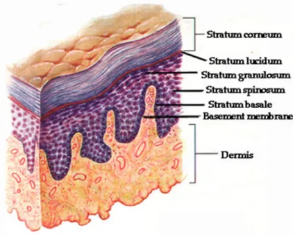
The skin is the largest organ of the body. In a 70 kg individual the skin weights over 5 kg covering a surface of 2 m2. Human skin consists of a stratified epidermis and an underlying dermis of connective tissue, which is organized into basal (stratum basale), spinous (stratum spinosum), granular (stratum granulosum) and cornified layers (stratum corneum), each layer consisting of keratinocytes of a specific morphology and state of differentiation (Figure 9), [154], [155], [156], [157], [158].
1 Introduction
24
Figure 9: Structure of the human skin (www.physioweb.org)
The keratinocytes account for more than 80% of the cells of the epidermis. They function as a barrier and contribute to skin repair and regeneration [159]. Important structural proteins of the vertebrate epidermis are keratins constituting up to 85%
of differentiated keratinocytes [160]. 20 different keratins are described for the human skin [161], [162], [163]. Typical keratins expressed in the mitotically active cells of the basal layer are keratins K5 and K14, which are considered to be biochemical markers of the epidermis [164]. K5 and K14 form intermediate filaments that assemble into strong networks, and anchor the epidermis to underlying layers of the skin. The network of keratin intermediate filaments provides strength and resiliency to the skin and provides protection from being damaged by friction and other everyday physical stresses [164], [165], [166]. Other important keratins are K1 and K10, which are the most abundant proteins in the upper epidermis where they polymerize to form intermediate filaments. In addition to their well-established function in providing epidermal stability, K1/K10 intermediate filaments are supposed to be important for terminal epidermal differentiation and barrier formation [167]. Point mutations of keratin genes can lead to severe diseases, many of which manifest as blistering skin diseases [166].
The most prominent of these inherited skin fragility disorders is epidermolysis bullosa simplex (EBS), of which the various variants are caused by a spectrum of point mutations of K5 or K14 [168], [169], [170].