Combinatorial engineering of adeno associated virus vectors
I n a u g u r a l – D i s s e r t a t i o n
zur
Erlangung des Doktorgrades
der Mathematisch-Naturwissenschaftlichen Fakultät
der Universität zu Köln
vorgelegt von
Stephan Märsch
aus Willich
Köln 2009
Berichterstatter/in: Prof. Dr. Dagmar Mörsdorf
Prof. Dr. Sigrun Korsching
Prof. Dr. Siegfied Roth
Tag der letzten mündlichen Prüfung: 02.07.2009
Die vorliegende Arbeit wurde in der Zeit vom September 2005 bis zum April 2009 unter Betreuung von Dagmar Mörsdorf im Genzentrum der Ludwig-Maximilians-Universität zu München und in der Medizinischen Klinik I des Universitätsklinikums zu Köln angefertigt.
Danksagung
Besonderer Dank gilt meinem Betreuer Dr. Luca Perabo, der mich durch intensive wissenschaftliche Betreuung und die vielen Freiheiten im Arbeiten, selbstständiges, wissenschaftliches Arbeiten gelerhrt hat.
Prof. Dr. Dagmar Mörsdorf möchte ich ganz besonders für die Bereitschaft danken, meine Promotionsarbeit trotz einiger aufregenden Situationen extern zu betreuen und sie so zu ermöglichen.
Ich danke Prof. Dr. M. Hallek für die Möglichkeit, meine Doktorarbeit in der Medizinischen Klinik I des Universitätsklinikums Köln anfertigen zu dürfen.
Prof. Dr. Sigrun Korsching danke ich für die Übernahme des Zweitgutachtens.
PD Dr. Hildegard Büning möchte ich für fruchtbare wissenschaftliche Diskussionen und die großzügige Hilfe durch Sie und Ihre Gruppe bei meiner Arbeit danken.
Ich danke Dr. Jan Endell für die praktische Einweisung in das Arbeitfeld der viralen Libraries.
Ich bedanke mich bei Dr. Paola Zigrino, Dr. Dirk Nettlebeck und Dr. Patrick Schmidt für die zur Verfügung gestellten Melanomzellen.
Ein ganz herzlicher Dank gilt den Kollegen aus meiner Arbeitsgruppe und den Kollegen aus den anderen Arbeitsgruppen der Ebene 4. Vor allem Anke, die mich in jeder Notlage selbstlos unterstützt hat, Miri, die meine Arbeit korrigiert hat, Jorge, T2 und Venkat, die meine Schwächen in WORD- formatierungs- und anderen EDV-Fragen geklärt haben, Hanna und Nina, mit denen man im richtigen Moment über etwas anderes als Arbeit sprechen kann und den vielen lieben Menschen, mit denen ich über die letzten Jahre Freud und Leid im LFI teilen durfte.
Ich danke vor allem meiner Freundin, meiner Familie und meinen Freunden, die mir Ruhe und Motivation gegeben haben, die man sich nur wünschen kann.
Die Natur kennt keine Probleme, nur Lösungen. Carl Amery
Für meine Familie
Table of Contents
1 Abstract ... 1
2 Zusammenfassung ... 2
3 Introduction ... 3
3.1 Human gene therapy ... 3
3.2 Adeno associated virus ... 4
3.2.1 Classification of adeno associated viruses ... 4
3.2.2 Genome organization of AAV2 ... 5
3.2.3 Capsid structure ... 7
3.2.4 Infection biology of AAV2 ... 8
3.3 AAV as a gene therapy vector ... 9
3.3.1 Production of recombinant AAV2 vectors ... 9
3.3.2 Characteristics of AAV ... 11
3.3.3 Immune responses to AAV vectors ... 12
3.3.4 Clinical trials with AAV vectors ... 13
3.4 Engineering of AAV vectors ... 15
3.4.1 Rational approaches ... 15
3.4.2 Evolutionary approaches ... 16
4 Results I ... 19
4.1 Aiming for selectivity ... 19
4.1.1 Design of a directed evolution protocol and a viral insertion library ... 19
4.1.2 Titration of adenovirus ... 21
4.1.3 Adjusting selective pressure ... 22
4.2 Selection of BLM specific mutants ... 24
4.3 Characterization of selected mutants ... 26
4.3.1 Packaging capacity of selected clones ... 26
4.3.2 Selectivity assay ... 27
4.3.3 Selected mutants are inhibited by heparin ... 30
4.3.4 Tropism for other cell types ... 31
5 Discussion I (Specificity selections) ... 33
6 Results II ... 37
6.1 Aiming for antibody evasion ... 37
6.1.1 Generation a complete randomization library (pdegen5Lib) ... 39
6.2 Selection of immune escaping mutants ... 40
6.2.1 Selection procedure ... 40
6.2.2 Selection results ... 43
6.3 Characterization of selected mutants ... 46
6.3.1 Packaging of GFP- rAAV vectors ... 46
6.3.2 Infection assays ... 48
6.3.3 Decoy assays... 51
6.3.4 Multiple cell infection and Heparin inhibition assay ... 53
7 Discussion II (Immune escape selections) ... 55
8 Conclusions and outlook ... 58
9 Materials ... 59
9.1 Chemicals, solutions and enzymes ... 59
9.2 Standard kits ... 60
9.3 Plasmids ... 60
9.4 Primers ... 61
9.5 Antibodies and sera ... 62
9.6 Bacteria strains ... 62
9.7 Eukaryotic cells ... 63
9.8 Data treating software ... 63
9.9 Laboratory equipment, disposables ... 64
10 Methods ... 66
10.1 Bacteria culture ... 66
10.1.1 Cultivation of bacteria ... 66
10.1.2 Glycerol stocks ... 66
10.1.3 Preparation of chemically competent bacteria ... 66
10.1.4 Chemical transformation of bacteria ... 67
10.1.5 Preparation of electrocompetent bacteria ... 67
10.1.6 Electrical transformation of bacteria ... 68
10.2 DNA techniques ... 68
10.2.1 Plasmid amplification and extraction, DNA cleanup ... 68
10.2.2 DNA quantification ... 69
10.2.3 Restriction enzyme digest ... 69
10.2.4 Agarose gel electrophoresis ... 69
10.2.5 Sequence analysis ... 69
10.2.6 Cloning of the pdegen5Lib library ... 70
10.2.7 Library ligation ... 73
10.3 Eukaryotic cell culture ... 74
10.3.1 Cultivation of cells ... 74
10.3.2 Trypsinisation ... 74
10.3.3 Seeding / passaging ... 74
10.3.4 Freezing and thawing cells ... 74
10.3.5 Cell counting ... 75
10.4 Production of AAV2-vectors ... 75
10.4.1 AAV-vector packaging ... 75
10.4.2 Iodixanol gradient purification ... 76
10.4.3 Virus titration ... 77
10.4.4 Capsid titer (ELISA) ... 79
10.5 Infection assays and selection procedures ... 80
10.5.1 Titration of adenovirus on BLM ... 80
10.5.2 Decoy assay on fibroblasts and keratinocytes ... 80
10.5.3 Selection protocol for BLM-specific mutants ... 80
10.5.4 Immune escape selection protocol ... 81
10.5.5 Infection assay (BLM cell specificity) ... 82
10.5.6 Infection assays (Immune escape mutants) ... 82
10.5.7 Decoy assay ... 82
10.5.8 Heparin inhibition assay ... 83
11 References ... 84
12 Abbreviations ... 100
Erklärung ... 103
Publikationen ... 104
Lebenslauf ... 105
1
1 Abstract
The use of viral vectors as delivery vehicles for therapeutic genes (gene therapy) is being investigated and improved since two decades. Despite exciting advances and promising results in animal studies and clinical trials, technical refinements are still needed to improve efficiency and safety of gene transfer. One of the most popular vector types derives from adeno associated virus of type 2 (AAV2). This virus has attracted the interest of gene therapists being safe, easy to produce at high titers and offering the possibility to target a plethora of cell types and to maintain detectable levels of transgene expression for long times.
However, two major limitations hamper the use of this vector type. First, the broad tropism of AAV means that vectors are rather unspecific and therefore likely to infect neighboring tissues. Second, high seroprevalence in human populations of antibodies directed against wtAAV2 capsids reduces or abolishes transduction rates in individuals with pre-existing immunity.
The goal of my work was to address these two major issues and develop new methods to engineer AAV capsid variants with increased target selectivity and decreased antibody recognition.
Within a first project, I established for the first time a straight-forward in vitro protocol for the selection of viral mutants with increased selectivity for a melanoma cell line (BLM) out of a combinatorial viral library. The selection procedure consisted of a negative selection step on non-target cells and a positive selection step on BLM cells. Selected mutants showed almost wt levels of infectivity on BLM cells while transducing several tested non-target cells with a significantly lower efficiency, resulting in a up to 3.7-fold increase of target specificity.
Within the second project I generated a novel AAV2 capsid library by randomizing five amino acid residues that had been previously reported to play a role in antibody docking to the viral shell. Screening this library for particles that remained infectious despite pre- incubation with neutralizing antibodies allowed the isolation of several capsid variants that remained highly infectious even in the presence of antibody concentrations that completely neutralize wtAAV2.
2
2 Zusammenfassung
Die Verwendung viraler Vektoren als Genfähren zum Transfer therapeutische Gene in menschliche Zellen wird seit über zwei Jahrzehnten erforscht. Trotz aufregender Neuerungen und vielversprechender Ergebnisse aktueller Tierexperimente und klinischer Studien muss die Sicherheit und Effektivität dieser neuen Technologie noch weiter verfeinert werden. Das Adeno-assoziierte Virus vom Serotyp 2 (AAV2), ein sicheres, effektiv herstellbares Virus ist einer der beliebtesten Vektoren zum Einsatz in der Gentherapie, da es eine stabile Genexpression in einem breiten Spektrum verschiedener Zelltypen über lange Zeit ermöglicht.
Zwei Nachteile behindern den Einsatz dieses Virus jedoch in besonderem Maße: Auf der einen Seite zieht der breite Tropismus bei systemischer Gabe die unspezifische Transduktion von Geweben nach sich, die nicht infiziert werden sollen. Zum Zweiten ist ein Großteil der Menschheit schon unbemerkt mit dem Virus in Kontakt gekommen und verfügt über neutralisierende Antikörper, welche die Transduktion der Zielzellen durch die Genfähren verhindern.
Ziel der beiden Projekte meiner Arbeit war die Entwicklung neuer kombinatorischer Strategien um die oben genannten Einschränkungen zu beseitigen.
In einem Projekt wurde ein in-vitro Protokoll entwickelt, mit dem Virusmutanten mit gesteigerter Spezifität für eine Melanomzelllinie (BLM) aus einer kombinatorischen
„Viruslibrary“ selektiert werden konnten. Das Protokoll besteht aus einem Negativselektionsschritt auf Fibroblasten und einem anschließenden Positivselektionsschritt auf BLM-Zellen. Die selektierten Mutanten zeigten mit Wildtyp vergleichbare Infektionslevel auf BLM-Zellen und signifikant herabgesetzte Transduktion der Fibroblasten und weiterer Nicht-Zielzellen.
Im zweiten Projekt habe ich eine neuartige „AAV2-Kapsidlibrary“ generiert, in der fünf zuvor als wichtig für Antikörperbindung charakterisierte Aminosäurepositionen vollständig randomisiert wurden. Nach vier Selektionsrunden mit neutralisierendem Serum konnte ich mehrere Mutanten isolieren, die trotz Anwesenheit von Serumkonzentrationen infektiös waren, welche den Wildtyp vollständig neutralisierten.
3
3 Introduction
3.1 Human gene therapy
Delivery of therapeutic genes to diseased tissues (gene therapy) holds the potential to heal a broad range of genetic and non-genetic pathologies by correcting genetic defects or by controlling the cellular metabolism by regulating gene expression. Different methods to bring foreign DNA into human cells ex vivo or in vivo are being developed. These techniques can be divided in two main groups. Non-viral methods exploit the possibility to deliver naked DNA/RNA or to couple this genetic information with different compounds (e.g. coating the nucleic acid with polyethylenglycole or lipofectamine). The other possibility is to use viruses as delivery vehicles. Until today, more than 1340 clinical trials have been conducted worldwide (Figure 1), aiming at delivering a plethora of different genes to address a wide variety of diseases.
Figure 1: Overview over clinical trials with gene therapy vectors (taken from http://www.wiley.co.uk/genetherapy/clinical/)
Over 65% of clinical trials worldwide use viral vectors as vehicles. Natural viruses have evolved to deliver their genetic cargo into host cells and these characteristics can be exploited to deliver therapeutic genetic material in gene therapy approaches. However, before they can
4
be successfully employed, natural viruses need to be adapted to medical usage. In particular, two aspects are fundamental: safety and efficacy.
In 1999, Jesse Gelsinger, an 18 years old Ornithine Transcarbamylase (OTC) deficiency patient died in Pennsylvania, USA, after treatment with a high dosage of adenoviral vector containing a normal copy of the OTC gene. The following investigation allowed to conclude that the subministred vector triggered a severe immune reaction leading to multiple organ system failure (Somia and Verma 2000).
In 2002, a retroviral vector containing the common γ-chain gene for the cytokine receptors IL- 2R, IL-4R, IL-7R, IL-9R and IL-15R was inoculated to ten X-linked severe combined immunodeficiency (X-SCID) patients. X-SCID normally leads to death within the first year of life. More than seven years later, 3 out of 11 patients have developed T-cell Leukemia as a consequence of the vector administration. One of them died of the cancer. The others live without SCID symptoms, two of them still fighting against leukemia (Cavazzana-Calvo et al.
2005; Cavazzana-Calvo and Fischer 2007). This example emphasizes the promises but also the challenges of this technology.
Advances in understanding diseases at molecular level and a better understanding of strengths and weaknesses of viral vectors is improving success rates of gene therapy protocols. Since the issues in this context are peculiar for each different vector type, the following paragraphs will focus on summarizing the relevant aspects with regard to the viral vector that has been the subject of my work: the adeno associated virus (AAV).
3.2 Adeno associated virus
3.2.1 Classification of adeno associated viruses
Adeno associated viruses (AAV) are members of the family of the parvoviridae. These non- enveloped viruses consist of a small icosahedral capsid with a diameter of 18-26 nm. The capsid contains a single-stranded DNA genome of approximately 4.7 kilobases. Parvoviruses can be divided into vertebrate- and insect-infecting viruses, classified as parvovirinae and densovirinae respectively. Human parvoviruses are not known to cause any disease except for erythrovirus B19 (Brown 2000) that was isolated by J. R. Pattison in the 1970s and can cause erythema infectiousum, hydrops fetalis and abortion. The first isolate of AAV was identified as contaminant in a simian adenovirus 15 preparation and therefore named adeno associated
5
virus. To date, thirteen other serotypes and more than 100 variants have been isolated in humans, other primates, rodents, or invertebrates. The serotypes have different levels of sequence homology causing differences in host and cell tropism and recognition by antibodies (Lukashov and Goudsmit 2001; Gao et al. 2002; Calcedo et al. 2009). In addition, AAV is classified in the genus of dependoviruses and in contrast to autonomous parvoviruses it requires for replication external helper functions that can be supplied by other viruses (e.g.
adenovirus, herpes simplex virus, vaccine virus, human cytomegalovirus or papilloma virus) or by physical or chemical factors (e.g. UV- or γ-radiation, heat shock, cancerogenic compounds) (Atchison, Casto et al. 1965; (Richardson and Westphal 1981; Schlehofer et al.
1986; Yalkinoglu et al. 1991). AAV2, the best analyzed serotype, was found in an adenovirus 12 contamination (Hoggan et al. 1966). Its atomic structure is resolved to three angstrom (Xie et al. 2002) (see 3.2.3).
3.2.2 Genome organization of AAV2
In 1982, the complete nucleotide sequence of AAV2 was determined (Srivastava et al. 1983).
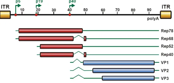
The single stranded DNA genome consists of 4675 base pairs (bp) coding for two open reading frames (ORFs). The cap gene codes for three structural proteins that build the viral shell while the rep gene encodes four non-structural proteins needed for replication and encapsidation of the viral genome. The ORFs are flanked by 145 bp long inverted terminal repeats (ITRs) that build a T-shaped hairpin structure at both ends of the viral genome and are used as origin of replication, as packaging signals and for the integration or excision of the viral DNA into the host genome (McLaughlin et al. 1988; Kotin et al. 1990)(see Figure 2).
The four multifunctional proteins Rep78, Rep68, Rep52 and Rep40 (named according to their molecular weight in kilodalton) are expressed under control of promoters p5 (Rep78; Rep68) and p19 (Rep 52; Rep 40), named after their position in the genome, which was divided into 100 map units. Rep expression results in four proteins, Rep68 and Rep40 being splice variants of their larger counterparts. All Rep proteins share helicase and ATPase activities, while Rep78 and Rep 68 also have site specific DNA-binding, and endonuclease activities (Im and Muzyczka 1990; Berns and Giraud 1996; Smith and Kotin 1998; Collaco et al. 2003). Rep72 and Rep68 contain nuclear localization signals essential for replication, transcription and site- specific integration into the host genome (Cassell and Weitzman 2004) while Rep 52 and Rep40 are responsible for helicase-dependent packaging of the viral genome into the preformed capsid through pores (Becerra et al. 1985; Wistuba et al. 1997; Dubielzig et al.
6
1999; King et al. 2001; Bleker et al. 2005). Rep proteins also control the splicing of cap and their own expression in the absence of helper effects by repressing p5 and p19 transcription (Pereira et al. 1997; Kronenberg et al. 2001; Qiu and Pintel 2002).
Figure 2: Organisation of the AAV2 genome and gene products. Two ORFs rep and cap are expressed under the control of three promoters p5, p19 (rep) and p40 (cap) named after their map unit position in the 4675 bp genome that is divided into 100 map units. The ORFs are flanked by inverted terminal repeats . A shared poly adenylation signal at unit position 96 is shared by all transcripts, while use of alternative splicing, alternative promoters and alternative start codons results in four rep genes, responsible for replication and three cap genes that built up the viral shell. Open reading frames are shown in cylinders, untranslated regions as solid lines and introns as kinks.
Three structural proteins VP1, VP2 and VP3 that share the same C-terminus are expressed from the cap ORF under the control of a promoter at map position 40 (p40). With respective molecular masses of 87, 72 and 62 kilodaltons (kDa), the three proteins are translated from two transcripts that arise from alternatively splicing of the cap gene. The longer one is translated into VP1, the largest of the three proteins that carries a phospholipase A2 domain at its N-terminus required for efficient infection (Zadori et al. 2001; Girod et al. 2002). VP2 and VP3 are translated from the shorter transcript from two separate start codons that supposedly have different translational activity. This gene architecture results in a VP1, 2 and 3 protein production at a 1:1:8 ratio respectively, which provides the appropriate proportion of capsid subunits (Becerra et al. 1988) to form a viral capsid of 60 subunits which is assembled with T
= 1 icosahedral symmetry (Xie et al. 2002) by 5 VP1, 5 VP2 and 50 VP3 subunits. The numbering of amino acid positions in capsid proteins refers to the VP1 counting in the following.
7 3.2.3 Capsid structure
Recently the atomic structure of AAV-2 has been determined to 3 Å resolution by x-ray crystallography. Each viral capsid is composed of 60 subunits arranged with T=1 icosahedral symmetry of the three structural proteins VP1, VP2, and VP3 (Xie et al. 2002). They share overlapping sequences and differ only at
their N-termini but these portions could not be resolved in the 3-D structures due to low electron density. Between the strands of a β-barrel core (Fig. 2) which is highly conserved among parvoviruses, large loop insertions are found that share only low similarity among the parvovirus family. These loops comprise two-thirds of the capsid structure and constitute the capsid surface features that interact with antibodies and cellular receptors.
Three of these loops contribute to the formation of the 3-fold-proximal peaks, which cluster around the 3-fold symmetry axis. In the valleys separating the three peaks of one 3-fold axis, clusters of positive charges are located, which are implicated in receptor binding. Mutational analyses have identified these locations being involved in binding to the primary receptor of AAV-2 (Wu et al. 2000). The basic aa R487, R585, R588 and H509, which are at the side of the peak seem to play a crucial role (Wu et al. 2000; Grifman et al. 2001; Xie et al. 2002;
Opie et al. 2003). Mapping of antibody binding epitopes (Wobus et al. 2000) (Huttner et al.
2003) have suggested the importance of the 3-fold proximal peaks not only in receptor binding, but also for the recognition of the viral particles by antibodies. Moreover, it is possible that other important viral functions are also located at this prominent feature.
Since the VP1 and VP2 regions of the capsid proteins are located within the lumen of the viral particle (Kronenberg et al. 2005), all virus:cell interactions that take place before entry and the interations between capsid and antibodies only depend on the surface of the VP3 protein.
Fig. 2: 3D structure of the VP3 capsid protein (Xie et al. 2002). See text for details. Blue numbers and lines indicate the symmetry axes.
8
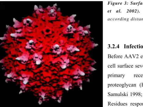
Figure 3: Surface topology of AAV-2 (Xie et al. 2002). The capsid is colored according distance from the viral center.
3.2.4 Infection biology of AAV2 Before AAV2 enters a cell, it contacts the cell surface several times and binds to its primary receptor heparan sulfate proteoglycan (HSPG) (Summerford and Samulski 1998; Seisenberger et al. 2001).
Residues responsible for HSPG binding have been described to be R484; R487; K532; R585 and R589 (Kern et al. 2003; Opie et al.
2003). For efficient uptake, the virus must also interact with additional secondary receptors.
Human fibroblast growth factor receptor 1, hepatocyte growth factor receptor, laminin receptor, the integrins αvβ5 or α5β1 have been proposed as secondary receptors (Qing et al.
1999; Summerford et al. 1999; Kashiwakura et al. 2005; Akache et al. 2006; Asokan et al.
2006) but a role of other molecules cannot be excluded.
After binding its receptors, the virus can enter the cell by receptor-mediated, dynamin dependent endocytosis through clathrin coated pits (Duan et al. 1999; Bartlett et al. 2000).
The exact events leading to internalization however are not completely understood. After moving along microtubules and microfilaments towards the nuclear area (Douar et al. 2001), AAV2 particles escape from endosomes after their acidification and accumulate in the perinuclear area (Bartlett et al. 2000). Rac1 and the PI3K pathway play an essential role in the efficiency of this step of infection (Sanlioglu et al. 2000).
It is not clear whether only genomes, protein-DNA complexes or whole particles of AAV2 enter the nucleus (Sanlioglu et al. 2000; Lux et al. 2005). Due to their small diameter, whole particles could enter through nuclear pore complexes (NPC) and an interaction of the viral capsid with a nuclear shuttle protein has been reported (Qiu and Brown 1999). However, NPC independent pathways have been suggested by other experimental data (Hansen et al. 2001). It is also important to note that different molecular pathways could be used for infection
9
depending on cell type, virus load, presence or absence of helper function and kind of helper function (Hansen et al. 2001; Xiao et al. 2002; Lux et al. 2005).
In the absence or helper effects, AAV2 converts its single stranded DNA into a double stranded form and integrates it into the host genome or stay in an episomal state. Integration occurs only in the presence of Rep proteins and mostly at the so called AAV site 1 (AAVS1) on chromosome 19 (Kotin et al. 1992; Nakai et al. 2001) which resides rep binding element and terminal resolution site sequences identical to those of AAV2 (Weitzman et al. 1994;
Linden et al. 1996).
After co-infection with helper viruses or in the presence of a helper effect like chemical or physical stress of the cell, integrated provirus can be rescued and initiate a lytic life cycle (Berns and Giraud 1996). In this case, a double stranded genome is produced to induce gene expression and replication (Goncalves 2005).
.
3.3 AAV as a gene therapy vector
3.3.1 Production of recombinant AAV2 vectors
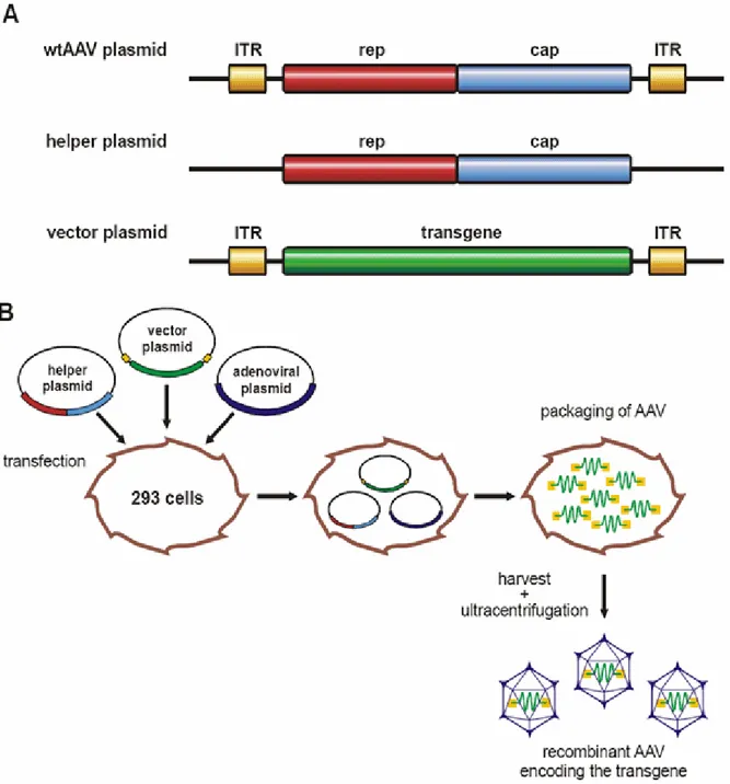
In our days, the genomes of AAV2 and other serotypes are available on plasmids. In gutless vectors, the viral genome is replaced by a desired transgene of up to 6 kb, only flanked by viral ITRs that are needed for packaging and gene expression. Producer cells (e.g. HEK293, transgenic for adenoviral genes E1A, E1B) are transfected with plasmids providing AAV rep and cap genes plus additional helper functions. For production of viral libraries or cloning constructs of wtAAV2, the rep and cap genes must be flanked by ITRs. When gutless vectors are produced, only the transgene, added to the transfection mixture on an additional plasmid is flanked by ITRs (see Figure 4)(Laughlin et al. 1983; Rolling and Samulski 1995; Xiao et al.
1998; Collaco et al. 1999; Allen et al. 2000; Grimm et al. 2003).
The viral progeny can be harvested by freeze and thaw cycles and purified on affinity columns (e.g. heparin-columns), iodixanol- or CsCl- gradients (Hermens et al. 1999;
Zolotukhin et al. 1999; Anderson et al. 2000). Preparation quality and yield can be quantified by quantitative polymerase chain reaction (qPCR) and enzyme linked immunosorbent assay (ELISA).
10
Figure 4: Production of adeno virus free rAAV vectors. HEK293 cells are transfected with three plasmids carrying rep and cap genes without ITRs in trans on a helper plasmid, adenoviral helper functions on a adenoviral plasmid and the desired transgene flanked by ITRs on a vector plasmid. The virions package within 48 hours and can then be isolated and purified.
Since second strand synthesis is a limiting step for gene expression, self-complementary genomes have been developed (McCarty et al. 2001), enhancing the expression rate in vitro and in vivo but limiting the size of the transgene to half the size. Other optimizations have been made to enhance purity and scale of AAV preparations during the last years (Blouin et al. 2004; Van Vliet et al. 2008).
11 3.3.2 Characteristics of AAV
Depending on the aims of gene therapy trials, requirements for a vector can differ. Therefore, it is hard to judge general advantages and disadvantages of some of AAV´s features. Anyway, I want to point out AAV-specific characteristics in this chapter to reveal its adequacy for gene therapy.
The first and most important requirement for a gene therapy vector is safety. No human disease could be related to AAV serotypes and the only known disease is caused by human parvoviral pathogene iserythrovirus B19 (Brown 2000). AAV has been used in over twenty clinical trials conducted to target a variety of diseases and to date no severe toxicity or immune reactions (see 3.3.3) have been associated with vector subministration. With helper virus free production methods and gutless vectors, AAV vectors are not contaminated with other viruses and cannot replicate themselves (Samulski et al. 1987). They integrate specifically at AAVS1 site in vitro if rep is given in trans (Huttner et al. 2003). Integration ath this site does not perturb host cell biology as it can be the case for retroviruses. However, recently some studies have reported occasional events of non-specific chromosomal integration. In vivo, stable, circular double-stranded episomes allocate the majority of wild- type AAV DNA (Kotin et al. 1992; Duan et al. 1998; Nakai et al. 2001; Miller et al. 2002;
Philpott et al. 2002; Schnepp et al. 2005).
AAV vectors are capable of long term transduction of targeted tissues. Expression of vector transgene was detectable up to over six years after application in some cases (Romano 2005;
Warrington and Herzog 2006).
Due to the availability and heterogeneity of different AAV serotypes and variants a broad range of target organs like brain, heart, muscle, liver and lung; dividing and non-dividing cells can be targeted (Flotte et al. 1993; Kaplitt et al. 1994; Podsakoff et al. 1994; Alexander et al.
1996; Kaplitt et al. 1996; Fisher et al. 1997; Manno et al. 2006).
The broad tropism of AAV vectors might be explained by the wide diffusion of their receptors (e.g. HSPG and integrins) on the surface of many cell types. While this allows the use of AAV vectors for a large number of therapeutic applications, it represents a disadvantage for applications requiring specific and selective transduction of target tissues. A typical example is cancer therapy with suicide genes, where unspecific transduction of neighboring tissues would cause severe collateral damage. In addition, unspecific vectors are dispersed in undesired organs and therefore higher doses of the vector must be applied.
12
Another limitation of AAV vectors is their packaging capacity of 5-6 kb. Recently, different techniques showed that larger transgenes can be split and delivered by so called “trans- splicing vectors” (Dong et al. 1996; Grieger and Samulski 2005; Ghosh et al. 2007).
3.3.3 Immune responses to AAV vectors
Up to 96% of humans are seropositive to AAV2 and up to 60% have neutralizing antibodies (Nabs) against the virus. The prevalence of Nabs is spread worldwide but depends on age, country, and ethnic group generally being highest in Africa for common serotypes (Erles et al.
1999; Calcedo et al. 2009). Although non-hazardous for the patient, neutralization of gene therapy vectors in the blood would prevent transduction of target cells or organs (Scallan et al.
2006; Lin et al. 2008). Some newly isolated, non-human viruses like AAV8 or AAVrh32.33, have a much lower prevalence of neutralizing antibodies (less than 10% in the human population), do not cross react with anti-AAV2 antibodies and have therefore attracted the interest of the scientific community as alternatives to circumvent pre-existing immunity (Gao et al. 2002; Scallan et al. 2006; Calcedo et al. 2009). However, not all serotypes are suitable for human gene therapy due to low infectivity or undesired tropism.
Animal studies also suggest, that even naïve patients would develop antibodies against any serotype or variant after a first treatment. Re-administration of AAV is therefore restricted to serotypes that do not cross-react with antibodies against the serotype used in the first treatment and that can provide sufficient transduction rates in the targeted tissue (Halbert et al.
2000; Moskalenko et al. 2000). For chronic diseases and long-time treatment, immunologic reactions can be suppressed efficiently for every consecutive treatment (Manning et al. 1998).
This treatment however is cumbersome, uncomfortable and risky for the patient. Quarantine is required since infection with other pathogens during treatment can lead to serious complications without an intact immune system.
In contrast to other viruses, AAV has minimal inflammatory potential and does not trigger severe immune responses. AAV2 is able to infect dendritic cells but due to an unknown post- entry block, they are not able to efficiently activate a T-cell response (Zhang et al. 2000). The cellular immune response against AAV was disregarded for years until liver inflammation was observed in patients after AAV2 treatment (Manno et al. 2006). Because human is a natural host of AAV2 and liver damage was not described in mice, it was suggested that cellular immune response was triggered by former co-infections of the patients with wtAAV
13
and immune activating helper viruses that occurred incidentially before the begin of therapy (Mingozzi et al. 2007). However, cellular immune responses against the capsid have been reported in human and animal studies (Manno et al. 2006; Mingozzi et al. 2007;
Vandenberghe and Wilson 2007; Wang et al. 2007).
Since gutless AAVs do not express viral capsid or replicative genes, activation of CD8+ T- cells after infection of cells and degradation of the capsid must occur through exogenous antigen presentation. It has been demonstrated that AAV-capsid specific T-cells do not lyse rAAV-infected cells (Li et al. 2007; Li et al. 2007; Wang et al. 2007). In addition, immunologic reactions against endogenous transgenes have not been reported, indicating that effective long-time expression can be reached with AAV vectors by immunosuppression during treatment until the clearance of vector capsid is complete.
3.3.4 Clinical trials with AAV vectors
Being considered a safe and efficient vector with long-time gene expression potential, AAV is being adopted in an increasing number of clinical trials aiming to cure a wide spectrum of diseases like Parkinson, Canavan`s disease, α1-antitrypsin deficiency, rheumatoid arthritis, cystic fibrosis and Hemophilia B (Goncalves 2005).
The first disease that was treated with AAV was a phase I study of patients with mild lung disease of cystic fibrosis transmembrane conductance regulator deficiency (Flotte et al. 1996).
The deficiency leads to chronic inflammation and lung damage which lead to early death because of loss of respiratory function. Clinical phase I and II trials have been conducted today with promising outcome for the future but no therapeutically relevant long-time expression has been reported so far.
Interesting results are announced from trials concerning hemophelia B (blood factor IX deficiency), a monogenic defect leading to failure in blood clotting. Tested doses of administered vectors were not causing immunologic side effects but were also too low to raise serum factor IX levels above 1% of normal, the minimal concentration needed for effective blood clotting. Newer results show however that increasing vector doses and switching from intramuscular to intraportal administration leads to liver transduction and therapeutical relevant levels of factor IX expression but also to liver inflammation accompanied by decline of factor IX levels after eight weeks (Manno et al. 2006).
14
Similar results are reported from lung disease trials against α-1 antitrypsin deficiency (ATT).
Transient gene expression was reported, but higher levels are needed for therapeutic treatment (Brantly et al. 2006).
In Leber congenital amaurosis (LCA), a severe retinal dystrophy causing blindness or severe visual impairment before the age of 1 year, AAV therapies have already achieved promising results. One form of the disease, LCA2, is caused by mutations in the retinal pigment epithelium-specific 65-kDa protein gene (RPE65). No severe side effects were diagnosed and patients reported increased sensitivity of sight after AAV-RPE65 treatment (Jacobson et al.
2006; Hauswirth et al. 2008; Maguire et al. 2008; Cai et al. 2009).
Lately, a patient treated with an AAV2 vector in a trial concerning rheumatoid arthritis died but the following investigation allowed to demonstrate that the death was not related to the administered vector but as consequences to anti-TNF therapy drug treatment that caused disseminated histoplasmosis in conjunction with retroperitneal hematoma instead (Kaiser 2007).
Anti-cancer gene therapies studies have not yielded broad success in the clinic. However, since being promising for future therapies, some of the general ideas are listed in the following paragraph. Anti-cancer gene therapies can be classified by different approaches used. Immunotherapies aim at enhancement of tumor cell recognition by the immune system.
Malignant cells can be loaded in vivo or ex vivo with immunostimulatory molecules (e.g.
tumor necrose factor α (TNF-α) or CD 40 ligand). Also vaccination of dendritic cells with tumor antigens or ex vivo transduction of T-cells with tumor specific T-cell receptors activate the immune system and increase recognition of tumor tissue (Nestle et al. 2005; Morgan et al.
2006; Aiuti et al. 2007; Alexander et al. 2007). Some naturally occurring oncolytic viruses and their laboratory derived relatives selectively replicate in cancer cells, causing cell death and infecting neighboring cells. However, non of the tested viruses was applicable systemically but reported effects were always local (Hu et al. 2006; Aiuti et al. 2007). Suicide gene therapy is a promising approach to effectively abolish tumor cells, especially for metastatic tumors that are hard to treat with irradiation, surgery or local drug application. The concept is to deliver suicide genes exclusively to tumor cells or tumor vascular system cells to directly kill the tumor cells or cut the tumor cells off from oxygen and nutrients with a prodrug that is applied systemically after infection of the target cells with the suicide gene (e.g. herpes simplex thymidine kinase (Hstk) and glanciclovir, a prodrug that becomes toxic
15
only after being metabolized by Hstk) (Plautz et al. 1991; Niculescu-Duvaz and Springer 2005; Alexander et al. 2007).
In summary, AAV clinical trials for several diseases are at a stage where safety has been proven and vector doses need to be adjusted to gain therapeutic relevant gene expression levels (Romano 2005; Warrington and Herzog 2006). Optimization of vector construction for effective targeting, immune escape and long time transgene expression of the transgene has to be refined and immune modulation during treatment considered.
3.4 Engineering of AAV vectors
The growing understanding of mechanisms underlying the infectious biology of the AAV capsid and its role in the infectious process have paved the way for efforts aiming at engineering viral particles at molecular level in order to tailor them with optimized characteristics required by the envisioned application.
To control packaging efficacy, host immune reactions against AAV, tropism, specificity, infectivity, genome size, intracellular trafficking and gene expression of AAV are major goals that would dramatically contribute to the breakthrough of AAV vectors in clinical routine.
Vector tropism depends on the surface architecture and chemical composition of the capsid surface. The structures presented to the external surface of the viral shell mediate interactions with the humoral immune system and with cellular receptors responsible for the binding and uptake of the particle into the cell. These structures can be chemically or genetically modified to modify the way they interact with the host environment. Viral particles can be engineered by two major approaches: rational approaches and evolutionary approaches.
3.4.1 Rational approaches
In a rational approach, scientists make an educated guess which amino acids or protein domains are to be changed in a defined way. Substitutions, insertions or deletions are chosen on purpose to yield a specific, intended phenotype of the generated mutants.
On the on hand, wtAAV vectors were coated with chemical reagents such as polyethylenglycol or biological molecules, e.g. bispecific antibodies or avidin linked ligands to change their tropism (Bartlett et al. 1999; Ponnazhagan et al. 2002). Although showing
16
altered tropism in vitro, critics argue that binding of coated reagents might not persist in vivo and attached molecules could interfere with post entry steps of the vector.
In rational genetic approaches, amino acids have been substituted or peptides have been inserted into the capsid. A single-chain antibody against human CD34 was genetically inserted at the N-terminal region of capsid proteins in an effort to target hematopoietic progenitor cells (Yang et al. 1998). In another case, both genetical modification and chemical coupling were combined: Ried et al. inserted an immunoglobulin (IgG) binding domain at position 587 of the capsid protein to enable rAAV to bind different antibodies via their Fc regions. The Fab region of bound IgGs thus remains free to function as a ligand directed against a specific cell surface receptor (Ried et al. 2002). in vitro results show that the tropism of AAV vectors could be expanded to non-permissive cell types by inserting a 14-amino-acid peptide, L14 (QAGTFALRGDNPQG), at position 587 of the AAV2 capsid protein (Girod et al. 1999). In a different approach, single or multiple amino acid residues on the surface of the capsid were substituted or deleted and the resulting phenotype was characterized. Mutation of two crucial arginines led to the disruption of AAV binding to its primary receptor HSPG and therefore detargeting of a number of cell types; other substitutions caused increased resistance against neutralizing serum (Opie et al. 2003; Lochrie et al. 2006). Combination of this detargeting and retargeting strategies were combined to create mutants with new tropism.
AAV mutants with inserted RGD-4C peptide at positions 520 and 584 showed loss of Heparin-binding and altered tropism (Shi et al. 2006).
As an additional possibility, capsid proteins of different serotypes were mixed, leading to the formation of chimeric capsids with altered phenotype (Gigout et al. 2005; Wu et al. 2006).
A general drawback of rational design approaches is the unpredictable change of the nature of insertions after integration into the capsid. To build up a viable capsid, the VP protein structure has to fulfill numerous specifications; educated guesses in mutagenesis of the viral shell often leads to non-functional VP protein. Insertions tend to not function in the originally expected way, because the course of capsid assembly and interactions between essential structure domains are not well-enough understood.
3.4.2 Evolutionary approaches
Controversy to rational approaches, evolutionary techniques create a very large number of randomly mutagenised variants of a biomolecule and thereafter screen this “library” for beneficial mutations without trying to predict the specific changes needed for the desired
17
phenotype. The general advantage of this technique is that it does not require detailed understanding of relationship between protein structure and function. On the other hand, generated libraries must be complex enough to include beneficial mutants and screening methods must be stringent enough to identify the latter ones.
Evolutionary techniques have proven to be useful for protein optimization before being applied to viruses (Stemmer 1994; Vasserot et al. 2003). Directed evolution approaches for viral vectors are based on a general principle: the capsid surface is mutated randomly and large libraries of variants are generated. In this way, viral libraries are constructed carrying mutants with properties differing from those of naturally occurring serotypes or variants. The number of all serotypes and variants known until now, taken together is exceeded more than thousand-fold by the produced number of variants in one such library. Out of this large pool of mutants, beneficial variants can be selected using high-throughput screening methods even if specific knowledge of capsid biology is limited. Packaging-, infection- and replicative- defective or -poor mutants can easily be ruled out during selections and mutants with beneficial phenotype for the desired application can be selected in clonal purity. Different techniques that have been previously applied in the field of AAV vectors are shortly introduced in this paragraph.
Selection of novel peptides from phage libraries and subsequent insertion into AAV:
tissue specific targeting peptides are identified by screening phage display libraries and subsequently inserted genetically in the capsid of AAV vectors (Nicklin et al. 2001; Work et al. 2005). Enhanced transduction of vascular endothelia cells was observed after insertion of a 7mer SIGYPLP peptide at position 587 (Nicklin et al. 2001). Grifman et al. have incorporated a tumor-targeting peptide previously identified from phage display into rAAV2, which successfully shifted vector tropism (Grifman et al. 2001). However, as for rational design insertions, peptides identified in different viral contexts often lose or abate biological function once inserted in the architectural context of AAV particles. Moreover, AAV virions can lose stability or functionality as consequence of the peptide insertion (Kwon and Schaffer 2008).
Insertion libraries: In this approach, randomized peptides are introduced at specific AAV capsid sites that have been previously proven to tolerate insertions in rational design studies (Girod et al. 1999). Site 587 was used by Perabo et al. to insert a random peptide library and select mutants with up to 100-fold infectivity on M-07e, a human megakaryocytic and a B- cell chronic lymphocytic leukemia cell line (Perabo et al. 2003). Human coronary artery endothelial cells were targeted in an analougous approach (Muller et al. 2003). These
18
insertion libraries can also be panned to select mutants with immune escaping phenotype (Muller et al. 2003; Perabo et al. 2003; Maheshri et al. 2006; Perabo et al. 2006). In these insertion library approaches, the selected peptides are optimized for functioning in the architectural environment of the AAV capsid.
Random mutagenesis libraries: Long stretches of the viral capsid can be randomly mutagenised, e.g. by error-prone PCR in order to introduce random point mutations. Point mutations were spread all over the viral capsid and immune escaping mutants were isolated out of such libraries (Perabo et al. 2006). These libraries also can be used to select mutants with altered receptor binding, tropism and transduction efficiency (Maheshri et al. 2006).
Recently, such a library has been used for in vivo selection for tumor and lung targeting mutants (Michelfelder et al. 2009).
Shuffling libraries: Natural biodiversity among AAV serotypes can be exploited by applying shuffling protocols, to swap capsid domains between serotypes via homologous recombination and to generate chimeric capsids. This procedure was used to generate mutants with mixed or novel phenotypes. AAV2 and AAV3 capsids were shuffled together and subjected to a Marker rescue assay, which reports the restoration of gene function by replacement of a defective gene with a normal one by recombination, resulting in chimeric particles with new characteristics such as altered receptor binding and infectivity (Bowles et al. 2003; Hauck et al. 2003). Hauck and colleagues combined capsids of AAV1 and AAV2, obtaining a library they were able to isolate mutants with combined beneficial features of both serotypes out of. A clone containing capsid fragments of serotypes 1, 2, 8 and 9 was isolated from an integrin minus hamster melanoma cell line previously shown to have low permissiveness to AAV. The mutant showed increased transduction rates on this celline and decreased antibody recognition compared to wild type serotypes (Li et al. 2008).
All combinatorial methods can be transferred to other serotypes and vectors, enhancing their phenotypes for the desired purposes.
19
4 Results I
4.1 Aiming for selectivity
Application of viral vectors with wide and unspecific tropism in clinical applications may translate in dispersion of vector particles in non-target tissue and unleash side effects in unspecifically transduced organs. AAV2-derived vectors that carry suicide genes as transgenes for example can be used in anti cancer therapies only if they specifically target malignant tissue. Combinatorial techniques have already been used for selection of AAV2 capsid mutants targeting refractory cells (Perabo et al. 2003; Yang et al. 2009). Despite showing high transduction rates however, these mutants lacked target selectivity and no protocol for isolation of cell type-specific mutants has been described yet. For technical reasons, it is easier to broaden vector tropism instead of restricting it for more specificity (Buning et al. 2008; Perabo et al. 2008; Michelfelder 2009). Recently, in vivo selection protocols have yielded clones with increased selectivity by sceening AAV libraries in mice (Arap et al. 2002; Grimm et al. 2008; Yang et al. 2009) or phage display libraries and eventually incorporating selected peptides in AAV capsids (Work et al. 2006). Since in vivo selections in human are ethically not feasible, I aimed to develop a straight forward in vitro protocol for the isolation of target cell specific AAV2 mutants. To achieve this goal, I tested the possibility to subject the viral library to positive and negative selection rounds in order to identify clones that efficiently infect a cell type used in the positive selection and inefficiently the cell type used in the negative selection step.
4.1.1 Design of a directed evolution protocol and a viral insertion library
The selection protocol for the isolation of specific mutants from a viral library consists of two steps. In the first step the library is incubated with non-target cells to deplete from the pool mutants that binding to these cells (negative selection). Eventually, the supernatant is transferred to target cells for 48 h. In this second step, the remaining virions can infect the cells and replicate (positive selection). After this step the viral progeny is harvested lysating the infected cells. Repeating this procedure several times, the initial pool is progressively enriched with mutants that do not efficiently bind to non-target cells but efficiently infect and replicate in target cells. After several selection rounds, the biodiversity of the pool is reduced to a level that allows identification of predominant clones (Figure 5).
20
Figure 5: Scheme of the selection protocol. The viral insertion library is produced in HEK 293 cells. To select BLM specific mutants, the library is incubated with non-target cells (fibroblasts) before the supernatant is used for the infection of target cells (BLM). 48 h post infection, viral progeny is collected, analyzed and eventually introduced to the next selection round.
As a target cell I used BLM, a human melanoma cell line which is highly permissive for wtAAV2. For the establishment of the protocol I decided for a cell line because homogeneous cell populations and large amounts of material were needed. The use of primary melanoma material might have led to problems in homogeneity of samples and sufficient material supply.
To get rid of mutants that unspecifically infect BLM, a negative selection protocol was needed. I tested fibroblasts and keratinocytes for their ability to trap AAV particles. These cells naturally coexist with melanocytes in the same microenvironment in the body.
The viral library used in this project was obtained by genetic insertion of 7 random amino acids on a peak of the viral capsid protein at amino acid position 587 (VP1-numbering) (Perabo et al. 2003) (see Figure 6). Since being present on all sixty VP proteins that build up the capsid, also the insertion is present sixty times on each capsid.The pool contains approximately 4x10⁶ different clones, expressing each a different type of peptide at the insertion position. Since this position hosts the major receptor-binding site of the AAV2 capsid (Xie et al. 2002), and since it is well exposed on the surface of the viral particle, mutants carrying a 7-mere peptide inserted at this site, depending on the particular peptide should have an altered tropism compared to wtAAV2.
21
Figure 6A: 3D structure of the VP3 capsid protein (Xie et al. 2002). The 587 site is indicated by the arrow. B: 3D structure of the AAV2 capsid, some of the 587 sites are indicated by yellow circles.
4.1.2 Titration of adenovirus
AAV belongs to the dependovirus family, a group of viruses that need the expression of helper genes in the host cell for replication. Among the proteins that can provide this function, gene products of adenovirus are potent effectors and adenovirus has been the helper agent of choice in all AAV library screening procedure described so far. The amount of adenovirus to be used in each different selection procedure requires exact dosage. If the load of adenovirus is to low, the helper effect provided is sub-optimal and AAV replication will be reduced. Too much adenovirus causes cytolysis and therefore reduces the production of AAV particles. The optimal amount of adenovirus to support AAV replication in BLM cells was identified in a single titration experiment (Figure 7). HeLa cells were infected with 103 wtAAV2 particles per cell and increasing amounts of adenovirus. The total viral progeny was measured after 48 h by quantitative PCR (qPCR). The progeny titer increased proportionally to the amount of adenovirus from 0-1400 pfu/cell and reached the highest level (3.6x10¹⁰ genomic particles) when an adenovirus titer of 2800 pfu/cell was used. Beyond this concentration the production of AAV particles decreased (1.1x10¹⁰ genomic particles at 5600 pfu/cell). The MOI of 2800 pfu/cell was used for the below described selection protocols.
22
Figure 7: Determination of the optimal adenovirus concentration for the replication of AAV in BLM cells. Cells were coinfected with same amounts of wtAAV2 and increasing amounts of adenovirus (plaque forming units per cell pfu/cell). AAV progeny was measured by qPCR 48 h post infection (single experiment).
4.1.3 Adjusting selective pressure
To optimize the selection pressure needed for the negative selection, the decoy ability of primary human fibroblasts and primary human keratinocytes was tested. Fibroblasts effectively depleted viral progeny during the negative selection step. After the following positive selection step the relative viral progeny was more than 100x (6.6x103) lower as compared to no negative selection (1.5x10⁶) or negative selection on keratinocytes (1.2x10⁶) (Figure 8). The reason that prevented keratinocytes to deplete the viral pool was not further investigated. For the successive screening experiments we used exclusively human primary fibroblasts as cellular decoys.
23
Figure 8: Decoy ability of keratinocytes and fibroblasts. Same amounts of display library were incubated in 6-wells containing no cells, keratinocytes or fibroblasts. After two hours, the virus-containing medium was transferred to BLM cells. The viral progeny produced of BLM cells was measured by qPCR 48 h post infection.
A crucial parameter for the success of this kind of selection procedures is the amount of AAV-library to be used. If the initial amount of virions is too low, biodiversity will be poor and the chance of the pool to contain interesting mutants decreases. Conversely, using too many virions per cell could lead to multiple infections of a single cell, resulting in the formation of chimeric particles and therefore in the uncoupling of genotype from phenotype in the viral progeny produced in these cells. The chimeric mutants that are produced in this case would impair the selection process. In addition, the negative selection capacity of cultured cells is limited: cells can only bind a finite number of virions, before surface receptors are oversaturated.
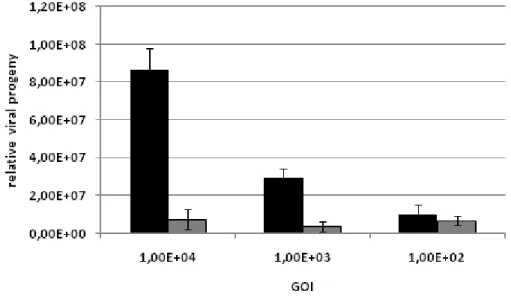
In order to determine the range in which the decoy effect of fibroblasts is effective, I measured the total viral progeny after a test-selection round (consisting of a negative successive a positive selection) either with or without the negative selection step, using different initial amounts of genomic particles (Figure 9).
Increasing the initial amount of AAV from 102 to 104 genomic particles per cell (GOI) resulted in increasing total viral outcome if no negative selection was carried out. In contrast, viral progeny after negative selection on fibroblasts was efficiently decoyed even when the highest amount of virus was applied. Input of more viral particles increases biodiversity and therefore the chance of the pool to contain interesting clones. Although the negative selection
24
potential was not yet saturated, no higher amounts of initial virus load were tested in order to avoid formation of chimeric particles that form at multiple infection of the same cell by different insertion mutants. The resulting uncoupling of genotype from phenotype impairs the selection process.
Figure 9: Decoy effect of Fibroblasts cells at different GOIs. The viral library was amplified on BLM cells without (black bars) or with (grey bars) negative selection on human primary fibroblasts. Relative progeny titers were measured by quantitative PCR 48 h post infection.
4.2 Selection of BLM specific mutants
To compare the effect of using different initial amounts of viral particles, two selection procedures were performed in parallel.
In one setting, a low number of particles (GOI = 100) was applied and three consecutive selection rounds were performed. Sequencing of the viral pools showed that one mutant G2 (carrying the insertion RSGQSLS) dominated the pool already after the first selection round accounting for more than 50% of the total number of clones. This mutant persisted in the following selection rounds. When tested in an infection assay it showed no increased specificity compared to wtAAV2 (see Table 1). This experiment suggested that the selection pressure had depleted the low biodiversity of the pool too sharply. In this conditions apparently, the evolution of the pool had the characteristics of a genetic drift instead that of a real selection, in which stochastic events led to the surviving of a mutant that showed no selective advantage.
25
In the second setting, four consecutive selection rounds (each consisting of a negative successive a positive selection) were performed using a GOI of 104. Evolution of the pools was monitored by qPCR and sequencing. Twelve clones of the first round´s progeny pool were sequenced to avoid preceding a selection with a pool in which one clone has already ruled out the others (as described above). All sequences appeared only once, suggesting that biodiversity was still high. The pool was amplified on BLM cells to cover the titer needed for the next cycle.
After the second selection round, sequencing of the pool showed that biodiversity was reduced. 12.5% of 24 sequences were represented by three mutants. Titers of the progeny for both initial GOIs were in a range of one log and had again more than doubled (3.2-fold; 2.1- fold), compared to the preceding selection round, indicating the increasing overall fitness of the pool.
Figure 10: Selection procedure monitored by qPCR.
In order to analyze the pools for accumulated clones a total of 98 samples of all viral pools was sequenced. No redundant clone was found after the first selection round. The insertions found here did also not reappear in the sequences of later selection rounds suggesting that the biodiversity of the pool was still high.
In contrast to this, sequencing of at least 20 clones from each of the later selection rounds showed a gradual decrease in biodiversity and the appearance of clones in multiple copies.
The redundancy of clones in the sequenced pools was 0% after the first round, 12.5% after the
26
second, 33% after round three and 66.7% after selection round 4. The observed increase in viral titers (despite the lower initial amounts of viral particles used in the last two selection rounds), and the concomitant decrease of biodiversity are strong indicators of a successful pressure-driven selection.
45% of the sequenced viral pool after the fourth selection round was composed of five clones (A7, C4, D8, D10, E10) that were packaged with GFP to further analyze their characteristics.
4.3 Characterization of selected mutants 4.3.1 Packaging capacity of selected clones
GFP-expressing vectors carrying the selected 587 capsid insertions were produced and analyzed for their specificity as described in materials and methods. An additional clone that was only found once in the pool after the fourth selection round (A5) was also characterized as an example of a mutant that was not strongly selected (see Table 1). Titers were measured via qPCR showing that mutants A5, C4, D10 and E10 packaged with efficiencies comparable to wtAAV2. Conversely, A7 and D8 viral preparations yielded genomic titers 103- and 211- fold lower than wtAAV2, respectively. This observation could be confirmed after repeating the viral packaging procedure in two additional experiments. Since these low titers allowed performance of infections only at GOIs that failed to yield detectable transduction rates on BLM cells (data not shown), mutants A7 and D8 were not further characterized. The low titer yield of these mutants can be due to ineffective packaging of the transgene or some other difference between selection and packaging that is disadvantageous for the packaging efficiency of A7 and D8. Mutants A5, C4, D10 and E10 were subjected to further characterization.
27
vector
amino acid sequence of insertion
% of sequenced
clones observed in selection round:
genomic particles/µl
wt - (2.10 ± 0.26) x 108
G2* RSGQSLS - 1 (1.91 ± 0.45) x 108 A5** DAANNPR 1x 4 (8.71 ± 1.51) x 107 A7 NDPRKPS 10.5 2, 3, 4 (2.04 ± 0.15) x 106 C4 PRGTNGP 5.8 2, 3, 4 (1.27 ± 0.26) x 108 D8 AGKAGIG 15.1 3, 4 (9.93 ± 1.12) x 105 D10 SRGATTT 8.1 2, 3, 4 (1.67 ± 0.18) x 108 E10 DGSGPTR 5.8 2, 3, 4 (8.88 ± 2.64) x 107
Table 1: Characterization of selected mutants. Titers were calculated as mean of 3 independent measurements.(*:G2 mutant was selected after using an initial GOI of 100;**: A5 mutant was only observed once in the last selection round)
4.3.2 Selectivity assay
To estimate the selectivity of the mutans, target and non-target cells were infected with GFP- vectors. GFP is expressed only in the infected cells so that the percentage of transduced cells can be distinguished from non-infected ones by cytofluorimetry 48 h post infection (see Figure 11 A-E). To exclude that results were influenced by the particular amount of genomic particles per cell used in the experiment, triplicate experiments were performed at six different GOIs. For quantification of target cell specificity ratio, three GOIs were chosen that fulfilled the following requirements: more than 25% and less than 75% of BLM cells and more than 3% of fibroblasts had to be infected by the given GOI (marked in Figure 11 A-E by caskets). It turned out that the values of infectivity ratios in the GOI range identified by these parameters were similar (Figure 12) and the final ratio of infected BLM and fibroblast cells was calculated as mean of these three values for each mutant (Figure 11 F).
28
Figure 11: A-E: Transduction efficiencies of wtAAV2 and selected mutants on BLM cells (black) and human primary fibroblasts (white). Infections were performed with six different multiplicities of infection (x-axis). Transduction rates were calculated as percentage of fluorescent cells. Values are given as average of three independent infection experiments, error bars represent standard deviation. The ratios marked in the caskets were used to quantify specificity ratios of each mutant (F).
29
Figure 12 Ratios for all tested mutants of infectivity of BLM and fibroblast cells at three different GOIs that were used to quantify specificity.
The mutants´ infectivity ratios were 1.8, 1.8, 2.8, 5.7 and 6.6 for wtAAV2, mutants A5, E10, C4 and D10, respectively (Figure 12 F). Specificity of the mutants was calculated comparing mutants´ratios with those of wtAAV. The respective values are 1.0, 1.6, 3.2 and 3.7 times higher for A5, E10, C4 and D10 (Figure 13).
Figure 13: Selectivity of mutants on BLM cells and fibroblasts. Infection ratios were quantified for wt and mutants and related to specificity of wtAAV2.
These results indicate that a lower unwanted transduction of non-target cells occurs when using the mutants. This outcome is depicted in Figure 14 comparing directly the curves of D10 and wtAAV infection demonstrating that a virus dose that infects 54% of BLM cells
30
would collaterally infect 30% fibroblasts when using wtAAV2 but only 8% when using D10 mutant.
Figure 14: Comparison of transduction efficiencies between wtAAV2 (black) and mutant D10 (white) on BLM cells (circles) and human primary fibroblasts (triangles) with increasing amounts of virus.
4.3.3 Selected mutants are inhibited by heparin
To further characterize the mutants, their ability to interact with AAV2 primary receptor, heparan sulfate proteoglycan (HSPG) was tested. Heparin is the sugar content of HSPG and the soluble form of this molecule can bind to the viral capsid of wt AAV2 suppressing its ability to bind HSPG on the cellular surface in a competitive way. Since the randomized peptides are inserted in the HSPG-binding region of the capsid, depending on the amino acid composition of the peptide, the HSPG binding phenotype of the virions could be altered with important consequences for the tropism of the viral particles (Perabo et al. 2006).
To assess the ability of the selected clones to bind HSPG, GFP-vectors derived from wtAAV2 and from insertion mutants were incubated for 30 min with HeLa cells at a concentration of 6x10³ genomic particles per cell in the presence or absence of 85 I.E./ml heparin. 48 h later, the percentage of infected cells was analyzed by cytofluorimetry (Figure 15). All mutants were inhibited by heparin. wtAAV2 and A5 mutant were almost completely blocked (3.1%;
3.5% residual infectivity) whilst E10, C4 and D10 showed higher residual infection rates