Ubiquitin C-terminal hydrolase-L1 (UCH-L1) stabilizes NOXA and impacts on cancer chemosusceptibility
Inaugural-Dissertation zur
Erlangung des Doktorgrades
der Mathematisch-Naturwissenschaftlichen Fakultät der Universität zu Köln
vorgelegt von Kerstin Brinkmann aus Kirchheimbolanden
Berichterstatter: Prof. Dr. Thomas Langer Prof. Dr. Thorsten Hoppe
Tag der mündlichen Prüfung: 14.05.2012
Content
Abstract ... I Zusammenfassung ... II Abbreviations ... IV
1 Introduction ... 1
1.1 Hallmarks of cancer ... 1
1.2 Apoptosis ... 2
1.3 The Bcl2 protein family ... 9
1.4 p53-mediated apoptosis in response to DNA damage ... 12
1.5 The Ubiquitin-Proteasome System ... 14
1.6 Aim of the work ... 17
2 Material and methods ... 19
2.1 Chemicals ... 19
2.2 DNA constructs ... 19
2.3 Cell culture and transfection ... 21
2.4 Cytotoxic treatments and cell viability ... 22
2.5 qPCR ... 23
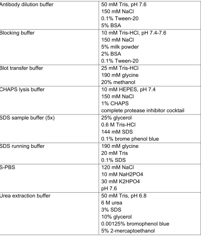
2.6 Sample preparation and immunoblotting (IB) ... 23
2.7 Immunoprecipitations (IP) ... 26
2.8 In vitro deubiquitylation and binding assays ... 27
2.9 Fluorescence microscopy ... 27
2.10 Flow cytometry (FACS) ... 28
2.11 Tissue immunohistochemistry (IHC) ... 28
3 Results ... 30
3.1 Proteasome inhibition induces mitochondrial apoptosis in a cohort of tumour entities ... 30
3.2 NOXA accumulates at the post-transcriptional level after proteasome inhibition ... 34
3.3 NOXA protein is less stable in bortezomib-sensitive tumour cells ... 36
3.4 NOXA is increasingly ubiquitylated in bortezomib-sensitive tumour cells ... 37
3.5 The deubiquitylating enzyme (DUB) UCH-L1 is absent in bortezomib- sensitive tumour cells ... 39
3.6 The DUB UCH-L1, binds, deubiquitylates and stabilizes NOXA in vitro ... 41
3.7 UCH-L1-expression correlates with increased NOXA-expression in tumour tissue sections of melanoma and CRC patients ... 45
3.8 UCH-L1 depletion is associated with chemoresistance in tumour cells ... 47
4 Discussion ... 50
4.1 NOXA accumulation as a key event in proteasome inhibition- induced apoptosis ... 50
4.2 Post-transcriptional regulation of NOXA by the UPS ... 53
4.3 Increased NOXA degradation as a result of lacking UCH-L1 expression, the specific DUB of NOXA ... 53
4.4 UCH-L1 as a modulator of apoptosis ... 55
4.5 UCH-L1 as an important component of the DNA damage response by stabilization of NOXA ... 56
4.6 Conclusion ... 57
5 References ... 59
6 Appendix ... 67
6.1 Microarray ... 67
6.1 Patient data ... 69
6.2 Vector maps ... 72
6.3 Danksagung ... 73
6.4 Erklärung ... 74
6.5 Lebenslauf ... 75
Abstract
Resistance to apoptosis is a hallmark of tumour cells. It constitutes an important clinical problem since chemotherapy and irradiation act primarily by inducing apoptosis. Compounds inhibiting proteasomal activity are able to selectively induce tumour cell apoptosis and a growing body of evidence suggests the up-regulation of NOXA (a BH3-only protein) upon proteasome inhibition to be one of the essential events. However, the underlying molecular mechanisms giving rise to the selective NOXA up-regulation in tumour cells is poorly understood.
The current work shows that in cells susceptible to proteasome inhibition NOXA protein was significantly less stable and continuously degraded by the Ubiquitin- Proteasome System (UPS). Amongst others, the degradation of substrates by the UPS is regulated by E3-ubiquitin ligases and deubiquitylating enzymes (DUBs). In the current work UCH-L1 was identified as a responsible DUB of NOXA, which was epigenetically silenced in tumour cells susceptible to proteasome inhibition. UCH-L1 binds and stabilizes NOXA by removing the Lys48-linked polyubiquitin chains that normally mark NOXA for proteasomal degradation. In line with these observations, increased UCH-L1 expression correlated with increased NOXA protein expression in human melanoma and colorectal cancer patient samples. As one of the genes responsive to genotoxic stress, NOXA has been considered as the BH3-only protein to be involved in the fine-tuning of apoptosis. Correspondingly, the current work demonstrates that UCH-L1 expression is important for DNA-damage induced apoptosis. Down-regulation of UCH-L1 results in decreased susceptibility to DNA damage-induced apoptosis accompanied by a decreased NOXA accumulation.
Taken together, the current work identified UCH-L1 as an important component of the DNA damage response that impacts on the susceptibility of cancer cells toward chemotherapy by stabilizing NOXA.
Zusammenfassung
Tumore zeichnen sich unter anderem durch eine Apoptoseresistenz aus, die ein gravierendes Problem in der Krebsbehandlung darstellt, da sowohl Chemotherapie als auch Bestrahlung primär über die Induktion der Apoptose wirken.
Inhibitoren des humanen 26S Proteasoms sind beschrieben, selektiv in Tumorzellen Apotose zu induzieren. Essentiell für die Induktion der Apoptose nach Behandlung mit Proteasominhibitoren ist die Akkumulation des pro-apoptotischen "BH3-only"- Proteins NOXA, wobei der Mechanismus nicht vollständig aufgeklärt ist.
Die vorliegende Arbeit zeigt, dass die Fähigkeit von Proteasominhibitoren, Apoptose zu induzieren unter Tumourzellen von verschiedenen Individuen stark variiert.
Außerdem wurde gezeigt, dass die Stabilität des essentiellen "BH3-only"-Proteins NOXA ebenfalls unter Tumourzellen von verschiedenen Individuen stark variiert. In Tumourzellen die nach Proteasominhibition Apoptose induzieren ist die Stabilität von NOXA stark verringert. Es wurde demonstriert, dass die verringerte Stabilität von NOXA die Folge einer verstärkten Ubiquitinierung und somit Degradation durch das Ubiquitin-Proteasome System (UPS) ist. Die Degradation von Proteinen durch das UPS wird unter anderem durch E3-Ubiquitin-Ligasen und deubiquitinierende Enzyme (DUB) reguliert. In der vorliegenden Arbeit wurde Ubiquitin-C-terminal hydrolase-L1 (UCH-L1) als spezifische DUB von NOXA identifiziert. In Tumourzellen, die eine verringerte Stabilität von NOXA aufweisen wurde UCH-L1 nicht exprimiert. Als direkter Interaktionspartner von NOXA war UCH-L1 in der Lage, NOXA zu deubiquitinieren und somit zu stabilisieren. In Übereinstimmung mit biochemischen und zellbiologischen Ergebnissen zeigten Expressionsanalysen von NOXA und UCH-L1 in Geweben von 81 Melanom- und 26 Coloncarcinompatienten ebenfalls eine Korrelation der Expression. Gewebe in den denen UCH-L1 exprimiert war, wiesen eine erhöhte Expression von NOXA auf.
NOXA ist ursprünglich als ein Protein beschrieben, das für die zelluläre Antwort zu genotoxischem Stress wichtig ist, indem es an der Feinabstimmung der mitochondrialen Membranpermeabilisierung (MOMP) beteiligt ist. Die vorliegende Arbeit zeigt, dass auch UCH-L1 für den Ablauf von Apoptose nach DNS-Schädigung
wichtig ist, indem es die Stabilität von NOXA kontrolliert.. Herunterregulation der UCH-L1 Expression in UCH-L1-exprimierenden Tumorzellen verringerte deren Fähigkeit nach Schädigung der DNS Apoptose zu induzieren. Dies war die Folge einer verringerten Fähigkeit NOXA zu akkumulieren.
Zusammenfassend wurde in dieser Arbeit UCH-L1 als spezifische DUB von NOXA identifiziert. Durch die Fähigkeit NOXA zu stabilisieren spielt UCH-L1 in der zellulären Antwort auf DNS-Schädigung eine entscheidende Rolle und beeinflusst die Sensitivität von Tumorzellen gegenüber Chemotherapie.
Abbreviations
Only abbreviations are listed that have not been described in the text
α anti
°C Degree Celsius
µg Microgramm
µl Microliter
µM Micromolar
aa amino acids
ATP adenosine triphosphate
bp basepares
BSA bovine serum albumin
C-terminal carboxyterminal
Da, kDa Dalton, Kilodalton
DMSO Dimethylsulfoxid
DNA Deoxyribonucleic acid
ds double stranded
DTT Dithiothreitol
E.coli Escherichia coli
DTT Dithiothreitol
EDTA ethylene diamine tetraacetic acid et al. et alteri /-a /-um (and others)
FCS fetal calf serum
Fig. figure
GFP green fluorescent protein
FITC fluorescein-5-isothiocyanat
HEPES 4-(2-hydroxyethyl)-1-piperazineethanesulfonic acid
His, H Histidine
HRP horse radish peroxidase
IgG Immunglobuline G
K Lysine
kb kilo base
kDa kilo dalton
N Asparagin
NaCl Sodium chloride
nm Nanometer
N-terminal aminoterminal
MEFs mouse embryonic fibroblasts
MW molecular weight
ORF open reading frame
ON over night
PBS phosphate buffered saline
PCR polymerase chain reaction
pH potentium hydrogenii (lat.)
pmol Picomol
RT room temperature
SDS sodiumdodecylsulfat
SDS-PAGE SDS polyacrylamid gel electrophoresis
Tab. table
Tris/HCl tris[hydroxymethyl]aminoethane
V Volt
1 Introduction
1.1 Hallmarks of cancer
The formation of malignant tumours is characterized by defects in cell proliferation and homeostasis eventually resulting in overgrowth and thereby loss of function of healthy tissue.
It is thought that the transformation of a healthy cell into a malignant cell is a multistep process with alterations in major signalling pathways. The vast majority of cancer cell genotypes is a manifestation of eight essential alterations in cell physiology that collectively dictate malignant growth: self-sufficiency in growth signals, insensitivity to growth-inhibitory (antigrowth) signals, evasion of programmed cell death (apoptosis), limitless replicative potential, sustained angiogenesis, tissue invasion and metastasis, reprogramming of energy metabolism and evading immune destruction (Hanahan and Weinberg, 2000, 2011). Underlying those alterations are dynamic disorders within the genome. Previous analyses of malignant tissues have identified several genes, which are involved in malignant formation, either by a gain–
of-function, referred to as oncogenes, or by a loss-of-function, referred to as tumour suppressors (Weinberg, 1996).
Furthermore, tumours are more than just a vast array of uncontrolled proliferating cancer cells. Rather they are a complex construction composed of multiple distinct - also non-malignant - cell types, which facilitate the acquisition of the hallmarks of cancer by forming the tumour microenvironment. How developing tumours acquire the hallmarks of cancer is a diverse process leading to hundreds of distinct types of cancer and numerous subtypes of tumours within specific organs. Nevertheless, the recognition of these common hallmarks enabled the introduction of mechanism- based targeted therapies, such as the reactivation of the apoptotic machinery (Hanahan and Weinberg, 2011).
1.2 Apoptosis
Apoptosis describes programmed cell death or cell suicide (Kerr et al., 1972). It is crucial for sculpting the embryo, maintaining tissue homeostasis, shaping the immune repertoire, terminating immune responses and restricting the progress of infections. The average adult human body generates approximately 60 billion cells per day, and as a consequence an equal number of cells must die by apoptosis to maintain tissue homeostasis. Disturbed regulation of this vital physiological process can result in numerous diseases including autoimmunity, degenerative disorders as well as cancer. Resistance to apoptosis can also permit tumour cells to escape from immune surveillance.(Strasser et al., 1991; Watanabe-Fukunaga et al., 1992), (Barr and Tomei, 1994; Thomson, 1995) (McDonnell and Korsmeyer, 1991; Strasser et al., 1990). Moreover, because chemotherapy and irradiation act primarily by inducing apoptosis, defects in the apoptotic pathway contribute to the resistance of cancer cells to conventional therapy, which constitutes an important clinical problem (Kerr et al., 1994).
The process of a programmed cell death was first described by Carl Vogt in 1842 (Vogt, 1842). The German scientist studied the development of tadpoles, and described the programmed cell death as a crucial step in the embryonic development of tadpoles. The drawings by Walther Flemming in 1885 provided the first morphological description of the programmed cell death. Those drawings clearly show cell shrinkage, nuclear fragmentation and apoptotic body formation, which are by now accepted hallmarks of apoptosis. However, a more detailed description was lacking until 1972 when Kerr, Wyllie and Curie were able to distinguish the programmed cell death and the traumatic or necrotic cell death (Necrosis) by electron microscopic analysis (Kerr et al., 1972). They introduced the word "apoptosis"
(ἁπόπτωσισ) which in Greek describes the "dropping off" or "falling off" of petals from flowers, or leaves from trees. 30 years later, in 2002 Bob Horvitz received the Nobel Prize in Physiology and Medicine for his pioneering work unravelling the fundamental aspects of the biology of apoptosis, using the nematode Caenhorhabditis elegans (C.
elegans) as a model system (Horvitz, 2003a, b).
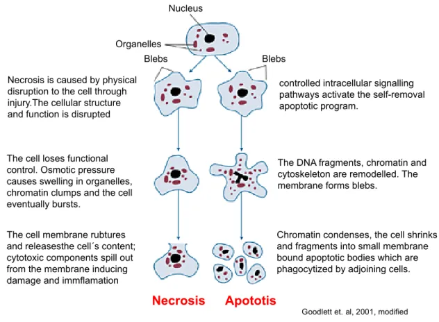
Morphological apoptosis is characterized by defined cellular changes. These changes include blebbing, cell shrinkage, nuclear fragmentation, chromatin condensation, chromosomal DNA fragmentation (Wyllie et al., 1980) and the
exposure of phosphatidyl-serines on the plasma membrane (Fadok and Henson, 1998). Apoptosis produces cell fragments called apoptotic bodies that are engulfed and quickly removed by phagocytic cells before the contents of the cell can spill and cause damage. In contrast, necrotic cell death is characterized by cell swelling, chromatin digestion, and disruption of the plasma membrane and organelle membranes (Enari et al., 1998; Fadok and Henson, 1998; Kerr et al., 1972; Wyllie et al., 1980). Leaky necrotic cells release intracellular contents, thereby causing inflammation (Fig. 1.1). While apoptosis often provides beneficial effects to the organism, necrosis is almost always detrimental and can be fatal. Necrosis is caused by factors external to the cell or tissue, such as infections, toxins, or trauma. In contrast, apoptosis is a naturally occurring cause of cellular death and tightly regulated by intracellular signalling pathways in response to cellular stress (Ellis and Horvitz, 1986; Vaux et al., 1988).
Figure 1.1 | Morphology of apoptosis and necrosis. Apoptosis is characterized by nuclear and DNA fragmentation, cell shrinkage and the formation of apoptotic bodies. Phagocytic cells engulf apoptotic bodies without activating the inflammatory response (right). Necrotic cell death is characterized by cell swelling, chromatin digestion and disruption of the plasma membrane resulting in the release of intracellular material and the activation of the inflammatory response (left) (Goodlett and Horn, 2001).
Goodlett et. al, 2001, modified The cell membrane rubtures
and releasesthe cell´s content;
cytotoxic components spill out from the membrane inducing damage and immflamation
Chromatin condenses, the cell shrinks and fragments into small membrane bound apoptotic bodies which are phagocytized by adjoining cells.
Organelles
Nucleus
Blebs Blebs
Necrosis Apototis
The DNA fragments, chromatin and cytoskeleton are remodelled. The membrane forms blebs.
controlled intracellular signalling pathways activate the self-removal apoptotic program.
The cell loses functional control. Osmotic pressure causes swelling in organelles, chromatin clumps and the cell eventually bursts.
Necrosis is caused by physical disruption to the cell through injury.The cellular structure and function is disrupted
1.2.1 Apoptotic signalling
In principle, there are two alternative pathways that initiate apoptosis: one is mediated by death receptors on the cell surface — referred to as the "extrinsic pathway"; the other is mediated by mitochondria — referred to as the "intrinsic or mitochondrial pathway". The initiation of extrinsic and mitochondrial apoptotic pathways occur by engagement of extracellular "death receptors" and intracellular stress (e.g. inadequate cytokine support, diverse types of cellular damage), respectively (Strasser et al., 2000).
Both pathways involve the activation of cysteine aspartidyl-specific proteases (caspases) - the main executioners of the apoptotic process (Kumar and Lavin, 1996;
Nicholson and Thornberry, 1997).
1.2.1.1 Caspases
Caspases cleave cellular substrates and thus initiate the biochemical and morphological changes that are characteristic of apoptosis. For instance, caspases process the inhibitor of the caspase-activated-DNase (CAD) resulting in active CAD that is responsible for DNA fragmentation (Sakahira et al., 1998). Moreover caspases induce the processing of the nuclear protein lamin and the poly (ADP-ribose) polymerase (PARP), which plays an important role in DNA damage repair, and its processing serves as a marker of ongoing apoptosis (Masson et al., 1995).
Furthermore, regulators of the cytoskeleton and adhesion proteins are processed by caspases resulting in cell shrinkage and disruption of the extracellular matrix (Sanghavi et al., 1998).
Caspases are a highly conserved family of cysteine proteases that cleave their substrates specifically after certain aspartate residues (Alnemri et al., 1996). Based on their intracellular function, caspases can be divided into three subgroups: cytokine activators (caspase 1, 4, 5, 11, 12, 13, 14), the initiator caspases (caspase 2, 8, 9, 10) and the effector caspases (caspase 3, 6, 7), while only initiator- and effector caspases play a role during apoptotic signalling (Denault and Salvesen, 2002).
Caspases are constitutively expressed in most cell types and reside as zymogenes in the cytosol (Nicholson, 1999). Structurally caspases consist of an amino-terminal (N- terminal) pro-domain, a large and a small subunit (Denault and Salvesen, 2002).
Upon activation, the large and the small subunit are separated by proteolytic
cleavage, and in a second step the pro-domain is removed. Active caspases form tetramers consisting of two small and two large subunits, which form two active sites (Widlak et al., 2003). The large subunit comprises the catalytic cysteine residue (Walker et al., 1994; Wilson et al., 1994) (Fig. 1.2). Some caspases also possess interaction domains such as the caspase associated recruitment domain (CARD) or the death effector domain (DED) (Ashkenazi and Dixit, 1998).
The activation of the caspase cascade is a point of no return. Once initiator caspases are activated, they activate the effector caspases by proteolytic cleavage resulting in degradation of the apoptotic substrates (Nicholson and Thornberry, 1997).
Figure 1.2 | Activation of caspases. Caspases consist of a pro-domain, a large and a small subunit. Upon activation the large and small subunit are separated by proteolytic cleavage and the pro-domain is removed (Denault and Salvesen, 2002). Active caspases form dimers comprising two large and two small subunits that form two active sides with the catalytic cystein residing in the large subunit (Widlak et al., 2003).
1.2.1.2 The extrinsic apoptotic signalling pathway
The extrinsic apoptotic-signalling pathway plays an important role during the development of T- and B-cells and is essential for the immune response.
Extrinsic apoptotic signalling is initiated through ligand binding (e.g. TNFα, CD95) to the extracellular domain of a death receptor of the TNF superfamily such as TNFR1 or CD95/FAS receptor (Ashkenazi, 2002). Characteristic for death receptors are a
large subunit small subunit calalytic cystein
N-terminal pro-domain proteolytic
processing
proteolytic processing
dimerization activation
cytosolic death domain (DD) and a death effector domain (DED) that upon activation initiate receptor-oligomerization and interaction with adaptor proteins (e.g. TRADD (TNF associated death domain) or FADD (Fas-associated-death-domain)), respectively. The resulting death inducing signalling complex (DISC) in turn leads to the recruitment of several pro-caspase 8 - or in some cases also pro-caspase 10 - molecules through their DED. The proximity of the zymogens provokes their dimerization and subsequent autocatalysis (Boatright et al., 2003).
Figure 1.3 | The extrinsic apoptotic signalling pathway. Upon binding of a death ligand (e.g. FAS, TRAIL, TNFa) the death inducing signalling complex (DISC) is formed, resulting in active caspase 8 (casp8). Caspase 8 directly activates caspase 3 (casp3) or indirectly by engaging the mitochondrial apoptotic signalling pathway via members of the pro-apoptotic BH3-only family.
Active caspase 3 cleaves the apoptotic substrates and apoptotic cell death occurs.
--
apoptotic substrates
- F ADD
DD DD DD
DED DED
DED DISC
(F AS , T R A IL , T NF a)
death receptors
pro-casp8
casp8
casp3
mitochondrial apoptotic signalling pathway
B H3- B H3-
pro-apoptotic BH3-only
proteins Death ligands
Alternatively, the extrinsic apoptotic pathway can be triggered by cytolytic T-cells or natural killer (NK) cells with the release of perforine and granzyme B. Perforine- release results in extracellular membrane permeabilization of the target cell and allows granzyme B to enter the cell (Lord et al., 2003).
Active caspase 8 or intracellular granzyme B proteolytically activate effector caspases such as caspase 3 irrevocably resulting in apoptotic cell death. Besides the direct activation of caspase 3, caspase 8 and granzyme B induce the mitochondrial apoptotic signalling pathway through engagement of pro-apoptotic BH3-only proteins (Fig. 1.3) (Luo et al., 1998; Wei et al., 2000).
1.2.1.3 Intrinsic/mitochondrial apoptotic signalling pathway
The intrinsic apoptotic signalling pathway is also often referred to as the mitochondrial apoptotic signalling pathway since the release of pro-apoptotic molecules from the mitochondrial inter-membrane space is the central event. The release of mitochondrial pro-apoptotic molecules is regulated by pro- and anti- apoptotic members of the Bcl2 protein family (Kluck et al., 1997; Vander Heiden et al., 1997; Yang et al., 1997). Upon intracellular stress, pro-apoptotic Bcl2 proteins (BH3 only proteins, see 3.2) are activated which in turn leads to the activation of the pro-apoptotic multi-domain proteins BAX and BAK. Upon activation, BAX and BAK undergo conformational changes and form homodimers (Hsu et al., 1997) that are thought to form pores in the mitochondrial outer membrane. This event is also referred to as mitochondrial outer membrane permeabilization (MOMP).
Another model states that induction of MOMP might occur by direct interaction of BAX and BAK with channels in the outer mitochondrial membrane such as VDAC (voltage dependent anion channel) or PTPC (permeabilization transition pore complex), rather than forming pores by themselves. Although a lot of research has been conducted in this field, the precise mechanism of MOMP is still not understood (Tait and Green, 2010). However, there is agreement about the fact that Bcl2- mediated MOMP subsequently results in the release of pro-apoptotic molecules such as cytochrome c and SMAC (second mitochondrial activator) (Newmeyer and Ferguson-Miller, 2003). Cytosolic cytochrome c binds in an ATP or dATP-dependent manner to APAF1 resulting in the recruitment of pro-caspase 9 molecules. The generated multiprotein complex, referred to as apoptosome, triggers the activation of
the initiator caspase 9. The initiator caspase directly activates the executioner caspase 3 and mediates subsequent continuation of apoptosis (Green, 2000).
Figure 1.4 | The mitochondrial apoptotic signalling pathway. The mitochondrial apoptotic signalling pathway is induced upon intracellular stress.
This leads to the activation of pro-apoptotic BH3-only proteins and BAX/BAK- dependent MOMP with the release of cytochrome C (Cyt C) from the mitochondrial intermembrane space. Cyt C forms a complex, the apoptosome, with ATP, APAF1 and pro-caspase 9 (pro-casp9), resulting in the activation of caspase 9 (casp9). Caspase 9 activates caspase 3 (casp3) and apoptosis proceeds. The mitochondrial apoptotic signalling pathway can also be engaged by the extrinsic pathway. Active caspase 8 (casp3) cleaves and thereby activates the BH3-only protein BID, ultimately resulting in MOMP.
As mentioned above the mitochondrial apoptotic signalling pathway is also induced upon activation of the extrinsic pathway. Active caspase 8 or granzyme B can directly cleave BID thereby forming the active tBID (truncated BID), which in turn activates BAX and BAK resulting in MOMP (Fig. 1.4) (Luo et al., 1998; Wei et al., 2000).
Depending on the cell type, this interference of both signalling pathways either resembles an amplification loop or is essential for the progression of apoptosis. Cells which can undergo apoptosis independently of MOMP are referred to as type I cells
B AX B AX
Cyt C Cyt C
Apaf 1
AT P
apoptotic substrates
extrinsic apoptotic signalling pathway
pro-apoptotic BH3-only
proteins
mitochondria
anti-apoptotic Bcl2 proteins
casp9 casp8
casp3 pro-casp9
Apoptosome
intracellular stress (e.g. DNAdamge)
tBID
(e.g. hepatocytes, pancreatic cells) and cells which depend on MOMP to effectively induce apoptosis are referred to as type II cells (e.g. lymphocytes) (Yin et al., 1999).
1.3 The Bcl2 protein family
The Bcl2 protein family regulates stress-induced apoptosis by an evolutionary highly conserved mechanism found in species as distantly related as humans and nematodes. The family consists of anti-apoptotic (e.g. BCL2, BCLxl, BCLw, A1) and two groups of pro-apoptotic members: the multidomain or BAX/BAK like proteins (e.g. BAK, BAX) and the BH3-only proteins (e.g. BIM, BID, PUMA, BAD, NOXA).
Members of the Bcl2 protein family share at least one conserved Bcl2 homology domain (BH domain), which is characterized by several α-helical segments. The BH- domain does not possess enzymatic activity but it allows pro- and anti-apoptotic members to bind to and to inhibit each other (Adams and Cory, 1998; Cory and Adams, 2002). Binding affinity assays using BH3-only peptides revealed that not all pro- and anti-apoptotic Bcl2 proteins can antagonize each other, but the affinity differs within the family (Fig. 1.5). Thus, level and composition of pro- and anti- apoptotic Bcl2 proteins determine whether intracellular stress results in apoptotic cell death or not (Chonghaile and Letai, 2008).
Figure 1.5 | Binding-pattern of pro- and anti-apoptotic members of the Bcl2 protein family. The BH3-only proteins BIM, BID, PUMA and BMF can bind and antagonize all anti-apoptotic Bcl2 proteins. In contrast, BAD can only bind BCL2, BCLxl and BCLw, and NOXA is restricted in binding to MCL1 and A1.
Anti-apoptotic members of the Bcl2 protein family possess four BH domains and a carboxy-terminal transmembrane domain (TM), which targets them with different
NOXA BAD BCL2
BCLxl BCLw MCL1
A1 BIM
BID PUMA
BMF
affinities to at least three intracellular membranes: the outer mitochondrial membrane, the endoplasmic reticulum (ER) and the nuclear envelope.
Pro-apoptotic members of the BAX/BAK like subgroup (BAX, BAK, BOK, BCLxs) possess two or three BH domains and a TM-domain while BH3-only proteins (BIK, BAD, BID, BIM, NOXA, PUMA, BMF) share only the BH3 domain (Fig. 1.6). Both subgroups of pro-apoptotic Bcl2 protein family members are essential for the induction of apoptosis (Cory and Adams, 2002; Coultas and Strasser, 2003).
Figure 1.6 | Protein structure of the Bcl2 protein family. The Bcl2 protein family consists of anti- (A) and pro-apoptotic members (B) that share at least one conserved Bcl2 homology domain (BH domain).
The anti-apoptotic members (BCL2, BCLxl, BCLw, A1, MCL1) possess four BH domains and an amino-terminal (N-terminal) transmembrane (TM) domain.
The pro-apoptotic members can be subdivided in the multidomain proteins and the BH3-only proteins. The multidomain proteins (BAX, BAK, BOK) possess 3 BH domains and an N-terminal TM-domain, while the BH3-only proteins (BID, BIM, BIK, BMF, BNIP3, HRK, NOXA, PUMA) comprise only a single BH3 domain.
1.3.1 Regulation of apoptosis by the Bcl2 protein family
To date, there are two proposed models that explain how the Bcl2 protein family regulates MOMP: (i) the indirect activator model and (ii) the direct activator- derepressor model. Both models result in the activation of BAX and BAK and the permeabilization of the outer mitochondrial membrane.
BH4 BH3 BH2 BH1 TM
BH3
BH3 BH2 BH1 TM A: Anti-apoptotic Bcl2 proteins:
B: Pro-apoptotic Bcl2 proteins:
Multidomain proteins
BH3-only proteins
A1, BCL2, BCLxl, BCLw, MCL1
BAX, BAK, BOK
BID, BIM, BIK, BMF, BNIP3, HRK, NOXA, PUMA
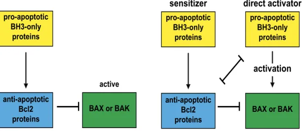
The indirect activator model postulates that BAX and BAK are bound in a constitutively active state to anti-apoptotic Bcl2 proteins. Competitive interactions with pro-apoptotic BH3-only proteins and anti-apoptotic Bcl2 proteins are sufficient to release active BAX and BAK and induce MOMP (Fig. 1.7).
Figure 1.7 | Regulation of MOMP by the Bcl2 protein family. A, The indirect activator model postulates that BAX and BAK are bound in a constitutively active state to anti-apoptotic Bcl2 proteins. Competitive interactions with pro- apoptotic BH3-only proteins and anti-apoptotic Bcl2 proteins are sufficient to release active BAX and BAK and induce MOMP. B, The direct-activator- derepressor or neutralization model implies that BAX and BAK are activated after binding to a subset of BH3-only proteins, referred to as direct activators.
Anti-apoptotic Bcl2-proteins prevent MOMP either by sequestering activated BAX and BAK or by inhibiting the direct activator BH3-only proteins. A second subset of BH3-only proteins cannot directly activate BAX or BAK but neutralize anti-apoptotic Bcl2 proteins. In the indirect activator model BAX and BAK are thought to be in a constitutively active state, and bound and thereby inhibited by anti-apoptotic Bcl2 proteins. In this model the competitive interaction of BH3- only proteins with anti-apoptotic Bcl2 proteins is sufficient to release of BAX and BAK and initiate MOMP.
In the direct activator-derepressor model (also called neutralization model), BAX and BAK are activated by the interaction with a subset of BH3-only proteins, such as BID and BIM, called direct activators. In this model, anti-apoptotic Bcl2 proteins either inhibit MOMP by antagonizing BAX or BAK directly or by sequestering the direct activator BH3-only proteins thus preventing them to activate BAX or BAK. A second subset of BH3-only proteins, called sensitizer, such as NOXA or BAD, cannot directly
pro-apoptotic BH3-only
proteins
BAX or BAK anti-apoptotic
Bcl2 proteins
active
pro-apoptotic BH3-only
proteins
pro-apoptotic BH3-only
proteins
BAX or BAK anti-apoptotic
Bcl2 proteins
A: indirect activator model B: direct activator-derepressor model sensitizer direct activator
activation
activate BAX or BAK but antagonize anti-apoptotic Bcl2 proteins and thereby release BAX and BAK for the activation by direct activator BH3-only proteins (Fig. 1.7).
Definitive proof for either model has been difficult to obtain, it is likely that certain aspects of both models are correct (Tait and Green, 2010).
1.4 p53-mediated apoptosis in response to DNA damage
In response to DNA damage, cells activate a highly conserved signalling network to arrest the cell cycle and initiate DNA repair. If the extent of damage is beyond repair capacity, additional pathways leading to the induction of apoptosis are activated to eliminate these potentially cancerous cells (Levine, 1997). The decision whether cell survival is sustained or the DNA damage induced apoptotic pathway (DDIA) is initiated depends on the nature of the DNA damage and the physiologic status of the damaged cell. For instance, thymocytes are highly primed to undergo DDIA. By contrast, primary fibroblasts appear to resist DDIA (Norbury and Zhivotovsky, 2004).
In germ cells, mechanisms for limiting genome alterations are required for faithful propagation of the species, whereas in somatic cells, responses to DNA damage prevent the accumulation of mutations that might lead to altered cell proliferation. Not surprisingly, several genes that regulate cellular responses to DNA damage function as tumour suppressors and defects in the DDIA contribute to tumourigenesis.
Central in the DDIA is the ATM (ataxia telangiectasia mutated) family of PI3 kinases such as ATM and ATR (Wang, 1998) responding to double strand breaks and single- stranded lesions (Shiloh, 2003). ATM and ATR are responsible for the post- translational stabilization and thus accumulation of p53 (Oren, 1999). ATM directly phosphorylates p53 at serine 15 and indirectly at serine 20 residues through the induction of the CHK2 kinase (Banin et al., 1998). Phosphorylation of p53 is believed to be critical for the stabilization and activation of p53. Activated p53 translocates to the nucleus where it induces the transcription of several pro-apoptotic molecules, including BAX and the BH3-only proteins PUMA and NOXA (Miyashita and Reed, 1995; Nakano and Vousden, 2001; Oda et al., 2000) which in turn initiate MOMP and apoptosis (Fig. 1.8). Interestingly, p53 is required for DDIA in certain but not all cell types (Clarke et al., 1993; Lowe and Gold, 1993; Strasser et al., 1994).
Figure 1.8 | DNA damage-induced apoptosis. Upon DNA damage ATM and ATR kinases get activated. ATM phosphorylates and thereby stabilizes p53 directly at serine 15 and indirectly at serine 20 by the induction of CHK2.
Activated p53 induces the transcription of BAX, PUMA and NOXA, ultimately resulting in MOMP. Activated CHK2 can also induce apoptosis by less investigated p53-independend signalling pathways. Besides apoptosis, ATM/ATR also induce signalling pathways resulting in cell-cycle arrest and DNA repair.
Common anti-cancer chemotherapeutics frequently engage the DDIA and thus defects in this critical regulatory mechanism promote resistance of cancer cells to a variety of therapeutic agents. Since de novo protein synthesis is supposed to be essential in mediating DDIA, it is widely believed that deregulation of transcription represents the main factor giving rise to chemoresistance. For instance, loss of p53 function is described in over 50% of human cancers, resulting in the inability of p53- mediated transcriptional up-regulation of pro-apoptotic molecules (Hollstein et al., 1994). However, disturbed post-transcriptional regulatory mechanisms, such as degradation of components of the DDIA pathway are now beginning to be recognized as a potential mediators of chemoresistance.
ATM ATR
CHK2
cell-cycle
arrest DNA repair p53
NOXA PUMA
Apoptosis
S20 S15P
P
DNA damage
1.5 The Ubiquitin-Proteasome System
The Ubiquitin-Proteasome System (UPS) is the main driver of regulated protein degradation in all eukaryotic cells (Ciechanover et al., 1984). Defects in UPS pathway have been implicated in a number of human pathologies, most notably in cancer and neurodegenerative diseases. In addition to the regulation of critical cellular pathways, including cell growth and proliferation, protein quality control, DNA repair, transcription, and immune response the UPS plays a critical role in regulating cell death (Eldridge and O'Brien, 2010). On the one hand the UPS is responsible for the direct degradation of several proteins involved in the regulation of apoptosis, on the other hand the UPS impacts on the transcription of several pro- and anti- apoptotic proteins through the degradation of their transcriptions factors such as p53 (Bernassola et al., 2010).
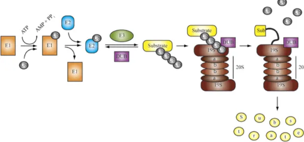
Degradation of a protein by the UPS involves two distinct and successive steps: (i) covalent attachment of multiple ubiquitin molecules to the target protein, and (ii) degradation of the tagged protein by the 26S proteasome.
Proteins are targeted for proteasomal degradation by the covalent attachment of multiple ubiquitin molecules (Ub). The Ub conjugating system operates in a three- step mechanism, with three distinct enzymes catalyzing each step. First, a Ub gets activated in its amino-terminal glycine by the Ub-activating enzyme (E1). Next, the activated Ub is transferred by one of several dozens of Ub-conjugating enzymes (E2) to one of several hundreds of substrate specific Ub-ligases (E3s) that finally attaches the Ub to the target protein (Pickart, 2001; Wilkinson, 1999). Finally, there are nearly 100 deubiquitylating enzymes (DUBs) that counter the activity of E3 ligases in regulatory pathways, as well as functioning in ubiquitin maturation and ubiquitin cleavage and -recycling at the 26S proteasome (Fig. 1.9) (Nijman et al., 2005).
The first Ub is either transferred to a ε-NH2 group of a lysine residue (K) of the target protein to generate an isopeptide bond, or in a linear manner to the N-terminal residue of the substrate. Subsequent Ub addition can occur through isopeptide linkage on all of ubiquitin’s seven lysine residues as well as its N-terminal primary amine, thereby generating a diverse range of chain topologies that can drive a variety of different protein fates (Breitschopf et al., 1998; Peng et al., 2003). K48-linked polyubiquitin chains serve as a specific tag for protein degradation by the 26S proteasome, while other polyubiquitin chains such as K63 chains are involved in
other signalling processes such as NFkB-activation and DNA repair (Chen, 2005;
Sun and Chen, 2004; Voges et al., 1999).
Figure 1.9 | The Ubiquitin Proteasome System. Ubiquitin is activated and conjugated to target proteins by a conserved series of E1 (ubiquitin-activating enzyme), E2 (ubiquitin-conjugating enzyme), and E3 (ubiquitin ligase) activities.
In some cases, an isopeptidase or a deubiquitylating enzyme (DUB) may oppose the activity of the E3. Polyubiquitylated proteins are recruited (via ubiquitin receptors) to the 26S proteasome, a multisubunit, barrel-shaped cellular protease consisting of a 20S core particle bound at one or both ends by 19S cap particles. This 19S cap confers both ATP- and ubiquitin-dependency to proteolysis by the 26S proteasome, and contains isopeptidase activities that remove ubiquitin from the substrate for recycling and ATPase activities that unfold the substrate and feed it into the 20S core for degradation (Eldridge and O'Brien, 2010).
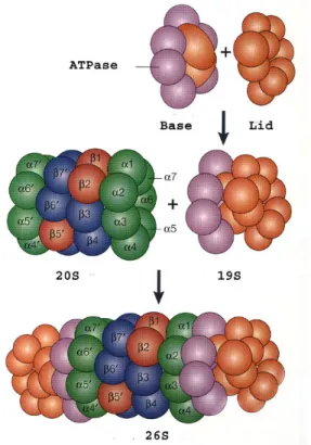
The 26S proteasome is a multisubunit protease complex, which in eukaryotic cells shows both nuclear and cytosolic localization. It is comprised of the 20S proteasome, which serves as the catalytic core, as well as two regulatory 19S subunits (cap) at either end. The regulatory 19S cap is composed of an 11-protein lid and a 10-protein base. The lid is responsible for recognition of polyubiquitylated substrates and detachment of the poly-Ub-chain. The base is essential for substrate-unfolding and entry of the substrate into the 20S core. The 20S core is responsible for proteolysis of the substrates (Voges et al., 1999). It compromises three different catalytic subunits (β1, β2, β5), each with a different proteolytic specificity: the trypsin-like- activity (β1), caspase-like-activity (β2) and chymotrypsin-like-activity (β5) (Fig. 1.10).
In all three β-subunits, a threonin-residue in the catalytic centre is essential for
Eldridge and O´Brien, 2010
catalysis, with the γ-hydroxy-group serving as nucleophil and the α-amino-group serving as proton-donor/acceptor during hydrolysis (Dick et al., 1998; Nussbaum et al., 1998).
Figure 1.10 | Structure of the 26S proteasome. The 26S proteasome is comprised of two 19S regulatory subunits and a 20S core unit.
The 19S complex consists of two sub-complexes: (i) the base (purple), which includes six AAA-ATPases and two non-ATPase proteins, is responsible for protein-unfolding and entry into the 20S core; and (ii) the lid (orange) consisting of 9 non-ATPase proteins, which is responsible for poly-Ub-recognition and detachement.
The 20S catalytic core complex is assembled of 28 subunits (14 different) building the outer α-subunits (green) and the inner β-subunit (blue). The β- subunit includes the catalytic centres β1, β2, β5 (red) responsible for caspase- like, trypsin-like and chymotrypsin-like activity, respectively.
1.5.1 Inhibition of the UPS as a therapeutic approach
Given the important role of the UPS in maintaining protein homeostasis and in regulating cellular processes such as apoptosis, the inhibition of components of the UPS has been seen as a promising target in the development of new therapeutics in the anti-cancer treatment. In 1994 inhibitors of the proteasome have first been described to induce apoptosis (Rock, Gramm et al. 1994), probably by changing the expression level of pro- and anti-apoptotic proteins, for instance pro-apoptotic members of the Bcl2 protein family. Accordingly, several pro-apoptotic members,
Klötzel et al., 2001
such as BAX, BIK, BIM and NOXA have been reported to accumulate upon proteasome inhibition. While BAX and BIK are thought to accumulate due to a block in their proteasomal degradation (Liu et al., 2008; Zhu et al., 2005), NOXA and BIM accumulation is reported to occur at the transcriptional level as a result of stabilization of their transcription factors, such as p53 (Fernandez et al., 2005; Perez- Galan et al., 2006).
The proteasome inhibitor bortezomib (PS-341, Velcade) (Adams, 2002) was the first drug that targets a component of the UPS and was first approved for clinical use in the United States (Chauhan, Hideshima et al. 2005). Bortezomib functions as a reversible inhibitor of the chymotrypsin-like activity of the 26S proteasome with a Ki of 0.6 nM. It binds the catalytic threonine in the active site of the β5 subunit, which also results in a reduction of the catalytic activities of the caspase-like and trypsin-like subunits. Developed by ProScript Inc in 1995, Bortezomib entered clinical trials in 1997 and was approved by the Federal Drug Administration (FDA) in 2003.
Bortezomib-treatment results in clinical benefit in the treatment of haematological malignancies, such as multiple myeloma or mantle cell lymphoma. Bortezomib is currently undergoing Phase III clinical trials for follicular non-Hodgkin’s lymphoma, in Phase II trials for diffuse large B cell lymphoma, and a great many other clinical trials (Yang et al., 2009). Interestingly, bortezomib-induced apoptosis preferentially occurs in malignant but not in non-malignant cells of some tumour tissues, although the detailed mechanism is still under investigation (Fernandez et al., 2005; Qin et al., 2005).
Besides the clinical benefit, substantial side-effects and resistance have been observed following bortezomib-treatment. Furthermore, the effect of bortezomib in the treatment of solid tumours has been less promising. Nevertheless, the surprising efficacy (and rapid clinical approval) of bortezomib for the treatment of multiple myeloma and mantle cell lymphoma has encouraged researchers to explore the possibility of targeting other components of the UPS in order to introduce more specific anti-cancer therapies.
1.6 Aim of the work
Numerous reports show that compounds inhibiting proteasomal activity are able to selectively induce tumour cell apoptosis (Rock, Gramm et al. 1994). However the
underlying mechanisms giving rise to the selective cytotoxicity of tumour cells toward proteasome inhibitors are incompletely understood. Bortezomib, a pharmacological inhibitor of the proteasome, is already successfully used in the clinical treatment of several types of cancer. However, besides beneficial effects substantial side-effects and resistance have been reported (Yang et al., 2009). Nevertheless, the initial beneficial effects clearly highlight the promising role of the UPS as a valuable therapeutic target in the treatment of cancer.
The current work aims to understand the detailed mechanisms of bortezomib- induced apoptosis. Moreover, the knowledge of why tumour cells are more susceptible to proteasome inhibitors may contribute to understand the pathophysiology of malignant transformation and may facilitate efforts aiming to design a novel anti-cancer therapy.
2 Material and methods
2.1 Chemicals
Unless indicated otherwise, all chemicals were from Roth (Karlsruhe, Germany) or Sigma (Deisenhofen, Germany).
Trichostatin A, 5-Aza-2´Deoxycytidine, etoposide, doxorubicin, and LDN-57444 were from Sigma (Deisenhofen, Germany) and bortezomib from Teva (Berlin, Germany).
2.2 DNA constructs
For construction of GFP-fusion proteins, open reading frames (ORF) encoding human NOXA-wt, NOXA-K0 or UCH-L1, were amplified by PCR and cloned into pEGFP-C3 vector (Clontech, Takara Bio Europe, Saint-German-en-Laye, France).
GFP-UCH-L1D176N was obtained by site directed mutagenesis PCR using GFP-UCH- L1 as template and UCH-L1-mut primer.
For the construction of myc-fusion proteins ORF encoding human NOXA-wt or NOXA-K0 were amplified by PCR and cloned into pCDNA3.1+ vector (Invitrogen, Karlsruhe, Germany) already containing an amino-terminal myc-sequence.
For the construction of HA-fusion proteins ORF encoding human NOXA-wt or NOXA- K0 were amplified by PCR with primers encoding an amino-terminal HA-sequence and cloned into pCDNA3.1+ vector.
NOXA-wt and NOXA-K0 cDNA were obtained from Addgene (Cambridge, USA).
Myc-UCH-L1 cDNA was a kind gift from Prof. K.J. Lee (Ewha Womans University, Seoul, Korea).
Myc-XIAP has been described previously (Kashkar et al., 2007).
All PCR amplifications have been performed with appropriate primers containing specific restriction sequences using Phusion™ High Fidelity DNA Polymerase. After restriction with the appropriate enzyme PCR-Fragments and vectors have been
separated by argarose gel electrophoresis (Agarose from Peqlab, Erlangen, Germany) in TE buffer (10 mM Tris-Cl, pH 7.5. 1 mM EDTA) using ethidiumbromide for nucleic acid staining, cleaned up with NucleoSpin® Gel and PCR Clean-up Kit and ligated using the Rapid DNA Ligation Kit according to the instructions of the manufacturer with a molecular template:vector ratio of 3:1.
Plasmid-DNA has been propagated in E.coli XL1-blue and preparated using NucleoSpinPlasmid (Mini) or NucleoBondPlasmid (Maxi) according to the instructions of the manufacturer. All constructs used in this work have been sequenced by GATC (Tübingen, Germany).
Table 2.1 | Kits and enzymes used for the construction of DNA constructs
Name Company
Phusion™ High Fidelity DNA Polymerase
Finnzymes, Fisher Scientific GmbH, Schwerte, Germany
Rapid DNA Ligation Kit Fermentas, Leon-Roth, Deutschland Restriction enzymes Fermentas, Leon-Roth, Deutschland NucleoSpinPlasmid (Mini)
NucleoBondPlasmid (Maxi)
MACHEREY-NAGEL GmbH & Co. KG, Düren, Germany
NucleoSpin® Gel and PCR Clean-up MACHEREY-NAGEL GmbH & Co. KG, Düren, Germany
Table 2.2 | DNA constructs used in this work with specific primers Construct in
pEGFP-C3
MW (kDa)
aa (bp)
5’ primer with restriction side
3’ primer with restriction side
GFP-NOXA-wt 35,9 915 5' EcoR1-NOXA 3' BamH1-NOXA
GFP-NOXA-K0 35,9 915 5' EcoR1-NOXA 3' BamH1-NOXA
GFP-UCH-L1 52,7 1413 5' HindIII-UCH-L1 3' BamH1-UCH-L1 GFP-UCH-L1D176N 52,7 1413 5' UCH-L1-mut 3' UCH-L1-mut Construct in
pCDNA3.1+
MW (kDa)
aa (bp)
5’ primer with restriction side
3’ primer with restriction side HA-NOXA-wt 7,8 192 5' HindIII-HA-NOXA-wt 3' XhoI-NOXA HA-NOXA-K0 7,8 192 5' HindIII-HA-NOXA-K0 3' XhoI-NOXA
Myc-NOXA-wt 8,2 210 5' BamH1-NOXA 3' XhoI-NOXA
Myc-NOXA-K0 8,2 210 5' BamH1-NOXA 3' XhoI-NOXA
Table 2.3 | Primer used for the generation of DNA constructs used in this work Primer name Sequence (5'-3')
5' BamH1-NOXA CCC CCC CCC GGA TCC ATG CCT GGG AAG AAG GCG CG
3' XhoI-NOXA CCC CCC CCC TCG AGT CAG GTT CCT GAG CAG AAG
5' EcoR1-NOXA CCC CCC CCG AAT TCT GAT GCC TGG GAA GAA GGC GCG
3' BamH1-NOXA CCC CCC CCG GAT CCT CAG GTT CCT GAG CAG AAG
5' HindIII-UCH-L1 GAT CAA GCT TAT GCA GCT CAA GCC GAT GGA G 3' BamH1-UCH-L1 GAT CGG ATC CTT AGG CTG CCT TGC AGA GAG C 5' HindIII-HA-NOXA-wt GAT CAA GCT TAC ATG TAC CCA TAC GAT GTT CCA
GAT TAC ATG CCT GGG AAG AAG GCG CGC
5' HindIII-HA-NOXA-K0 GAT CAA GCT TAC ATG TAC CCA TAC GAT GTT CCA GAT TAC ATG CCT GGG AGA AGA GCG CGC
5' UCH-L1-mut GGC CAC CTC TAT GAA CTT AAT GGA CGA ATG CCT TTT C
3' UCH-L1-mut ATC CAC GTT GTT AAA CAG AAT AAA ATG G
2.3 Cell culture and transfection
All cell culture media and additives were from Biochrom (Berlin, Germany). Plastic material was obtained from TPP (Trasadingen, Switzerland), Nunc (Roskilde, Denmark) or BD Biosciences (Falcon™, Franklin Lakes, USA).
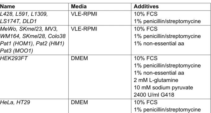
The establishment and maintenance of the following cells and cell lines has been described previously: L428, L591, L1309 (Kashkar et al., 2007), MeWo (Seeger et al., 2010), SKmel23, SKmel28, MV3, WM164 (Zigrino et al., 2005), Colo38 (Giacomini et al., 1986), and primary melanoma cells derived from Pat1 (HOM1), Pat2 (HM1), Pat3 (MOO1) (Schmidt et al., 2011). HEK293FT, HeLa, DLD1, HT29 and LS174T were from ATCC (CRL-11268, Rockville, MD, USA) and maintained according to the instructions of the manufacturer.
In brief, cells were expanded to an adequate amount and several aliquotes were frozen by -150°C in FCS containing 10% DMSO. Experiments were performed after resawing and expanding of cells to 4 passages. Cells were cultured at 37°C in the following media: