Outcome of a penetrating keratoplasty in a 3-month-old child with sclerocornea
Abstract
Sclerocornea is a rare congenital anomaly with clouding of the peripheral cornea that possibly extends up to the center of the cornea. Character-
Dominika Pohlmann
1Mirjam Rossel
1istically, a clear distinction (limbus) between sclera and cornea is lacking.
Daniel J. Salchow
1Early surgical treatment is essential for preventing amblyopia, but
Eckart Bertelmann
1penetrating keratoplasty in children carries a relatively high risk of complications. Especially for sclerocornea, penetrating keratoplasty has generally been reported to have a poor surgical outcome and a high
1 Charité – Universitätsmedizin Berlin, corporate member of risk of complications, including corneoscleral adhesions. Here, we report
the 4-year follow-up on a child with sclerocornea, who was successfully
Freie Universität Berlin, operated on at the age of 3 months and had a favorable outcome. Our Humboldt-Universität zu findings suggest that in some cases, penetrating keratoplasty may be
an option to treat sclerocornea in young children.
Berlin, and Berlin Institute of Health, Berlin, Germany Keywords:congenital corneal opacity, graft survival, pediatric
keratoplasty, sclerocornea
Introduction
A congenital corneal opacity occurs in 3 of 100,000 new- borns [1] and may cause visual impairment and even blindness. In children, deprivation amblyopia may occur if a visually significant corneal opacity is not treated early on. A first report of a penetrating keratoplasty (PK) in a 9-month-old infant with sclerocornea was published in 1970 [2]. Still, PK in children is considered to carry a high risk for complications [2], [3], [4], [5], [6], [7], [8], and PK in sclerocornea is not recommended by some surgeons because of possible catastrophic results including irido- corneal adhesions and graft failure [2], [9]. The surgery is technically challenging in infants due to the elasticity of sclera and cornea, the shallow anterior chamber, an- terior displacement of the lens-iris diaphragm, and the smaller size of the eye [10]. Difficult postoperative exam- inations and postoperative care contribute to a poor outcome compared to PK in adults. Postoperative compli- cations include suture loosening, corneal infiltrates, and higher rates of graft failure [3], [6], [7], [10], [11], [12].
Repeated examinations under anesthesia may be re- quired for postoperative management. Zhang et al. sug- gest that the main determinants for the outcome of PK in children include indication and types of surgery per- formed [10]. In their study conducted in Beijing, the most common indication for PK in children younger than 12 years was congenital corneal opacity which was not further specified. In the same study, the graft survival was 68.1% with a mean follow-up of 33.7 months in in- fants and children [10]. Graft failure occurred in 26 eyes of 79 (26.6%) in infants aged ≥3 months to 4 years [10].
Lin et al.’s study on children with congenital corneal opacities found that most eyes with clear grafts achieved
ambulatory vision (≥20/960), especially children with bilateral opacity [13]. Of all surgical indications for PK in children, unilateral sclerocornea was associated with the worst visual outcome. Lin et al. proposed that sclero- cornea might be associated with denser opacification and anterior segment dysplasia [13]; and average graft survival after PK for sclerocornea has been reported to be 36.4 months for children under 5 years [9]. Despite the concern connected with performing PK at an early stage, it may be considered and of value in select pa- tients.
We report our case of a 3-month-old child with bilateral sclerocornea with 4-year follow-up after PK.
Case description
A 3-month-old girl presented in March 2015 with bilateral dense corneal opacity since birth. The family history was positive for anterior segmental mesenchymal dysgenesis.
Her brother was born in 2019 and also showed corneal opacity, but only in one eye. The mother had a congenital cataract associated with a PITX3 gene mutation; the father had no pertinent ocular history. The children were not genetically examined.
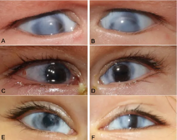
At the first presentation, the girl showed dense corneal opacity and limbal vascularization on both eyes. The opaque corneas of the girl precluded examination of deeper anterior segment structures (Figure 1A, B).
Horizontal corneal diameter was 10 mm bilaterally, ver- tical corneal diameter was 9 mm on the right eye (R) and 8.5 mm on the left eye (L). Axial length was 17.28 mm (R) and 17.55 mm (L), anterior chamber depth was 2.05 mm (R) and 1.99 mm (L). The retina was
Figure 1: Pre- and postoperative stages of penetrating keratoplasty (PK) of the presenting case. The right (A) and left eye (B) revealed a congenital corneal opacity with scleral tissue extension in the center and the periphery of the cornea, and discreet limbal vascularisation. The histological findings confirmed sclerocornea. The corneal grafts were completely clear after PK at 4 days on the right eye (C) and at 5 months on the left eye (D). In the left eye, the sutures had already been removed. After
3 years, both eyes (E, F) showed a clear corneal transplant in the center.
attached on B-scan ultrasound in both eyes. The girl did not fixate or follow objects, and a conjugate horizontal pendular nystagmus as well as esotropia on the left eye were noted. Intraocular pressure (IOP) was normal (R 7.8 mm Hg, L 16.5 mm Hg by Schiötz tonometry).
A PK was performed on the left eye in April 2015 and on the right eye in September 2015 by an experienced corneal specialist (EB). Each trepanation of cornea of the recipient was 5.5 mm and each corneal transplant amounted to 6.0 mm with an endothelial cell density of
≥2500 cells/mm2, and was obtained from the Eye Bank Berlin, Charité – Universitätsmedizin Berlin. The first donor cornea was grafted with two continuous sutures using 10-0 and 11-0 monofilament nylon sutures (Resorba®, Nürnberg, Germany) and single sutures using 10-0 monofilament nylon (Resorba®, Nürnberg, Germany) on the left eye. The right eye was grafted with single su- tures using 10-0 monofilament nylon sutures (Resorba®, Nürnberg, Germany) from the beginning.
Postoperatively, prednisolone acetate 1% (Pred Forte, Allergan, Irvine, CA, USA) and unpreserved ofloxacin 0.3%
(Floxal® EDO®, Bausch & Lomb, Berlin, Germany), both 5 times daily, were administered until the corneal sutures were removed on the left eye after 5 months, when the second keratoplasty surgery on the right eye was per- formed. Then, the treatment was changed to Loteprednol- etabonat 0.5% (Bausch & Lomb, Berlin, Germany) 2 times
a day. An epithelial defect of the left eye was treated with a bandage contact lens for four weeks. An anterior syne- chia at 4 o’clock and a loose suture were observed in the left eye one month after surgery. They were replaced, and the synechia was lysed with a spatula through a paracen- tesis. Throughout the postoperative course, the IOP was at a normal level, ranging from 11 to 18 mm Hg (iCare).
The girl fixated large objects with her left eye after one month. One month after the second PK, the refractive error was +8.0 –8.0 x 135° (R) and +4.0 sph (L), so that glasses were prescribed. The anterior synechia was still present in the left eye, the lenses were clear, and the fundoscopic examination revealed a normal optic disc with a cup-to-disc ratio of 0.1, and a normal retina bilat- erally. It was recommended that the girl wears the glasses and patches the left eye 1 hour a day. Five months after the second surgery on the right eye, all sutures were re- moved in both eyes. 17 months after surgery, visual acuity (VA) was 0.16 (Cardiff Acuity Test, CAT) on the left eye. A VA for the right eye could not be measured because the occlusion was not tolerated on the left eye.
In April 2017, the girl could fixate and follow objects with the right eye; VA of the left eye was 0.4 with CAT. Com- plete anterior synechia were noted on the right eye, which were lysed surgically. Under general anesthesia, reti- noscopy did not yield a sufficient reflex to measure the
refractive error. Part-time occlusion of 1 hour a day of the left eye was continued.
Four years after the first operation, VA was 0.03 (LEA Vision Test in 1 meter) on the right eye, and 0.2 (LEA Vision Test in 6 meters) on the left eye. There was a right esotropia. Horizontal corneal diameter was 9 mm (R) and 10 mm (L), vertical corneal diameter was 10.5 mm bilat- erally. Axial length was 21.13 mm (R) and 19.04 mm (L).
IOP was 15 mm Hg in both eyes (iCare). The clinical presentation revealed well-adapted corneal transplants with a clear central graft in both eyes with otherwise normal anterior and posterior segment findings.
Discussion
Sclerocornea is a primary anomaly of the eye in which the cornea blends with the sclera, having no clear boundary [14]. In our case, the mother of the child had a mutation in PITX3, a transcription factor containing homeodomain (HD) that causes cataract and anterior segment mesenchymal dysgenesis in several families [15], [16].
In general, corneal opacity such as Peters anomaly and sclerocornea have been considered as the main indication for PK in young children. However, there seem to be dif- ferences in regional clinical practice regarding the use of PK in this age group. The European corneal experts are more conservative and do not recommend PK in sclero- cornea due to poor surgical outcomes [3], [4], [9], [17], in contrast to, for example, Asian colleagues [7], [10], [11], [12], [13].
To note, anatomic success after PK does not always cor- relate with visual outcome, because visual improvement depends on accurate optical correction and strict adher- ence to amblyopia therapy, if necessary [18]. The diagno- sis and surgical treatment of a congenital corneal opacity may be challenging for several reasons. First, examination of the patient may be limited, necessitating an exam un- der general anesthesia. Glaucoma should be ruled out in any child with corneal opacities. Additional diagnostic modalities such as ultrasound of the anterior and posteri- or segment and anterior segment optical coherence tomography (OCT) may be required to identify the correct diagnosis. In young children, sclera and cornea are smaller and more elastic, and the anterior chamber is shallower, making watertight closure during a PK more difficult in children compared to adults [4], [10]. Increased vitreous pressure in children may result in protrusion of the iris-lens diaphragm when the globe is open during surgery. Postoperatively, pronounced inflammation may lead to synechiae and elevation of IOP [4]. However, the main complication is the graft failure, which is more common in children than in adults due to the difficulties of postoperative examinations. The reported graft survival rate varied from 32.6% to 78.6% in children for more than one year follow-up [12], [19]. Lin et al. revealed that in children aged 0 to 7 years, the transparent graft rate of congenital corneal opacities was about 55.6% during
the 6- to 82-month follow-up period [13]. Michaeli et al.
reported a graft survival rate of 78% in 38 children with congenital opacities with a mean follow-up of 40.4 months [19]. A study by Yang et al. demonstrated a graft clarity rate of 56% at 6 months and 44% at 3 years after PK in Peters anomaly [20]. A recent study by Zhang et al. showed a graft survival in 68.1%, with a mean follow-up of 33.7 months [10].
The main cause of corneal graft failure is irreversible rejection. Zhang et al. accounted a graft rejection in 33.8% of 160 eyes in infants and children, 52% occurred in regrafted patients [10]. Kim et al. emphasized that the graft survival largely depended on the type of congenital corneal anomaly [9]. In their study, 7 out of 8 grafts sur- vived in patients with Peters anomaly, whereas 9 out of 12 failed in patients with sclerocornea [9]. These results were concordant with previous reports demonstrating a poor outcome of PK in sclerocornea [21], [19]. The differ- ence is that Peters anomaly has a clear cornea in the periphery, whereas sclerocornea has scleral tissue exten- sion and vascularization in the periphery of the cornea.
This likely contributes to the poorer outcome of PK in sclerocornea and may be a key factor.
Our case also showed peripheral vascularization of the cornea. Postoperatively, the girl developed many risk factors for a potential rejection, including loosening of sutures, anterior synechiae, and new vascularization of the graft. However, there was no increased inflammation, fibrin reaction, or IOP elevation during the postoperative course. We had to change some loosened sutures, and for this reason, the fixation of the graft in children should be done by interrupted sutures as already recommended [3], [4].
Age may also be an important key factor regarding graft survival after PK in children. The infant’s immune system is still developing and is modulated in order to coexist with the mother’s immune system in a newborn for 3 months. While humoral immunity is transferred from the mother, T cell-mediated cellular immunity is being developed by the infant. Regulatory T cells (Tregs) have been described as T suppressor cells with a role of ocular privilege [22] and tolerance induction [23]. Two subsets of Tregs, natural Tregs (nTregs, which are developed in the thymus), and inducable Tregs (iTregs, which differen- tiate from naïve CD4 T cells in the periphery under the influence of particular environmental conditions) have been described. It is known that iTregs can induce toler- ance in models of graft versus host disease and solid organ transplantation [24]. In an animal model, it was shown that subconjunctival application of naïve Tregs supports corneal graft survival in baby rats [25]. Further studies are needed to examine the immune system in children under 1 year of age after PK.
Conclusion
In our case report, we presented a successful PK in a 3-month-old infant without rejections and a well-
developed VA after 4 years. We think that in some indi- vidual cases a PK in children with sclerocornea is reason- able to prevent lifelong visual impairment.
Notes
Competing interests
DP discloses that she has financial relationships with Bayer and Allergan. EB discloses that he has financial relationships with Alcon, Johnson & Johnson, Zeiss, and Human Optics. MR and DJS declare that they have no competing interests.
Scientist program participation
DP is participant in the Berlin Institute of Health (BIH) Charité clinician scientist program funded by Charité – Universitätsmedizin Berlin and BIH.
References
1. Bermejo E, Martínez-Frías ML. Congenital eye malformations:
clinical-epidemiological analysis of 1,124,654 consecutive births in Spain. Am J Med Genet. 1998 Feb;75(5):497-504. DOI:
10.1002/(sici)1096-8628(19980217)75:5<497::aid- ajmg8>3.0.co;2-k
2. Wood TO, Kaufman HE. Penetrating keratoplasty in an infant with sclerocornea. Am J Ophthalmol. 1970 Oct;70(4):609-13.
DOI: 10.1016/0002-9394(70)90897-4
3. Seitz B, Hager T, Szentmáry N, Langenbucher A, Naumann G.
Die perforierende Keratoplastik im Kindesalter – das ewige Dilemma [Keratoplasty in children – still a dilemma]. Klin Monbl Augenheilkd. 2013 Jun;230(6):587-94. DOI: 10.1055/s-0032- 1328653
4. Bachmann B, Avgitidou G, Siebelmann S, Cursiefen C.
Hornhautchirurgie und Hornhauttransplantation bei Kindern [Pediatric corneal surgery and corneal transplantation].
Ophthalmologe. 2015 Feb;112(2):110-7. DOI: 10.1007/s00347- 014-3053-9
5. Karadag R, Chan TC, Azari AA, Nagra PK, Hammersmith KM, Rapuano CJ. Survival of Primary Penetrating Keratoplasty in Children. Am J Ophthalmol. 2016 Nov;171:95-100. DOI:
10.1016/j.ajo.2016.08.031
6. Kusumesh R, Vanathi M. Graft rejection in pediatric penetrating keratoplasty: Clinical features and outcomes. Oman J Ophthalmol.
2015 Jan-Apr;8(1):33-7. DOI: 10.4103/0974-620X.149862 7. Low JR, Anshu A, Tan AC, Htoon HM, Tan DT. The outcomes of
primary pediatric keratoplasty in Singapore. Am J Ophthalmol.
2014 Sep;158(3):496-502. DOI: 10.1016/j.ajo.2014.05.020 8. Limaiem R, Chebil A, Baba A, Ben Youssef N, Mghaieth F, El Matri
L. Pediatric penetrating keratoplasty: indications and outcomes.
Transplant Proc. 2011 Mar;43(2):649-51. DOI:
10.1016/j.transproceed.2011.01.055
9. Kim YW, Choi HJ, Kim MK, Wee WR, Yu YS, Oh JY. Clinical outcome of penetrating keratoplasty in patients 5 years or younger: peters anomaly versus sclerocornea. Cornea. 2013 Nov;32(11):1432-6. DOI: 10.1097/ICO.0b013e31829dd836
10. Zhang Y, Liu Y, Liang Q, Miao S, Lin Q, Zhang J, Pan Z, Lu Q.
Indications and Outcomes of Penetrating Keratoplasty in Infants and Children of Beijing, China. Cornea. 2018 Oct;37(10):1243- 8. DOI: 10.1097/ICO.0000000000001695
11. Medsinge A, Speedwell L, Nischal KK. Defining success in infant penetrating keratoplasty for developmental corneal opacities.
Am Orthopt J. 2014;64:81-8. DOI: 10.3368/aoj.64.1.81 12. Zhu AY, Marquezan MC, Kraus CL, Prescott CR. Pediatric Corneal
Transplants: Review of Current Practice Patterns. Cornea. 2018 Aug;37(8):973-80. DOI: 10.1097/ICO.0000000000001613 13. Lin Q, Shi W, Miao S, Zhang Y, Li L, Pan Z. Visual Outcomes and
Prognostic Factors of Successful Penetrating Keratoplasty in 0- to 7-Year-Old Children With Congenital Corneal Opacities.
Cornea. 2018 Oct;37(10):1237-42. DOI:
10.1097/ICO.0000000000001689
14. Elliott JH, Feman SS, O’Day DM, Garber M. Hereditary sclerocornea. Arch Ophthalmol. 1985 May;103(5):676-9. DOI:
10.1001/archopht.1985.01050050068020
15. Berry V, Yang Z, Addison PK, Francis PJ, Ionides A, Karan G, Jiang L, Lin W, Hu J, Yang R, Moore A, Zhang K, Bhattacharya SS.
Recurrent 17 bp duplication in PITX3 is primarily associated with posterior polar cataract (CPP4). J Med Genet. 2004
Aug;41(8):e109. DOI: 10.1136/jmg.2004.020289
16. Semina EV, Ferrell RE, Mintz-Hittner HA, Bitoun P, Alward WL, Reiter RS, Funkhauser C, Daack-Hirsch S, Murray JC. A novel homeobox gene PITX3 is mutated in families with autosomal- dominant cataracts and ASMD. Nat Genet. 1998 Jun;19(2):167- 70. DOI: 10.1038/527
17. Kampik A, Lund OE, Hälbig W. Perforierende Keratoplastik bei kongenitalen Hornhauttrübungen [Penetrating keratoplasty in congenital corneal opacities]. Klin Monbl Augenheilkd. 1986 Mar;188(3):188-92. DOI: 10.1055/s-2008-1050611 18. Huang C, O’Hara M, Mannis MJ. Primary pediatric keratoplasty:
indications and outcomes. Cornea. 2009 Oct;28(9):1003-8. DOI:
10.1097/ICO.0b013e3181a186c0
19. Michaeli A, Markovich A, Rootman DS. Corneal transplants for the treatment of congenital corneal opacities. J Pediatr Ophthalmol Strabismus. 2005 Jan-Feb;42(1):34-44.
20. Yang LL, Lambert SR, Drews-Botsch C, Stulting RD. Long-term visual outcome of penetrating keratoplasty in infants and children with Peters anomaly. J AAPOS. 2009 Apr;13(2):175-80. DOI:
10.1016/j.jaapos.2008.10.007
21. Rezende RA, Uchoa UB, Uchoa R, Rapuano CJ, Laibson PR, Cohen EJ. Congenital corneal opacities in a cornea referral practice.
Cornea. 2004 Aug;23(6):565-70. DOI:
10.1097/01.ico.0000126317.90271.d8
22. Whittum JA, McCulley JP, Niederkorn JY, Streilein JW. Ocular disease induced in mice by anterior chamber inoculation of herpes simplex virus. Invest Ophthalmol Vis Sci. 1984 Sep;25(9):1065-73.
23. Thurau SR, Caspi RR, Chan CC, Weiner HL, Nussenblatt RB.
Immunologische Suppression der experimentellen Autoimmunuveitis (EAU) [Immunologic suppression of experimental autoimmune uveitis]. Fortschr Ophthalmol.
1991;88(4):404-7.
24. Chauhan SK, Saban DR, Lee HK, Dana R. Levels of Foxp3 in regulatory T cells reflect their functional status in transplantation.
J Immunol. 2009 Jan;182(1):148-53. DOI:
10.4049/jimmunol.182.1.148
25. Hildebrand A, Jarsch C, Kern Y, Böhringer D, Reinhard T, Schwartzkopff J. Subconjunctivally applied naïve Tregs support corneal graft survival in baby rats. Mol Vis. 2014;20:1749-57.
26. Dana MR, Schaumberg DA, Moyes AL, Gomes JA. Corneal transplantation in children with Peters anomaly and mesenchymal dysgenesis. Multicenter Pediatric Keratoplasty Study. Ophthalmology. 1997 Oct;104(10):1580-6. DOI:
10.1016/s0161-6420(97)30093-1
27. Lowe MT, Keane MC, Coster DJ, Williams KA. The outcome of corneal transplantation in infants, children, and adolescents.
Ophthalmology. 2011 Mar;118(3):492-7. DOI:
10.1016/j.ophtha.2010.07.006
Corresponding authors:
Dr. Dominika Pohlmann, MD
Department of Ophthalmology, Charité – Universitätsmedizin Berlin, Campus Virchow,
Augustenburger Platz 1, 13353 Berlin, Germany, Phone:
+49 30 450 554202, Fax: +49 30 450 554900 dominika.pohlmann@charite.de
Eckart Bertelmann, MD
Department of Ophthalmology, Charité – Universitätsmedizin Berlin, Campus Virchow,
Augustenburger Platz 1, 13353 Berlin, Germany, Phone:
+49 30 450 554202, Fax: +49 30 450 554900 eckart.bertelmann@charite.de
Please cite as
Pohlmann D, Rossel M, Salchow DJ, Bertelmann E. Outcome of a penetrating keratoplasty in a 3-month-old child with sclerocornea. GMS Ophthalmol Cases. 2020;10:Doc35.
DOI: 10.3205/oc000162, URN: urn:nbn:de:0183-oc0001629
This article is freely available from
https://www.egms.de/en/journals/oc/2020-10/oc000162.shtml Published:2020-08-07
Copyright
©2020 Pohlmann et al. This is an Open Access article distributed under the terms of the Creative Commons Attribution 4.0 License. See license information at http://creativecommons.org/licenses/by/4.0/.