Eur. J. Clin. Chem. Clin. Biochem.
Vol. 31, 1993, pp. 129-134
© 1993 Walter de Gniyter & Co.
Berlin · New York
A Comparative Study of Urinary Xanthopterin and Neopterin in Liver Diseases
By Akira Fukuda
1*), Toshio Mazda
2, William L. Gyure
3, Teruhiko lino*, Hideharu Harada
1, Michiyasu Yakura
1, Hiroshi Kamitsukasa
1, Akira Ohbayashi
1, Teruaki Oka
5and Motoo Tsusiie
61
Department of Gastroenterology, Liver Unit, Tokyo National Sanatorium Hospital, Kiyose, Tokyo, Japan
2
Japanese Red Cross Tokyo Metropolitan Blood Center, Musashino, Tokyo, Japan
3
Seton Hall University, South Orange, New Jersey, USA
4
Department of General Education, Nihon University, Setagaya-ku, Tokyo, Japan
5
Department of Pathology, Medical School, Tokyo University, Bunkyo-ku, Tokyo, Japan
6
Biological Laboratory, School of Liberal Arts and Sciences, Kitasato University, Sagamihara, Kanagawa, Japan
(Received September 18, 1992)**)
Summary: By adsorption to activated charcoal, various pteridine derivatives in human urine are oxidized to xanthopterin. Following this oxidation, xanthopterin in urine from healthy subjects and from patients with liver diseases was assayed by high performance liquid chromatography. The mean values for xanthopterin in healthy subjects were 532 ±116 μηιοΐ/mol creatinine (mean + SD) in males and 585 ±153 μπιοΐ/mol creatinine in females; the difference was statistically significant (p < 0.01). Xanthopterin concentrations in patients with liver disease were significantly higher than those in normal subjects. When compared with urinary neopterin, which is a marker of activated cell immunity, xanthopterin was significantly increased even in fatty liver disease. These findings suggest that increased concentrations of urinary xanthopterin in liver diseases reflect not only the status of activated cell-mediated immunity, but also injury to liver cells.
Introduction
Many pteridine derivatives have been found in human urine (1). Among these pteridines, neopterin has been used as a biochemical marker of the activated state of cell-mediated immunity, and it has been used to monitor and screen for some clinical disorders (2 — 6).
Some current studies show that urinary neopterin concentrations in patients with acute or chronic viral hepatitis are higher than in normal controls (7, 8).
Urinary neopterin has also been utilized to distinguish between chronic non-A,non-B hepatitis and liver stea- tosis (9).
*) Present address: First Department of Internal Medicine, Osaka Medical College, Takatsuki, Osaka, Japan.
**) Submitted February 10/August 31, 1992 to "Pteridines", which discontinued with Vol. 3, No. 3, 1992
On the other hand, little information is available on urinary xanthopterin. An early report describes the occurrence of the compound in human urine (1).
Urinary xanthopterin and neopterin concentrations are found to be higher in some cancer patients (10, 11). Plesner & Kalckar (12) showed that many pteri-~
dine derivatives in urine can be oxidized, adsorbed
and eluted from activated charcoal to yield stable
xanthopterin. Because pteridine metabolism takes
place mainly in liver (13), the excretion of other pter-
idine derivatives, besides neopterin, may possibly be
an indicator of liver disease. In our preliminary study,
applying Plesner & Kalckar's extraction method, we
assayed urinary xanthopterin in patients with chronic
non-A, non-B hepatitis and in patients with fatty liver
disease (14). In the present paper we assayed urinary
xanthopterin in different types of liver disease. Uri-
nary neopterin was also determined for comparison.
Patients and Methods Healthy individuals
As controls, urine samples were collected from 178 apparently healthy individuals (age 21 to 64 years, mean age 42), 64 were males (age 24 to 63, mean age 44) and 114 females (age 21 to 64, mean age 40). The samples were collected during routine medical examinations for health care workers at the Tokyo National Sanatorium Hospital.
Patients with liver disease
Urine samples were collected from in- and out-patients in the liver unit, Tokyo National Sanatorium Hospital. The patients were divided into 5 groups as follows;
1) acute viral hepatitis 8 cases, 2) chronic viral hepatitis 53 cases, 3) liver cirrhosis 21 cases,
4) alcohol-induced liver disease 18 cases and 5) non-alcoholic fatty liver 13 cases.
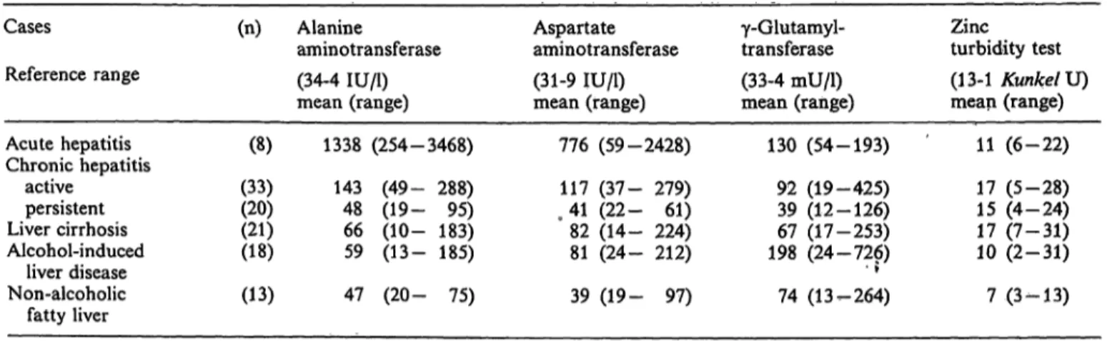
The results of routine biochemical liver tests in each group are shown in table 1.
Among the acute viral hepatitis cases (4 males and 4 females, mean age 37 years), 5 were type A (anti-IgM HAV antibody positive), 1 was type B (HBs antigen and anti-IgM HBc anti- body positive) and 2 were type C (both above HAV and HBV markers negative and anti-clOO-3 positive). Of the chronic viral hepatitis cases (27 males and 26 females, mean age 57 years), 10 were type B, 33 were type C and the other 10 were type non-A,non-B,non-C. Among the chronic viral hepatitis cases, histological features on liver biopsy showed chronic persistent hepatitis in 20 and chronic active hepatitis in 33. Of the liver cirrhosis cases (16 males and 5 females, mean age 60 years), 12 were type B, 2 were type C and 7 were type non-A,non-B,non- C. All the cases were non-alcoholic with biopsy results showing postnecrotic cirrhosis. The alcohol-induced liver disease cases (all 18 were males, mean age 52 years) had a long history of alcohol abuse, with histological features showing either fatty liver or alcoholic fibrosis with or without fat deposits. The non- alcoholic fatty liver cases (12 males and 1 female, mean age 43 years) were considered to be due to obesity and/or metabolic abnormalities such as hyperlipaemia or non-insulin-dependent diabetes.
Renal failure was not present in any of the study subjects, according to urinary and serologic tests. In addition no drugs
such as azathiopurine, corticosteroids or interferon were given in the 6 months prior to this study.
Urine and blood sample analysjs
After the measurement of creatinine concentrations by the alkaline picrate method, urine samples were immediately stored at -30°C in the dark until used for the measurement of xanthopterin and neopterin.
Blood samples were taken from all test subjects at the Tokyo National Sanatorium Hospital at the time of urine collection.
Serum tests were performed for the following enzymes; alanine aminotransferase, aspartate aminotransferase, γ-glutamyl transferase. As a screening test, the zinc turbidity test was also performed.
All analyses were conducted using a 736-40E multi-channel autochemicai analyser (Hitachi Co., Ltd., Tokyo, Japan).
Urinary xanthopterin assay
Urinary xanthopterin was measured applying the isolation method of Plesner & Kalckar (12) and using high performance liquid chromatography (HPLC) for quantitation as follows: To 6 mg of activated charcoal (Norit A or Norit SX-3) 10 μΐ of 1 mol/1 acetic acid and 1 ml of urine were added successively, mixed, and allowed to stand at room temperature for 10 min- utes. The mixture was centrifuged at 6700 g for 5 minutes. The resulting precipitate was washed once with distilled water, then resuspended in 500 μΐ of 0.05 mol/1 NaOH containing 20 ml/1 pyridine. This mixture was centrifuged at 6700 g for 5 minutes and after a 10 fold dilution of the supernate with distilled water, 50 μ! of the solution was injected into the HPLC system.
For xanthopterin determinations, the Twincle HPLC system (Japan Spectroscopic Co., Ltd., Tokyo, Japan) was used. For fluorescence detection, a Jasco FP-110 spectre-photometer (Ja- pan Spectroscopic Co., Ltd., Tokyo, Japan) was used with excitation at 390 nm and emission measurement at 460 nm. A cut-off filter was used to eliminate detection of emission wave lengths shorter than 460 nm. Peaks were recorded on a D-2000 chromato integrator (Hitachi Co., Ltd., Tokyo, Japan). The HPLC column was packed with Asahipac GS 320H (Asahi Chemical Industry Co., Ltd., Kawasaki, Japan). The elution fluid was methanol (volume fraction 0.10) containing 10 mmol/1 potassium phosphate buffer pH 7.0 at a flow rate of 1 ml per minute. The main peak, which showed a retention time of 10.5 minutes, was identified as xanthopterin by comparison with authentic xanthopterin using the HPLC system described above.
The values were expressed as μιηοΐ xanthopterin per mol cre- atinine.
Tab. 1. Biochemical liver tests in each group of liver diseases Cases
Reference range Acute hepatitis Chronic hepatitis
active persistent Liver cirrhosis Alcohol-induced
liver disease Non-alcoholic
fatty liver
(n)
(8) (33)(20) (21)(18) (13)
Alanine
aminotransferase (34-4 IU/1) mean (range)
1338 (254-3468) 143 (49- 288) 48 (19- 95) 66 (10- 183) 59 (13- 185) 47 (20- 75)
Aspartate aminotransferase (31-9 IU/1) mean (range)
776 (59-2428) 117 (37- 279) 41 (22- 61) 82 (14- 224) 81 (24- 212) 39 (19- 97)
γ-Glutamyl- transferase (33-4 mU/1) mean (range)
130 (54-193) 92 (19-425) 39 (12-126) 67 (17-253) 198 (24-726)• ? 74 (13-264)
Zincturbidity test (13-1 Kunkel U) mean (range)
11 (6-22) 17 (5-28) 15 (4-24) 17 (7-31) 10 (2-31) 7 (3-13)
U r i n a r y neopterin assay
Urinary neopterin was directly assayed without oxidation (15) using a RIA kit "IMMUtest Neopterin" (Henning Berlin GmbH, Berlin, Germany). Radioactivity was counted by a programmed gamma-counter "ANSR" (Dinabot Co., Ltd., To- kyo, Japan).
The values were expressed as μπιοί neopterin per mol creatinine.
Statistical analysis
The differences of xanthopterin values in healthy subject be- tween different age groups were assessed by the Kruskal-Wallis test. Using urinary xanthopterin and neopterin assay values as data, median values for these pteridines were determined under various conditions and evaluated by the Mann-Whitney U-test.
Comparisons of abnormal levels of xanthopterin and neopterin were assessed by the %2-test for the different groups of patients separately.
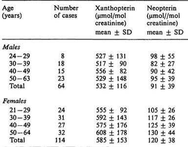
Tab. 2. Urinary healthy Age(years)
Males
24-29 30-39 40-49 50-63 Total
Females21-29 30-39 40-49 50-64 Total
excretion of individuals
Number of cases
188 1523 64
2431 2732 114
xanthopterin and Xanthopterin (μηιοΐ/mol creatinine) mean ± SD 527 + 131 517 + 90 556 + 82 529 + 148 532 + 116
555 ± 92 592 ± 143 575 + 176 608 + 178 585 + 153
neopterin in Neopterin (μπιο1//ιηο1 creatinine) mean ± SD
98 ± 55 82 + 27 90 + 42 95 + 39 91 ± 39
105 ± 26 117 + 26 125 + 39 130 + 44 120 + 38
Results
Urinary xanthopterin in healthy individuals In 178 healthy subjects, the mean values of xantho- pterin were 566 ±143 μπιοΐ/mol creatinine (mean
± SD). In 64 males the values were 532 ±116 μπιοί/
mol creatinine and in 114 females 585 ±153 μηιοί/
mol creatinine. Although a statistical difference in urinary xanthopterin concentrations for males and females was found in this present study (p < 0.01), there were no statistically significant age group dif- ferences (p > 0.05), as shown in table 2.
Urinary xanthopterin in liver disease patients
As shown in table 3, urinary xanthopterin concentra- tions in patients with liver disease including non- alcoholic fatty liver were significantly higher than those in healthy subjects. The highest concentrations of xanthopterin were found in the acute hepatitis
patients, and xanthopterin concentrations were also high in chronic active hepatitis. Urinary xanthopterin concentrations in the active stages of chronic hepatitis were statistically higher than in the persistent stages (chronic active hepatitis vs. chronic persistent hepa- titis: ρ < 0.01).
Comparison with urinary neopterin
Urinary neopterin concentrations for the same sub- jects are also shown in table 3. Neopterin was signif- icantly elevated in cases of acute hepatitis, chronic hepatitis, liver cirrhosis and alcohol-induced liver dis- ease, when compared with normal subjects. In non- alcoholic fatty liver cases, however, there was no significant elevation of neopterin.
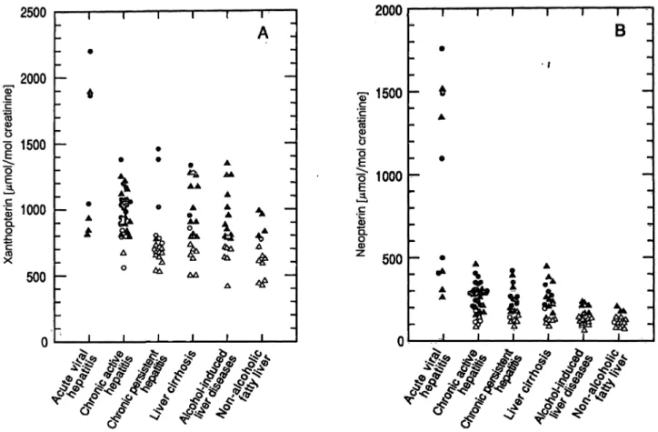
Urinary concentrations of xanthopterin and neopterin in all groups of liver disease are shown in figure 1.
Tab. 3. Urinary excretion of xanthopterin and neopterin
Group (n) Xanthopterin P
(μιηοΐ/mol creatinine)
Control Acute hepatitis Chronic hepatitis
active persistent Liver cirrhosis Alcohol-induced
liver disease Non-alcoholic
fatty liver
1788 3320 2118 13
Mean + SD 566 + 143 1305 + 579 978 ± 174 807 ± 261 934 ± 251 868 ± 248 689 ± 183
Range 250 - 902 813 - 2201 564 - 1377 536 - 1462 505- 1325 418 - 1347 435 - 993
Neopterin
(μιηοΐ/ηιοί creatinine) P Mean + SD Range
< 0.001
< 0.001
< 0.001
< 0.001
< 0.001
<0.05
109 + 40 769 ± 571 251 + 92 224 ± 94 227 ± 99 154+ 48 125 ± 42
266 -27- 91 -91 - 90-71 - 74-
1791232 463422 452240 207
< 0.001
< 0.001
< 0.001
< 0.001
< 0.001 NS NS: not significant compared with controls
SUU
_2000
ΈΓ
'cC
*3
s b
1500 :>
ja.
| 1000
Je
1
X500
n
ι ι ι ι ι ι
- A I _ — *
» —
""
•
— "—
• · β Α "
^ AAA A
_ Φ Υ · Α Α . _
. A a^ IA ^ * . - A & sh 5k a ^, A -
Δ SH ^ ^ Δ
^Δ ^^τ -1
_ ^ ΔΔ —0 Αν
Δ ^Γ _
Ι Ι Ι Ι Ι Ι
2000
•S· 1500
'c ο
b
ε Ι
1000
500
Ι Ι Ι Ι
Β
• \
t JL i ϊ
Fig. 1. Urinary concentrations of xanthopterin (A) and neopterin (B) in all groups of liver disease.
Δ: males within, Δ: males outside the range χ + 2 SD of healthy male controls, o: females within, o: females outside the range χ Η- 2 SD of healthy female controls.
Tab. 4. Abnormal rates of xanthopterin and neopterin excre- tion in all groups of liver disease. The relative and absolute frequency of patients having a measured value greater than the mean + 2 SD of normal controls are shown
Disease
Acute hepatitis Chronic hepatitis
active persistent Liver cirrhosis Alcohol-induced
liver disease Non-alcoholic
fatty liver
Xanthopterin
% 88 8225 6767
31 n
01 8)
(27/33) (5/20) (14/21) (12/18) (4/13)
Neopterin
% n
100 (8/ 8) 79 (26/33) 70 (14/20) 62 (13/21) 39 (7/18) 23 (3/13)
When the normal upper limit of each pteridine was set at the mean + 2 SD in healthy controls, then abnormal rates were highest in acute hepatitis and chronic active hepatitis as seen in table 4. Mean + 2 SD for xanthopterin was 764 μηηοΐ/mol creati- nine in males and 891 μηιοΐ/mol creatinine in females, and for neopterin it was 169 μιηοΐ/mol creatinine in males and 196 μηιοΐ/mol creatinine in females. There was a significant difference in xanthopterin concen-
trations between chronic active hepatitis and chronic persistent hepatitis (82% and 25%, p < 0.001), while neopterin did not show this difference (79% and 70%, p > 0.05).
Discussion
In the present paper a new method of urinary xan- thopterin assay after treatment with activated char- coal is presented. Using this method we assayed uri- nary xanthopterin in healthy individuals and in pa- tients with liver disease. In healthy subjects xantho- pterin and neopterin concentrations when expressed in μπιοΐ/πιοί creatinine were higher in females than in males, but there were no significant age-differences in this study. In our study populations, the concentra- tions of urinary xanthopterin in all groμps with liver disease were significantly higher when compared with healthy subjects. Neopterin concentrations were also raised, but in agreement with Prior et al. (9), there was no significant elevation of neopterin in non-al- coholic fatty liver.
Urinary or serum neopterin concentrations are raised
in infectious or malignant disorders with activated
cell-mediated immunity (4—6). Neopterin is mainly
produced in human monocytes/macrophages, and its release is accelerated upon stimulation by interferon-y (3). In our study, urinary neopterin concentrations were very high in acute hepatitis, and high in virus- induced chronic liver diseases. In alcoholic liver dis- eases, neopterin concentrations were only slightly higher than in controls. This finding may support the view that alcoholic hepatitis successively activates cell- mediated immunity, and that the progress of liver damage in spite of alcohol abstinence, is responsible for the hyperimmune reactivity (16, 17).
The biochemistry of xanthopterin production and ex- cretion is still scarcely known. Nevertheless, we as- sayed xanthopterin together with neopterin. Surpris- ingly, xanthopterin concentrations were significantly high in all the liver disease groups mentioned above.
The increase in urinary xanthopterin, like that of neopterin, in liver diseases may also be caused by activated cell immunity, because the concentrations of both are roughly correlated.
However, abnormally elevated concentrations of xan- thopterin were found in 82% of patients with chronic active hepatitis, but in only 25% of patients with chronic persistent hepatitis (p < 0.001). These two groups were histologically differentiated from those with severe parenchymal destruction. With respect to neopterin excretion, however, these two groups were not significantly different (79% and 70%, p > 0.05).
Moreover, the rate of occurrence of abnormally ele- vated urinary xanthopterin in fatty liver and other chronic liver diseases appear to parallel the mean values of serum aminotransferase activities. This cor-
relation does not exist for neopterin excretion. These findings indicate that increased urinary xanthopterin reflects liver-cell damage rather than the hyperim- mune state.
When solutions (10~~
5mol/1) of dihydroxanthopterin, xanthopterin, dihydroneopterin or tetrahydrobiop- terin were mixed with equal volumes of urine and assayed by the present method, the xanthopterin con- centrations were found to be 1.8, 1.5, 1.3 and 1.4 times the basal concentration (unpublished data).
However, when neopterin, biopterin or dihydrobio- pterin were examined by this method, no increase in urinary xanthopterin was observed. Thus, the present method represents an assay for dihydroxanthopterin, dihydroneopterin and tetrahydrobiopterin in urine.
After treatment with activated charcoal, eluates ob- tained by this method were fairly pure, and an HPLC column could be used for hundreds of samples with- out column washing. A further convenient feature of this HPLC method is that it can be utilized in labo- ratories where only a fluorometer is available (12).
Further studies will be undertaken of the biochemical processes leading to the raised excretion of xantho- pterin.
Acknowledgements
We wish to thank Dr. K. Shibata, Hoechst Japan Co., Ltd., for the generous supply of RIA kits; Mr. K. Fukuhara, Mr. M.
Nagashima and his coworkers at Tokyo National Sanatorium Hospital for generous assistance; and Dr. T. Katayama, Director of the Tokyo National Sanatorium Hospital, for his help in this study.
References
1. Fukushima, T. & Shiota, T. (1972) Pterins in human urine.
J. Biol. Chem. 247, 4549-4556.
2. Wächter, H., Hausen, A. & Grassmayr, K. (1979) Erhöhte Ausscheidung von Neopterin im Harn von Patienten mit malignen Tumoren und mit Viruserkrankungen. Hoppe- Seyler's Z. Physiol. Chem. 360, 1957-1960.
3. Huber, C, Batchelor, J. R., Fuchs, D., Hausen, A., Lang, A., Niederwieser, D., Reibnegger, G., Swetly, P., Troppmair, J. & Wächter, H. (1984) Immune response-associated pro- duction of neopterin. Release from macrophages primarily under control of interferon-gamma. J. Exp. Ivied. 160, 310-316.
4. Hausen, ., Bichler, ., Fuchs, D., Hetzel, H., Reibnegger, G. & Wächter, H. (1985) Neopterin, a biochemical indicator of cellular immune reactions, in the detection and control of patients with neoplastic diseases. Cancer Detection and Prevention 8, 121 — 128.
5. Fuchs, D., Hausen, ., Reibnegger, G., Werner, E. R., Dierich, M. P. & Wächter, H. (1988) Neopterin as a marker for activated cell-mediated immunity: Application in HIV infection. Immunol. Today P, 150—155.
6. Hausen, ., Fuchs, D., Reibnegger, G., Werner, E. R. &
Wächter, H. (1989) Neopterin in clinical use. Pteridines 7, 3-10.
7. Farci, A. M. G., Laconi, R., Cabras, F., Loviselli, A., Cappai, G., Balestrieri, A., Garau, V. L., Tocco, M. &
Casula, D. (1985) Urinary neopterin in acute and chronic liver diseases. In: Biochemical ami Clinical Aspects of Pter- idines (Wächter, H., Curtius, H. C. & Pfleiderer, W., eds.) Berlin—New York, Walter de Gruyter, pp. 453-460.
8. Reibnegger, G., Auhuber, L, Fuchs, D., Hausen, ., Jud- maier, G., Prior, C., Werner, E. R. & Wächter, H. (1988)- Urinary neopterin levels in acute viral hepatitis. Hepatology
£,771-774.
9. Prior, C., Fuchs, D., Hausen, A., Judmaier, G., Reibnegger, G., Werner, E. R., Vogel, W. & Wächter, H. (1987) Potential of urinary neopterin excretion in differentiating chronic non-A,non-B hepatitis from fatty liver. Lancet //, 1235 — 1237.
10. Rokos, H., Rokos, K., Frisius, H. & Kirstaedter, H.-J.
(1980) Altered urinary excretion of pterlidines in neoplastic disease. Determination of biopterin, neopterin, xantho- pterin, and pterin. Clin. Chim. Acta 705, 275 — 286.
11. Stea, B., Halpern, R. MM Halpern, B. C. & Smith, R. A.
(1981) Urinary excretion levels of unconjugated pterins in cancer patients and normal individuals. Clin. Chim. Acta
7/5,231-242.
12. Plesner, P. & Kalckar, H. M. (1956) Enzymic micro deter- minations of uric acid, hypoxanthine, xanthine, adenine, and xanthopterine by ultraviolet spectrophotometry. Meth- ods of Biochemical Analysis J, 97—110.
13. Fukushima, K., Eto, L, Mayumi, T., Richter, W., Goodson, S. & Shiota, T. (1975) Biosynthesis of pterins in mammalian systems. In: Chemistry and Biology ofPteridines (Pfleideref, W., ed.) Berlin-New York, Walter de Gruyter, pp. 247- 14. Mazda, T, Ogasawara, K., Fukuda, A., Gyure, W. L. &263.
Tsusue, M. (1990) Urinary xanthopterin and derivatives as indicators of liver disease. In: Chemistry and Biology of Pteridines 1989 (Curtius, H. C, Ghisla, S. & Blau, N., eds.) Berlin-New York, Walter de Gruyter, pp. 579-582.
15. Fuchs, D., Milstien, S., Krämer, A., Reibnegger, G., Wer- ner, E. R., Goedert, J. J., Kaufman, S. & Wächter, H.
(1989) Urinary neopterin concentrations vs total neopterins for clinical utility, din. Chem. 55, 2305-2307.
16. Kanagasundaram, N., Kakumu,' S., Chen, T. & Leevy, C.
M. (1977) Alcoholic hyalin antigen (AHAg) and antibody (AHAb) in alcoholic hepatitis. Gastroenterology 73,1368—
1373.
17. Izumi, N., Hasumura, Y. &Takeuchi, J. (1983) Lymphocyte cytotoxicity for autologous human hepatocytes in alcoholic liver disease. Clin. Exp. Immunol. 53, 219—224.
Toshio Mazda, PhD.
Japanese Red Cross
Tokyo Metropolitan Blood Center 1-26-1, Kyonan-cho
Musashino Tokyo 180 Japan