Hendriks et al.: A carboxypeptidase in human serum different from carboxypeptidase N 277 J. Clin. Chera. Clin. Biochem.
Vol. 27, 1989, pp. 277-285
© 1989 Walter de Gruyter & Co.
Berlin · New York
Characterisation of a Carboxypeptidase in Human Serum Distinct from Carboxypeptidase N
By D. Hendriks, S. Schärpe, M. van Sande and M. P. Lommaert
Department of Medical Biochemistry, Faculty of Medicine, University ofAntwerp, Wilrijk, Belgium
(Received August 15/November 24, 1988)
Summary: Arginine carboxypeptidase activity in human serum, measured with the hippuryl-L-arginine sub- strate, is about three times higher than in human plasma. This difference is much smaller when hippuryl-L- lysine is used äs the Substrate. When fresh serum is incubated at 30 °C, the arginine and lysine carboxypeptidase activity decreases until a stable activity, close to the plasma activity, is reached. This stable carboxypeptidase activity is attributed to carboxypeptidase N. The unstable carboxypeptidase differs from carboxypeptidase N in pH-optimum, esterase activity, Substrate specifity, Co
2+-activation and dithiotreitol activation. Blood cells are not responsible for the release of this enzyme during coagulation. No activator of carboxypeptidase N was detectable in human serum. -exchange chromatography on DEAE-cellulose confirms the presence of two different molecular forms of arginine carboxypeptidase activity.
Introduction
Carboxypeptidase N (arginine carboxypeptidase, kin- inase I)
1) cleaves carboxy-tefminal arginine and lysine from various peptides in human plasma. Some of the most interesting Substrates for this enzyme are the kinins, bradykinin and kallidin (1), the anaphylatox- ins C
3a, C
4aand C
5a(2, 3), fibrinopeptides 6A and 6D (4), the creatine kinase MM-isöenzyme (5, 6), hexa- peptide enkephalins (7) and the atrial natriuretic pep- tide atriopeptin II (8).
A number of papers deal with the determination of the activity of carboxypeptidase N in human serum (9 -=-23) or in plasma (24) in various pathological conditions. Here we describe and characterize the impprtant difference in carboxypeptidase activities be- tween fresh serum, on the one hand, and older serum or heparinized plasma on the other, depending on the Substrate used. We present evidence of a new carboxy- peptidase activity in fresh human serum which has some characteristics in common with carboxypeptid- ase N.
Materials and Methods Chemicals
Hippuryl-L-arginine (Hip-Arg), hippuryl-L-lysine (Hip-Lys), 3- (2-furylacryloyl)-alanyl-L-lysine (furylacryloyl-Ala-Lys), p-hy- droxy-hippuryl-jL-arginine (pOH-Hip-Arg), and/?-hydroxy-hip- puryl-L-lysine (pOH-Hip-Lys) were from Bachern Feinchemi- kalien (Bubendorf, Switzerland). jD,L-2-Mercaptomethyl-3- guanidinoethylthiopropanoic acid and 4-(2-hydroxyethyl)-l-pi- perazineethane-sulphonic acid (HEPES) were purchased from Calbiochem (La Jolla, CA, USA). Phenylmethylsulphonylfluor- ide, diisopropylfluorophosphate, heparin (Na-salt), and DEAE- cellulose (microgranülar form) were obtained from Sigma (St.
Louis, MO, USA) and dithiotreitol from Janssen Chimica (Beerse, Belgium).
CoCl2 · 6H2O was from Aldrich Chemical Company (Milwau- kee, WI, USA). 3-(2-Fuiylacryloyl)-alanyl-L-arginine (furyla- cryloyl-Ala-Arg) was a kind gift of Dr. Thomas ff. Plununer, Jr., New York State Department of Health (Albany, NY, USA) and hippuryl-L-argininic acid was kindly provided by Dr. Ye- huda Lavin of the Weizmann Institute of Science, Rehovot, Israel. o-Methylhippuric acid was synthesized from glycine and o-methylbenzoylchlbride (UCB, Drogenbos, Belgium) by a pro- cedure analogous to that used for the synthesis of hippuric acid (25).
All other reagents used were of high purity grade and were from Merck, (Darmstadt, FRG).
Enzyme
Carboxypeptidase N, EC 3.4.17.3
J. CHn. Chem. Clin. Biochem. / Vol. 27,1989 / No. 5
Instruments
The pipetting of serum samples and reagents was performed with a Dilutrend dispenser (Boehringer, Mannheim, FRQ). For colorimetric determinations a Hewlett-Packard 8540 ultravi- olet/visible diode-array spectrophotometer was used with a quartz flow cell (10 mm optical pathway). The high perform- ance liquid chromatography System consisted of a 303 solvent- delivery System, an 802 C manometric module, a model 231 — 401 autosampling injector (all from Gilson, Paris, France), an LKB 2140 diode-array UV detector (LKB, Bromma Sweden), and a 100 χ 8mm (i. d.) Ct8 reversed-phase μ-Bondapak col- umn fitted in a radial compression module (Millipore, Br ssels, Belgium).
Routine coagulation analyses were performed on a Coag lab 40A automatic coagulation analyser (Ortho Diagnostics, Rar- iton, NJ, U.S.A.).
Blood Samples
From five healthy individuals (laboratory staff, three males, two women) blood samples were taken (in no additive, silicone coated tubes, and in lithium heparin tubes (Vacutainer, Becton and Dickinson, Rutherford, NJ, U.S.A.). All samples were placed in ice for l h to allow clotting for the serum preparation, then centrifuged (10 min, 2000 g). Sera and heparinized plasma were collected^ divided into 200 μΐ portions and stpred at -80°C. Part of the serum, before storage at -80°C, was placed in a waterbath at 30 °C for 15h. This was arbitrarily defined s "conditioned" serum. Before assaying enzyme activ- ities, the samples were thawed in ice.
Preparation of homogenates from human blood cells Leukocyte-rich fraction
Buffy coat cells obtained from citrated human blood (33 ml) were mixed with 8 ml of Volex (Mc Graw Laboratories, Irvine, CA, U. S. A.) in a measuring cylinder. After Standing at room temperature for l h, the leukocyte-rich layer was collected from the top and the cells were washed twice with phosphate buffered saline.
Erythrocytes
Erythrocytes were obtained from 5 ml of heparinized blood and were washed three times with phosphate buffered saline.
Enzyme assays
The enzyme activity with the Substrates Hip-Arg and Hip-Lys was determined by a high performance liquid chromatography- assisted assay s described elsewhere (26), with the following modifications: the buffered Substrate Solutions were both pre- pared at pH 8.0 (pH measurements were made at room tem- perature, 18—21 °C) and the incubation time was 30 min.
/?OH-Hip-Arg and ^pOH-Hip-Lys-splitting activity was meas- ured by means of a colorimetric assay f(27).
Hydrolysis of the furylacryloyl-substrates (furylacryloyl-Ala- Arg and furylacryloyl-Ala-Lys) was determined by the method ofPlummer & Kimmel (28), adapted to the following conditions:
for furylacryloyl-Ala-Lys: 15 μΐ of sample was mixed with 615 ul of buffer (50 mmol/1 HEPES/250 mmol/1 NaCl pH 7.80) previously brought to 37 °C and preincubated for 2min at 37 °C; 70 μΐ of a solution of 6 mmol/1 furylacryloyl-Ala-Lys (0.6 mmol/1 final concentr tion) in distilled water was added.
After mixing, the solution was transferred into a cuvette ther- mostatted at 37 °C, and the decrease in absorbance at 340 nm was continuously measured for 400 s (reading against distilled water). For furylacryloyl-Ala-Arg, a siinilar procedure was fol- lowed with 35 μΐ of sample, 595 μΐ of buffer and 70 ul of 6 mmol/1 furylacryloyl-Ala-Arg solution. One unit of enzyme activity was defined s the amount of enzyme required to hydrolyse l μηιοί pf Substrate per niinute at 37 °C s determined by the Δε. The esterase activity, determined with the Substrate Hip-argininic acid, was measured s follows: to 40 μΐ of sub- strate solution (10 mmol/1 Hip-argininic acid/50 mmol/1 HEPES pH 8.0) were added 10 μΐ of the sample; this mixture was incubated for 10 min at 37 °C and the reaction stopped with 50 μΐ of l mol/1 HC1. After addition of 10 μΐ of the internal Standard -methylhippuric acid, the hippuric acid was extracted and determined by liquid chromatography s described else- where (26). Triplicate determinations were performed for all enzyme assays.
Activators and inhibitors
The effects of all the enzyme inhibitors and activators were evaluated with the Hip-Arg Substrate, sing the HPLC-assisted assay (26). Preincubations were performed s follows:
Cobalt Chloride
Samples, diluted l plus 4 with physiological saline, were prehv cubated with Co++ (5 mmol/1 solution of CoCl2 in physiological saline) for l h on ice (final Co++-concentration during assay was l mmol/1).
Thrombocytes
Thrombocyte concentrates (60 · 1012/0 were washed three times with phosphate buffered saline.
Extracts were obtained in two ways from all cells:
Extraction in presence ofdetergent: A 100 g/i solution of Non- idet P40 (LKB, Bromma, Sweden) in water was added to the cells to obtain a concentr tion of 5 g/l Nonidet P40; the cell suspensions were vortexed for 15 s and centrifuged at 10000#
for 5min. The supernatant was divided into 200 μΐ portions and stored at -80°C.
Extraction in absence ofdetergent: The cells were subjected to 2 cycles of freezing and thawing by keeping them at ^80 °C for 15 min and at room temperature for 5 min, then sonicated for 10 s. The supernate, obtained after centrifugation at 10 000 g for 5min, was divided into 200 μΐ portions and stored at -80°C.
Dithiotreitol
A 2 mmol/1 solution of dithiotreitol in HEPES, 10 mmol/1 (pH 7.4) was mixed in equal parts with the samples and then prein- cubated for l h in ice (final dithiotreitol concentr tion during the assay was 0.2 mmol/1).
Diisopropylfluorophosph te
A solution of 100 g/l in isopropanol was diluted with physio^
logical saline to obtain a 10 mmol/1 diisoprppylfluorophosphate concentr tion. Ninety microlitres of eaeh sample were mixed with 10 μΐ of this 10 mmol/1 diisopropylfluorophosphate solu- tion, preincubated in ice for 2 h and then assayed for enzyme activity (diisopropylfluorophosphate concentr tion during preincubation was l mmol/1).
Appropriate blanks containing the same amount of isopropanol were assayed to exclude any effect of the isopropanol on enzyme actiyity.
J. Clin. Chem. Cli . Biochem. / Vol. 27,1989 / No. 5
Hendriks et al.: A carboxypeptidase in human serum different from carboxypeptidase N
279
PhenylmelhylsulphonylfluorideA 400 mmol/1 solution of phenylmethylsulphonylfluoride in ethanol was diluted with physiological saJine to obtain a 2 mmol/1 phenylmethylsulphonylfluoride solution. Samples were mixed in equal parts with this 2 mmol/1 phenylmethylsul- phonylfluoride solution and preincubated for 2 h in ice before assaying enzyme activity. Appropriate blanks containing the same amount of ethanol were assayed. The phenylmethylsul- phonylfluoride solution should be used within 30 min to avoid spontaneous hydrolysis of the inhibitor (29).
1 ,10-Phenanthroline
Samples were diluted l plus 2 with a solution of 15 mmol/1 1,10 phenanthroline in physiological saline, then preincubated for 2 h in ice (phenanthroline concentration during preincuba- tion was 10 mmol/1; the final concentration during assay was 2 mmol/1).
D,L-2-mercaptomethyl-3-guanidinoethylthiopropanoic acid 10 μΐ of freshly prepared stock solution of 1200 μπιοΐ/ΐ in distilled water (which was first freed of oxygen by passing N2
for 30 min) was added to 40 ul of the Substrate solution (Hip- Arg) before assaying the samples for enzyme activity. (The final concentration of D,L-2-mercaptomethyl-3-guanidinoethylthio- propanoic acid was 200 μηιοΙ/1). The stock solution of £>,/>2- mercaptomethyl-3-guanidinoethylthiopropanoic acid should be used within the hour, to avoid loss of inhibitory capacity.
Ion exchange ehromatography
Two ml of fresh human serum diluted l plus 4 with the equi- libration buffer was applied at a flow rate of 21 ml · cm""2 · h"1 to a column (1.6 x 35 cm) of DEAE cellulose previously equilibrated in Tris, 0.05 mol/1, pH 7.2. The enzymes were eluted with a linear gradient of NaCl from 0 to 0.25 mol/1 in Tris 0.05 mol/1, pH 7.2 during 8 h. The effluent was collected in 10 ml fractions. All operations were performed at 4°C.
Protein was determined by its absorbance at 280 nm. Aliquots of the fractions (100 μΐ) were diluted l + l in serum, which had previously been in ctivated by keeping it at 56 °C for 12 h (this serum contained no arginine carboxypeptidase activity after inactivation). Samples were then assayed for arginine carboxypeptidase activity using the HPLC-assisted assay. The same samples were also assayed for arginine carboxypeptidase activity after placing them at 37 °C for 2 h.
Results
Arginine and lysine carboxypeptidase activ- ity in serum and plasma
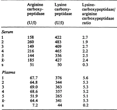
The results of arginine carboxypeptidase and lysine carboxypeptidase activities of the serum and plasma of the 5 healthy subjects are s nimarized in table 1.
Carboxypeptidase activities, when rneasured with the Hip-Arg Substrate, are three times higher (287%) in the fresh serum samples than in the plasma samples.
The lysine carboxypeptidase activity, however, is only 25% elevated in serum s compared with plasma (125%). This is also confirmed by the lysine carboxy- peptidase/arginine carboxypeptidase ratio.
Tab. 1. Arginine and lysine carboxypeptidase activity in serum and plasma
Serum 21 34 x·5 s Plasma 2\ 43 5x·
s
Arginine carboxy- peptidase (U/l) 260158 216149 144185 51 64.867.7 69.068.6 51.964.4 7.2
Lysine carboxy- peptidase (U/l) 422483 409465 427356 50 344376 363357 265341 44
Lysine-
carboxypeptidase/
arginine
carboxypeptidase ratio
2.71.9 2.72.2 2.52.4 0.3 5.65.3 5.35.2 5.15.3 0.2
From five healthy individuals (laboratory staff), blood was taken s described in the Methods section. Carboxypeptidase activities in serum and heparinized plasma were determined with both Substrates Hip-Arg (arginine carboxypeptidase) and Hip-Lys (lysine carboxypeptidase) using the HPLC-assisted as- say.
Stability
Stability during assay
Serum samples were assayed for arginine carboxypep- tidase and lysine carboxypeptidase activity (26) using different incubation times at 37 °C. The activities ob- tained with arginine carboxypeptidase for incubation times of 10, 20 and 30 min were: 194, 191, and 197 U/l; with lysine carboxypeptidase: 476, 461, and 455 U/L
Even with prolonged incubation, Stability was good:
arginine carboxypeptidase: 160, 158, and 151 U/l (other serum, incubation times 30, 60 and 90 min respectively). This clearly demonstrates that both ar- ginine carboxypeptidase and lysine carboxypeptidase activities measured in fresh serum are stable under a$s y conditions, i. e. in the presence of the Substrate.
Stability s afunction oftime and temperature
Fresh serum was kept in a water bath at 30 °C and assayed for arginine carboxypeptidase and lysine car- boxypeptidase activities at different time intervals.
The same experiment was performed with fresh hep- arinized plasrna.
J. Clin. Chem. Clin. Biochem. / Vol. 27,1989 / No. 5
As shown in figure l, arginine carboxypeptidase and lysine carboxypeptidase activity in serum is unstable:
during the first two hours of storage at 30 °C, there is a rapid decrease in their activities, which reach a plateau after about 9 h, then remain stable. The extent of this decrease is much more pronounced for arginine carboxypeptidase then for lysine carboxypeptidase ac- tivity. In contrast, in fresh human plasma, the arginine carboxypeptidase and lysine carboxypeptidase activ- ities remain stable over the 21 h of storage at 30 °C.
It is also remarkable that the plateau of serum argi- nine carboxypeptidase and lysine carboxypeptidase activities shows values comparable with those of the plasma (serum: arginine carboxypeptidase: 68 U/l, lysine carboxypeptidase: 340 U/l; plasma: arginine carboxypeptidase: 65 U/l, lysine carboxypeptidase:
350 U/l).
The lysine carboxypeptidase/arginine carboxypeptid- ase ratio, which is initially low (2.1) for the fresh serum, gradually increases with time until it reaches a stable value of 5.1, which is comparable to the plasma ratio (5.4).
The same experiment was repeated keeping the sam- ples at room temperature and at 37 °C. At room temperature, the decrease in arginine carboxypeptid- ase and lysine carboxypeptidase activities progresses much more slowly and reaches a plateau after about four days of storage, while at 37 °C both activities stabilize after 2 h. These stable plasma arginine car- boxypeptidase and lysine carboxypeptidase activities and unstable serum enzyme activities, which stabilize at about the same value s the plasma enzyme activ- ities, were seen in all the samples studied.
In order to facilitate the precise description of further experiments, we define arbitrarily the unstable frac- tion of the arginine carboxypeptidase and lysine carb- oxypeptidase activities in serum s carboxypeptidase U activities, whereas the stable arginine carboxypep- tidase and lysine carboxypeptidase activities in plasma and "conditioned" sera are carboxypeptidase N activ-
ities. ''
fInfluence of heparin
To 90 μΐ of the fresh serum samples and the "condi- tioned" serum satnples were added either 10 μΐ of distilled water (a), or 10 μΐ of heparin in distilled water: 2 g/l (b) or 10 g/l (c). The normal heparin concentration in hepafinized plasma is about 0.2 g/l (30). All samples were preincubated in ice f f 2h, then assayed for arginine carboxypeptidase and lysine carboxypeptidase activities (HPLC-assay, pH 8.0, 30 min incubation time).
Heparin in c ncentrations of 0.2 and l .0 g/l inhibits the arginine carboxypeptidase N activity by 5%, and the arginine carboxypeptidase U activity by 25%.
Lysine carboxypeptidase activities, both carboxypep- tidase N and carboxypeptidase U, were not influenced by heparin. These results show that heparin cannot be held responsible for the absence of carboxypeptid- ase U activity in heparinized plasma. The small degree of Inhibition of the arginine carboxypeptidase U ac- tivity by heparin explains why plasma arginine car- boxypeptidase activities are about 5% to 10% lower than arginine carboxypeptidase activities in "condi- tioned" serum.
-L J_
400
200
10 15 Preincubation time at 30*C[h]
20 Fig. 1. Stability of arginine and lysine carboxypeptidase activity in human serum and plasma.
Human serum (—-) and plasma ( ) were kept in a water bath at 30 °C and assayed for arginine (·) and lysine (Δ) carboxypeptidase activity at different time intervals. The r nge for the triplicate determinati ns is indicated.
J. Clin. Chein. Clin. Biochem. / Vol. 27,1989 / No. 5
Hendriks et al.: A carboxypeptidase in human serum different from carboxypeptidase N 281
Possibility of an activator
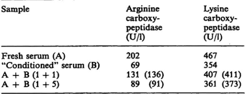
To evaluate whether the carboxypeptidase U activity in fresh human serum could be due to the presence of an activator — which should be unstable — ad- ditional experiments were performed. Resülts show that the fresh serum does not contain an activator or an inhibitor for these carboxypeptidase activities (tab. 2).
Tab. 2. Experiments to evaluate the presence of a carboxypep- tidase N activator or inhibitor in human serum Sample
Fresh serum (A)
"Conditioned" serum (B) A + B (1 4- 1)
A + B (1 + 5)
Arginine carboxy- peptidase (U/l) 20269 131 (136)
89 (91)
Lysine carboxy- peptidase (U/l) 467354 407 (411) 361 (373) Fresh serum and "conditioned" serum were mixed l plus l and l plus 5, then assayed for arginine carboxypeptidase and lysine carboxypeptidase activities. The values between parentheses indicate the theoretical enzyme activities if no activation or Inhibition between A and B occurs.
Enzyme activity in human blood cells
In order to exclude the possibility that the carboxy- peptidase U activity generated during serum forma- tion is derived from human blood cells, we determined enzyme activity in human leukocytes, erythrocytes and thrombocytes using Hip-Arg and Hip-Lys äs sub- strates.
Leukocytes and erythrocytes did not contain any measurable enzyme activity on these Substrates, and thrombocytes showed a very low carboxypeptidase activity, only detectable with prolonged incubatiön.
These findings are in accordaiice with earlier studies where human polymorphonuclear leukocytes did not hydrolyze Hip-Lys (31), haemolyzed red blood cells showed no activity with Hip-Arg (32), and throm- bocytes split Hip-Lys very slowly (32).
Influence of recalcification of citrate pläsma For direct proof that the carboxypeptidase U activity is generated during the coagulation process, we re- calcified a citrate pläsma (0.129 mol/1 Na-citrate) with an equal volume of CaCl
220 mmol/1 (in HEPES 20 mmol/1, pH 7.4) and placed it in ice for l h. After eentrifugation we detennined an arginine carboxy- peptidase activity of 149 U/l in the supernate serum, while the original citrated pläsma had an activity of 59.6 U/1. Ca"
1"*· 20 mmol/1 did not effect arginine carboxypeptidase activity.
Characterization of the carboxypeptidase U activity
pH Optimum
Fresh serum and "conditioned" serum were assayed for arginine carboxypeptidase activity in HEPES 50 mmol/1 at pH-values ranging from 6.5 to 8.5 (fig. 2).
The difference between the two pH-curves obtained shows the pH-profile of the arginine carboxypeptidase U activity. Arginine carboxypeptidase N showed a pH Optimum in the ränge of 8.0 to 8.2, which is the same äs that termined before with the same assay (26).
At pH 8.0 to 8.2 however, the arginine carboxypep- tidase U activity is about 15% below its Optimum, which is around pH 7.5 to 7.8. Thus, arginine car- boxypeptidase N has a different pH Optimum from that of arginine carboxypeptidase U activity.
300
200
S
o;i 100
6.5 7.5
pH
8.5 Fig. 2. Effect of pH on the arginine carboxypeptidase activity
of fresh serum and "conditioned" serum. Fresh serum (A) and "conditioned" serum (B) were assayed for ar- ginine carboxypeptidase activity in 50 mmol/1 HEPES buffer at different pH. The difference between the two pH-curves obtained shows the pH-proflle of the arginine carboxypeptidase U activity (C). The ränge for the triplicate determinations is indicated.
Activation and Inhibition
The effect of several enzyme inhibitors and activators on arginine carboxypeptidase N and U activities was determined using Hip-Arg äs the Substrate (tab. 3).
Cp
2+ (l mmol/1), a well known activator of carboxy-
peptidase N (32), increased the arginine carboxy-
peptidase N activity, but inhibited the arginine carb-
J. Clin. Chem. Clin. Biochem, / Vol. 27,1989 / No. 5Tab. 3. Activation and Inhibition of arginine carboxypeptidase
Addition Concentration Arginine carboxypeptidase activity (%)
"Conditioned" serum (arginine carboxy- peptidase N)
Fresh serum (arginine carboxy- peptidase N + U)
Difference (arginine carboxy- peptidase U) NoneCo2+
Dithiothreitol
Diisopropylfluorophosphate Phenylmethylsulphonylfluoride
-Phenanthroline
D,L-2-mercaptomethyl-3-guanidino ethylthiopropanoic acid
_
1 mmol/1 0.2 mmol/1 1 mmol/1 1 mmol/1 2 mmol/1 200 μηιοΐ/ΐ
244 (225-267)100 311 (282-339) 98 (97- 99) 99 (97-100) 0 (0- 0) 0 ( 0 - 0 )
100139 (133-155) 151 (133-166) 93 (92- 93) 101 (99-102) 16 (14- 20) 4 (4- 4)
10032 (28- 39) 'r 87 (73- 96) 90 (89- 91) 101 (100-103) 25 (23- 27) 6 ( 6 - 6 ) Satnples were incubated with the inhibitors or activators s described in the Materials and Methods section, then assayed for arginine carboxypeptidase activity using the HPLC-assisted assay. The data represent the mean for three separate experiments.
The values in brackets indicate the r nge.
oxypeptidase U activity. Diisopropylfluorophosphate (l mmol/1) and phenylmethylsulphonylfluoride (l mmol/1) had no influence on either enzyme activity, which indicates that carboxypeptidase U is not a serine protease. Dithiotreitol (0.2 mmol/1) strongly enhanced the activity of arginine carboxypeptidase N, but did not increase arginine carboxypeptidase U activity. o-Phenanthroline (2 mmol/1) completely in- hibited arginine carboxypeptidase N, but carboxypep- tidase U showed a residual activity of 25%. The carboxypeptidase N Inhibitor Z),L-2-mercaptomethyl- 3-guanidinoethylthiopropanoic acid (36) inhibited ar- ginine carboxypeptidase N and U to the same extent.
Substrate specificity
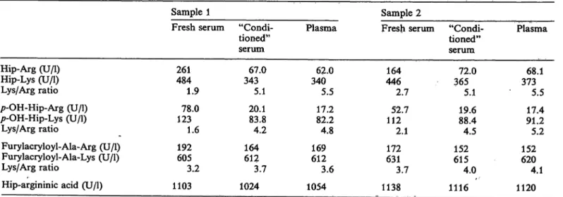
Table 4 summarizes the results of the Substrate spec- ificity of both carboxypeptidases, N and U.
As already shown in the first experiments, carboxy- peptidase U activity is much more pronounced for Hip-Arg than for Hip-Lys. We see sinailar results for the /7-hydroxy-hippuryl Substrates. In contrast, the carboxypeptidase U activity was low with furylacry- loyl-Ala-Arg, and no carboxypeptidase U activity could be demonstrated with furylacryloyl-Ala-Lys.
This is in sharp contrast with carboxypeptidase N itself, which is very active towards these Substrates.
The esterase activity of carboxypeptidase N measured with Hip-argininic acid, first described by Erd s et al.
(32), is high in comparison with its peptidase activity.
Nearly no carboxypeptidase U esterase activity could be demonstrated with the Hip-argininic acid sub- strate. Activities measured in "conditioned" sera and in plasma are very similar, confirming that the carb- oxypeptidase N activity is, indeed, measured in these samples. For the Hip-Arg and pOH-Hip-Arg sub-
Tab. 4. Substrate specificity of carboxypeptidases in human serum and plasma Sample 1
Fresh serum "Condi- tioned"
Hip-Arg (U/l) Hip-Lys (U/l) Lys/Arg ratio p-OH-Hip-Arg (U/l) p-OH-Hip-Lys (U/l)
Lys/Arg ratio
Furylacryloyl-Ala-Arg (U/l) Furylacryloyl-Ala-Lys (U/l) Lys/Arg ratio
Hip-argininic acid (U/l)
261484 1.9 12378.0
1.6 192605
3.2 1103
serum 34367.0
5.1 20.183.8 4.2 164612
3.7 1024
Plasma
34062.0 5.5 17.282.2 4.8 169612
3.6 1054
Sample 2
Fresh serum "Condi- tioned"
446164 2.7 11252.7
2.1 631172
3.7 1138
serum 36572.0
5.1 19.688.4 4.5 152615
4.0 1116
Plasma
37368.1 5.5 91.217.4 5.2 620152
4.1 1120 Carboxypeptidase activities with different synthetic Substrates currently used for carboxypeptidase N activity determinations (26 — 28, 32-25) were determined in fresh sera, "conditioned" sera and heparinized plasma froni two donors.
J. Clin. Chem. CHn. Biochem. / Vol. 27,1989 / No. 5
Hendriks et al.: A carboxypeptidase in human serum different from carboxypeptidase N 283
strates, activities in "conditioned" serum are about
10% higher than activities in plasma, while for the Hip-Lys and pOH-Hip-Lys Substrates, the activities are about equal. This can be explained by the effect of heparin, which was found to have a small inhibitory effect on arginine carboxypeptidase activities.
Influence of Z),L-2-mercaptomethyl-3-guan- idinoethylthiopropanoic acid on routine co- agulation tests
Since the carboxypeptidase U activity appears during coagulation and consequently only is present in serum, it would be of interest to know if an inhibitor of this enzyme could disturb the coagulation cascade.
Three citrated plasma samples were mixed with a solution of Z>,L-2-mercaptomethyl-3-guanidinoethyl- thiopropanoic acid in oxygen-free distilled water to obtain a final Z>,L-2-mercaptomethyl-3-guanidino ethylthiopropanoic acid concentration of 185 μιηοΐ/l, which completely inhibited carboxypeptidase N and U activities. In these plasma samples with and without
£>,L-2-mercaptomethyl-3«guanidinoethylthiopropa- noic acid, we determined the following routine co- agulation parameters: the prothrombin time by the
Quick one-stage method, the partial thromboplastintime with kaolin, and the Thrombotest™ (37). All three tests showed normal values for all samples as- sayed, and no significant difference was found be- tween the samples with or without Z>,L-2-mercapto- methyl-3-guanidinoethylthiopropanoicacid.
Ion exchange chromatography
In order to find out if another carboxypeptidase be*·
sides carboxypeptidase N is present in human serum,
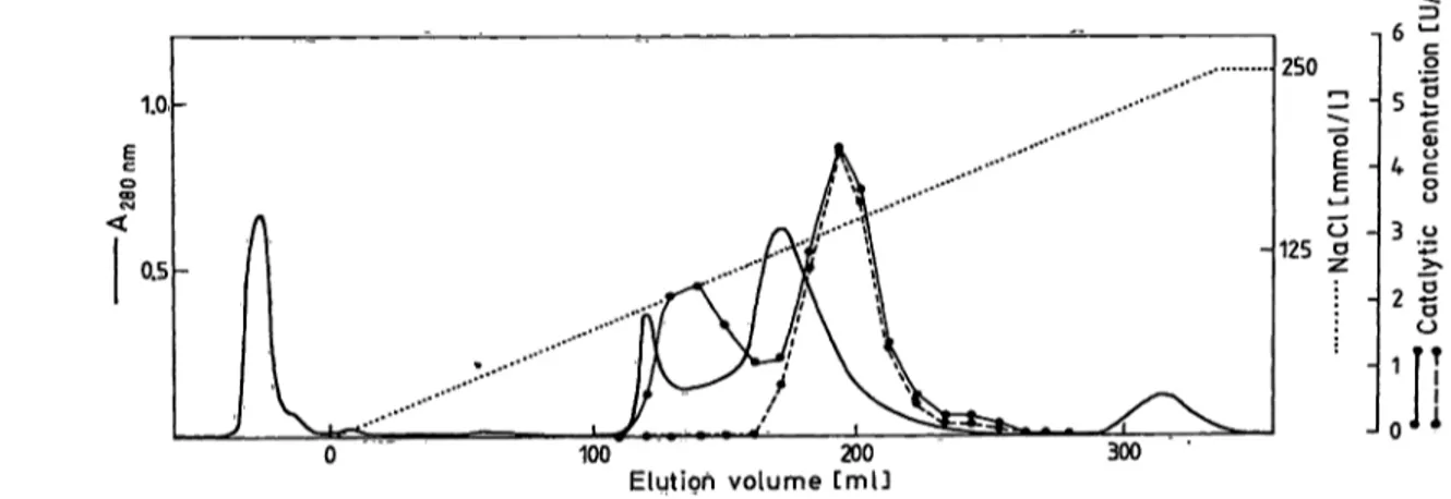
we performed DEAE-cellulose ion-exchange chro- matography on fresh human serum. To detect the unstable carboxypeptidase activity (carboxypeptidase U), it is necessary to use fresh serum, and to work at 4 °C, measuring enzyme activity in the fractions after mixing with serum devoid of carboxypeptidase activ- ity. Inactivation of the fractions by placing them at 37 °C for 2 h reveals the stable carboxypeptidase ac- tivity (carboxypeptidase N). From the results shown in figure 3, we can conclude that ion-exchange chro- matography demonstrates the presence of two carb- oxypeptidases in human serum, one of them being thermolabile (carboxypeptidase U). The overall yield of enzymatic activity was 75%. Measurement of Co
2+activation and esterase activity on fractions of Peak I (carboxypeptidase U) and Peak II (carboxypeptidase N) confirmed our previous results: carboxypeptidase N was activated by Co
2+(l mmol/1) and showed a high esterase activity, while carboxypeptidase U showed 50% Inhibition with Co
2+and yielded no esterase activity.
Discussion
The presence of a carboxypeptidase B-type enzyme in human plasma that inactivates bradykinin was first described by Er d s & Sloane in 1962 (33) and named carboxypeptidase N. Since changes in the blood level of this enzyme may be significant in various disease states, carboxypeptidase N activities in serum or plasma were previously determined in a variety of pathological conditions.
During our studies on arginine carboxypeptidase de- terminations in sera, we observed a marked instability of these activities. This instability has not been pre-
1.0.
0.5
250 οΕ -|
125 οο - 6 ^
4 cου 3 ο">»
100 200
Elutioh volume CmU 300
Fig. 3. DEAE-cellulose chromatography of human serum. 2ml of fresh human serum diluted l plus 4 with the equilibration b ffer was applied at a flow rate of 21 ml · cm~2 · h"1 to a column (1.6 χ 35 cm) of DEAE-cellulose previously equilibrated in 0.05 mol/1 Tns, pH 7.2. The enzymes were eluted with a linear gradient of NaCl from 0 to 0.25 mol/1 in 0.05 mol/1 Tris, pH 7.2 during 8 h. The effluent was collected in 10 ml fractions. 100 μΐ aliquots of the fractions were diluted 1 + 1 in serum which was previously inactivated by keeping it at 56 °C. Samples were then assayed for arginine carboxypeptidase activity (· - ·). The same samples were also assayed for arginine carboxypeptidase activity after keeping them at 37 °C for2h(· -- ·).
J. Clin. Chem. Clin. Biochem. / Vol. 27, 1>989 / No. 5
viously reported in the literature. In further experi- ments, it became clear that fresh sera contain a high enzymatic activity äs regards carboxy-terminal argi- nine removal, while this activity is much lower in heparinized plasma or in sera kept at room temper- ature for several hours. In this report, we describe for the first time the presence of an unstable arginine carboxypeptidase activity in fresh human serum, which we call carboxypeptidase U activity.
The carboxypeptidase U activity has some character- istics in common with the human plasma carboxy- peptidase N: both enzymes cleave Hip-Arg and Hip- Lys, are not inhibited by the serine proteinase inhib- itors diisopropylfluorophosphate and phenylmethyl- sulphonylfluoride, and, most importantly, both are inhibited by the carboxypeptidase N Inhibitor D,L-2- mercaptomethyl-3-guanidinoethylthiopropanoicacid.
Arginine carboxypeptidase U is inhibited by 0-phen- anthroline, but not to the same extent äs the arginine carboxypeptidase N activity.
Hpwever, some characteristics clearly differentiate carboxypeptidase U activities from carboxypeptidase N activities: the carboxypeptidase U activity towards Hip-Arg is higher than towards Hip-Lys, while carb- oxypeptidase N cleaves Hip-Lys about five times faster than Hip-Arg. Furylacryloyl-Ala-Arg was cleaved only very slowly and furylacryloyl-Ala-Lys not at all by carboxypeptidase U. Moreover, carboxy- peptidase U showed no esterase activity when meas- ured with the Substrate Hip-argininic acid. The pH Optimum of the arginine carboxypeptidase U activity (pH 7.7) was also different from that of carboxypep- tidase N (pH 8.1).
We directly demonstrated that, during the process of clot formation, a new enzyme activity appears, clearly different from normal carboxypeptidase N in terms of stability, Substrate specificity, effect of activators and inhibitors, and pH-optimum. Ion exchange chro- matography on DEAE-cellulose is used äs the first step in the purification of carboxypeptidase U, be- cause it provides a good Separation of carboxypeptid- ase N and carboxypeptidase U, and the yield is high.
Purification of carboxypeptidase U to homogeneity will be difficult, owing to its marked instability.
A hypothesis for the appearance of this unstable en- zyme activity during coagulation lies within the struc- ture of the carboxypeptidase N itself. Carboxypeptid- ase N is a 280000 relative molecular mass tetrameric enzyme consisting of two M
r48 000 and two M
r83 000 subunits (38, 39).
The M
T48000 subunit possesses the füll enzymatic activity of the intact enzyme with ester and small
peptide Substrates, but loses 75% of its activity when stored for 2 h at 37 °C (39). Proteolysis of the intact enzyme or of the M
r48 000 subunit with plasmin, trypsin or urinary kallikrein yielded fragrnents with increased esterase and peptidase activity, but with decreased stability (39). During the coagulation proc- ess, many proteolytic enzymes are activated, and this could cause a partial pro teolysis'of carboxypeptidase N, resülting in increased activity and a lesser stability.
However, the carboxypeptidase U in our experiments had no esterase activity. Moreover, if a part of the carboxypeptidase N is converted into unstable sub- units, the carboxypeptidase N activity measured in
"conditioned" serum should be considerably lower than in plasma. It therefore seems unlikely that the carboxypeptidase U activity originates from a subunit formation of carboxypeptidase N.
Another hypothesis is that one of the enzymes that takes part in the coagulation cascade, and is activated during clot formation, exhibits a carboxypeptidase U activity. Many of these coagulation enzymes, being serine proteinases, are inhibited by diisopropylfluorq- phosphate or phenylmethylsulphonylfluoride. This was not the cäse for the carboxypeptidase U activity.
Addition of jD,L-2-mercaptomethyl-3-guanidino ethylthiopropanoic acid to citrate plasma did not cause a significant increase in coagulation time (pro- thrornbin time, partial thromboplastin time with ka- olin, thrombotest). This excludes a major role for thi$
enzyme in the coagulation System.
Release of the carboxypeptidase U activity from hu- man blood cells seems unlikely, since we could not detect any carboxypeptidase N or carboxypeptidase U activity in leukocytes or erythrocytes and only very little activity in thrombocytes.
The carboxypeptidase U is also clearly different from the recently identified urinary carboxypeptidase, which is stable at 37 °C and exhibits an esterase ao- tivity (40).
Sheikh & Kaplan (41) reported that arginine removal from bradykinin in serum occurs at a rate exceeding that of heparinized plasma and is more rapid than can be attributed to carboxypeptidase N. This im- portant observation is consistent with the presenee of the carboxypeptidase U activity we have demon- strated, which preferentially removes C-terminal ar- ginine.
Carboxypeptidase N is recognized to be an important enzyme, mainly because of its anaphylatoxin-inhibit- ing characteristics. Many papers deal with the deter- minatioa of carboxypeptidase N activities, üsing dif- ferent Substrates, in human serum of patients with
J. Clin. Chem. Clin. Biochem. / Vol. 27,1989 / No. 5
Hendriks et al: A carboxypeptidase in human serum different from carboxypeptidase N 285
various pathologies. Those working in this field
should pay great attention in collecting samples. In order to determine only the carboxypeptidase N ac- tivity, we can recommend
(1) keeping serum samples at 37 °C for 2 h before assay;
(2) using heparinized plasma samples; or
(3) measuring esterase activity in either sera or plasma.
Current data on carboxypeptidase N activities in sera should be carefully evaluated.
References
1. Erdös, E. G. (1979) In: Bradykinin, Kallidin and Kalükrein, 21.
Handbook of Experimental Pharmacology, Vol. 25 (Suppl.), pp. 427-487. 22.
2. Bokish, V. A. & Müller-Eberhard, H. J. (1970) J. Clin.
Invest. 49, 2427-2436. 23.
3. Gorski, J., Hugli, T. & Müller-Eberhard, H. J. (1979) Proc.
Natl. Acad. Sei. USA 76, 5299-5302. 24.
4. Belew, M., Gerdin, B., Lindeberg, G., Porath, J. & Wallin, R. (1980) Biochim. Biophys. Acta 621, 169-178. 25.
5. Perryman, M., Knell, D. & Roberts, R. (1984) Clin. Chem.
30,662-664. 26.
6. Hendriks, D., Soons, J., Schärpe, S., Wevers, R., van Sande, M. & Holmquist, B. (1988) Clin. Chim. Acta 772, 253- 27.
7. Skidgel, R. A., Johnson, A. R. & Erdös, E. G. (1984) 28.260.
Biochem. Pharmacol. 33, 3471-3478.
8. Hendriks, D., Verkerk, R., Vanhoof, G., De Meester, I., 29.
van Sande, M. & Schärpe, S. (1987) Biochem. Soc. Trans.
75,359-360.
9. Corbin, N. C., Hugli, X E. & Müller-Eberhard, H. J. (1975) 30.
Anal. Biochem. 73, 41 — 51.
10. Erdös, E. G., Wohler, I. M., Levine, M. I. & Westerman, 31.
M. P. (1965) Clin. Chim. Acta 77, 39-43. 32.
11. Gerhardt, W., Kallos, P. & Lundquist, A. (1982) Int. Archs.
Allergy Appl. Immun. 69, 206—209. 33.
12. Koheil, A., Corey, M. & Forstner, G. (1979) Clin. Invest.
Med. 2, 99-103. 34.
13. Lieberman, J. (1975) Am. Rev. Resp. Dis. 777, 100-102.
14. Mathews, K. P., Pan, P. M., Gardner, N. J. & Hugli, T. E. 35.
(1980) Ann. Int. Med. 93, 443-445.
15. Rohatgi, P. K. & Ryan, J. W. (1982) Am. Rev. Resp. Dis. 36.
725, 115.
16. Schweisfurth, H., Heinrich, J., Brugger, E., Wernze, H., 37.
Wichmann, J. & Ertl, G. (1983) Praxis Klin. Pneumql. 37, 562-564. 38.
17. Delk, A., Durie, P., Fletcher, T. & Largman, C. (1985) Clin.
Chem. 31, 1294-1300. 39.
18. Schweisfurth, H. & Burghardt, W. (1983) Z. Gastroenterol.
24, 397. 40.
19. Schweisfurth, H., Heinrich, J., Brugger, E. & Burghardt, W. (1984) Dtsch. Med. Wochenschr. 709, 166-169. 41.
20. Schweisfurth, H., Schmidt, M., Brugger, E., Maiwald, L.
& Thiel, H. (1985) Clin. Biochem. 18, 242-246.
Schweisfurth, H. & Thiel, H. (1987) Atemw. Lungenkrkh.
13, 138-140.
Schweisfurth, H., Pickert, E., Gramer, E. & Reiners, C.
(1987) Clin. Biochem. 20, 43-46.
Sommer, H., Schweisfurth, H. & Schultz, M. (1986) Enzyme 35, 181-188.
Streeten, D. H., Kerr, L. R, Kerr, C. B., Prior, J. C. &
Dalakos, T. G. (1972) Lancet 77, 1048-1053.
Vogel, A. J. (1970) In: Practical Organic Chemistry, p. 584, Longman, London.
Hendriks, D., Schärpe, S. & van Sande, M. (1985) Clin.
Chem. 37, 1936-1939.
Hendriks, D., Schärpe, S., van Sande, M., Lommaert, M.
P. & Kasahara, Y. (1987) Anal. Biochem. 164, 90-95.
Plummer, T. H. & Kimmel, M. T. (1980) Anal. Biochem.
108, 348-353.
Barrett, A. J. & Salvesen, G. (1986) In: Proteinase Inhibi- tors, Research Monographs in Cell and Tissue Physiology, Vol. 12, p. 88.
Tietz, N. W. (1986) In: Textbook of Clinical Chemistry, p. 485, W. B. Saunders Company, PA.
Nakahara, M. (1980) Biochem. Pharmacol. 29,2235-2239.
Erdös, E. G., Yang, H. J., Tague, L. L. & Manning, N.
(1967) Biochem. Pharmacol. 16, 1287-1297.
Erdös, E. G. & Sloane, E. M. (1962) Biochem. Pharmacol.
77, 585-592.
Hendriks, D., van Sande, M. & Schärpe, S. (1986) Clin.
Chim. Acta 757, 103-108.
Marceau, F., Drumheller, A., Gendreau, M., Lussier, A. &
St.-Pierre, S. (1987) J. Chromatog. 266, 173-177.
Plummer, t. H. & Ryan, T. J. (1981) Biochem. Biophys.
Res. Commun. 98, 448-454.
Dacie, J. V. & Lewis, S. M. (1975) In: Practical Haematol- ogy, Churchill Livingstone, New York, NY.
Plummer, T. H. & Hurwitz, M. J. (1978) J. Biol. Chem.
255, 3907-3912.
Levin, J., Skidgel, R. A. & Erdös, E. G. (1982) Proc. Natl.
Acad. Sei. USA 79, 4618-4622.
Skidgel, R. A., Davis, R. M. & Erdös, E. G. (1984) Anal..
Biochem. 740,520-531.
Sheikh, J. A. & Kaplan, A. P. (1986) Biochem. Phannacol.
35, 1957-1963.
Prof. Dr. S. Schärpe
Department of Medical Biochemistry University of Antwerp
B-2610 Wilrijk
J. Clin. Chem. Clin. Biochem. / Vol. 27,1989 / No. 5