Carboxypeptidase N and Creatine Kinase-MB Isoforms in Acute Myocardial Infarction
Martina Zaninotto1, Sara Altinier2, Mattia Lachin1 and Mario Plebani1'2
1 Servizio di Medicina di Laboratorio, Azienda Ospedaliera di Padova, Padova, Italy
2 Centre Regionale di Ricerca Biomedica, Castelfranco Veneto (TV), Italy
Summary: The aims of our study were to evaluate the plasma Carboxypeptidase N activity in normal subjects and in patients with acute myocardial infarction and to delineate its relationship with creatine kinase-MB isoforms in monitoring of acute myocardial infarction, Carboxypeptidase N being the major determinant of creatine kinase isoform conversion in plasma. The study was carried out in 34 healthy subjects and 19 patients with acute myocar- dial infarction diagnosed according to the World Health Organization (WHO) criteria in which the blood samples were collected immediately upon admission to the coronary care unit (median time 3.5 hours), every 4 to 6 hours for 24 hours, and every 12 hours until the third day post admission. Carboxypeptidase N activity, total creatine kinase, creatine kinase-MB mass concentration and creatine kinase-MB isoforms were determined in each sample from acute myocardial infarction patients, whereas only Carboxypeptidase N and total creatine kinase activities were assayed in samples from healthy subjects. The results showed a high variability in Carboxypeptidase N values among healthy subjects (median = 220 U/l; interquartile range = 190—247 U/l) and in the first available samples from acute myocardial infarction patients (median = 213 U/l; interquartile range = 197—234 U/l) without signifi- cant differences between groups and without a correlation between Carboxypeptidase N and creatine kinase activities either in healthy subjects or in acute myocardial infarction patients; in the latter group, however, a significant correlation (p < 0.01) with creatine kinase-MB calculated on all samples, was observed. In acute myocardial infarc- tion patients Carboxypeptidase N showed time-related variations, reaching the highest levels about 48 h after onset of chest pain. A statistically significant difference in Carboxypeptidase N values (p = 0.0001) was found before and after creatine kinase-MB peak values as well as before and after MB2/MB! normalization. Worthy of note is the finding that in two acute myocardial infarction patients presenting MB2/MBj ratios lower than the cutoff value (1.5) throughout the period of observation, the baseline values for Carboxypeptidase N were higher than in other patients studied. Our results suggest that the increase of Carboxypeptidase N activity after infarction could be induced by an increase in endogenous substrate concentrations, in particular creatine kinase-MB released from damaged myocardium. Furthermore, high baseline levels of Carboxypeptidase N will reduce the diagnosis efficiency of creatine kinase-MB isoforms in the diagnosis of acute myocardial infarction.
Introduction myocardial infarction as well as for the assessment of The creatine kinase-MB isoforms and, in particular, coronai^ rePerfi*ion after thrombolytic therapy (1-4).
the MB2 (tissue isoform containing C-terminal lysine) Moreover> now that a real-time, fully-automated method to MB! (circulating des-lysine isoform) ratio is a sensi- is available for their ™asurement, this assay is also an tive and specific marker for the early diagnosis of acute mterestmS too1 for the early diagnosis of acute Wocar-
dial infarction in emergency (5, 6). Previous studies (1—9) indicated that the tissue creatine kinase-MB2 iso- 1) Enzymes: form released from the damaged myocardium is con- Carboxypeptidase N (arginine Carboxypeptidase, EC 3.4.17.3); verted in the blood to creatine kinase-MB! by carboxy- Creatine kinase (ATP : creatine N-phosphotransferase, EC 2.7.3.2). oeotidase χ,κ an enzvme synthesized in the liver and
2) Remark of the Managing Editor: peptmase IN ), an enzyme syntnesizea in me nver ana The absorption coefficient at 336 nm is given by 1. c. (15) with secreted as a Mr = 280 000 tetramer and which is known Δε = -1300 Μ'1 cnr1, corresponding to 130m2 x mor1. In to be an important inactivator of peptide hormones such
I.e. (13) "an absorption coefficient of 0.13 L · nmol ' · min '"is , , , . . , ι ι ^ · A 1^.1 u ^
given. This was repeated by Skidgel RA, Erdos EG. Carboxypepti- as bradykimn and anaphylatoxms. Although the enzyme dase N (arginine Carboxypeptidase) peptidyl-Z,-arginine hydrolase, appears to be present in all humans, its levels in plasma EC 3.4.17.3. In: Bergmeyer HU, editor. Methods of enzymatic vary considerably among subjects. In normal subjects, analysis. 3rd English ed., vol. V. Weinheim: Verlag Chemie, . ., , . , , . . . .
1988:60-72. In the latter two publications it should read: 0.131 m fact' UP to a threefold variation in enzyme activity x mmor1 x mm~', also corresponding to 130m2 χ mol"1. has been observed; during pregnancy and during the
course of certain types of cancer, elevated enzyme con- centrations have been reported while low concentrations have been observed in patients with cirrhosis of the liver (10, 11). The reported variability in Carboxypeptidase N activities may therefore be important in the use of the creatine kinase-MB isoform ratio for the diagnosis of myocardial infarction: in fact, the sensitivity and the diagnostic accuracy of isoform analysis for the detection of infarction will likely depend, in part, on the rate of conversion of isoforms in plasma (12). However, despite some evidence correlating variations of Carboxypepti- dase N and creatine kinase-MM in acute myocardial in- farction (13), no data exist on the relationship between plasma Carboxypeptidase N activity and creatine kinase- MB isoforms in acute myocardial infarction.
The aim of this study was to delineate the relationship between creatine kinase-MB isoforms and carboxypepti- dase N activity in patients with acute myocardial infarc- tion.
buffer (0.05 mol/1 HEPES, pH 7.75, 0.25 mol/1 NaCl) containing 0.5 mmol/1 of substrate, previously equilibrated at 37 °C. Absor- bance was measured at 336 nm over a 6 minute period; the enzyme activities, calculated using a conventional method with a molar absorption coefficient2) of 130 m2 X mol"1 (13), were expressed as U/l (l U = amount of enzyme hydrolyzing 1 μιηοΐ of furylacry- loyl-alanyl-lysine per minute).
The within-day coefficient of variation (n = 11) was 4.8% at a mean value of 217 U/l and 3.6% at 251 U/l. In the same sera, between-day coefficients of variation of 7.5% and 6.3% respec- tively were obtained.
Total creatine kinase catalytic activity concentration
The catalytic concentration of creatine kinase was determined at 37 °C (Bracco, Milano, Italy) according to the method recom- mended by the IFCC (16). The upper reference limits were > 160 U/l for females and > 190 U/l for males.
Creatine kinase-MB mass concentration
The mass concentration of creatine kinase-MB was measured using a commercially available fluorometric enzyme immunoassay (Stra- tus CK-MB; Baxter Dade, Milano, Italy) with a sensitivity of 0.4 μ§/1. The upper reference limit was 5 μ^Ι.
Materials and Methods Subjects and blood collection
The study was carried out on 34 healthy subjects (14 males and 20 females, aged 23 to 55 years) from the laboratory staff and 19 patients (12 men and 7 women, aged 46 to 74 years) with acute myocardial infarction diagnosed according to WHO criteria (char- acteristic chest pain, unequivocal ECG changes and serial increases in creatine kinase and creatine kinase-MB mass concentration with peak values twice the upper limit of the reference interval); 14 had Q-wave and 5 non-Q-wave indicated infarctions. The patients were admitted to the coronary care unit within 9 hours after the onset of symptoms (range 0.5—9 h; median 3.5 h); 12 underwent thrombo- lytic treatment while 7 were not treated depending on clinical cir- cumstances; 10 were classified as reperfused on the basis of their clinical pictures (symptoms resolved rapidly, ST-segment changes improved, presence of reperfusion arrhythmias) (14). Blood sam- ples were collected from patients immediately upon admission to the coronary care unit and every 4 to 6 hours thereafter for 24 hours and then every 12 hours until the third day of hospitalization.
All patients gave their informed consent for extra blood samples to be drawn. Carboxypeptidase Ν activity, total creatine kinase, creatine kinase-MB mass concentration and creatine kinase-MB isoforms were determined in each sample from acute myocardial infarction patients, whereas only Carboxypeptidase Ν and total cre- atine kinase activities were assayed in samples from the healthy subjects.
Venous blood samples for the measurement of creatine kinase, cre- atine kinase-MB mass and Carboxypeptidase Ν were drawn in lith- ium heparinate-containing tubes, centrifuged at 2000 g for 15 min- utes and analyzed immediately. Samples for creatine kinase iso- form determination were collected in tubes containing ethylene glycol bis ( -aminoethyl ether)-N,N,N',N'tetracetic acid (EGTA, final concentration 30 mmol/1) and 2-mercaptoethanol (final con- centration 10 mmol/1) to inhibit Carboxypeptidase N-mediated iso- form conversion after blood collection, and the EGTA-plasma was stored at -20 °C until analysis (1).
Methods
Carboxypeptidase N activity
Spectrophotometric assay at 37 °C using furylacryloyl-alanyl-ly- sine as the substrate (Sigma Diagnostic, Milano, Italy) according to Plummer & Kimmel (15) was used to measure Carboxypeptidase N activity, 40 μΐ of sample being added to 3 ml of the reaction
Creatine kinase-MB isoforms
Using high-voltage electrophoresis on agarose gels with the auto- mated REP/EDC system (Helena Laboratories, Milano, Italy) cre- atine kinase-MB isoforms were separated, and then detected by fluorescence densitometry. This procedure also enables the separa- tion of creatine kinase-MM isoforms, but only the more specific cardiac creatine kinase-MB isoforms were considered in our study, two creatine kinase-MB isoforms being separated with this pro- cedure: according to their electrophoretic mobilities, isoform cre- atine kinase-MB2 is the most cathodic band while creatine kinase- MB! is the fastest moving, most anodic band in the creatine ki- nase series.
The within-run and between-run precision (CVs) calculated in a sample with MB2/MBj ratio of 0.93 and MB2 activity of 5.8 U/l were 9.1% and 11.6% respectively. The minimum MB activity for isoform analysis using this procedure was 4 ± 1 U/l. The upper reference limit for the MB2/MBi ratio was 1.5 (5).
Statistics and calculation
Medians and percentiles were calculated to described continuous variables. Two-way unpaired t-tests were used for between-group comparison of the results, while one-way paired t-tests were used to compare the results from acute myocardial infarction patients: p values < 0.05 were considered to indicate statistical significance.
Results
The Carboxypeptidase N activities in plasma of normal subjects and in the first blood sample of patients with myocardial infarction collected 3.5 h (median) after the onset of chest pain show a high variability over a two- fold range among subjects, without a significant differ- ence between controls (median = 220 U/l; interquartile range = 190—247 U/l) and acute myocardial infarction patients (median = 213 U/l; interquartile range = 197—
234 U/l).
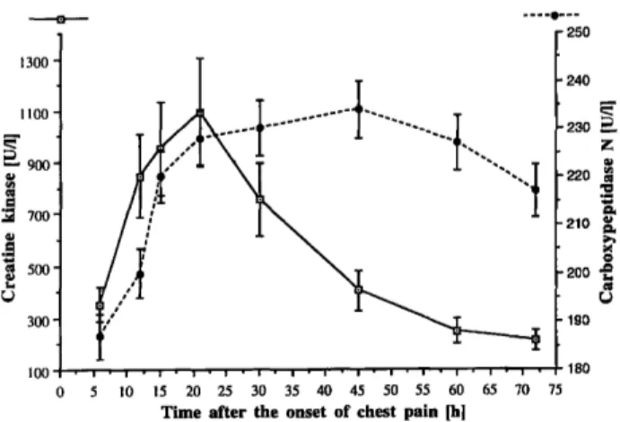
The pattern of Carboxypeptidase N values in patients with myocardial infarction (fig. 1) shows time-related variations, the highest levels (ranging from 10 to 72%
iioo- Γ - 900- l
! 700-
ί
! 500-
Γ 250 240 U 230 h 220 210 Γ 200 190
180 Ο 5 10 15 20 25 30 35 40 45 50 55 60 65 70 75
Time after the onset of chest pain [h]
Fig. 1 Time-related variations of Carboxypeptidase Ν and total creatine kinase in patients with acute myocardial infarction (mean values ± SE).
to initial values) being reached about 48 hours after the onset of chest pain, and maintained for more than 60 hours. The increased values in Carboxypeptidase Ν con- centrations following infarction and the maximum level therefore occurred in each patient later also in respect to total creatine kinase, the last marker of acute myocardial infarction to be considered in our study. Furthermore, no correlation was found between Carboxypeptidase Ν and total creatine kinase values either in healthy subjects (n = 34; r = 0.051, p = 0.774) or in all samples of acute myocardial infarction patients (n = 171; r = 0.346, p = 0.061), while in the latter group a significant corre- lation between Carboxypeptidase N and creatine kinase- MB concentrations (r = 0.479; p < 0.01) was obtained.
300 280
260
T
z ^u
S 220- α« 180·
g 160- am 140·
υ 120- 100 ·
-τ-
ο
Before creatine kinase MB peak value
I
I
— β—
After creatine kinase MB peak value
Fig. 2 Distribution of Carboxypeptidase N values from patients with acute myocardial infarction in relation to creatine kinase-MB peak concentrations.
Figure 2 shows the relationship between carboxypepti- dase N values and creatine kinase MB mass concentra- tion (median time to peak concentration 15 h; interquar- tile range 10—26 h) in patients studied. The reported re- sults show that the values of Carboxypeptidase N ob- tained from each patient in the sample collected before creatine kinase-MB peak concentrations (median = 201 U/l, interquartile range: 184—223 U/l), were signifi-
cantly lower (p = 0.0001) than those obtained from the samples collected after creatine kinase-MB peak values (median = 227 U/l, interquartile range: 211-242 U/l).
In the same patients the relationship between Carboxy- peptidase N and the MB2/MB! ratio shows, as expected, a statistically significant difference (p = 0.0001) be- tween Carboxypeptidase N values before (median =184 U/l, interquartile range: 171-201) and after (median
= 209 U/l, interquartile range: 203-243 U/l) MB2/MBl
ζφ<αη η
Carboxypeptl
300 280 260 240 220 200 180 160·
140·
120·
-T- -r
0
W
MB isoform MB isoform ratio >1. 5 ratio <1. 5
Fig. 3 Distribution of Carboxypeptidase N values from patients with acute myocardial infarction in relation to the creatine kinase- MB isoform ratio.
normalization (fig. 3). However, in two out of the 19 acute myocardial infarction patients monitored in our study, the creatine kinase-MB isoform ratio was lower than the cutoff value (x ± SD: 0.39 ±0.13 and 0.51
±0.12 respectively) throughout the period of observa- tion. In these two patients the first blood samples were obtained shortly after the onset of chest pain (2.5 and 2.0 hours respectively), with creatine kinase-MB con- centrations ranging from 6.3 to 246 μg/l and from 7.8 to 351 μg/l (baseline and peak values respectively). Worthy of note is the finding that in these patients the baseline Carboxypeptidase N values (222 U/l and 228 U/l respec- tively) were higher compared to those from patients (n = 8) with similar hospital admission time (2—3.5 h) in which the median baseline value was 189 U/l (inter- quartile range: 174—206 U/l). These observations sug- gest that the baseline values of Carboxypeptidase N aciti- vity in acute myocardial infarction patients were respon- sible for early conversion of tissue MB2 isoform into plasma MB! isoform allowing an early shift of the MB2/
ratio.
Discussion
Analysis of isoforms of creatine kinase-MB in plasma was recently shown to enable the detection of acute myocardial infarction and of coronary recanalization very early after their onset, before a significant elevation in total creatine kinase activity and, in some cases, ere-
atine kinase-MB (1—6). The sensitivity of isoform analysis results from the early release from the damaged myocardium of the tissue isoenzyme of MB (MB2), which is converted in plasma into an additional subform (MB}) with a similar specific activity but different iso- electric point (isoforms). Thus, the release in plasma of a modest amount of creatine kinase-MB2 after myocardial infarction causes a prompt and marked increase in the MB2 to MBi ratio (17, 18, 20-22).
Several studies have shown that carboxypeptidase N is the main factor accounting for isoform conversion in human plasma under physiological conditions (7—11) and after myocardial infarction, elucidating in particular the relation between carboxypeptidase N activity in plasma and observed rates of creatine kinase-MM iso- form conversion (13).
On the basis of these suggestions, the aim of our study was to ascertain the variations in plasma carboxypepti- dase N activity in normal subjects and in patients under- going acute myocardial infarction and to delineate its relationship with creatine kinase-MB isoforms. The car- boxypeptidase N levels were assessed using a sensitive and specific method, showing good analytical perfor- mance, as previously demonstrated (15).
Time-related variations of carboxypeptidase N activity in patients with acute myocardial infarction were found with the highest level being reached about 48 hours after the onset of pain. In none of the subjects studied was a correlation observed between carboxypeptidase N and total creatine kinase values. However, a significant cor- relation between carboxypeptidase N and creatine ki- nase-MB concentrations has been found in patients with myocardial infarction, carboxypeptidase N values being significantly different in relation to creatine kinase-MB peak concentrations.
These results suggest that the increase in carboxypepti- dase N after infarction might be induced by the increase in endogenous substrate concentrations, in particular creatine kinase-MB, released from the damaged myocar- dium and emphasize the need of further studies for bet- ter understanding the behaviour of this enzymatic activ- ity.
Moreover, our findings underline the relevance of car- boxypeptidase N determination for the accurate diagno- sis of acute myocardial infarction using the assay of cre- atine kinase-MB isoforms. In fact, two evaluated pa- tients, who during monitoring had MB2 to MB ι ratios, lower than the cutoff value, had high baseline carboxy- peptidase levels yielding false negative results. This evi- dence, in conjunction with the already described influ- ence of low carboxypeptidase Ν levels in normal sub- jects leading to false positive results for the MM isoform ratio (13), emphasizes the need for caution in evaluating the creatine kinase-MB isoform results in acute myocar- dial infarction.
Conclusion
The determination of creatine kinase-MB isoforms in plasma provides considerable advantages for the early, non-invasive detection of acute myocardial infarction as well as for the evaluation of coronary recanalization.
Nevertheless, findings made by us and by other authors (12, 13) indicate that the biological variations in carb- oxypeptidase N activity may be of importance in the clinical use of the creatine kinase-MB isoform ratio.
Thus, the determination of carboxypeptidase N concen- trations using a rapid and analytically accurate pro- cedure may increase the specificity and the sensitivity of this test in the diagnosis of acute myocardial infarction.
References
1. Puleo PR, Guadagno PA, Roberts R, Scheel MV, Marian AJ, Churchill D, et al. Early diagnosis of acute myocardial infarc- tion based on assay for subforms of creatine kinase-MB. Circu- lation 1990; 82:759-64.
2. Abendschein DR. Rapid diagnosis of myocardial infarction and reperfusion by assay of plasma isoforms of creatine kinase isoenzymes. Clin Biochem 1990; 23:399-407.
3. Bhayana V, Cohoe S, Leung FY. Jablonsky G, Henderson AR.
Diagnostic evaluation of creatine kinase-2 mass and creatine kinase-3 and -2 isoform ratios in early diagnosis of acute myo- cardial infarction. Clin Chem 1993; 39:488-95.
4. Christenson RH, Ohman EM, Topol EJ, O'Hanesian MA, Sig- mon KN, Duh SH, et al. Creatine kinase-MM and MB iso- forms in patients receiving thrombolytic therapy and acute an- giography. Clin Chem 1995; 41:844-52.
5. Puleo PR, Meyer D, Wathen C, Tawa CB, Wheeler S, Ham- burg RJ, et al. Use of a rapid assay of subforms of creatine kinase-MB to diagnose or rule out acute myocardial infarction.
N Engl J Med 1994; 331:561 -6.
6. Secchiero S, Altinier S, Zaninotto M, Lachin M, Plebani M.
Evaluation of a new automated system for the determination of CK-MB isoforms. J Clin Lab Anal 1995; 9:359-65.
7. Michelutti L, Falter H, Certossi S, Marcotte B, Mazzuchin A.
Isolation and purification of creatine kinase conversion factor from human serum and its identification as carboxypeptidase N. Clin Biochem 1987; 20:21-9.
8. Billadello JJ, Fontanet HL, Strauss AW, Abendschein DR.
Characterization of MB creatine kinase isoform conversion in vitro and in vivo in dogs. J Clin Invest 1989; 83:1037—43.
9. Prager NA, Suzuki T, Jaffe AS, Sobel BE, Abendschein DR.
Nature and time course of generation of isoforms of creatine kinase-MB fraction in vivo. J Am Coll Cardiol 1992;
20:414-9.
10. Erd s EG, Wohler IM, Levine MI, Westermann MR Carboxy- peptidase in blood and other fluids: values in human blood in normal and pathological conditions. Clin Chim Acta 1965;
11:39-43.
11. Levin Y, Skidgel RA, Erd s EG. Isolation and characterization of the subunits of human plasma carboxypeptidase N (kininase I). Proc Natl Acad Sei USA 1982; 79:4618-22.
12. Erd s EG, Skidgel RA. More on subforms of creatine kinase- MB. N Engl J Med 1995; 333:390.
13. Abendschein DR, Serota H, Plummer ΤΗ, Amiraian Κ, Strauss AW, Sobel BE, et al. Conversion of MM creatine kinase iso- forms in human plasma by carboxypeptidase N. J Lab Clin Med 1987; 110:798-806.
14. Pasternak RC, Braunwald E, Sobel BE. Acute myocardial in- farction. In: Braunwald E, editor. Heart disease. Philadelphia:
WB Saunders, 1988:1222-313.
15. Plummer TH, Kimmel MT. An improved spectrophotometric assay for human plasma carboxypeptidase N. Anal Biochem 1980; 108:348-53.
16. International Federation of Clinical Chemistry. IFCC methods for the measurement of catalytic concentration of enzymes;
Part 7. IFCC method for creatine kinase (ATP : Creatine N- phosphotransferase, EC 2.7.3.2). IFCC recommendation. Clin Chim Acta 1990; 190:S4-S40; Eur J Clin Chem Clin Bio- chem 1991; 29:435-56.
17. Panteghini M. Serum isoforms of creatine kinase isoenzymes.
Clin Biochem 1988; 21:211-8.
18. Puleo PR, Guadagno PA, Roberts R, Ferryman MB. Sensitive, rapid assay of subforms of creatine kinase-MB in plasma. Clin Chem 1989; 35:1452-5.
19. Abendschein DR, Seacord LM, Nohara R, Sobel BS, Jaffe AS.
Prompt detection of myocardial injury by assay of creatine kinase isoforms in initial plasma samples. Clin Cardiol 1988;
11:661-4.
20. Wu AHB. Creatine kinase-MM and MB isoforms. Lab Med 1992; 23:303-5.
21. Harker CC, Wu AHB. Early diagnosis of acute myocardial infarction (MI) upon initial hospital admission using CK-MB2 isoform analysis. Clin Chem 1990; 36:1128.
22. Panteghini M, Bonora R, Pagani F. An immunoinhibition assay for determination of creatine kinase isoforms in serum. Eur J Clin Chem Clin Biochem 1994; 32:383-9.
Received September 10, 1996/January 6, 1997
Corresponding author: Dr. ssa Martina Zaninotto, Servizio di Medicina di Laboratorio, Azienda Ospedaliera di Padova, Via Giustiniani 2, 1-35128 Padova, Italy