scale in human bone by means of Synchrotron Radiation CT
Dissertation
zur Erlangung des akademischen Grades doctor rerum naturalium
(Dr. rer. nat.) im Fach Biophysik
eingereicht an der
Mathematisch-Naturwissenschaftlichen Fakultät I der Humboldt-Universität zu Berlin
von
Diplom-Physiker Bernhard Hesse
Präsident der Humboldt-Universität zu Berlin:
Prof. Dr. Jan Hendrik Olbertz
Dekan der Mathematisch-Naturwissenschaftlichen Fakultät I:
Prof. Stefan Hecht, Ph.D.
Gutachter/in: 1. Kay Raum 2.Pascal Laugier 3. Klaus Rademann 4. Birgit Kanngießer
Datum der mündlichen Prüfung: 24.03.2014
The present thesis was carried out within a bi-national program (Co-tutelle de thèse) between L’Institut National des Sciences Appliquée de Lyon and the Humboldt-Universität zu Berlin.
The present manuscript is submitted to INSA de Lyon and to the Fakultät 1 of the Humboldt- Universität zu Berlin at the same time. The cover page is adjusted to the institute where this manuscript is submitted to. The cover page of the respective other institute can be found in the appendix.
Abstract
Under healthy conditions, human bone undergoes permanent remodeling in order to adjust to mechanical demands, to repair microcracks and to maintain mineral homeostasis. This process of remodeling is performed by osteoblasts and osteoclasts, the bone-forming and bone-resorbing cells, respectively. The activity of osteoclasts and osteoblasts is triggered by the osteocytes, the most frequently occurring type of bone cell, via mechanosensation processes. Mineralization of the newly formed bone tissue has an initial rapid phase, within which up to 90% of the final mineralization level is reached. This is followed by a secondary mineralization process, which continues for several years until full mineralization is achieved.
It is known that the mineral available from nutrition and from the remodeling process is insufficient to cover the daily mineral fluctuation in the blood. It has been proposed that the mineral in the bone matrix adjacent to the extracellular fluid in the pore network serves as rapid exchangeable calcium pool used to maintain mineral homeostasis. In this context, enlarged osteocyte-lacunae volumes have been reported in a lactating mice model.
In the present work, the volume distribution of osteocyte lacunae of human cortical jaw bone has been investigated, and a comparison between healthy subjects and patients treated with bisphosphonates was carried out by means of synchrotron radiation µCT. Bisphosphonates prescribed during treatment for osteoporosis or bone metastasis inhibit osteoclast activity and thus decrease the bone turnover. We hypothesized that patients treated with bisphosphonates would exhibit larger volumes of osteocyte lacunae, since tissue at the surface of the peri- lacunar region has been resolved in a process called osteocyte osteolysis in order to maintain mineral homeostasis, because the mineral available via the remodeling process has decreased.
Interestingly, we could not confirm this hypothesis. It remains unclear if the potential process of osteocytic osteolysis results in decreased peri-lacunar mineralization instead of enlarged osteocyte lacunae. We found that the apparent mass density of jaw bone was significantly smaller compared to that of tibia, consistent with a higher bone turnover in the jaw bone. In a comparison of the volume distribution between the jaw and the tibial/femoral sites, we observed a higher value of large osteocyte lacunae volumes in the jaw. We concluded that bisphosphonates are deposited into jaw bone at a higher rate, due to its higher bone remodeling rate. Moreover, if deposited into osteocyte lacunae during the mineralization of
osteocyte lacunae (micropetrosis), a toxic concentration of bisphosphonates may be reached following the de-solution of minerals, caused by a decreased pH level in the course of infections, for example. This process potentially explains one of the serious side effects of bisphosphonate treatment: osteonecrosis of the jaw. However, more in-depth studies are necessary to support this hypothesis, in particular with samples originating from more homogeneous groups than those being used in the present thesis.
In a second approach, we used synchrotron radiation nano-CT in combination with phase retrieval to investigate the morphology of the canalicular network and the bone tissue properties in the vicinity of the lacuna-canalicular network of human jaw bone, originating from both healthy subjects and patients treated with bisphosphonates. We hypothesized that the secondary mineralization process takes place via a diffusion process through the fluid- matrix interface at both the lacunar and the canalicular surfaces. The proposed diffusion process should result in mass density gradients with respect to the distance to the pore boundary.
We observed that the hypothesized mass density gradients indeed exist at both the lacunar and canalicular interfaces. However, based on our finding that these gradients exist at the lacunar as well as at the canalicular surface, we further hypothesized that the process of mineral exchange between the extracellular fluid and the mineralized matrix occurs at all bone surfaces, including the canalicular network. Our data suggested that the capacity of the pore network to exchange minerals with the extracellular mineralized bone matrix would increase by one order of magnitude if the canalicular surface is taken into account. Based on the morphology of the lacuna-canalicular network, we could then show that the mass density gradients are most likely unaffected by short-term fluctuations of mineral in the fluid, and are instead affected solely by secondary mineralization processes.
To conclude, the findings concerning the effective diffusion processes during secondary mineralization and also that the canalicular surfaces are likely to be involved in this process, provide an important contribution in terms of understanding and optimizing drug delivery.
However, more sophisticated studies should be performed, targeting not only the changes of tissue properties during secondary mineralization, but also during fluctuations of mineral concentration in periods of high mineral demand.
Kurzfassung
Gesunder humaner Knochen unterliegt einem permanenten Umbau, um sich den mechanischen Anforderungen anzupassen, Mikrofrakturen zu reparieren und das Minerliengleichgewicht zu erhalten. Dieser Umbauprozess wird durch Osteoblasten- und Osteoklastenaktivität realisiert, den knochenbildenden bzw. knochenresorbierenden Zellen. Es wird angenommen, dass die Aktivität der Osteoklasten und Osteoblasten durch Osteozyten reguliert wird, wobei dem Oteozyten-Netwerk mechanosensorische Fähigkeiten zugesprochen werden. Osteozyten befinden sich in Lakunen die über Kanäle verbunden sind, sie bilden das lakuno-kanalikuläre Netzwerk (LKN). Neu gebildeter Knochen erreicht innerhalb weniger Tage bis zu 90% seiner Mineralisierung. Dieser primären Mineralisierung folgt ein sekundärer, mehrjähriger, zur endgültigen Mineralisierung führender Prozess. Mineralien, verfügbar aus den ständigen Umbauprozessen des Knochens, sind nicht ausreichend und die zugehörigen Prozesse zu langsam, um Schwankungen der Kalziumkonzentration im Blut zu erklären. Eine Hypothese zum Kalziumgleichgewicht schlägt vor, dass Mineralien in unmittelbarer Nähe der extrazellulären Flüssigkeit des LKN entfernt bzw. abgelagert werden.
In diesem Zusammenhang wurde bereits über vergrößerte Osteozyten-Lakunen (OL) in einem stillendem Mausmodell berichtet.
Im ersten Teil dieser Arbeit wurden morphologische Eigenschaften der OL in humanem Knochen mittels Synchrotron-µCT untersucht. Dabei wurden einerseits gesunde Probanden und andererseits mit Bisphosphonaten (BP) behandelte Patienten verglichen. BP, Medikamente die bei der Behandlung von Osteoporose oder Knochenmetastasen eingesetzt werden, hemmen die Aktivität der Osteoklasten und erhöhen somit die Knochenumsatzzeit.
Wir haben vermutet, dass die mit BP behandelten Patienten vergrößerte OL aufweisen, da ein Teil des peri-lakunaren Knochengewebes für das durch die gehemmten Osteoklasten veränderte Mineralgleichgewicht im Körper verwendet werden könnte. Interessanterweise konnten wir diese Hypothese nicht bestätigen. Es blieb unklar, ob der potentielle Prozess des Abbauens des peri-lakunaren Gewebes zu einer verminderten peri-lakunaren Mineralisierung führt, statt zu vergrößerten OL. Unsere Untersuchungen haben gezeigt, dass humaner Kieferknochen im Vergleich zu Tibia einen geringeren Mineralisierungsgrad aufweist, was auf einen höheren Knochenumsatz des Kiefers hinweist. Wir schlussfolgerten daraus, dass BP aufgrund des höheren Knochenumsatzes in größerem Umfang im Kieferknochen abgelagert werden können. Beim Vergleich der Volumenverteilung der OL des Kieferknochens mit der
von Tibia und Femur fanden wir größere OL im Kiefer. OL können, nach dem Tod der Osteozyten, mit Mineral gefüllt werden. Wir folgerten, dass BP während dieses Prozesses zu hohen Konzentrationen in den OL abgelagert werden könnten, und es schließlich zu einer toxischen Konzentration von BP kommen kann, wenn die BP-belasteten Mineralien wieder herausgelöst werden (z.B. durch einen verringerten pH Wert während einer Infektion). Diese Hypothese könnte ein wichtiger Schlüssel im Verlauf einer Nebenwirkung der Behandlung mit BP sein: der Osteonekrose des Kiefers (BRONJ). Allerdings muss diese Hypothese in einer weiterführenden Studie genauer untersucht werden.
Im zweiten Teil dieser Arbeit haben wir Synchrotron-Nano-CT in Kombination mit Phasen- Kontrast angewandt, um die Morphologie des LKN und die Gewebeeigenschaften humanen Kieferknochens in der Umgebung des LKN von gesunden Probanden und mit BP behandelten Patienten zu untersuchen. Wir nahmen an, dass der sekundäre Mineralisierungsprozess mittels eines Diffusionsprozesses durch die Grenzfläche der extrazellulären Flüssigkeit im LKN stattfindet. Ein solcher Diffusionsprozess, der von der Diffusionskonstante und der Morphologie des LKN abhängig wäre, sollte in Gradienten der Massendichte in der Umgebung des LKN resultieren. Unsere Untersuchungen haben gezeigt, dass sowohl an den lakunären als auch an den kanalikulären Oberflächen Massendichte-Gradienten existieren.
Daraus schließen wir, dass der Mineralienaustauschprozess zwischen der extrazellulären Flüssigkeit und der mineralisierten Matrix an der gesamten Oberfläche des LKN stattfindet.
Wir schätzten, dass die Kapazität der Knochenmatrix Mineralien aufzunehmen oder bereitzustellen etwa eine Größenordnung höher ist, wenn die kanalikulären Grenzflächen einbezogen werden, gegenüber der Annahme, dass der Austausch lediglich an den Grenzflächen der OL stattfindet. Basierend auf der Morphologie des LKN konnten wir zeigen, dass die Massendichtegradienten wahrscheinlich nicht auf kurzfristige Schwankungen der Kalziumkonzentration im Blut oder den extrazellularen Flüssigkeiten zurückzuführen sind, sondern nur durch sekundäre Mineralisierungsprozesse verursacht werden. Unsere Erkenntnisse zu den Diffusionsprozessen während der sekundären Mineralisierung sind für das Verständnis und die Optimierung von neuen Medikamenten von großer Bedeutung.
Weiterführende Studien sollten nun durchgeführt werden, um nicht nur die Veränderungen der Gewebeeigenschaften während der sekundären Mineralisierung zu untersuchen, sondern auch Schwankungen der Mineralienkonzentration bei hohen Kalziumanforderungen des Körpers zu analysieren.
Résumé Français étendu
En fonctionnement normal, les os subissent des remodelages permanents afin de s’adapter aux différentes contraintes mécaniques, de réparer leurs micro-fractures potentielles et de maintenir l’homéostasie des minéraux. Ces transformations s’opèrent grâce aux ostéoblastes et aux ostéoclastes qui sont des cellules respectivement responsables de la synthèse et de la résorption du tissu osseux. Les activités ostéoclastiques et ostéoblastiques sont orchestrées par les ostéocytes, qui sont les cellules osseuses les plus abondantes, logées dans des cavités appelées “lacunes“ ostéocytaires. Les ostéocytes sont connectés entre eux via des dentrites se situant à l’intérieur de petits canaux de quelques centaines de nanomètres de diamètre, appelées canalicules. Ce réseau d‘ostéocytes joue un rôle primordial dans le processus de mécano-sensation et de mécano-transmission. La morphologie du réseau lacuno-canaliculaire (RLC) est supposée être liée aux processus de mécano-sensation et mécano-transduction (Schneider et al., 2010, Lanyon, 1993, Burger & Klein-Nulend, 1999, Zhou et al., 2009, Wang et al., 1993b, Weinbaum et al., 1994, McCreadie et al., 2004, Vatsa et al., 2008, van Hove et al., 2009, Currey, 2003, Mullins et al., 2007). Ce réseau RLC assure aussi le transport des déchets et des substances nutritives au sein des cellules osseuses (Burger & Klein-Nulend, 1999). Il joue, de plus, un rôle essentiel dans la résorption de micro-fissures en stimulant le remodelage osseux (Currey, 1984b). En plus de leurs fonctions mécaniques, les ostéocytes sont supposées participer à la régulation du métabolisme minéral osseux tel que le métabolisme du phosphore (Westbroek et al., 2002, Nakashima et al., 2011).
Les études sur le réseau lacuno-canaliculaire ont surtout été réalisées à partir de coupes 2D imagées par microscopie optique ou électronique. La microscopie électronique à balayage (SEM, cf. (Reznikov et al., 2013) permet aussi d’atteindre des résolutions inférieures au micron mais a pour l’instant été implémentée pour la représentation de tissus déminéralisés.
Récemment, des études sur des tissus osseux de souris ont montré que la microscopie par rayons X permettait d’avoir accès au degré de minéralisation au voisinage du RLC avec une taille de pixel de 208 nm (Nango et al., 2013). Les quelques représentations 3D ont jusqu’alors essentiellement été obtenues par microscopie confocale (Kerschnitzki et al., 2011, Verbruggen et al., 2012) avec cependant des images de très faible profondeur de champ. La faisabilité de la ptychographie par rayons X a été démontrée pour la représentation 3D du RLC avec des résolutions en dessous du micron (Dierolf et al., 2010), toutefois les temps
d’acquisition restent trop long pour envisager une application à des séries d’échantillons. Une étude récente a montré que la nano-tomographie par rayonnement synchrotron en contraste de phase (SR-PNT) permettait de réaliser des reconstructions 3D de la densité volumique de l’échantillon avec une taille de voxel d’environ 60 nm de côté pour des champs de vue supérieurs à 100x100x100 µm3 (Langer et al., 2012b). Comparée aux méthodes conventionnelles de tomographie par atténuation, l’imagerie par contraste de phase est beaucoup plus sensible aux changements de masse. Cette technique a permis d’observer des détails sur le RCL et de quantifier pour la première fois en 3D les orientations des fibres de collagène dans les lamelles du tissu osseux (Varga et al., 2013).
Une bonne régulation des niveaux de minéralisation de l’os est cruciale dans les tissus sains mais peut se trouver altérée dans le cadre de maladies osseuses telle l’ostéoporose. Une solution courante pour lutter contre l’ostéoporose ou le développement de métastase osseuse est de recourir à un traitement par bisphosphonates (BP) qui va permettre de freiner voire d’annihiler la résorption osseuse par inhibition de l’activité ostéoclastique (Dhillon & Lyseng- Williamson, 2008, Liberman et al., 1995). Un surdosage de BP peut avoir un effet inverse et irréversible, entrainant une ostéonécrose de la mâchoire. Même si plusieurs hypothèses ont été récemment avancées, les mécanismes physiopathologiques des bisphosphonates responsables de l’ostéonécrose de la mâchoire sont encore mal compris (Otto et al., 2010, Bertoldo et al., 2007).
Dans le cadre de mes travaux de thèse, j’ai utilisé la tomographie par rayonnement synchrotron afin d’analyser et d’étudier la morphologie du RLC ainsi que la distribution de masse densité des tissus osseux minéralisés provenant de sujets contrôle ou atteints d’ostéonécrose de la mâchoire.
L’ostéonécrose ne survient qu’au sein des os de la mâchoire. Chez les animaux, le taux de renouvellement des os de la mâchoire est plus élevé que dans les autres parties du corps, à cause des différentes contraintes physiques exercées sur celle-ci et notamment par les dents.
Nous pensons que l’absorption de BP par les cellules osseuses est étroitement liée à ce taux de renouvellement osseux. Dans un premier temps (i), nous étudierons si la densité moyenne des os de la mâchoire humaine est inférieure aux autres régions anatomiques, ce qui confirmerait l’hypothèse d’un taux de renouvellement plus important des os de la mâchoire. Les BPs réduisent l’activité ostéoclastique et donc ralentissent le renouvellement de l’os. Nous
étudierons dans un second temps (ii) si la masse volumique moyenne des os de la mâchoire est plus importante chez les patients atteints d’ostéonécrose. Une activité ostéoclastique réduite restreint les stocks de minéraux disponibles pour le remodelage osseux. Nous étudierons enfin (iii) si le volume des lacunes ostéocytaires est plus important chez les patients atteints d’ostéonécrose du fait du remodelage direct des parois lacunaires afin de maintenir l’homéostasie des minéraux.
La première partie rappelle les éléments nécessaires à la compréhension de ce travail, au niveau de l’os de l’imagerie osseuse et de la tomographie X.
Dans un premier temps, nous rappelons les connaissances de base au niveau de la structure et de la fonction de l’os. Nous nous focalisons sur la description du système ostéocytaire et des processus de remodelage, incluant la résorption et la formation osseuse. Compte tenu de notre sujet d’étude, nous décrivons également les BP et leurs mécanismes d’actions. Dans un deuxième temps, nous décrivons les méthodes d’imagerie du tissu osseux à l’échelle micrométrique ou sub- micrométrique. Dans un troisième temps, nous détaillons les principes de la micro-tomographie par rayons X et par rayonnement synchrotron. Après une description des phénomènes d’interaction X-matière, nous expliquons la formation de l’image en tomographie par rayons X. Nous décrivons brièvement le principe des méthodes de reconstructions d’images en tomographie par rayons X, et l’algorithme par rétro projection filtrée. Nous nous intéressons ensuite à la tomographie de phase qui sera utilisée dans la suite de cette étude et décrivons le contraste de phase obtenu en particulier par propagation d’un faisceau cohérent de rayons X tel que ceux qui peuvent être obtenus à partir d’une source synchrotron. Cette technique nécessite l’acquisition d’une ou de plusieurs images radiographiques à différentes distances de propagation qui doivent ensuite être traitées par un algorithme de reconstruction de phase. L’algorithme le plus simple est celui de Paganin. Il permet de reconstruire la phase à partir d’une seule distance sous l’hypothèse que l’objet est homogène et que l’on connaît le rapport delta/beta, i.e. le rapport entre la partie réelle et la partie imaginaire de l’indice de réfraction complexe dans l’échantillon. Des algorithmes exploitant différentes distances de propagation, basés sur une linéarisation du problème direct ont également été proposés, notamment l’approche mixte. La méthode de reconstruction de phase utilisée dans la suite utilise la méthode mixte suivie d’une séquence d’itérations prenant en compte le problème non linéaire. Après cette étape, les radiographies de phase sont utilisées dans un algorithme de reconstruction tomographique par rétroprojection filtrée pour
reconstruire l’image 3D de l’indice de réfraction complexe. Finalement, nous décrivant les systèmes expérimentaux d’imagerie tomographiques qui ont été utilisés à l’ESRF: le système de micro-tomographie parallèle de la ligne ID19 de l’ESRF, ainsi que le système de nano- tomographie de phase divergent de la ligne ID22.
La deuxième partie est consacrée aux trois études qui ont été menées pendant cette thèse.
Dans un premier temps (Chapter 3.1), nous avons analysé à l’échelle sous-micrométrique les propriétés géométriques des lacunes ostéocytaires des os de mâchoire humaine. Notre étude se fonde sur des données collectées à partir de 5 échantillons osseux prélevés chez 2 sujets sains ainsi que chez 3 patients atteints d’ostéonécrose. Les échantillons ont été scannés en micro-tomographie par rayonnement synchrotron sur le système parallèle de la ligne ID19 (taille de voxel de 300nm). Ces images ont été exploitées pour caractériser les lacunes ostéocytaires. Un ensemble de 19208 lacunes ostéocytaires ont été analysées. Les volumes moyens étaient V=296 - 502 µm3 et leur écart type 153 – 234 µm3, avec notamment des volumes moyens et des écart-types moindre dans les tissus ostéonaux. Ces lacunes avaient globalement une forme ellipsoïdale définie par 3 axes a>b>c, tels que a=2.2b et a=4c. La densité lacunaire variait entre 15800 et 50200 1/mm3. Nous avons de plus, quantifié la distribution spatiale du réseau lacunaire en calculant une distance relative des lacunes à la matrice minérale. Notre étude montre qu’environ 50% de cette matrice est situé à moins de 11.9 µm de la paroi lacunaire la plus proche. Enfin, nous avons quantifié la fréquence d’occurrence de matière minérale dans les lacunes ostéocytaires qui est apparue plus élevée dans les échantillons de tissu osseux ostéonécrosé. Dans cette étude pilote, le nombre trop restreint d’échantillons ne nous a pas permis de conclure sur les causes et l’évolution de l’ostéonécrose mais nous a permis de dégager différentes propriétés morphologiques pour chaque échantillon analysé.
A noter que cette étude ne nous a pas permis de confirmer que les patients traités par bisphosphonate présentaient des lacunes ostéocytaires plus larges que la normale. Nous ne pouvons donc conclure si les processus de résorption découlent d’une diminution de la minéralisation des zones péri-lacunaires plutôt que d’une quantité plus importante de lacunes ostéocytaires.
Dans le Chapitre 3.2, incluant plusieurs échantillons et les différents sites anatomiques, il est montré que les bisphosphonates se déposent plus facilement dans les os de la mâchoire grâce à un renouvellement osseux plus rapide. Si ces bisphosphonates se déposent dans les lacunes ostéocytaires pendant le processus de minéralisation, un seuil de toxicité de BP peut être atteint après la dissolution minérale provoquée par une diminution du pH pendant une infection. Ce procédé permet d’expliquer en partie l’effet secondaire du bisphosphonate qui n’est autre que l’ostéonécrose de la mâchoire. Cependant, ceci nécessiterait une étude plus approfondie notamment en considérant un plus grand nombre échantillons plus homogènes.
Dans le Chapitre 3.3, nous avons utilisé la nano tomographie de phase sur la ligne ID22 de l’ESRF (taille de voxel = 50 nm) afin d’étudier et comparer la morphologie des réseaux canaliculaires ainsi que les propriétés des tissus osseux au voisinage du réseau lacuno- canaliculaire de la mâchoire humaine dans des patients sains et/ou des patients traités au bisphosphonate. Cette technique permet d’avoir accès à la fois à l’architecture du RLC et fournit une cartographie 3D de la densité électronique de l’échantillon, qui permet d’avoir une idée de la minéralisation du tissu. De récentes études ont supposé que les canalicules seraient aussi impliquées dans les processus d’homéostasie minérale (Qing & Bonewald, 2009).
Cependant, aucun échange de minéraux aux frontières canaliculaires n’a encore été observé.
Nous avons fait l’hypothèse que la maturation du tissu osseux s’opérait par diffusion à travers l’interface lacune/fluide extra-cellulaire et la surface canaliculaire par gradients de densité.
Ces gradients doivent dépendre de la constante de diffusion effective et de l’âge du tissu osseux.
Pour savoir si ce processus de diffusion se produit uniquement aux frontières lacunaires ou à toutes les frontières du réseau RLC, nous avons analysé la densité des tissus au voisinage des lacunes ostéocytaires et au voisinage des canalicules pour des échantillons osseux d’âges différents en considérant des tissus ostéonaux, des tissus interstitiels ainsi que des tissus osseux provenant de patients traités au bisphosphonate. Nous avons aussi pris en compte la morphologie du réseau RLC pour identifier à la fois l’impact des fluctuations à court-terme des minéraux dans le fluide et l’impact de la maturation des tissus osseux pendant la deuxième phase de minéralisation.
Concernant la morphologie du réseau canaliculaire, la porosité canaliculaire a été estimée à environ 2% avec des différences mineures entre les différents tissus analysés. Dans un
deuxième temps, nous avons observé que les diamètres canaliculaires Cn.Dm étaient sensiblement plus grand pour les tissus ostéonécrosés (Cn.Dm = 0.40 ± 0.04 µm) que pour les tissus ostéonaux (Cn.Dm = 0.36 ± 0.01 µm) ou interstitiels (Cn.Dm = 0.37 ± 0.04 µm). Enfin, nous avons mesuré à partir de la carte de distance 3D sur l’image des lacunes, la distance correspondant à 50% de la distribution, paramètre appelé Cn.Dist. Nous avons trouvé que la distance moyenne Cn.Dist50 de toutes les régions analysées était Cn.Dist50 = 1.3 ± 0.4 µm.
En ce qui concerne les propriétés tissulaires, nous avons montré que les gradients de densité induits étaient présents aux interfaces lacunaires et canaliculaires. A partir de ces résultats nous avons fait l’hypothèse que les procédés d’échange de minéraux entre le fluide extra- cellulaire et la matrice minérale se produisait sur chaque surface osseuse et donc du réseau canaliculaire. Nous avons aussi montré que la capacité de la matrice à résorber ou à fournir des minéraux pour ou à partir des matrices extra-cellulaires augmentait (environ 1 ordre de grandeur) par rapport aux cas où aucun échange ne se produisent au niveau de la surface canaliculaire. Selon les morphologies des réseaux lacuno-canaliculaires, nous avons enfin montré que les gradients de densité ne sont pas induits par des fluctuations court-terme des minéraux dans le fluide mais par les processus de minéralisation secondaire.
Les résultats de ces travaux montrent la participation des surfaces canaliculaires dans la diffusion de minéraux, phénomène qui peut être expliqué par un processus de diffusion pendant le procédé de minéralisation. Cette observation est totalement originale et la compréhension de ces phénomènes constitue une avancée significative pour remédier à l’ostéoporose et proposer des traitements adéquats.
Une étude plus approfondie permettrait d’étudier les changements de propriétés tissulaires non seulement pendant les épisodes de minéralisation secondaire mais aussi pendant les fluctuations de minéraux.
Chapitre 4: Dans le but d'étudier la morphologie du RLC et la distribution de la densité massique de l'os cortical de la mâchoire humaine et d'établir une comparaison entre les sujets sains et les patients traités au moyen de bisphosphonate, on a pu montrer que la micro/nano- tomographie par rayonnement synchrotron en contraste de phase est un outil adapté à l'analyse des os à l'échelle micrométrique et sous-micrométrique. Les détails sur la morphologie de la micro/nano-porosité ainsi que les propriétés du tissus minéralisé
environnant à cette échelle présentent un grand intérêt pour la compréhension des fonctions osseuse à toutes les échelles.
Content
1 Introduction ... 17
2 Background ... 21
2.1 Bone ... 21
2.1.1 General details on bone structure and function ... 21
2.1.2 Modeling and remodeling of bone ... 22
2.1.3 Bone Metabolism beyond osteoblast/osteoclast activity ... 24
2.1.4 The osteocyte-lacunar canalicular network ... 25
2.1.5 Bisphosphonate Related osteonecrosis of the jaw ... 28
2.2 Imaging of Bone at the micron and submicron scale ... 31
2.2.1 2D imaging at the micron and sub-micron scale ... 31
2.2.2 3D imaging at the micron and sub-micron scales... 32
2.3 X-ray computed tomography ... 34
2.3.1 Fundamental Principles of X-rays ... 34
2.3.2 X-ray interaction with matter ... 36
2.3.2.1 Scattering ... 36
2.3.2.2 The refractive index ... 41
2.3.3 Synchrotron radiation ... 43
2.3.4 Fundamentals of Computed tomography ... 45
2.3.4.1 Principle of CT ... 45
2.3.4.2 Fourier slice theorem ... 48
2.3.4.3 Filtered back projection ... 49
2.3.5 Retrieving the phase shift ... 51
2.3.5.1 Paganin approach ... 51
2.3.5.2 X-ray in-line phase tomography towards heterogeneous objects ... 55
2.3.6 Tomography setups at end stations ID19 and ID22 at the ESRF ... 58
2.3.6.1 ID19 setup ... 58
2.3.6.2 ID22NI setup ... 58
3 Experimental studies ... 60
3.1 osteocyte lacunar geometrical properties in human jaw bone on the sub-micron length scale 60 3.1.1 Abstract ... 60
3.1.2 Methods ... 60
3.1.2.1 Specimen preparation ... 60
3.1.2.2 SR-µCT ... 61
3.1.2.3 Image analysis ... 61
3.1.2.4 Statistical analysis: ... 67
3.1.3 Results ... 68
3.1.4 Discussion ... 72
3.1.5 Conclusion ... 76
3.2 Alterations of mass density and 3D osteocyte lacunar properties in bisphosphonate-related osteonecrotic human jaw bone ... 77
3.2.1 Abstract ... 77
3.2.2 Material and Methods ... 78
3.2.2.1 Specimen preparation ... 78
3.2.2.2 Synchrotron Radiation phase contrast µCT ... 78
3.2.2.3 Image segmentation ... 79
3.2.2.4 Extraction of quantitative parameters ... 79
3.2.2.5 Statistical analysis ... 81
3.2.3 Results ... 81
3.2.3.1 Differences between anatomical sites ... 85
3.2.3.2 Differences between BRONJ and control jaw bones ... 89
3.2.3.3 Estimating the absolute number of osteocyte lacunae for human subject ... 89
3.2.4 Discussion ... 89
3.3 Canalicular network morphology is the major determinant of the spatial distribution of mass density in human bone tissue - evidence by phase-contrast Synchrotron Radiation nanoCT. ... 94
3.3.1 Abstract ... 94
3.3.2 Materials and Methods ... 95
3.3.2.1 Sample preparation ... 95
3.3.2.2 Image acquisition and reconstruction ... 95
3.3.2.3 Segmentation ... 96
3.3.2.4 Quantification of the LCN morphology, the peri-canalicular and peri-lacunar mass density 97 3.3.2.5 Impact of LCN morphology on peri-LCN mass density ... 98
3.3.2.6 Statistical analysis ... 100
3.3.3 Results ... 100
3.3.3.1 Canalicular morphology ... 103
3.3.3.2 Peri-canalicular and peri-lacunar mass density properties ... 103
3.3.3.3 Impact of LCN morphology on peri-LCN mass density ... 107
3.3.4 Discussion ... 108
3.3.4.1 Lacunar and canalicular morphology ... 108
3.3.4.2 Peri-canalicular and peri-lacunar mass density distributions ... 109
3.3.4.3 Diffusive mineralization of bone tissue through all pore boundaries ... 109
3.3.4.4 Impact of LCN morphology on peri-LCN mass density ... 111
3.3.4.5 Limitations... 111
3.3.4.6 Conclusions ... 112
3.3.4.7 Appendix A: Mathematical modelling on the tissue distribution around the canaliculi 113 4 Conclusion ... 114
5 References ... 117
6 Appendix ... 129
1 I NTRODUCTION
Bone is a biological material structured in a hierarchical way over several length scales, from the molecular level of collagen to the organ level (Fratzl & Weinkamer, 2007, Weiner &
Traub, 1992, Rho et al., 1998). Apparent bone quality in terms of strength, for example, depends on bone properties at all scale lengths. Thanks to recent advances in imaging technologies the three-dimensional ultrastructure of bone became assessable (Langer et al., 2012b). Details on the morphology of the micro/nano porosity as well as properties of the surrounding mineralized tissue at this scale are of high interest in understanding bone function at all length scales.
Under healthy conditions human bone undergoes permanent remodeling in order to adjust its structure to mechanical demands, repair microfractures and maintain mineral homeostasis.
This process is called remodeling, which is performed by osteoblasts and osteoclasts, the bone-forming and bone-resorbing cells, respectively. The activity of these cells is believed to be orchestrated by osteocytes, the most frequently occurring type of bone cell, which are housed in cavities of the bone matrix called lacunae. Osteocytes are interconnected through slender canals called canaliculi, which are only several hundred nm in diameter. The osteocyte network plays a central role in sensing the mechanical signals (mechanosensation).
The morphology of the lacuno-canalicular network (LCN) is believed to be related to the mechanosensation and mechanotransduction processes of osteocytes (Schneider et al., 2010, Lanyon, 1993, Burger & Klein-Nulend, 1999, Zhou et al., 2009, Wang et al., 1993a, Weinbaum et al., 1994, McCreadie et al., 2004, Vatsa et al., 2008, van Hove et al., 2009, Currey, 2003, Mullins et al., 2007). Furthermore, the LCN ensures the transport of cellular waste and nutrients (Burger & Klein-Nulend, 1999). Additionally, the LCN has been reported to be essential for micro-crack repair by triggering bone remodeling (Currey, 1984a).
In addition to their mechanosensitive function, osteocytes are hypothesized to regulate the metabolism of bone mineral (Westbroek et al., 2002, Nakashima et al., 2011). Well-balanced osteoblast and osteoclast activity is crucial in maintaining bone mass and structure in healthy bone tissue and is altered in bone diseases such as osteoporosis. In the course of osteoporosis or the development of bone metastasis, treatment with bisphosphonates (BP) is a common intervention to suppress bone resorption by inhibiting osteoclast activity (Dhillon & Lyseng-
Williamson, 2008, Liberman et al., 1995). A severe and most often irreversible adverse effect of high-dosage BP treatment is the potential occurrence of osteonecrosis of the jaw (Mercer et al., 2013, Allen & Ruggiero, 2009). Although multiple hypotheses have been formulated recently, the underlying pathophysiological mechanisms of bisphosphonate-related osteonecrosis of the jaw (BRONJ) are still not completely understood (Otto et al., 2010, Bertoldo et al., 2007). In animal models it was possible to show that the bone turnover of jaw bone is higher in comparison to other sites (Huja et al., 2006, Vignery & Baron, 1980), which is potentially explained by high stress and tooth movement (Bertoldo et al., 2007). Osteocytes might be involved in the pathogenesis of BRONJ.
Given its increasingly acknowledged importance, interest in 3D imaging of the lacuno- canalicular network (LCN) has recently increased, mainly carried out using confocal microscopy (Kerschnitzki et al., 2011, Verbruggen et al., 2012), which has the disadvantage of a limited penetration depth. Ptychographic X-ray CT has been demonstrated as a feasible method to resolve the 3D LCN with sub-micron resolution (Dierolf et al., 2010). Serial section scanning electron microscopy (Reznikov et al., 2013) also provides sub-micron resolution but has so far only been applied to demineralized tissues.
The degree of mineralization in the vicinity of the LCN in mice bone has recently been shown to be accessible using an X-ray microscope with a 208 nm pixel size (Nango et al., 2013).
Recently, it has been demonstrated that synchrotron phase nano-tomography (SR-PNT) allows the 3D imaging of mass density with an isotropic voxel size ranging down to 60 nm in a field of view larger than 100x100x100 µm3 (Langer et al., 2012b, Varga et al., 2013). In comparison to conventional attenuation tomography, phase imaging is several orders of magnitude more sensitive to changes in mass density.
In an exploratory study (Chapter 3.1) we used synchrotron radiation CT with attenuation contrast to investigate the osteocyte lacunar geometrical properties in human jaw bone on the sub-micron length scale, based on five jaw bone samples originating from two healthy subjects and three patients suffering from BRONJ.
In a second study (Chapter 3.2) we included a larger set of human bone sections originating from the femoral site (N=7), the tibial site (N=3), BRONJ (N=10) and healthy jaw bone (N=9) and investigated the alterations of mass density and 3D osteocyte lacunar properties using synchrotron µCT in combination with a phase retrieval.
We speculated that the uptake of BP is linked to the turnover time and addressed the following research questions:
(i) Given the higher turnover rate of the jaw, we analyzed whether the mean mass density of human jaw bone is also lower than that of other anatomical sites.
(ii) Since BP suppresses osteoclast activity and therefore turnover time, we then asked if the mean mass density of jaw bone originating from patients suffering BRONJ was higher compared to jaw sections originating from controls.
(iii) Reduced osteoclast activity results in a decreased amount of mineral being available from remodeling processes. We thus raised the question as to whether the volumes of osteocyte- lacunae are increased in BRONJ when compared to controls, due to the direct remodeling of the lacunar wall in order to maintain mineral homeostasis.
Moreover, in a further study presented in Chapter 3.3. we hypothesized that the morphology of the LCN is altered by BP treatment and may have relevance for the course of BRONJ.
Thus, (iv) we quantified the morphology of the canalicular network and the bone tissue properties in the vicinity of the LCN using synchrotron radiation nano-CT in combination with a phase retrieval and compared these properties between human jaw bone originating from both healthy subjects and patients treated with bisphosphonates.
It has been speculated that not only the lacunae but also the canaliculi are involved in processes related to mineral homeostasis (Qing & Bonewald, 2009). However, a direct mineral exchange at the canalicular boundaries could not be shown so far. We hypothesized that tissue maturation takes place via a diffusion process occurring through the interface of the extracellular fluid with the mineralized matrix of both the lacunar and canalicular surfaces, resulting in mass density gradients with respect to the distance to these pore boundaries.
These gradients would depend on the (unknown) effective diffusion constant and tissue age.
Aiming to assess whether diffusion occurs at all LCN boundaries or only at the lacunar surface we (v) investigated tissue properties independently in the lacunar and canalicular vicinities for bone regions with different tissue ages, namely osteonal tissue, interstitial tissue and tissue originating from BP treated patients. We included considerations regarding the morphology of the LCN to potentially explain and differentiate the impact of short-term
mineral fluctuations in the fluid and the impact of tissue maturation during secondary mineralization on the appearance of mass density gradients.
2 B ACKGROUND 2.1 BONE
In this chapter, a brief overview of how bone is structured and how it works to fulfill its multi-fold functions will be given. It must be noted that the considerations focus on cortical human bone and on the micron to sub-micron scale.
2.1.1 GENERAL DETAILS ON BONE STRUCTURE AND FUNCTION
According to Wolff’s law, it is believed that form follows function. The shape of bone on the organ level depends on the anatomical site and the function it has to fulfill. The frontal bone of the skull has to protect the brain, while the femur, for example, is a load bearing bone, which at the same time enables walking, in interaction with muscles and tendons.
Furthermore, bone is important for mineral homeostasis. In summary, bone has to support, protect, allow for locomotion and participate in mineral homeostasis.
Bone marrow is found inside long bones such as the femur. The bone marrow is a mature component for the lymphatic system and also the site of hematopoiesis, the generation of blood cells.
Bone is a biological material structured in a hierarchical way over several length scales, from the molecular level of collagen to the organ level (Fratzl & Weinkamer, 2007, Weiner &
Traub, 1992, Rho et al., 1998), see Fig. 2-1. Apparent bone quality in terms of strength, for example, depends on bone properties at all scale lengths. Bone is able to adapt to mechanical conditions through permanent remodeling (Fig. 2-2).
The main constituents of bone are water, collagen and hydroxyapatite (HA). Bone is infiltrated by bone cells, the most abundant ones are osteocytes. There are approximately as many as 20000 – 40000 osteocytes per mm3 in cortical bone.
Fig. 2-1 The hierarchical structure of bone from the macro-scale to the sub-nanoscale. (from (Rho et al., 1998))
2.1.2 MODELING AND REMODELING OF BONE
Under healthy conditions bone undergoes continuous remodeling to adapt to spatially and temporarily (such as after a period of long bed rest or a space flight (LeBlanc et al., 2007)) variable demands. This takes places through a delicate equilibrium of resorption and formation which is performed by osteoclast and osteoblast cells, respectively. Osteoclasts, the bone resorbing cells, are large multi-nuclei cells of up to 40 µm in diameter and with 15-20 nuclei. A thorough description of osteoclast function can be found in (Vaananen et al., 2000).
The mono-nuclei osteoblasts are bone forming cells. Osteoid, the extracellular matrix that consists mainly of Type I collagen, is formed by osteoblasts. After osteoid is formed, the osteoblast is also responsible for mineralization of the extracellular matrix. The osteoblast differentiates into osteocyte after it is trapped in its own formed extracellular matrix.
Osteocytes are interconnected with each other via cell processes housed in canals called canaliculi. Newly formed osteonal bone maturation takes place during a rapid primary and a slower secondary increase in mineralization. This increase requires the supply and precipitation of mineral into the bone matrix. The delivery of mineral can only occur via
interstitial fluid, through interfaces such as the Haversian system and the pore network of osteocytes. Stimulated by biological and mechanical signals (Klein-Nulend et al., 2013), osteocytes are believed to orchestrate the activity of osteoclasts and osteoblasts.
The fundamental functional unit of human bone is the osteon (or Haversian system). An osteon is cylindrical in shape and about 0.1 µm - 0.5 µm in diameter. The central tunnel, called the Haversian canal, contains the blood vessels and nerves. Concentric layers of bone matrix containing the bone cell network surround the Haversian canal. The regions between adjoining osteons are called interstitial tissue. Each osteon is separated from the surrounding bone matrix by a highly mineralized wall called the cement sheath.
Bone remodeling results in a heterogeneous distribution of mineralized tissue units with variable degrees of mineralization. This heterogeneity can be assessed from the distribution of bone mineralization density.
The apoptosis of osteoytes, the process of programmed cell death, is believed to have an important role in initiating the remodeling process (Jilka et al., 2013). In micropetrosis (Frost, 1960), the lacuna is filled up with hypermineralized tissue. The appearance of highly mineralized occlusions (pearls) in the lacunae has been proposed as an intermediate step of this process (Carpentier et al., 2012) and has been observed in both aging (Busse et al., 2010) and bone diseases (Carpentier et al., 2012).
Fig. 2-2 Schematic drawing of the remodeling process performed by osteoclasts resorbing the mineralized bone matrix. New bone is subsequently laid down by osteoblasts. With kind permission from: http://www.york.ac.uk/res/btr/Image%20Library/Bone%20remodelling.jpg
2.1.3 BONE METABOLISM BEYOND OSTEOBLAST/OSTEOCLAST ACTIVITY
After the process of initial mineralization, which is still part of the process of remodeling described in the previous section, a secondary mineralization process takes place, which adds about 10 % more mineral over a period of several years (Parfitt, 2003). In (Parfitt, 2003) it is also outlined that an average turnover of about 12 % per year at a total body calcium of 1000 g and a bone loss of about 1 % per year leads to a net calcium flux of about 30 mg out of an adult skeleton each day. However, much larger fluxes of calcium between the extracellular fluid (ECF) and the bone matrix have been reported than could be explained through osteoblast/osteoclast activities (Parfitt, 2003, Marenzana et al., 2005, Pirklbauer & Mayer, 2011).
It is believed that osteocytes resolve calcium from the peri-lacunar space and replace it according to demand, in order to maintain calcium homeostasis (Atkins & Findlay, 2012).
The process of calcium removal from the peri-lacunar matrix is called osteocytic osteolysis (Belanger et al., 1967) and it has recently became the object of research again (Qing &
Bonewald, 2009, Teti & Zallone, 2009). It is believed that the calcium concentration in the ECF is controlled by the parathyroid hormone (PTH) (Talmage & Mobley, 2008), a non- collagenous protein. This study also reports that it is via PTH that the concentration of
calcium in the ECF is elevated from the basic level of 35 mg/l to the required physiological level of 50 mg/l. Excess mineral ions are limited by the renal threshold for calcium which has to be coordinated with the processes of mineral exchange at the ECF-bone matrix interface by PTH (Talmage & Mobley, 2008). A large amount (~ 90 %) of the ECF-bone interface makes up the lacuna-canalicular surface (Atkins & Findlay, 2012).
The role of non-collagenous proteins in regulating the mineralization process is of great interest in literature (Hunter et al., 1996). For example, non-collagenous proteins which are found at the borders of the collagen matrix attached to the minerals in the ECF inhibit crystal growth, while crystals that are not in contact with non-collagenous protein in the collagen matrix grow normally (Talmage & Mobley, 2008). The authors of (Talmage & Mobley, 2008) conclude that the calcium movement into and out of hydroxyapatite is due to crystal growth and crystal dissolution, respectively. These two processes appear through equilibrium processes at the surfaces (Talmage & Mobley, 2008) and potentially explain the stability of the calcium supply demanded by all kinds of physiological processes.
2.1.4 THE OSTEOCYTE-LACUNAR CANALICULAR NETWORK
Osteocytes are the most abundant bone cells, embedded in small cavities (lacunae) of the extracellular matrix during the deposition of new tissue during remodeling. They are interconnected and communicate via cell processes extending into slender tunnels called canaliculi, see Fig. 2-3 and Fig. 2-4. Lacunae are some hundreds of µm3 in size, while the diameter of the canaliculi in human bone is in the range of 200-900 nm (Marotti et al., 1995).
Osteocytes are involved in bone remodeling and mineralization as they orchestrate the delicate equilibrium between osteoclast and osteoblast activity through their ability to sense biological and mechanical signals (Bonewald, 2011, Klein-Nulend et al., 2013). However, the details of mechanosensation and mechanotransduction processes are not yet fully understood (Klein-Nulend et al., 2013, Schneider et al., 2010, Zhou et al., 2009, Wang et al., 1993a). The morphology of the lacuno-canalicular network (LCN) plays a crucial role in mechanosensation (Anderson & Knothe Tate, 2008, Knothe Tate, 2003, Knothe Tate, 2011) and is related to bone tissue quality (Schneider et al., 2010, Kerschnitzki et al., 2013) and has been reported to be adapted to the anatomical location (Carter et al., 2013b, Mishra & Knothe Tate, 2003). Furthermore, given the close relationship between bone diseases and remodeling, the osteocyte is believed to have particular importance and has recently gained increasing
attention. LCN morphology is believed to be altered in various bone diseases although little data is available (Mullender et al., 1996, Qiu et al., 2003, Mullender et al., 2005, van Hove et al., 2009). Furthermore, the LCN ensures the transport of cellular waste and nutrients (Burger
& Klein-Nulend, 1999). The LCN has also been reported to be essential for micro-crack repair by triggering bone remodeling (Currey, 1984b). The role of osteocytes in the metabolism of phosphate has recently been investigated (Nakashima et al., 2011, Feng et al., 2013). Moreover, a recent study in mice demonstrated osteocytic osteolyis during lactation (Qing et al., 2012), confirming that osteocytes also actively participate in calcium homeostasis. Alterations in lacunar size have also been observed in response to changes in the mechanical environment, e.g. enlarged lacunae have been reported in mice after space flights (Blaber et al., 2013), or after glucocorticoid treatment (Lane et al., 2006). Both lacunar size and density were found to be altered in newly-formed bone after antiresorptive and anabolic pharmaceutical treatment in ovariectomized rats (Tommasini et al., 2012). The distribution of osteocytes is thus not only crucial for proper sensing of mechanical signals across the bone matrix, but also for easy access to mineral reservoirs. The distance between the extracellular tissue and the LCN is therefore of particular importance and has recently been demonstrated to be strongly related to the thickness and orientation of mineral particles (Kerschnitzki et al., 2013). The canalicular network provides a direct interface with the bone tissue which is much larger and much closer to the mineralized matrix compared to the interface formed by the lacunar walls (Kerschnitzki et al., 2013). It has therefore been hypothesized that cell dendrites are involved in the active role of the osteocytes in tissue remodelling (Kerschnitzki et al., 2013, Qing et al., 2012, Qing & Bonewald, 2009). However, no measurement could support this hypothesis so far, due to the lack of adequate three-dimensional quantitative imaging modalities at the length scales of the canaliculi (Webster et al., 2013).
Whether bone cell dendrites can penetrate through the cement line is controversial: cell dendrites are connected through the cement wall (Milovanovic et al., 2013), and the cement wall disrupts the connections of the cells at the boundary of the osteon (Kerschnitzki et al., 2011).
Fig. 2-3 Reconstructed slice obtained through synchrotron radiation µCT of human cortical jaw bone, for details of image acquisition see section 3.1.2.2 . The osteocyte-lacunae and the canaliculi network can be seen (indicated). The gray color corresponds to attenuation, with brighter gray corresponding to a higher attenuation. The osteonal tissue separated from the interstitial tissue by the cement line (indicated) appears less bright, indicating a higher mineralization of the interstitial tissue.
Fig. 2-4 A minimum intensity projection illustrating the high connectivity of the osteocyte lacunae through the canalicular network. The cement line separating bone units formed at different points in time is visible and no intersections of the canaliculi are apparent. For details of image acquisition see section 3.3.2.2B: Volume rendering of an osteocyte lacuna and its canaliculi. (Reprinted from (Langer et al., 2012b))
2.1.5 BISPHOSPHONATE RELATED OSTEONECROSIS OF THE JAW
A well-adjusted level of osteoclast vs. osteoblast activity is crucial in healthy tissue and is altered in bone diseases such as osteoporosis. In the course of osteoporosis or development of bone metastasis, treatment with bisphosphonates (BP) is one common intervention to suppress bone resorption by inhibiting osteoclast activity (Dhillon & Lyseng-Williamson, 2008, Liberman et al., 1995). A severe and usually irreversible adverse effect of high-dosage BP treatment is the potential occurrence of osteonecrosis of the jaw (2009, Ruggiero et al., 2009, Mercer et al., 2013, Kuhl et al., 2012, Allen & Ruggiero, 2009). Although multiple hypotheses have been formulated recently, the underlying pathophysiological mechanisms of bisphosphonate related osteonecrosis of the jaw (BRONJ) are still not completely understood (Lesclous et al., 2009, Otto et al., 2010, Meiller et al., 2012, Bertoldo et al., 2007).
In animal models it has been shown that the bone turnover of jaw bone is higher compared to other sites (Huja et al., 2006, Vignery & Baron, 1980), a potential explanation for which is
high stress and tooth movement (Bertoldo et al., 2007). Whether or not the uptake of BP in the jaw bone is increased compared to other anatomical sites is controversially discussed in a pilot study. Using a rat model, the uptake of BP in the jaw bone was found to be similar to the uptake of BP in long bones (Bauss et al., 2008) while others concluded that the uptake of BP depends on bone turnover (Cremers et al., 2005, Bertoldo et al., 2007). In fact, in (Bauss et al., 2008) the BP concentration was determined in whole-bone hydrolyzates using gas chromatography, thus local concentration could not be assessed. In addition to this, the administration time of 9 days could be too short in comparison to the bone turnover time in order to observe the impact of turnover on BP deposition. In (Bertoldo et al., 2007) the potential cause of BRONJ is proposed to be based on an increase of BP concentration over time, until a critical concentration is reached and a triggering event such as tooth extraction occurs. The trigger event causes locally high doses of BP to be released, which induces osteoclast apoptosis and slows wound healing. The wound region becomes more sensitive to infection and finally results in osteomyelitis and osteonecrosis (Fig. 2-5).
Another potential explanation for the cause of BRONJ relies on the toxicity of BP at high doses (Allen & Burr, 2008), which are reached in the jaw bone due to the increased deposition.
Another hypothesis is that the BP accumulation of bone is sufficient enough to be directly toxic for the oval epithelium, causing the healing of soft tissue lesions to fail, e.g. due to injuries during tooth removal. The failure of wound healing could then lead to secondary infection of the underlying bone and BRONJ (Reid et al., 2007).
Otto et al. (Otto et al., 2010) speculated whether the pH level is the “missing part in the pathogenesis puzzle”. They argue that BP binds to bone at a neutral pH level, and the dissociation between BP and bone mineral takes place during bone resorption under acid pH conditions. Further, they conclude that an infection can lead to a reduced pH level, resulting in an increased release of BP from the bone. Eventually, “it is conceivable that BP-derivatives specific toxic levels are exceeded in response to a prolonged or localized acidification, which in turn may trigger the cascade of pathways that cumulate in BRONJ” (Otto et al., 2010).
Fig. 2-5 Mechanism of bisphosphonate accumulation in the jaw and a hypothetical pathogenic role in osteonecrosis. During BP treatment, dental procedures or periodontal pathology that induce high cellular turnover in alveolar bone facilitate the preferential accumulation of BPs in the maxillary and/or mandibular bone. When a critical concentration of BP in the bone is achieved, a trigger event (e.g. a tooth extraction) activates bone remodeling and may release locally pharmacological doses of BPs that then inhibit osteoclast-driven bone healing. The contamination of the bony wound by oral microflora (i.e.
by a member of the Actinomycesgenus) induces necrotic osteomyelitis. The timing of BP treatment is provided as an example. Abbreviation: BP, bisphosphonate. Figure and caption from (Bertoldo et al., 2007), reprinted with kind permission from NPG.
2.2 IMAGING OF BONE AT THE MICRON AND SUBMICRON SCALE
Since bone is a highly hierarchical material the apparent properties (such as resistance against external forces, or the degree of mineralization) of which may depend on substructures:
properties at the coarser length scale are determined by the structure at the finer scales.
Assessment on the micro/nano length scales may help to understand how bone adapts to fulfill its multifold functions. In this thesis, we imaged bone in 3D by means of synchrotron radiation computed tomography on the micron and sub-micron scale. In this chapter, a brief overview of other imaging modalities on the micron and sub-micron length scale will be given and compared in terms of strengths and weaknesses. A more detailed comparison may be found in (Webster et al., 2013).
2.2.1 2D IMAGING AT THE MICRON AND SUB-MICRON SCALE
There are various ways of imaging bone in 2D on the micron length scale. All microscopy modalities using visible light are limited in spatial resolution, due to diffraction to about 0.2 µm. In histology, where thin sections of tissue are stained, different tissue types can be differentiated.
When imaging with monochromatic light, such as light emitted by LASER, electronic transitions can be investigated, giving access to the molecular properties of the probed specimen, such as in Raman spectroscopy (Raghavan, Sahar et al. 2012). For many molecules the electronic transitions are tabulated and characterization of the sample then becomes possible. This method can be extended by using polarized light because the probability that electronic transitions can be induced by the light depends on the polarization of the incoming light. However, the principle remains the same. Many studies based on Raman spectroscopy were performed on bone, e.g. (Hofmann et al., 2006), (Isaksson et al., 2010), (Paschalis et al., 2001). Similarly to Raman, infrared light absorption spectroscopy gives access to the chemical properties of the probed volume (Boskey & Pleshko Camacho, 2007, Paschalis et al., 1996).
When moving away from visible light, which has an energy in the order of 1 eV, towards waves with shorter wavelengths such as X-rays, which have energies in the order of 1000 to 100 000 times higher than visible light (Fig. 2-6), much smaller features can be observed as the wavelength is reciprocally proportional to the energy. The high energy does not only enable the investigation of states of the outer electrons but also those of the inner electrons.
While the energy levels of outer electrons are specific to the chemical composition and environment of the probed material, the energy-levels of the inner electrons are specific to the element the electron belongs to. Using X-ray fluorescence techniques, elemental composition can be obtained (Pemmer et al., 2013), with the spatial resolution depending on the optics and investigated elements. At the same spatial resolution, chemical properties such as coordination and bonding length can be obtained using X-ray absorption spectroscopy (Laurencin et al., 2010). Crystalline morphology, for example the thickness of HA, can be obtained using X-ray diffraction techniques (Li et al., 2010, Kerschnitzki et al., 2013).
Electron microscopy has been extensively used to study bone on the micron and sub-micron length scale, for example using techniques such as quantitative backscattered electron imaging (qbei) (Ruffoni et al., 2007, Roschger et al., 2008, Bach-Gansmo et al., 2013). To image bone on the sub-micron scale and gain access to the ultrastructure, scanning electron microscopy has proved to be suitable (Mahamid et al., 2011b, Reznikov et al., 2013).
Alteration of the canalicular network with tissue age was recently investigated across two- dimensional planes using an acid etching technique in combination with electron microscopy (Milovanovic et al., 2013).
Mapping of mechanical properties of bone can be obtained using techniques such as nano- indentation (Rodriguez-Florez, Oyen et al. 2013), atomic force microscopy (Milovanovic et al., 2012) or scanning acoustic microscopy (Raum, 2008).
2.2.2 3D IMAGING AT THE MICRON AND SUB-MICRON SCALES
The 3D assessment of the LCN and its surrounding bone matrix is of great interest for understanding bone functions (e.g. mechanosensation, osteocyte osteolysis). In the past, 3D imaging of the LCN has been mainly carried out using confocal microscopy (Kerschnitzki et al., 2011, Verbruggen et al., 2012, McCreadie et al., 2004) with the disadvantage of limited in-depth information. Ptychographic X-ray CT proved to be feasible in order to 3D resolve the LCN on the sub-micron scale (Dierolf et al., 2010), with the disadvantage of long acquisition times. Serial section scanning electron microscopy (Reznikov et al., 2013) allows for sub-micron resolution but is still challenging in terms of measuring time due to the sectional nature of the technique and thus fully destructive. The degree of mineralization in the vicinity of the lacuanar-canalicular network in mice bone was recently shown to be accessible using an X-ray microscope at 208 nm pixel size (Nango et al., 2013).
Synchrotron Radiation micro-computed tomography (SR µCT) enables 3D imaging of bone tissue at the cellular length-scale and has been shown to be an appropriate tool for investigating 3D lacunar morphology (Carter et al., 2013b, Dong et al., 2013a, Carter et al., 2013a, Hesse et al., 2013 UNDER REVIEW, Dong et al., 2013b, Vatsa et al., 2008, van Hove et al., 2009, Hannah et al., 2010).
Recently, it has been demonstrated that synchrotron phase nano-tomography (SR-PNT) enables 3D images of mass density with an isotropic voxel size ranging down to 60 nm in a field of view larger than 100x100x100 µm3 (Langer et al., 2012b, Varga et al., 2013). Another key property of this technique is that it is orders of magnitude more sensitive to differences in mass density than conventional synchrotron absorption tomography. The amount of time for collecting a full holotomographical scan is approximately 2 hours.
2.3
X-
RAY COMPUTED TOMOGRAPHYIn this chapter a brief introduction will be given concerning X-rays and the interactions of X- rays with matter, followed by an introduction into X-ray computed tomography. A short background description of synchrotron radiation will be given, describing in particular the tomography setups installed at ID19 and ID22 (now ID16) at the ESRF.
2.3.1 FUNDAMENTAL PRINCIPLES OF X-RAYS
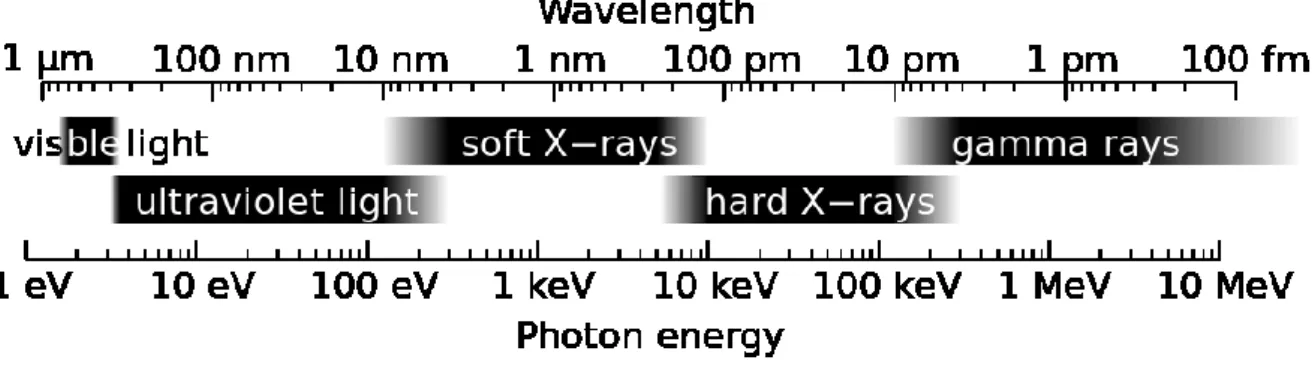
Like visible light, X-rays are electromagnetic waves (Fig. 2-6) that can be characterized by their wavelength and amplitude. X-rays, first discovered by W.C. Röntgen1 in 1895, became an important tool, not just for medical applications or material research. See Fig. 2-7 for one of the first X-ray images ever. Ever since the discovery of X-rays, X-ray sources have been constantly increasing their power, ranging from rather simple X-ray tubes to synchrotrons and free electron lasers. Electromagnetic waves with energies higher than those of ultraviolet light are called soft X-rays. X-ray energies above 5 -10 keV are called hard X-rays. The energy, E, of an electromagnetic wave depends on its wavelength: , with being Planck’s constant2, c being speed of light, and the wavelength. The wavelength and angular frequency, , are related via .
1 Wilhelm Conrad Röntgen (1845 - 1923) was a German physicist. He earned the first Nobel Prize
in Physics in 1901 for his discovery of the X-rays.
2 Max Karl Ernst Ludwig Planck (1858 – 1947) was a German theoretical physicist who won the Nobel Prize in Physics in 1918 for his quantum theory.
h= 6.62606957(29) 10–34 Js
Fig. 2-6. Part of the electromagnetic spectrum showing photon energies and their corresponding wavelengths. (http://commons.wikimedia.org/wiki/File:X-ray_range.svg)
Fig. 2-7 One of the first x-ray pictures ever. It shows the hand of Röntgen’s wife and was presented in Freiburg in January 1896. (from Wikimedia commons:
http://commons.wikimedia.org/wiki/File:First_medical_X-
ray_by_Wilhelm_R%C3%B6ntgen_of_his_wife_Anna_Bertha_Ludwig%27s_hand_-_18951222.gif)