Contents lists available atScienceDirect
Fish and Shell fi sh Immunology
journal homepage:www.elsevier.com/locate/fsi
Full length article
Expansion and loss events characterized the occurrence of MIF-like genes in bivalves
Umberto Rosani
a,b,*, Stefania Domeneghetti
a, Marco Gerdol
c, Alberto Pallavicini
c, Paola Venier
a,*aDepartment of Biology, University of Padova, via U. Bassi 58/b, 35121, Padova, Italy
bAWI Alfred Wegener Institute, Coastal Ecology, Hafenstraße 43, 25992, List auf Sylt, Germany
cDepartment of Life Sciences, University of Trieste, via L. Giorgeri 5, 34127, Trieste, Italy
A R T I C L E I N F O
Keywords:
Macrophage migration inhibitory factor MIF
Cytokines Bivalves
Mytilus galloprovincialis
A B S T R A C T
Macrophage migration inhibitory factor (MIF) dynamically connects innate and adaptive immune systems in vertebrate animals, allowing highly orchestrated systemic responses to various insults. The occurrence ofMIF- likegenes in non-vertebrate organisms suggests its origin from an ancestral metazoan gene, whose function is still a matter of debate. In the present work, by analyzing available genomic and transcriptomic data from bivalve mollusks, we identified 137MIF-likesequences, which were classified into three types, based on phy- logeny and conservation of key residues: MIF, D-DT, and the lineage-specific type MDL. Comparative genomics revealed syntenic conservation of homologous genes at the family level, the loss of D-DT in theOstreidaefamily as well as the expansion ofMIF-likegenes in theMytilidaefamily, possibly underpinning the neofunctionalization of duplicated gene copies. InM. galloprovincialis, MIF and one D-DT were mostly expressed in haemocytes and mantle rim of untreated animals, while D-DT paralogs often showed very limited expression, suggesting an accessory role or their persistence as relict genes.
1. Introduction
An heterogeneous group of intercellular regulatory proteins known as cytokines contributes to the establishment of fast and effective re- sponses to pathogenic insults in vertebrates [1]. In these animals, re- markable evolutionary processes, likeen-blocgenome duplications and gene loss events, have contributed to the emergence of the adaptive immune system, together with complex cytokine networks [2–4]. The presence of acytokine-likesystem in the latest common bilaterian an- cestor is suggested by the presence of cytokine homologs in proto- stomes, in spite of at least 600 million years of independent evolution between this lineage and deuterostomes [5].
The Bivalvia class (Lophotrochozoa, phylum Mollusca) comprises several thousand species adapted to live in a broad range of environ- ments and also species of worldwide importance as seafood or for the ecosystem services they provide [6]. The involvement ofcytokine-like genes in bivalve immunity has been hypothesized since 1990 [7,8] and has been corroborated over the past two decades by the identification of several sequences homologous to vertebrate cytokines in bivalve gen- omes, as well as of homologs of downstream signaling elements, often reported as expanded gene families [9]. The repertoire of known cy- tokines shared by bivalves and vertebrates currently includes
transforming growth factor-beta [10], allograft inflammatory factor-1 [11], interleukin-17 [12], tumor necrosis factor-alfa (TNFα) [13] and macrophage migration inhibitory factor (MIF) [14]. Furthermore, im- munological research enabled the identification of a number of other cytokines independently developed in the protostome lineage [8], in- cludingspätzlein arthropods [15], astakinein crustaceans and in the bivalveCrassostrea gigas[16,17],CCFin the annelidEisenia foetida[18]
andVago in arthropods [19]. The latter two cytokines might act as functional homologues of vertebrate TNFαand interferon-gamma, re- spectively.
MIF was the first intercellular regulatory protein ever described [20]. Two paralogous genes are present in the human genome, namely MIF andD-dopachrome decarboxylase(D-DT) [21,22]. In mammals, both MIF and D-DT functionally connect the innate and adaptive immune systems [23] as well as the immune and endocrine systems [24]. In addition to macrophages, MIF expression has been reported in other cell types and tissues, including the nervous system and cancer cells [25]. The binding of MIF to its membrane receptor, the CD74−CD44 complex, promotes cell growth, the production of other inflammatory cytokines and the recruitment of immunocompetent cells [26]. In de- tail, the interactions triggered by MIF along the ERK1/2 MAPK cascade support the activity ofJUN N-terminal kinase, as well as the production
https://doi.org/10.1016/j.fsi.2019.07.019
Received 26 March 2019; Received in revised form 14 June 2019; Accepted 10 July 2019
*Corresponding authors. Department of Biology, University of Padova, via U. Bassi 58/b, Padova, Italy.
E-mail addresses:umberto.rosani@unipd.it(U. Rosani),paola.venier@unipd.it(P. Venier).
Available online 12 July 2019
1050-4648/ © 2019 Published by Elsevier Ltd.
T
of cytoplasmic phospholipase A2 (cPLA2), arachidonic acid and pros- taglandin E2 [27]. By inducing the production of TLR4 through the action of ETS transcription factors, MIF improves pathogen detection [28] and promotes cell-survival by suppressing apoptosis [29]. MIF is often upregulated in human tumors and MIF-induced TLR4 expression boosts the metastatic migration of malignant cells [28]. Moreover, MIF possesses a unique function as an antagonist of glucocorticoids, due to the direct counteraction of the immunosuppressive and anti-in- flammatory effects of these drugs, such as the inhibition of cPLA2, the promotion of the synthesis of inhibitor of nuclear factor-κB andMAPK phosphatase-1, and the degradation of cytokine mRNAs [30]. The bio- logical relevance of the D-dopachrome tautomerase enzymatic activity of D-DT, also shared by MIF [31], is unexplained, as the naturally oc- curring substrate of this reaction has not been identified yet. However, this activity has been tentatively connected to a vestigial function that may reflect the ancestral role of MIF in the invertebrate melanotic en- capsulation response [32]. The phenoloxidase (PO) system leads, through the activation of PO enzymes, to the melanization of pathogens and damaged tissues and it represents a major defense mechanism of non-vertebrates, particularly arthropods [33,34].MIF-likegenes have been identified in several domains of the tree of life. These include, among unicellular organisms, Bacteria [35], SAR (Stramenopiles-Al- veolates-Rhizaria) and parasites such asPlasmodium, whose MIF mimics the human molecule interacting with human receptors during malaria [36]. In multicellular organism, besides vertebrates,MIF-likegenes are present in plants [37], basal chordates [38,39] and protostomes, such as bivalves, nematodes [40] and arthropods [41,42]. Among the few functionally characterized MIF-like proteins of protostomes, BgMIF displays a cytokine behavior in the gastropod Biomphalaria glabrata [43], and MIFs from blood- and lymph-sucking ecto-parasites, such as ticks and aphids, are capable of inhibiting the host immune response to prolong the feeding period [44,45]. Protein databases include a high number of MIF-domain containing proteins (e.g. ~4 k in the Interpro database v.72), mostly of them derived from automated gene predic- tions of published genomes. Currently, just a very few studies have been focused on bivalve MIFs [46,47] and a perspective on the evolution of these genes is still lacking, since the available analyses only included very few sequences from mollusks (i.e. 4 in Ref. [48] and 6 in Ref.
[47]). In the present work we aimed to update the distribution ofMIF- like sequence in bivalves through the analysis of genomic and tran- scriptomic data, to define the phylogenetic relationships between ver- tebrate and bivalveMIF-likegenes and to provide afirst insight into the genomic context and the transcriptional behavior of these genes in bi- valves, with a focus on theMytilidae MIF-likegenes.
2. Materials and Methods
2.1. Sequence data retrieval and preliminary processing
Genomic scaffolds and gene models of 11 bivalve species were re- trieved from the corresponding public repositories. In detail, we con- sidered the genomic scaffolds of two Mytilus galloprovincialisgenome shotgun projects (APJB000000000.1 [49] and LNJA000000000.1 [50]) and scaffolds plus gene models ofBathymodiolus platifronsandModiolus philippinarum [51], Crassostrea gigas[52], C. virginica, Pinctada fucata [53],Ruditapes philippinarum(only gene models) [54],Saccostrea glo- merata[55],Mizuhopecten yessoensis[56] andLimnoperna fortunei[57].
The access to genomic data ofScapharca broughtoniiwere kindly pro- vided by Dr. Chang-Ming Bai [58]. All the predicted proteins of the Ensembl genome browser v.95 (https://www.ensembl.org/index.html) and Ensembl Metazoa v.42 (https://metazoa.ensembl.org/index.html) were downloaded from the corresponding ftp repositories (accessed in December 2018). To increase the sampling size of species pertaining to Lophotrochozoa, we additionally retrieved the gene models of 2 gas- tropods (Biomphalaria glabrata and Haliotis discus discus), one bra- chiopod (Lingula anatina), one nemertean (Notospermus geniculatus) and
one phoronid (Phoronis australis). RNA sequencing data of 47 bivalve species were downloaded from the NCBI SRA archive and trimmed for quality, setting the maximum number of ambiguous bases and minimum quality threshold to 2 and Q20, respectively.De-novotran- scriptome assemblies were constructed for each species with CLC genomics workbench v.9 (Qiagen, Germany), setting word and bubble size parameters to“automatic”and a minimal contig length of 200 bp.
The assembled contigs were subjected to open reading frame (ORF) prediction by theTransdecodertool (Trinity suite [59]) with a minimal ORF length of 100 codons. The details of the considered sequence da- tasets are included in Supplementary File 1.
2.2. Production of M. galloprovincialis RNA-seq data
To investigate the presence ofMIF-liketranscripts in greater detail in M. galloprovincialis, we sequenced one RNA library prepared from the gill tissue of mussels sampled in the North Adriatic Sea (Italy). In detail, 30 mussels were collected from the lagoon of Goro (20th of June 2011) and, soon after transportation to the laboratory, they were dissected, the gills were placed in Trizol (Thermofisher, US) and stored at−80 °C.
Total RNA was extracted following the producer's recommendations.
RNA quantity and quality were checked using a Nanodrop instrument (Thermofisher) and an Agilent RNA6000 Nanochip (Agilent Technologies, US), respectively. One microgram of high-quality RNA was used to construct libraries for Illumina high-throughput sequen- cing, subsequently carried out with a 2 × 100 read layout in a Hi-Seq 2000 instrument (BMR Genomics, Italy). The raw reads were deposited at the SRA archive under accession ID PRJNA526432.
2.3. Identification of MIF domain-containing proteins and other pathway elements
The Hidden Markov Model of MIF protein signature (HMM PF01187) was obtained from the Pfam-A database v.29 [60] and used to scan the protein datasets using HMMer [61]. Hits with an E-value lower than 10−10 were further screened for the presence of a signal peptide region using SignalP-4.0 [62]. All the sequences discussed in this paper are available in Supplementary File 2.
2.4. Phylogenetic analysis
Cd-hit [63] was used to reduce the redundancy of the genome-de- rivedMIF-likesequence dataset, applying a sequence identity cut-offof 90% for bivalve hits and of 75% for the other metazoan hits. The cd-hit step reduced the initial dataset ofMIF-likeproteins from 207 into 150 sequences, mostly removing redundant orthologous derived from phy- logenetic-related species. Bivalve transcriptomes generated 99 addi- tional non-redundantMIF-likesequences, bringing the full dataset to a total of 249 protein sequences, which were aligned with MUSCLE [64]
using default parameters. The multiple sequence alignment (MSA) was subsequently screened to retain only informative positions (cut-off: 0.63) and to remove phylogenetically poorly informative sequences (cut-off of 0.63) using GUIDANCE [65], and further manually in- spected. These steps further reduced the sequence dataset to 213 se- quences. ModelTest-NG v0.1.2 [66] was used to assess the best-fitting model of molecular evolution for the MSA (identified as the WAG + G model) and, accordingly, a Bayesian phylogenetic analysis was per- formed using MrBayes v3.2.5 [67]. Two independent Markov Chain Monte Carlo analysis were run with four chains each for 10,000,000 generations, with a sampling frequency of 1000 and a burn-in of 25%
sampled trees. The convergence of parallel runs was estimated by reaching an average standard deviation of split frequency < 0.05 and of a potential scale reduction factor equal to 1. Adequate posterior sam- pling was evaluated by the reaching of an effective sample size > 200 for each of the estimated parameters using Tracer v1.6 [68]. Thefinal consensus tree was visualized and edited using FigTree v1.4.3 (http://
tree.bio.ed.ac.uk/software/figtree/).
2.5. Reconstruction of the structure of bivalve MIF-like genes
We obtained the structure of bivalveMIF-likegenes and identified theflanking genes from the available genomic scaffolds. To investigate the peculiar expansion of MIF-likegenes we focused on species be- longing to the Mytilidaefamily and, due to the different quality and completeness ofMytilidaegenomes, we adopted different strategies to retrieve the MIF loci. In the case of complete genomes, we mostly relied on existing annotated gene models, whereas ablast-based strategy was adopted to recover MIF-likegenes in theM. galloprovincialis genome [50]. We used the MIF transcript sequences as queries (blastn) and the genomic scaffolds matching with anE-value lower than 10−20were extracted andde-novoassembled to resolve overlapping regions. Later, we mapped the RNA-seq reads generated in this study (see the “Pro- duction ofM. galloprovincialisRNA-seq data”section) on the obtained contigs to ascertain the correctness of the gene architecture and to predict intron/exon boundaries (using the CLC Genomic Workbench large gap mapping tool, with similarity and length fraction parameters set to 0.95). Similarly, not annotatedM. philippinarumandC. gigas MIF-like genes were recognized by a combination of blast and read mapping approaches (see Supplementary File 3 for further details). To visualize syntenic conserved regions, selected chromosomal regions were aligned using the progressiveMAUVE tool, implemented in MAUVE [69].
2.6. Mytilus galloprovincialis immuno-stimulation and tissue sampling M. galloprovincialis of commercial size (5.1 ± 0.3 cm of shell length) were collected from an outlet of the Venice lagoon (Italy) in September 2015, acclimated at 21 ± 1 °C in artificial sea water (32‰
of salinity) and fed with invertebrate food (Plancto Aqua Medic, Bissendorf, Germany). Six different tissues (haemolymph, gills, diges- tive gland, mantle rim, foot and adductor muscle) were collected from five mussels. Injections with either 0.1 ml of NaCl-enriched PBS (PBS–NaCl, used as control) or 0.1 ml of a mixture of heat-killed Gram- positive and -negative bacteria (108CFU/ml) were carried out in the posterior adductor muscle of at least 60 mussels. The bacterial cocktail was prepared from equal amounts ofMicrococcus lysodeikticus, Vibrio splendidusandVibrio anguillarum, separately grown overnight in Marine Broth at 22 °C (Vibrio spp.) or in Luria–Bertani medium at 30 °C (M.
lysodeiticus). Bacteria were collected from each culture by centrifuga- tion at 3000×gfor 10 min and their concentration, estimated by optical density at 600 nm, was adjusted by re-suspending them in PBS–NaCl.
The mixture was prepared, heated at 65 °C for 2 h and the complete inactivation of the bacteria cells was verified by plating on nutrient medium. At 3, 9, 24 and 48 h post-injection (hpi), haemolymph, gills, digestive gland, adductor muscle and mantle rim tissues were collected fromfive treated andfive control mussels. Haemocytes were collected by centrifugation carried out at 800 g and 4 °C for 15 min and re-sus- pended in 1 ml Trizol (Invitrogen). Total RNA was extracted following
the Trizol manufacturer's instructions, treated with DNAse I (Qiagen) to remove genomic DNA contamination and purified using a RNeasy MinElute Cleanup kit (Qiagen). RNA concentration and quality were ascertained with a NanoDrop ND-1000UV spectrophotometer and an Agilent 2100 Bioanalyzer (Agilent Technologies), respectively. RNAs of 5 individual mussels per tissue and condition were pooled and cDNAs were prepared starting from 1μg of total RNA using a SuperScript IV Reverse Transcriptase (Life Technologies, USA), purified (PureLink PCR Purification kit, Thermo Scientific, USA), quantified and diluted to the appropriate concentration.
2.7. Expression analysis
Available RNA-seq samples of selected bivalve species were used to compute the expression levels ofMIF-likegenes in different tissues and conditions (Supplementary File 1). In details, we used RNA-seq datasets of visceral mass, mantle, gills, foot and adductor muscle forM. phi- lippinarum[51], of ovary, visceral mass, mantle, gills, foot and adductor muscle forB. platifrons[51] and of mantle, gills (this study), adductor muscle, haemocytes and digestive gland forM. galloprovincialis[70,71].
Each dataset of clean reads was mapped to all the annotated genes of the corresponding species using CLC Genomics Workbench, with length and similarity fractions set to 0.95 and 0.95, respectively, whereas mismatch/insertion/deletion penalties were set to 3/3/3. For each dataset, the number of unique mapped reads were counted and used to calculate digital expression values as Transcripts Per Million (TPM) [72]. qPCR was carried out with the DyNAmo HS SYBR Green qPCR kit (Thermo Scientific) starting from 1μl of cDNA in a 15μl offinal reac- tion mixture (1X Master Mix, 1X Rox passive reference dye, 0.5μM of each specific primer). The housekeeping geneElongation factor-1-alpha (EF1-α) was chosen as reference. The stability of EF1-αas reference gene has been demonstrated by applying four different algorithms to data obtained in different experimental conditions including bacterial immune-stimulation [73]. Primer pairs (Table 1) were designed to amplify nine M. galloprovincialis MIF-like transcripts and EF1αusing Primer 3 (http://bioinfo.ut.ee/primer3-0.4.0/) with the following parameters: product size 100–250 bp, melting temperature Tm 60 ± 1 °C and G/C content≤55%. qPCR was performed on an Applied Biosystems 7900HT Fast Real-Time PCR System in a MicroAmp Fast Optical 96 or 348-Well Reaction Plate (Life Technologies) as follows:
95 °C for 15 min, followed by 40 amplification cycles at 95 °C for 30 s (denaturation) and 60 °C for 1 min (annealing and elongation). Each qPCR assay was carried out in triplicate on the same plate for each primer pair. The relative expression ratio of the selected target gene (RQ) was based on the delta Ct method [74]. Data are expressed as mean value ± standard deviation for tissue expression or as Ct ratio between bacteria-injected versus PBS-injected values, with significant differences calculated with a two-tailed pairedt-test (*p < 0.05).
Table 1
M. galloprovincialisprimer sequences used in the present work. NCBI ID, forward and reverse sequence of the primer pair and length of the amplicon are reported for Elongation factor 1-alfa, MIF, MDL, D-DT and D-DT2 genes.
Gene type NCBI ID Forward sequence Reverse sequence Amplicon length (bp)
EF1α AB162021.1 CAAGACCCACAGACAAAGC GGAGCAAAGGTAACAACCAT 130
MIF MH190398 GCCATTTACACCAATCTTCCT CCCCCATGACTCATCATTT 143
MDL MH190397 TGGATAAAAATGCCGGGGA CCGTGTTGAATTTTGTTGCT 120
D-DT MH190396 AATGCTGGAGACGAATCAC GTGCGGTTGAAGATCTGT 167
D-DT2 MH190399 GGCATGACGGGAATTATGAT AGGCTGGATAAGGGACAC 174
D-DT2 MH190400 ACAAAAGATGGGAGAGGTG AAAACCCTGTGTGGAGGA 215
D-DT2 MH190401 AGAAGTGGACTTACGAGATGA AGAAGTGAAGCAAAAAACGG 224
D-DT2 MH190402 CGAGCTTGAAAGATGCAGA CCATTGGTGTTTTCTTCCCA 162
D-DT2 MH190403 TGTCTGTAACCCCTGGTG ATATTCGGGTGCTCTTCTG 229
D-DT2 MH190404 ATTGACGTGTGGAGATAGGA TTGTAGTCTGGTGCTCGT 180
3. Results
3.1. MIF-like sequences in bivalves
The analysis of available transcriptomic and genomic data of 47
species representative of 13 bivalve families, as well as data mining of 61 million reads ofM. galloprovincialisgenerated in this study, allowed us to identify 137MIF-likesequences. These sequences are character- ized by the presence of a MIF domain identified with significantE-value support and no signal peptide, consistent with the non-classical secre- tion previously reported for vertebrate MIFs [75]. Both genome and transcriptome data supported the presence of a higher number ofMIF- likesequences in theMytilidaecompared to other bivalve families, i.e.
M. galloprovincialis(9 full-length transcripts),Perna viridis(7 full-length transcripts),B. platifrons(13 gene models) andM. philippinarum(9 gene models, including a previously not annotated gene described in Sup- plementary File 3).L. fortuneidisplayed just 4MIF-likegenes, possibly due to the partial completeness of its genome (Table 2). In addition to bivalve species, we could retrieveMIF-like sequences in most of the major metazoan taxa by exploring the Ensembl databases. Porifera and the rotiferAdineta vagawere the only species to lack a MIF sequence, among those analyzed in the present study (Supplementary File 1).
3.2. Phylogenetic relationships of bivalve MIF-like sequences
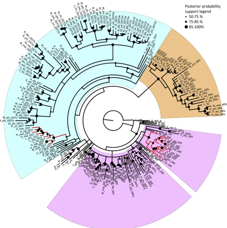
The Bayesian phylogeny analysis, performed on a non-redundant set of MIF-like sequences (see Materials and Methods), identified three main protein groups (Fig. 1). Thefirst two major groups were named MIF and D-DT, according to the presence of previously characterized human MIF and D-DT proteins. The MIF clade, which included both vertebrate and invertebrate sequences, displayed a short average branch length, which points to a rather low molecular diversity. We identified 34 bivalve MIF sequences, often present in a single copy in the genomes of these organisms (with some exceptions, i.e.C. virginica, R. philippinarum,Atrina pectinata,Meretix meretrixandB. platifrons).
The D-DT clade, which also included both vertebrate and in- vertebrate sequences, displayed a much higher molecular diversity, as pointed out by the complex branching patters observed. The number of bivalve sequences identified (69) was much higher that the number of MIF sequences, and their disordered clustering only partly mirrored the currently accepted species phylogeny. All fully sequenced bivalve genomes possessed at least one D-DT gene, except for those of the Ostreidaefamily, which apparently underwent a gene loss. However, most species possess multiple genes (up to 8 inB. platifrons), which relatively high pairwise distance revealed a high degree of molecular diversification. Within the D-DT clade, we could identify a highly supported subgroup of 33 D-DT sequences, named D-DT2, which show peculiar primary sequence features (described in detail in the following section). All DD-T2 sequences are of theMytilidaefamily, marking the occurrence of a lineage-specific expansion event.
Finally, the third clade exclusively contained molluscan sequences, i.e. 31 from bivalves and two from gastropods. These sequences, en- coded by single-copy genes in most, but not all species (i.e.Arcidae family underwent a gene loss event), clustered in agreement with the expected evolutionary relationship among species. Due to peculiar features of the primary amino acid sequences of the proteins pertaining to this clade, which allow a clear discrimination both from MIFs and D- DTs (see the next section for a detailed discussion), we named this subclade“MIF/D-DT-like”(MDL).
3.3. Conservation of bivalve MIF-like sequences
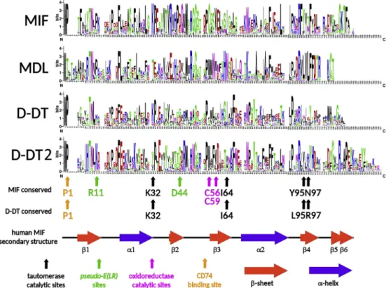
We investigated whether the clear-cut phylogenetic branching of bivalveMIF-likesequences in the MIF, D-DT, D-DT2 and MDL clades (Fig. 1) could depend on a peculiar organization of the primary protein sequence or on single conserved amino acid residues known to be functionally important in vertebrate MIFs and D-DTs. The sequence conservation logos presented inFig. 2highlight the most relevant fea- tures of each of the four groups ofMIF-likesequences. The bivalve MIFs closely resemble vertebrate MIFs, in particular due to the high con- servation of the residues involved in the interaction with the substrate Table 2
Bivalve MIF-like sequences. Family and species names, genomic (G) or tran- scriptomic (T) dataset of origin and related number of MIF-like sequences (MIF, D-DT or MDL, the latter is referred to MIF/D-DT-like sequences) are reported per species.
Family Species Dataset MIF D-DT MDL
Mytilidae Mytilus galloprovincialis G + T 1 7 1
Mytilus edulis T 0 3 0
Mytilus californianus T 1 5 1
Mytilus trossulus T 0 1 1
Mytilus coruscus T 0 4 0
Mytilus chilensis T 0 4 0
Perna viridis T 1 5 1
Limnoperna fortunei G 0 3 1
Bathymodiolus platifrons G 4 8 1
Bathymodiolus puteoserpentis T 0 5 0
Bathymodiolus azoricus T 0 0 1
Lithophaga lithophaga T 0 1 1
Modiolus philippinarum G + T 1 7 1
Ostreidae Crassostrea corteziensis T 1 0 1
Crassostrea virginica G 4 0 2
Crassostrea gigas G 1 0 1
Crassostrea hongkongensis T 1 0 1
Ostrea chilensis T 1 0 0
Ostrea edulis T 0 0 1
Ostrea lurida T 1 0 1
Saccostrea glomerata G 1 0 1
Pteriidae Pinctada fucata G 1 0 0
Pinnidae Atrina pectinata T 2 2 0
Pectinidae Mizuhopecten yessoensis G 1 2 1
Pecten maximus T 1 0 0
Amusium pleuronectes T 1 0 1
Argopecten irradians T 1 1 1
Mimachlamys nobilis T 1 0 1
Placopecten magellanicus T 1 1 1
Azumapecten farreri T 0 2 0
Solemyidae Solemya velum T 1 1 1
Neotrigoniidae Neotrigonia margaritacea T 1 0 0
Unionidae Elliptio complanata T 0 2 0
Pyganodon grandis T 1 0 1
Uniomerus tetralasmus T 1 0 0
Hyriopsis cumingii T 1 0 0
Villosa lienosa T 1 0 1
Nuculoida Ennucula tenuis T 0 1 0
Veneridae Corbiculafluminea T 0 2 1
Meretrix meretrix T 2 4 1
Paphia textile T 0 1 1
Astarte sulcata T 0 1 1
Ruditapes decussatus T 1 2 1
Ruditapes philippinarum T 2 3 1
Tellinidae Macoma baltica T 1 0 0
Arcidae Scapharca broughtonii G 1 1 0
Anadara trapezia T 1 0 0
Myochamidae Myochama anomioides T 0 1 0
Numbers in red indicate MIF with non-conserved key residues.
Underlined number refer to the newly annotated genes, see Supplementary File 3.
[32]. In fact, P1, K32 and, in minor measure, I64, Y95 and N97 (co- ordinates are referred to human MIF, NP_002406.1) are conserved in bivalve MIFs. Only one (C56) of the two cysteine residues, character- izing the catalytic center for the oxidoreductase activity of vertebrate MIFs [76], was invariably conserved in bivalves, whereas C59 was re- placed by different amino acids. The pseudo-(E)LRmotif (R11, D44), which mediates the non-canonical interaction with the CXCR2 receptor in humans [77], was not found in bivalve MIFs. Overall, the con- servation of these residues and the preservation of the chemico-physical properties of regions involved in the formation of secondary structures
in vertebrate MIFs are consistent with the detection of a highly sup- ported MIF domain in bivalve species (medianE-valueof 1.5−30) and support a certain degree of functional conservation between vertebrate and bivalve MIFs.
Although bivalve D-DT and D-DT2 sequences displayed, in most cases, a well-conserved P1 residue, these two groups were otherwise quite diverse. Indeed, D-DT2s are significantly longer than canonical D- DTs and MIFs, as they presented a C-terminal extension of a dozen of amino acids and lacked some of the conserved residues typical of ver- tebrate D-DTs, such as L95 and R95 (homologous to Y95, N97 of MIF).
Fig. 1.Bayesian phylogenetic tree ofMIF-likesequence. The clade highlighted in light violet includes MIF proteins, the one in light blue includes D-DT proteins, while the clade highlighted in orange includes MDL proteins. Red lines denote vertebrate sequences. Posterior probability of the node is depicted in a size-dependent way, whereas the nodes supported by poor posterior probability values (< 0.5) were collapsed. The sequences are labeled with thefirst genera letter followed by the three first species letters and the indication of the protein type (MIF, DD-T or MDL). The tree in nexus format is provided as Supplementary File. (For interpretation of the references to colour in thisfigure legend, the reader is referred to the Web version of this article.)
On the other hand, canonical D-DTs retained, in most cases, K32 and I64, which were not found in the mussel-specific D-DT2 sequences. As expected, neither the MIF-specific oxidoreductase center nor the pseudo-(E)LRmotif could be identified in bivalve D-DTs.
All bivalve MDL sequences were characterized by a unique organi- zation of the N-terminal region as, compared to MIF and D-DT, they presented an insertion of four amino acids and lacked P1. Moreover, none of the other residues enabling substrate binding in human MIF and D-DT was conserved, thus explaining the lowE-valueof detection of the MIF-likedomain in these proteins. Compared to MIF and D-DT, MDLs also lacked a few residues at their C-terminus and showed a highly conserved consensus stretch (DRVLVLFFDTRKCT) which did not match any vertebrateMIF-likesequence. Overall, the lack of conservation of all the key residues of the catalytic and receptor binding sites, justifies our classification of MDLs as a group distinct from MIFs and D-DTs.
3.4. Genomic organization of MIF-like genes in bivalve
We exploited the available bivalve genomes to investigate the genomic organization of the MIF loci, with a focus onMytilidae, in order to understand the possible mechanisms underpinning the expansion event which D-DT2 genes underwent in this family. In general, bivalve MIF-likegenes showed strong structural conservation, with 3 exons and two introns, whose length slightly varied between gene types (Table 3).
Thefirst exon included the 5′UTR and a coding region offixed length in MIF and D-DT genes (108 nucleotides), but slightly longer in MDLs (120 nucleotides). The second exon was 167–176 nucleotides long, and the third exon, comprised a variable number of coding nucleotides, being considerably longer (up to 172 nucleotides) in the mussel-specific D- DT2 genes, compared with MIFs, D-DTs and MDLs.
InC. virginicaboth MIFs and MDL are part of a duplicated chro- mosomal region: the two C. virginicaMIFs genes are tandemly dupli- cated on the same chromosome at a distance of around 400 kb, while MDL is duplicated at 600 kb of distance, for a total of 6 genes. We can also report a remarkable conservation of theflanking genes of MIF and
MDL in the Ostreidae family (C. gigas, C. virginica andS. glomerata, Supplementary File 4), whereas the overall syntenic conservation was low in the other bivalve species. Unfortunately, this type of study in Mytilidaewas hampered by high fragmentation and relatively low N50 values of genome assemblies. These technical features led to the scat- tering ofMIF-likegenes on different scaffolds and currently preclude a large-scale assessment of the association between MIF, D-DT, D-DT2 and MDL genes on larger contiguous chromosomal regions.
Nevertheless, preliminary data indicated that at least some MIF-like genes are found in a cluster organization and we could prove the lo- cation on the same genomic scaffold of MDL and D-DT2 genes inB.
platifrons. In addition, the evidence of microsynteny partly supports the inter-species conservation of the architecture ofMIF-likegene clusters, sinceB. platifronsandM. philippinarumshare CDC45 as the 3’flanking gene of D-DT.
3.5. Expression analysis of Mytilidae MIF-like genes
The expression levels of the 9M. galloprovincialis MIF-like genes were measured by qPCR in six different tissues and the results were compared within-silicoexpression values computed from the available RNA-seq data (Supplementary File 5). TheMIF-likeexpression levels in M. galloprovincialistissues were generally low (i.e. delta Ct values lower than 0.03, with the only exception of D-DT in the mantle rim that reached 0.1,Fig. 3A). While RNA-seq data showed a prevalence of MIF expression in digestive gland and gills, the qPCR data showed the highest levels in haemocytes, followed by digestive gland and gills.
Although at low levels, PCR data confirmed the prevalent expression of M. galloprovincialis MDL in gills. In agreement within-silico data, the expression of the expanded D-DT2 gene copies was low in all tissues, in some cases not even detectable. The comparison ofin-silico gene ex- pression values of other Mytilidae species (Supplementary File 5) showed similar expression trends, revealing an interesting peak of ex- pression for MIF in the ovary ofB. platifronsand a generalized lack of expression for duplicated D-DT2 gene copies.
Fig. 2.The conservation of bivalveMIF-likeproteins (MIF, MDL, D-DT and MsD-DT) is depicted with sequence logos. The conserved key residues have been linked to the function of vertebrate MIF and D-DT sequences are shown, together with the secondary structure of human MIF.
InM. galloprovincialis, we further evaluated the expression trend of the two most highly expressed transcripts (MIF and D-DT) at 3, 9, 24 and 48 h after injection (hpi) with 107heat-killed bacteria mix (treat- ment group) or PBS (paired controls) in digestive gland, gills, haemo- lymph and mantle rim tissues. Compared to controls, both MIF and D- DT showed similar expression trends in most of the analyzed tissues, with the induction of MIF peaking at 24 hpi in digestive gland (1.9x), gills (3.5x) and mantle rim (3.7x) and at 48 hpi in haemolymph (3.1x).
Similarly, the D-DT expression peaked at 24 hpi in mantle rim (5.8x) and at 48 hpi in haemolymph (2.5x), whereas this transcript was down- regulated in digestive gland up to 24 hpi, showing an opposite trend, compared to MIF. Remarkably, both MIF and D-DT were down-regu- lated in haemolymph at 3 hpi (7.2x and 5.1x, respectively). In this tissue, the expression of D-DT was quickly restored, while the down- regulation of MIF remained visible up to 24 hpi (Fig. 3B).
4. Discussion
The presence of cytokines in invertebrates has been long theorized [7,78,79] and the two alternatives hypotheses concerning their origins
either by extreme divergence from ancestral molecules shared with vertebrates, or by an independent lineage-specific evolutionary gain has been debated since the early 2000s [8,33]. Recent genetic and mole- cular studies have clarified that MIF is an ancient cytokine, derived from an ancestral gene which was present in the latest common an- cestor of bilaterian animals. The origin of MIF can be back-dated even before the radiation of animals, as evidenced by our detection of two MIF-likegenes ofTrichoplax adherensin Placozoa, a basal group in the metazoan tree of life [80] and also considering the presence of possible MIF paralog genes in plants [37]. Despite the remarkable molecular diversification of bivalveMIF-likegenes, now fully evidenced by the availability of complete genomics datasets, the evolutionary history of these sequences in metazoans has not been properly investigated yet.
Based on phylogenetic evidence (Fig. 1) and on the conservation of key residues important for receptor and substrate binding (Fig. 2), we classified bivalve MIF-likesequences in three groups, i.e. MIF, MDL (MIF/D-DT-like) and D-DT. In spite of some limitations, linked to the small size of these cytokines and the low number of phylogenetically informative sites (e.g. human MIF and D-DT only share 34% sequence identity), the phylogenetic tree strongly supported the distinction Table 3
BivalveMIF-likegenes. Species, gene ID and name, length in bp, number of exons, scaffold length and 5′, 3’flanking genes are reported.
Species Gene ID Name Length (bp) No. of coding exon Distribution of coding nt Scaffold length [kb] Flanking genes (5’/3′)
Scapharca broughtonii EVM0019218 MIF 10,901 3 107-172-72 43,747 unknown/SETD7
EVM0001330 D-DT 5567 3 102-176-61 43,747 unknown/C11orf54-like
Crassostrea virginica LOC111133621 MIF (a) 2975 3 108-167-67 98,698 MIF/YGR130c
LOC111133619 MIF (a) 4019 3 108-167-77 98,698 neural cadherin/unknown
LOC111127184 MDL (a) 2141 3 120-176-40 77,016 IAP/DMBT1 - (A)
Crassostrea gigas new MIF 4196 3 108-165-69 622 neural cadherin/unknown
CGI10009263 MDL 1732 2 120-173-43 228 IAP/unknown (A)
Saccostrea glomerata 19774 MIF 1538 3 108-166-66 5234 transmembrane pr/TRIMM33
4538 MDL 2686 3 120-176-40 1429 IAP/unknown (A)
Mizuhopecten yessoensis 1181 MIF 7036 3 112-168-40 2667 transmembrane pr/YGR130c
9574 MDL 28,846 3 120-176-40 469 na/unknown
Pincada fucata 10016766 MIF 2917 4 108-173-41-317 59,022 zinc-finger/lectin
568.566 MDL 4531 3 126-76-40 48,891 unknown/MAP
Mytilus galloprovincialis MH190398 MIF 3932 108-169-70 / /
MH190397 MDL 1836 3 120-176-40 / /
MH190396 D-DT 4400 3 108-173-52 / /
MH190399 D-DT2 4598 3 108-173-106 / /
MH190400 D-DT2 4001 1a 108-.. / /
MH190401 D-DT2 5489 2a 108-168-.. / /
MH190402 D-DT2 3629 3 108-176-104 / /
MH190403 D-DT2 11,575 1a 108-.. / /
MH190404 D-DT2 9832 2a / / /
Bathymodiolus platifrons Bpl_scaf_64320–2.0 MIF1 1908 2a 108–170 425 unknown/HNRNPU
Bpl_scaf_13589–0.15 MIF2 1911 2a 108–170 117 PKD1/-
Bpl_scaf_51474–1.3 MIF3 12,148 3 108-169-69 270 Mucin2/-
Bpl_scaf_55759–4.7 MIF4 15,412 2a 527 -/PATS1
Bpl_scaf_38338–3.7 MDL 10,870 3 120-175-39 1339 PRY3/unknown
Bpl_scaf_19807–0.14 D-DT 13,787 3 108-175-54 179 -/unknown
Bpl_scaf_40345–1.14 D-DT 6874 3 108-175-54 220 unknown/CDC45
Bpl_scaf_5477–0.6 D-DT 6787 3 282 unknown/unknown
Bpl_scaf_38338–9.11 D-DT2 8783 3 108-172-102 1339 MIF/unknown
Bpl_scaf_58766–0.17 D-DT2 11,745 3 108-172-156 194 unknown/-
Bpl_scaf_3150–0.12 D-DT2 7882 3 456 LRP6/unknown
Bpl_scaf_38338–9.7 D-DT2 6762 3 108-172-93 1339 NUPR1/MIF
Bpl_scaf_12362–2.6 D-DT2 5228 3 108-173-94 331 -/unknown
Modiolus philippinarum MH190405 MIF 37,247 3 108-176-66 300 unknown/-
Mph_scaf_36746–0.3 MDL 4704 2 150–216 34 -/-
Mph_scaf_9485–0.7 D-DT 4378 2a 108–198 203 MIF/CDC45
Mph_scaf_71627–0.10 D-DT 2001 3 108-176-55 66 unknown/-
Mph_scaf_9485–0.5 D-DT 10,970 3 108-176-76 203 unknown/MIF
Mph_scaf_71922–0.4 D-DT2 10,712 3 108-173-103 115 -/-
Mph_scaf_54981–3.21 D-DT2 6049 3 108-173-112 476 C Vgou1/unknown
Mph_scaf_17703–0.9 D-DT2 6512 3 108-173-97 422 -/unknown
Mph_scaf_9485–0.6 / 6841 9 .. 203 -/MIF
Limnoperna fortunei itr6_4720_pi.g1185.t1 MDL 3855 3 120-176-40 615 unknown/GRHPR
itr6_4889_pi.g435.t1 D-DT2 1804 3 108-173-103 297 MIF/TX1
itr6_4889_pi.g436.t1 D-DT2 6004 3 108-173-103 297 SULT1B1/MIF
itr6_779_pil.g1922.t1 D-DT2 16,994 3 108-173-172 200 P450/unknown
a Partial gene. (a)Duplicated gene.
between the three aforementioned clades. Whereas sequences per- taining to the MIF and D-DT clades are present in a broad range of animals, MDLs represent a lineage-specific acquisitions of the Mollusca lineage, as evidenced by their detection in gastropods, besides bivalves.
The analysis of genomic and transcriptomic data supported the presence of a core-set ofMIF-likegenes in bivalves, comprising at least one gene for each of the three protein types in nearly all species, with the notable exceptions of Ostreidae, which lacked D-DT genes, and Arcidae, which lacked MDL genes. The genomes of several bivalve species contain multiple MIF-like genes, with Mytilidae showing, in particular, a remarkable radiation of the sequences pertaining to the D- DT2 lineage-specific clade (Fig. 1), which is characterized by peculiar sequence features (Fig. 2), low constitutive expression levels and ap- parent non-responsiveness to bacterial challenges (Fig. 3). Based on phylogenetic evidence, the origin of D-DT2s needs to be placed to the latest commonMytilidaeancestor, dated 56–94 Mya [81].
In stark contrast with the relatively high number ofMIF-likegenes evidenced in bivalves, most animal genomes only contain single MIF and D-DT gene copies or, in some cases (e.g. insects) may even lack one of these two paralogs (D-DT). To the best of our knowledge, this is the first report of a relevant expansion and molecular diversification of MIF-likegenes, besides lymph-sucking ecto-parasites, such as ticks and aphids, where the functional differentiation of MIF has an adaptive value and supports efficient feeding [82]. Altogether, the classification of bivalveMIF-likesequences within the MIF, D-DT and MDL clades that we propose in this study clarifies the sequence diversity reported in in a previous study inM. galloprovincialis[46].
The 3-exon gene structure of MIF and D-DT and the position of splicing sites was found to be highly conserved throughout metazoan evolution, to the point that thefirst exon invariably included 108-nt of coding sequence from basal metazoans (T. adherentsandN. vectensis) to bivalves and vertebrates. The bivalve-exclusive MDL and the mussel- specific D-DT2 genes did not conform to this rule, displaying peculiar architectural features. While we demonstrated a considerable syntenic conservation of MIF and MDL loci inOstreidae, we can draw few con- clusions on the organizationMIF-likegenes inMytilidaeon a chromo- somal level, due to the low contiguity of their genome assemblies.
Among the few observations that can help to delineate the evolutionary scenario which led to the D-DT expansion in this bivalve family, we reported a cluster organization for D-DT2 genes, likely derived from tandem duplications of a single ancestral gene copy. Finally,
microsynteny analyses confirmed the conservation of the association between one of such clusters and the CDC45 gene. These genomic features, not shared byMIF-likegenes of other bivalve families, leave many open questions about the possible neo-functionalization of D-DT2 paralogs.
The biological implications of the presence of multipleMIF-likese- quences in bivalve mollusks are possibly connected to their function, which might be either exerted as a cytokine, through the interaction with a receptor, or as a tautomerase, through the enzymatic conversion of an unknown substrate related to dopachrome. With this respect, we showed that MDLs lack all the conserved sites involved in both receptor and substrate binding in vertebrate MIF and D-DT proteins (Fig. 2).
Therefore, the biological relevance of this group of sequences, re- presenting a taxonomically restricted gene family in bivalves, is un- known. To investigate the possible mechanisms by which the two other groups of bivalveMIF-likeproteins might exert their biological func- tions, we have to point out that none of MIF receptors and co-receptors (CD74, CXCR2 and CXCR4) are recognizable in bivalves and, more in general, in invertebrates [83]. Human CD74 is a member of the major histocompatibility complex (MHC) superfamily and no orthologs are known in invertebrates, where allorecognition is mediated by different receptors [84]. CD44 orthologs are only detectable in jawed vertebrates and no homologous sequence has ever been reported in basal deuter- ostomes. Consistent with the lack of invertebrate homologs for verte- brate interleukins, except IL-17, no CXCR2 (receptor for IL-8) nor CXCR4 (receptor for SDF-1) are recognizable among the many G pro- tein–coupled receptors currently found in bivalve genomes. In agree- ment with these observations, we could not identify the pseudo-(E)LR motif necessary for MIF binding to interleukin receptors [77] in any of the bivalveMIF-likesequences (Fig. 2). The only amino acid residue showing high conservation in most sequences (except for some D- DT2s), P1, is both required for the binding of MIF by CD74, and for the ability of MIF and DDT to tautomerize D-dopachrome. Assuming that other yet to be identified receptors cover a function homologous to the vertebrate CD74/CD44 complex, the expected immune signaling route would most certainly involve the MAPK cascade, an evolutionarily conserved signaling pathway in all metazoans [85], including bivalves [86–88]. In summary, the nature and the existence itself of the bivalve receptor of MIF remain to be investigated and any hypothesis con- cerning the cytokine-like function of bivalveMIF-likemolecules based on a presumed functional homology with their vertebrate orthologs Fig. 3. A.qPCR quantification of musselMIF-liketranscripts in digestive gland (DG), gills (G), haemolymph (HAE), mantle rim (MR), adductor muscle (AM) and foot (F) of mussels from one Venice Lagoon outlet (Italy). Transcript levels were normalized to the endogenous control (EF1α) and are reported as delta Ct values plus standard deviation.B.Fold change values (bacteria versus PBS-injection expression values) ofMytilus galloprovincialisMIF and D-DT in mussel digestive gland (DG), gills (G), haemolymph (HAE) and mantle rim (MR) during a time course experiment (3, 9, 24 and 48 h). Statistically significant values (p-value < 0.05) are indicated with an asterisk. The legend reports the type of MIF protein (MIF, D-DT, MDL or D-DT2) and the NCBI accession code.
would be, at this stage, a speculative exercise.
The absence of the cysteine residues involved in the inhibition of oxidative stress-induced apoptosis marks another important difference between bivalve and vertebrate MIFs [76]. Only one of the two residues was found in the bivalve MIF clade (but not in the D-DT clade,Fig. 2), matching the situation previously reported in jawlessfishes and sug- gesting that this situation reflects the configuration of the ancestral metazoan MIF sequence [39]. In contrast with the sites required for receptor binding and oxidoreductase activity, those involved in the interaction with the substrate for the D-dopachrome tautomerase ac- tivity were rather conserved (Fig. 2). Hence, the biological function of bivalve MIF and D-DT sequences might be connected to this evolutio- narily persisting, but still poorly understood, enzymatic activity. In- deed, human MIF and D-DT can catalyze the conversion of the non- physiological substrate D-dopachrome to 5,6-dihydroxyindole-2-car- boxylic acid (DHICA) [89]. If experimentally confirmed, the tauto- merase activity of bivalveMIF-likeproteins might be connected to the conversion of an unknown dopachrome-related substrate to 5,6-dihy- droxyindole (DHI) during melanogenesis, as previously hypothesized in the mud crab Scylla paramamosain [90]. The substrate specificity of human MIF depends on five residues, namely P1, K32, I64, Y95 and N97, with the latter two replaced by L95 and R97 in D-DT [32]. As mentioned above, the comparative analysis of bivalve sequences evi- denced the nearly universal conservation of P1 in bivalve MIFs and D- DTs. Similarly, K32 was present in all MIFs and most D-DTs, whereas it was replaced by a T or V in the D-DT2 group. I64 also showed a re- markable degree of conservation in both MIFs and D-DTs, but not in D- DT2s. Finally, Y95 and N97 (replaced by L95 and R97 in human D-DT) were moderately conserved in bivalve MIFs and D-DTs (in the latter, with either L, I or V in position 95 or histidine as the dominant amino acid in position 97). No correspondence was found between D-DT2 and human sequences in these two positions. In summary, the sequences pertaining to the bivalve MIF and D-DT clades showed a significant conservation of the five residues deemed to be important for the maintenance of a D-dopachrome tautomerase activity, whereas the maintenance of this function remains to be investigated for the mussel specific clade D-DT2 and can be excluded for MDLs. Only the functional testing of recombinant proteins could clarify whether bivalveMIF-like proteins can catalyze the production of DHI melanin, an important process to support bivalve shell formation [91], immune defense during wound healing [92] and, in some species, sclerotization of byssal threads [93,94]. To date, no homologs to dopachrome converting en- zyme (DCE) or (L-)dopachrome tautomerase (DCT), the two key en- zymes in melanin biosynthesis in insects and mammals [95], have been identified in mollusks. Indeed, our knowledge of the bivalve melani- zation pathways mainly derives from biochemical approaches, which have allowed the identification of intra- and extra-cellular pro-PO ac- tivity in different tissues of multiple species [96,97]. The use of specific inhibitors of the PO activity has clearly demonstrated the existence of such activity in oyster haemolymph [98] and, if experimentally con- firmed, the enzymatic function ofMIF-likegenes might be involved in similar processes.
Even in the absence of functional data derived from the study of recombinant proteins, the analysis of gene expression profiles and the experimental evaluation of their modulation in response to bacterial challenges can provide some preliminary indication about the possible role of bivalveMIF-likegenes. According to qPCR assays, these genes were constitutively expressed at low levels in multiple tissues in M.
galloprovincialis, except for D-DT, which displayed a very strong ex- pression in the mantle rim. Thisfinding might, to some extent, support the involvement of mussel D-DT in the melanization process, which has been reported to take place in the external epithelium of the mantle [92]. Previous studies had evidenced the lack of inducibility of MIF after bacterial immune stimulation inM. galloprovincialis[46] andM.
meretrix[47], and our preliminary data showed a weak up-regulation of MIF, with significant values reached only in haemocytes and gills. The
continuous production of MIF and its storage in granules would enable a rapid response to infection whenever needed. To test this hypothesis, specific antibodies for MIF could allow the detection of proteins asso- ciated to cytosolic granules in circulating haemocytes. On the other hand, D-DT was strongly upregulated at 24 hpi in the mantle rim, a finding consistent with its possible involvement in mantle-related im- mune processes. Although at limited levels, in digestive gland MIF and D-DT showed opposite expression trends, which may underpin different functional roles in this tissue. RNA-seq analysis showed that MIF fol- lowed a similar expression pattern inC. gigas, where no D-DT homolog is present due to a gene loss event (Supplementary File 6). The ex- pression of MDL was relatively high in some developmental stages ofC.
gigas, with a different expression pattern compared to MIF (Supple- mentary File 6). The role ofMIF-likegenes in larval development of bivalves is intriguing and requires further investigation, considering that the immune system of these organisms in the early development stages differs from that of adult stages, as it develops in conjunction with the maturation of the enkephalinergic system and with the for- mation of the digestive apparatus [99,100].
Acknowledgments
This research project was supported by the European project VIVALDI "Preventing and mitigating farmed bivalve diseases" (H2020 programme, GA n°678589). UR was supported by a biennial grant of the University of Padova (-BIRD168432).
Appendix A. Supplementary data
Supplementary data to this article can be found online athttps://
doi.org/10.1016/j.fsi.2019.07.019.
References
[1] I.B. McInnes, C.D. Buckley, J.D. Isaacs, Cytokines in rheumatoid arthritis - shaping the immunological landscape, Nat. Rev. Rheumatol. 12 (2016) 63–68.
[2] L.C. Borish, J.W. Steinke, 2. Cytokines and chemokines, J. Allergy Clin. Immunol.
111 (2003) S460–S475.
[3] M.E. DeVries, A.A. Kelvin, L. Xu, L. Ran, J. Robinson, D.J. Kelvin, Defining the origins and evolution of the chemokine/chemokine receptor system, J. Immunol.
176 (2006) 401–415.
[4] L. Abi-Rached, A. Gilles, T. Shiina, P. Pontarotti, H. Inoko, Evidence of en bloc duplication in vertebrate genomes, Nat. Genet. 31 (2002) 100–105.
[5] K. Buchmann, Evolution of innate immunity: clues from invertebrates viafish to mammals, Front. Immunol. 5 (2014).
[6] C. Gascon, T.M. Brooks, T. Contreras-MacBeath, N. Heard, W. Konstant, J. Lamoreux, F. Launay, M. Maunder, R.A. Mittermeier, S. Molur, et al., The im- portance and benefits of species, Curr. Biol. CB 25 (2015) R431–R438.
[7] T.K. Hughes, E.M. Smith, R. Chin, P. Cadet, J. Sinisterra, M.K. Leung, M.A. Shipp, B. Scharrer, G.B. Stefano, Interaction of immunoactive monokines (interleukin 1 and tumor necrosis factor) in the bivalve molluscMytilus edulis, Proc. Natl. Acad.
Sci. U.S.A. 87 (1990) 4426–4429.
[8] A. Beschin, M. Bilej, E. Torreele, P. De Baetselier, On the existence of cytokines in invertebrates, Cell. Mol. Life Sci. CMLS 58 (2001) 801–814.
[9] I.C. McDowell, T.H. Modak, C.E. Lane, M. Gomez-Chiarri, Multi-species protein similarity clustering reveals novel expanded immune gene families in the eastern oysterCrassostrea virginica, Fish Shellfish Immunol. 53 (2016) 13–23.
[10] C. Lelong, F. Badariotti, H. Le Quéré, F. Rodet, M.P. Dubos, P. Favrel, Cg-TGF-beta, a TGF-beta/activin homologue in the Pacific OysterCrassostrea gigas, is involved in immunity against Gram-negative microbial infection, Dev. Comp. Immunol. 31 (2007) 30–38.
[11] Y. Zhang, J. Li, F. Yu, X. He, Z. Yu, Allograft inflammatory factor-1 stimulates hemocyte immune activation by enhancing phagocytosis and expression of in- flammatory cytokines inCrassostrea gigas, Fish Shellfish Immunol. 34 (2013) 1071–1077.
[12] U. Rosani, L. Varotto, M. Gerdol, A. Pallavicini, P. Venier, IL-17 signaling compo- nents in bivalves: comparative sequence analysis and involvement in the immune responses, Dev. Comp. Immunol. 52 (2) (2015) 255–268.
[13] Y. Sun, Z. Zhou, L. Wang, C. Yang, S. Jianga, L. Song, The immunomodulation of a novel tumor necrosis factor (CgTNF-1) in oysterCrassostrea gigas, Dev. Comp.
Immunol. 45 (2014) 291–299.
[14] P. Venier, L. Varotto, U. Rosani, C. Millino, B. Celegato, F. Bernante, G. Lanfranchi, B. Novoa, P. Roch, A. Figueras, et al., Insights into the innate immunity of the Mediterranean musselMytilus galloprovincialis, BMC Genomics 12 (2011) 69.
[15] J.A. Hoffmann, J.-M. Reichhart,Drosophilainnate immunity: an evolutionary