THE RADIATION TOLERANCE OF IGNICOCCUS SPECIES
THEIR ASTROBIOLOGICAL RELEVANCE AND IMPLICATIONS TO DNA REPAIR PROCESSES
DISSERTATION
ZUR ERLANGUNG DES DOKTORGRADES DER NATURWISSENSCHAFTEN (DR. RER. NAT.)
DER FAKULTÄT FÜR BIOLOGIE UND VORKLINISCHE MEDIZIN
DER UNIVERSITÄT REGENSBURG
VORGELEGT VON DAGMAR KOSCHNITZKI
AUS DORMAGEN IM JAHR 2016
Das Promotionsgesuch wurde eingereicht am
• 29.06.2016
Die Arbeit wurde angeleitet von
• Prof. Dr. Reinhard Wirth (Universität Regensburg) in Zusammenarbeit mit
• Dr. Petra Rettberg (Deutsches Zentrum für Luft- und Raumfahrt in Köln).
Prüfungsausschuss: Vorsitzender: Prof. Dr. Björn Brembs 1. Gutachter: Prof. Dr. Reinhard Wirth 2. Gutachter: PD Dr. Ruth Hemmersbach 3. Prüfer: Prof. Dr. Reinhard Rachel
—Für Vadda—
“We will now discuss in a little more detail the Struggle for Existence!”
—Charles Darwin—
Table of Contents
Table of Contents ________________________________________________________________ I List of Figures __________________________________________________________________ V List of Tables _________________________________________________________________ VII Abbreviations _________________________________________________________________ VIII Abstract ______________________________________________________________________ X Zusammenfassung _____________________________________________________________ XII
1 Introduction _________________________________________ 1
1.1 Life on early Earth _____________________________________________ 1 1.1.1 Environmental conditions on early Earth ___________________________________ 1 1.1.2 Radiation levels on early Earth ___________________________________________ 3 1.1.3 Where LUCA may have felt at home ______________________________________ 4 1.1.4 LUCA´s potential mode of life ____________________________________________ 5 1.1.5 LUCA´s descendants __________________________________________________ 6 1.2 Hydrothermal vents ____________________________________________ 9 1.3 The genus Ignicoccus _________________________________________ 10 1.3.1 Ignicoccus islandicus and Ignicoccus pacificus _____________________________ 10 1.3.2 Ignicoccus hospitalis __________________________________________________ 11 1.3.3 “Ignicoccus morulus” _________________________________________________ 13 1.3.4 Tolerance of (hyper-) thermophilic archaea to radiation ______________________ 14 1.4 Radiation and its effects _______________________________________ 14
1.4.1 Non-ionizing radiation _________________________________________________ 14 1.4.2 Ionizing radiation ____________________________________________________ 16 1.4.3 Effects on biological systems ___________________________________________ 16 1.4.4 Effects on DNA ______________________________________________________ 17 1.5 DNA repair pathways in Archaea ________________________________ 18 1.6 Aim of this work ______________________________________________ 19
2 Material and Methods ________________________________ 21
2.1 Sources of supply ____________________________________________ 21 2.1.1 Chemicals __________________________________________________________ 21 2.1.2 Standards __________________________________________________________ 22 2.1.3 PCR reagents and cDNA synthesis ______________________________________ 22 2.1.4 Oligonucleotides _____________________________________________________ 23 2.1.4.1 RAPD primer ___________________________________________________ 23 2.1.4.2 qRT-PCR primers for gene expression and qPCR primers for
DNA damage detection after 60Co radiation exposure ____________________ 23 2.1.5 Buffers ____________________________________________________________ 26 2.1.6 Gas mixtures _______________________________________________________ 26 2.2 Strains and cultivation ________________________________________ 26
2.2.1 Strains _____________________________________________________________ 26 2.2.2 Media ______________________________________________________________ 27 2.2.2.1 SME medium (Synthetisches Meerwasser/synthetic sea water) ____________ 27 2.2.2.2 ½ SME+S0 medium for all Ignicoccus representatives ____________________ 27 2.2.3 Cultivation __________________________________________________________ 29 2.2.3.1 Stock cultures ___________________________________________________ 29 2.2.3.2 Anaerobic cultivation ______________________________________________ 29 2.2.3.3 Phase contrast microscopy of cultures ________________________________ 29 2.3 Determination of viable (culturable) and total cell numbers __________ 30
2.3.1 Total cell number _____________________________________________________ 30 2.3.2 Most probable number (MPN) technique to determine growth and reproduction ____ 30 2.3.3 Detection of metabolic activity by detecting metabolically produced
hydrogen sulfide (H2S) _________________________________________________ 30 2.3.4 Determination of survival after stress exposure _____________________________ 31 2.4 Exposure to radiation _________________________________________ 31
2.4.1 Non-ionizing radiation _________________________________________________ 31 2.4.1.1 UV-C source and determination of fluence rates ________________________ 31 2.4.1.2 Measuring the absorption of medium _________________________________ 32 2.4.1.3 UV-C irradiation in liquid suspension _________________________________ 32 2.4.1.4 DNA damage repair by photoreactivation ______________________________ 33 2.4.2 Ionizing radiation _____________________________________________________ 35 2.4.2.1 Heavy ions ______________________________________________________ 35 2.4.2.2 X-ray source and determination of dose rates __________________________ 36 2.4.2.3 X-ray irradiation in liquid suspension __________________________________ 37 2.4.2.4 Hot exposure ____________________________________________________ 38 2.4.2.5 The impact of cultivation temperature on X-ray tolerance __________________ 39 2.4.2.6 Sample preparation for gene expression studies after X-ray exposure
(qRT-PCR) ______________________________________________________ 39 2.4.2.7 Gamma ray (60Co radiation) source and dosimetry for Death by Radiation
(DbR #1, #2, #3) _________________________________________________ 41 2.4.2.8 60Co irradiation in liquid suspension __________________________________ 41 2.4.2.9 60Co irradiation of single ½ SME medium components ____________________ 43 2.4.2.10 Exposure of sulfur ________________________________________________ 44 2.4.2.11 Quorum sensing _________________________________________________ 44 2.5 Molecular biological methods ___________________________________ 45
2.5.1 Extraction of genomic DNA _____________________________________________ 45 2.5.1.1 Qubit® Fluorometric Quantitation of double stranded DNA for RAPD assays ___ 46 2.5.1.2 Agarose gel electrophoresis to determine the quality of extracted
genomic DNA ____________________________________________________ 47 2.5.1.3 Agarose gel electrophoresis for RAPD band pattern analyses ______________ 47 2.5.2 RNA extraction for qRT-PCR ____________________________________________ 47 2.5.2.1 Determination of total RNA concentrations using NanoDropTM ______________ 48 2.5.2.2 Agarose gel electrophoresis to determine RNA quality ____________________ 49 2.5.3 First strand cDNA synthesis for qRT-PCR _________________________________ 49 2.5.3.1 Removal of genomic DNA from RNA preparations _______________________ 49 2.5.3.2 First strand cDNA synthesis ________________________________________ 49 2.5.4 Analytical methods____________________________________________________ 50
2.5.4.1 RAPD (randomly amplified polymorphic DNA) to determine
genomic DNA integrity _____________________________________________ 50 2.5.4.2 qPCR (quantitative real-time PCR) to detect relative amounts of DNA lesions _ 51 2.5.5 DNA repair __________________________________________________________ 52
2.5.5.1 RAPD for DNA repair determination after 12.6 kGy exposure ______________ 52 2.5.5.2 Determination of gray-levels ________________________________________ 53 2.5.6 Gene expression by qRT-PCR (quantitative Reverse Transcription-PCR) ________ 53 2.5.6.1 Absolute Ct value and molecule number ______________________________ 54
3 Results ____________________________________________ 56
3.1 Non-ionizing radiation (UV-C) ___________________________________ 56 3.1.1 Measuring the absorption of the medium __________________________________ 56 3.1.2 Survival of Ignicoccus (0-300 J/m2) ______________________________________ 57 3.1.3 Survival of Ignicoccus (0-3000 J/m2) _____________________________________ 58 3.1.4 UV-C leveling _______________________________________________________ 59 3.1.5 Extraction of genomic DNA ____________________________________________ 59 3.1.6 Relative amount of DNA lesions, and genomic DNA integrity after
UV-C exposure of I. hospitalis __________________________________________ 60 3.2 Ionizing radiation _____________________________________________ 62
3.2.1 Heavy ions _________________________________________________________ 62 3.2.2 Electromagnetic radiation ______________________________________________ 64 3.2.2.1 60Co irradiation in liquid suspension __________________________________ 64 3.2.2.2 Survival of I. hospitalis and “I. morulus” after 60Co radiation exposure _______ 64 3.2.2.3 Comparing the impact of 60Co radiation exposed ½ SME+S0 medium to
I. hospitalis stationary phase cells which were serial diluted prior to exposure _ 71 3.2.2.4 60Co radiation exposure of elemental sulfur (dry and wet) _________________ 72 3.2.2.5 60Co irradiation of single ½ SME medium components ___________________ 73 3.2.2.6 Quorum sensing _________________________________________________ 75 3.2.2.7 DNA integrity after gamma ray (60Co radiation) exposure _________________ 77 3.2.2.8 X-ray exposure of I. hospitalis with and without soft X-rays ________________ 80 3.2.2.9 Survival of I. hospitalis after X-ray exposure ___________________________ 81 3.2.2.10 Influence of the cultivation temperature on X-ray tolerance ________________ 81 3.2.2.11 Hot exposure ___________________________________________________ 82 3.2.2.12 DNA integrity after X-ray exposure ___________________________________ 83 3.3 DNA repair __________________________________________________ 84
3.3.1 DNA repair of I. hospitalis after X-ray exposure _____________________________ 84 3.3.2 RNA extraction for qRT-PCR ___________________________________________ 85 3.3.3 Gene expression studies by qRT-PCR ___________________________________ 86 3.3.3.1 Test of experimental setup _________________________________________ 86 3.3.3.2 RNA transcription levels of potential housekeeping genes tested
under various experimental conditions ________________________________ 87 3.3.3.3 RNA transcription levels of DNA damage repair genes tested
under various experimental conditions ________________________________ 89 3.3.3.4 Determination of molecule numbers of putative DNA damage repair genes ___ 91 3.3.3.5 RNA transcription levels of genes involved in DNA replication _____________ 92 3.3.3.6 DNA damage repair by photoreactivation _____________________________ 93
4 Discussion _________________________________________ 96
4.1 Non-ionizing radiation _________________________________________ 96 4.2 Ionizing radiation tolerance ___________________________________ 100
4.3 DNA integrity and DNA repair of I. hospitalis after
ionizing radiation exposure __________________________________ 109
5 Conclusion and Outlook _____________________________ 114 6 References ________________________________________ 122
7 Appendix _________________________________________ 136
Primer design ________________________________________________________________ 136 Metabolic activity of “I. morulus” after 60Co radiation exposure___________________________ 152
60Co irradiation of single ½ SME medium compounds _________________________________ 153 Certified dosimetry data for DbR #1, #2, #3 _________________________________________ 154 Acknowledgement _____________________________________________________________ 158
List of Figures
Introduction
Figure 1: Schematic representation of environmental conditions present on early Earth... 3
Figure 2: Phylogenetic tree based on 16S rRNA sequence comparisons. ... 7
Figure 3: Main energy-yielding reactions used by chemolithoautotrophic hyperthermophiles. ... 8
Figure 4: Black smoker. ... 10
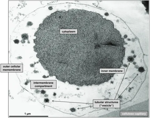
Figure 5: Transmission electron micrograph from I. hospitalis and N. equitans (ultrathin sections). ... 11
Figure 6: Ignicoccus and the mode of life. ... 12
Figure 7: Ultrathin section from an I. hospitalis cell shown as transmission electron micrograph. 13 Figure 8: Solar radiation spectrum reaching Earth´s surface compared to the action spectrum predominating on early Earth. ... 15
Figure 9: Schematic representation of ionizations caused by radiation with low, or high LET ... 16
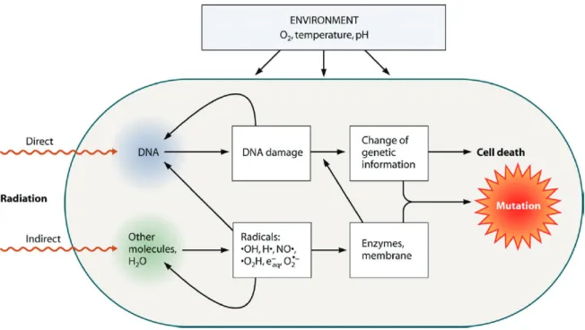
Figure 10: Damages on cellular level caused by ionizing radiation following direct or indirect interactions ... 17
Figure 11: Different types of DNA damages caused by either radiation or chemical agents. ... 18
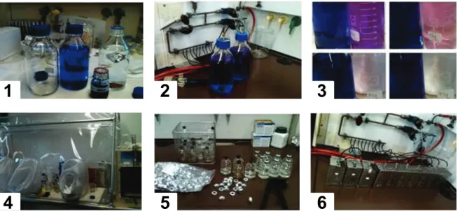
Material and Methods Figure 12: Exemplaric illustration of ½ SME+S0 medium preparation. ... 28
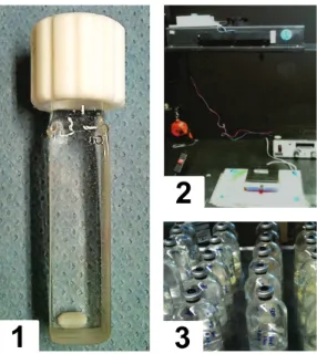
Figure 13: Serum bottle containing 20 ml ½ SME+S0 medium, and syringe with 0.6 x 30 mm needle used for inoculation. ... 29
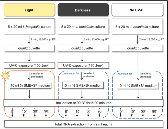
Figure 14: Experimental setup for UV-C exposure in liquid suspension. ... 33
Figure 15: Experimental setup for photoreactivation... 34
Figure 16: Schematic representation of photoreactivation experiment. ... 35
Figure 17: Experimental setup for X-ray exposure in liquid suspension. ... 37
Figure 18: Schematic representation of exposure/reference bucket. ... 38
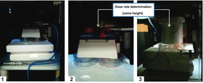
Figure 19: Dose rate determination for hot exposure experiment. ... 39
Figure 20: Schematic representation and comparison of experimental setups designed for the first and second radiation campaign. ... 42
Figure 21: Determination of gray-levels. ... 53
Results Figure 22: Absorption spectrum of different ½ SME medium combinations. ... 56
Figure 23: Survival curve of all Ignicoccus representatives after UV-C exposure (0-300 J/m2). ... 57
Figure 24: Survival curves of all Ignicoccus representatives after UV-C exposure (0-3000 J/m2). . 58
Figure 25: Survival curve of UV-C exposed I. hospitalis cells compared to E. coli cells exposed in either cuvettes or petri dishes. ... 59
Figure 26: Agarose gel of extracted genomic DNA from I. hospitalis and “I. morulus”. ... 60
Figure 27: Relative amount of DNA lesions vs. survival, and genomic DNA integrity of I. hospitalis after UV-C exposure... 61
Figure 28: Analysis of heavy ion exposed samples (I. hospitalis) on agarose gels. ... 63
Figure 29: 60Co radiation exposed stationary phase cultures of I. hospitalis. ... 65
Figure 30: Metabolic activity of I. hospitalis (IH1, IH2, IH3) after 60Co radiation exposure monitored on lead acetate paper. ... 66
Figure 31: Survival curve of I. hospitalis and “I. morulus” after 60Co radiation exposure. ... 67
Figure 32: Discrimination between culturable, and viable but nonculturable (VBNC) state. ... 69
Figure 33: Microscopic images of I. hospitalis cells at 1000-fold magnification. ... 69
Figure 34: Metabolic activity of three independent I. hospitalis stationary phase cultures
(IH1, 2, 3) after gamma ray exposure monitored on lead acetate tape. ... 70
Figure 35: Impact of 60Co radiation exposed ½ SME+S0 medium on the survival of I. hospitalis. ... 72
Figure 36: Relative survival of I. hospitalis when cultivated in 60Co radiation exposed or unexposed ½ SME medium supplemented by elemental sulfur. ... 73
Figure 37: Aliquots of substances needed for ½ SME-S0 medium preparations. ... 74
Figure 38: Quorum sensing with 60Co radiation exposed stationary phase cells of I. hospitalis. ... 76
Figure 39: Quorum sensing with samples from the last bottle giving a positive signal within the serial dilution after 60Co radiation exposure. ... 76
Figure 40: Analysis of the genomic DNA integrity of I. hospitalis after 60Co radiation exposure. .... 78
Figure 41: Analysis of the genomic DNA integrity of “I. morulus” after 60Co radiation exposure. .... 78
Figure 42: Survival of I. hospitalis after X-ray exposure with and without the use of a 0.1 mm Al filter. ... 80
Figure 43: HPLC vials. ... 80
Figure 44: Survival curve of I. hospitalis after X-ray exposure. ... 81
Figure 45: Survival curve of I. hospitalis after X-ray exposure when cultivated below/above Topt. .. 82
Figure 46: Survival curve of I. hospitalis after X-ray exposure at ~88 °C... 83
Figure 47: DNA repair of I. hospitalis after X-ray exposure. ... 84
Figure 48: Agarose gel of extracted total RNA. ... 86
Figure 49: qRT-PCR of X-ray (1500 Gy) exposed I. hospitalis cells using the sequence specific recB primer. ... 87
Figure 50: Recorded melting curves for gene specific primers (16S rRNA, Pol E´, Mips, Thermosome) using cDNA from experimental condition A ... 88
Figure 51: RNA transcription levels represented by absolute Ct values for different experimental conditions. ... 90
Figure 52: Standard curve generated with genomic DNA of I. hospitalis using the gene specific primer recB. ... 91
Figure 53: Calculated molecule or copy numbers for rad2, rad50, recB, and radA. ... 92
Figure 54: RNA transcription levels for genes involved in replication are represented by absolute Ct values using cDNA from experimental condition E. ... 93
Discussion Figure 55: Bacterial survival curve after UV-C exposure. ... 96
Figure 56: Different types of membranes used for sterile filtration... 106
Figure 57: Chemical substances with strongest color change after 60Co radiation exposure (117.1 kGy). ... 106
Figure 58: Colloidal sulfur. ... 108
Figure 59: Potential scenario for the origin and early evolution of life. ... 119
Appendix Figure 60: Test of primers ... 151
Figure 61: Metabolic activity of "I. morulus" (IM1, IM2, IM3) monitored on lead acetate paper ... 152
Figure 62: Aliquots of substances needed for ½ SME-S0 medium preparations. ... 153
Figure 63: Certificate of irradiation for DbR #1. ... 154
Figure 64: Certificate of irradiation for DbR #2. ... 155
Figure 65: Certificate of irradiation for DbR #3. ... 156
List of Tables
Material and Methods
Table 1: Chemicals used for experimentation. ... 21
Table 2: Primer used for RAPD analysis. ... 23
Table 3: Ignicoccus hospitalis specific primers used for qRT-PCR (housekeeping, DNA repair) . 24 Table 4: Ignicoccus hospitalis specific primers used for qRT-PCR (replication) ... 25
Table 5: Ignicoccus hospitalis specific primers used for qPCR to detect DNA damages after 60Co radiatin exposure ... 25
Table 6: Impact of 0.1 mm Al filter vs. filter-less exposure on the dose rate in dependence of an additional HPLC filter (here: glass). ... 36
Table 7: X-ray dose applied with or without 0.1 mm Al filter. ... 37
Table 8: Different experimental setups for qRT-PCR gene expression studies after X-ray exposure. ... 41
Table 9: Preparation of ½ SME medium from single components which were exposed to increasing 60Co radiation dose. ... 43
Table 10: Preparation of ½ SME medium from unexposed single components. ... 43
Table 11: Preparation of ½ SME medium (60Co radiation exposed or unexposed) which was supplemented by different sulfur combinations (dry/wet, exposed/unexposed). ... 44
Table 12: Scheme of steps needed for genomic DNA extraction. ... 46
Table 13: Scheme of steps needed for total RNA extraction. ... 48
Table 14: RAPD cycles. ... 51
Table 15: qPCR program for DNA damage detection.. ... 51
Results Table 16: qRT-PCR program for gene expression studies after stress exposure. ... 54
Table 17: Calculated F10-values for different Ignicoccus representatives. ... 58
Table 18: F10-values for model organisms. ... 58
Table 19: Calculated D10-values (60Co radiation) for different Ignicoccus representatives. ... 67
Table 20: D10-values (60Co radiation) for model organisms ... 67
Table 21: D10-values (60Co radiation) for I. hospitalis, serial diluted in exposed ½ SME+S0 medium or serial diluted prior to exposure in (unexposed) ½ SME+S0 ... 72
Table 22: Preparation of ½ SME medium from single components which were exposed to increasing 60Co radiation dose. ... 74
Table 23: Gray-levels of RAPD bands. ... 85
Table 24: RNA transcription levels of tested housekeeping genes listed as absolute Ct values for different experimental conditions. ... 88
Table 25: RNA transcription levels of Pol E´ (potential housekeeping gene), and photolyase listed as absolute Ct values. ... 94
Abbreviations
(6-4)PPs pyrimidine(6-4)pyrimidone photoproducts
“I. morulus” Ignicoccus morulus
°C degree centigrade
½ SME ½ synthetic sea water
½ SME+S0 medium ½ SME medium supplemented with elemental sulfur
½ SME-S0 medium sulfur-free ½ SME medium A. fulgidus Archaeoglobus fulgidus
Al aluminum
AP site apurinic/apyrimidinic site
BER base excision repair
BGS Beta Gamma Service
bp base pair
CFU colony forming unit
CPDs cyclobutane-pyrimidine dimer
Ct threshold cycle
D. radiodurans Deinococcus radiodurans
DbR Death by Radiation
ddH2O double distilled water
DLR Deutsches Zentrum für Luft- und Raumfahrt (German Aerospace Center)
DNA deoxyribonucleic acid
dsDNA double-strand DNA (breaks) E. coli Escherichia coli
Fe iron
for forward primer
Ga billion years
Gy gray
H. salinarum Halobacterium salinarum
HR homologous recombination
I. hospitalis Ignicoccus hospitalis I. islandicus Ignicoccus islandicus I. pacificus Ignicoccus pacificus
ICP-MS inductively coupled plasma mass spectrometry J/m2 joule per square meter
kGy kilo gray
kV kilo volt
LET linear energy transfer
LUCA last universal common ancestor
Mn manganese
MPN most probable number (technique)
NCBI National Center for Biotechnology Information NER nucleotide excision repair
NHEJ non-homologous end joining
O/N over night
P. furiosus Pyrococcus furiosus PBS phosphate-buffered saline PCR polymerase chain reaction PFGE pulsed-field gel electrophoresis qPCR quantitative real-time PCR
qRT-PCR quantitative Reverse Transcription-PCR RAPD randomly amplified polymorphic DNA RBE relative biological effectiveness
rev reverse primer
RIN RNA integrity number
RNA ribonucleic acid
ROS reactive oxygen species
RT room temperature
S. solfataricus Sulfolobus solfataricus ssDNA single-strand DNA (breaks) T. gammatolerans Thermococcus gammatolerans
Topt optimal growth temperature
UV ultraviolet
v/v volume per volume
VBNC viable but nonculturable
Abstract
The environmental conditions on early Earth were harsh and hostile for life compared to environmental conditions prevailing on present Earth. The atmosphere during the Archaean Age (3.8-2.5 Ga ago) was essentially anoxic and the lack of an UV-absorbing ozone layer enabled the solar ultraviolet radiation spectrum to penetrate Earth´s surface increasing the overall terrestrial UV stress. In addition, elevated radiation levels in terms of ionizing radiation contributed to this rugged terrestrial environment. The Late Heavy Bombardment of the planet took place heaviest until about 3.8 Ga and may have heated up the ocean partially over 100 °C. Nevertheless, life has evolved during the Archaean under these circumstances inhabiting our planet since about 3.8 Ga. The potential setting under which life has evolved fascinates and still encourages humans to think about it.
Different ideas, hypothesis and opinions about the Last Universal Common Ancestor (LUCA) and essential abilities needed for the propagation of life are still under debate.
The underlying work emphases a hot origin of life and focuses on representatives of the genus Ignicoccus isolated from (deep-sea) hydrothermal vents. All representatives of this genus belong to the crenarchaeal branch and follow a hyperthermophilic, chemolithoautotrophic mode of life, living as obligate anaerobes growing by sulfur reduction. Ignicoccus species are promising candidates for early Earth inhabitants because they combine several abilities which may have been advantageous to withstand early Earth´s harsh environmental conditions including elevated levels of radiation.
Results of this work show that Ignicoccus species tend to survive high doses of ionizing radiation. This observation was the starting point to investigate the resistance of all four representatives of this genus with respect to different radiation types, ionizing radiation (X- rays, γ-rays) and non-ionizing radiation (UV-C). All tested species demonstrated similar inactivation tendencies after non-ionizing radiation exposure resulting in a F10-value of
~300 J/m2. Additionally, I. hospitalis and “I. morulus” showed a high tolerance to ionizing radiation exposure with a D10-value of ~5 kGy. Besides this impressive radiation tolerance, it was possible to demonstrate for the first time, that a so called VBNC (viable but nonculturable) state may also exist for Archaea after ionizing radiation exposure. Viable and culturable cells were microscopically observed after exposure to 60Co radiation doses of <19 kGy, passing a transition state, and reaching the VBNC state after doses of
>27.2 kGy. This observed VBNC state was ascribed to the ongoing metabolic activity, thus H2S production could be monitored. Additional experiments showed that the ionizing radiation tolerance of I. hospitalis seemed to be unaffected by pre-cultivation temperature and the temperature during radiation exposure. However, the tolerance of I. hospitalis to ionizing radiation accompanied by active repair of radiation induced DNA damages was
investigated in more detail. It was shown that the PCR-based randomly amplified polymorphic DNA (RAPD) analysis method was a powerful tool to visualize radiation induced DNA damages thus inferring genomic DNA integrity. This method allowed monitoring the DNA repair over time. It was demonstrated that the overall genome integrity was highly affected by both types of radiation and that RAPD analysis represents an attractive alternative for the commonly used and time consuming pulse-field gel electrophoresis (PFGE). I. hospitalis showed fast DNA repair after ionizing radiation exposure; the repair seemed to be completed within one hour. Due to the fact that I. hospitalis was able to withstand these high radiation doses, it was of great interest to investigate whether classical genes involved in DNA repair (e.g. rad2, rad50, recB and radA) were up- or down-regulated upon irradiation. Gene specific primers were designed for qRT-PCR studies and tested under varying experimental conditions. An upregulation of gene expression was detected for the genes mentioned above after 1500 Gy with I. hospitalis cells, when exposed in their early exponential phase. These promising results gave the first indication in regards to its radiation resistance capabilities and further investigation in terms of transcriptomics is definitely warranted. It is very likely that additional mechanisms may support this unusual high radiotolerance. Post-translational modifications for example would point to a completely new way of thinking in terms of the radiation tolerance of I. hospitalis and would allow the regulation of potentially high levels of repair proteins present due to its hot lifestyle. The surprisingly high radiotolerance may also be supported by a potential polyploidy, an increased genome copy number resulting in an enhanced resistance against DNA-damaging conditions. Nevertheless, I. hospitalis has not yet been observed in terms of post-translational modifications or polyploidy; these are promising experiments for follow-up studies. All underlying results obtained with these studies add new pieces to the puzzle how life on Earth may have evolved and the successful propagation under harsh and life hostile conditions.
Zusammenfassung
Die Umweltbedingungen der frühen Erde, vor 3,8-2,5 Milliarden Jahren, waren im Vergleich zu den heutigen Umweltbedingungen hart und lebensfeindlich. Die Atmosphäre während des Archaikums war nahezu sauerstofflos und die UV-absorbierende Ozonschicht fehlte. Das solare ultraviolette Strahlenspektrum konnte ungehindert in die Erdoberfläche eindringen. Dieses hohe Strahlungsniveau wurde begleitet von einem ebenfalls erhöhten Anteil an ionisierender Strahlung, welche zu diesen schwierigen Umweltbedingungen maßgeblich beitrugen. Zudem führte das große Bombardement, das vor ca. 3,8 Milliarden Jahren sein Maximum erreichte, dazu, dass der Ozean durch die Meteoriteneinschläge teilweise auf über 100 °C erhitzt wurde. Dennoch hat sich das Leben unter den oben genannten Bedingungen während dieses Erdzeitalters entwickelt und besiedelt bis heute erfolgreich unseren Planeten. Diese Tatsache fasziniert Menschen noch immer und ermutigt sie, über die möglichen Umstände nachzudenken, unter denen sich das Leben entwickelt hat. Unterschiedliche Ideen, Hypothesen und Meinungen über den letzten gemeinsamen Vorfahren (LUCA) und seiner notwendigen Fähigkeiten, damit sich das Leben verbreiten konnte, werden noch immer kontrovers diskutiert.
Die zu Grunde liegende Arbeit stützt sich auf die Theorie eines heißen Ursprungs des Lebens. Die Stellvertreter der Gattung Ignicoccus, die von hydrothermalen Quellen isoliert wurden, werden im Nachfolgenden als mögliche Bewohnr der frühen Erde betrachtet. Alle Vertreter dieser Gattung gehören der crenarchaellen Abzweigung des phyolgenetischen Stammbaums an. Sie folgen als obligate Anaerobier einer hyperthermophilen, chemolithoautotrophen Lebensweise und gewinnen ihre Energie mit Hilfe der Schwefelreduktion. Diese Mikroorganismen vereinen mehrere Fähigkeiten, die vorteilhaft waren, um den damals vorherrschenden Umweltbedingungen, insbesondere der erhöhten Strahlungsintensitäten, zu trotzen. In dieser Arbeit wurde gezeigt, dass Ignicoccus Spezies hohe Dosen ionisierender Strahlung überleben können. Diese Beobachtung war ausschlaggebend für nachfolgende Untersuchungen mit allen bisher bekannten Vertretern dieser Gattung in Bezug auf ihre Toleranz gegenüber unterschiedlicher Strahlungsarten.
Alle untersuchten Spezies zeigten vergleichbare Inaktivierungstendenzen gegenüber nicht ionisierender Strahlung resultierend in F10-Werten von ~300 J/m2. Zudem zeigten I. hospitalis and “I. morulus“ eine hohe Strahlungstoleranz gegenüber ionisierender Strahlung resultierend in D10-Werten von ~5 kGy. Neben dieser beeindruckenden Strahlentoleranz war es zudem möglich zu zeigen, dass ein sogenannter VBNC Statuts (viable but nonculturable) nach Exposition gegenüber ionisierender Strahlung auch für
Archaeen zu existieren scheint. Lebensfähige und damit kultivierbare Zellen konnten nach
60Co Exposition mit Dosen <19 kGy beobachtet werden. Dem als Übergangszustand definierten Bereich folgte der VBNC Status nach Exposition mit Dosen >27,2 kGy. In diesem VBNC Status konnte eine fortlaufende metabolische Aktivität in Form von H2S Produktion verfolgt werden. Zusätzliche Experimente konnten zeigen, dass die Toleranz von I. hospitalis gegenüber ionisierender Strahlung unbeeinträchtigt von der Temperatur während der Anzucht und der Temperatur während des Experiments war.
Die Toleranz von I. hospitalis gegenüber ionisierender Strahlung einhergehend mit aktiver Reparatur von strahleninduzierten DNS-Schäden wurde im Detail untersucht. Es konnte gezeigt werden, dass die PCR-basierte RAPD (randomly amplified polymoprhic DNA)- Methode sehr gut geeignet ist, um strahleninduzierte DNS-Schäden zu veranschaulichen und erlaubt den Verlauf der Reparatur zu verfolgen. Somit stellt diese Methode eine attraktive Alternative zu der häufig verwendeten und zeitintensiven Puls-Feld- Gelelektrophorese (PFGE) dar. Es wurde demonstriert, dass die gesamte Integrität des Genoms stark durch beide Arten von Strahlung negativ beeinflusst wurde. I. hospitalis zeigte indes eine schnelle DNS-Reparatur nach ionisierender Strahlung. Basierend auf vorliegenden Experimenten wurde demonstriert, dass diese Reparatur binnen einer Stunde vollzogen war. Da gezeigt werden konnte, dass I. hospitalis fähig ist, hohe Dosen ionisierender Strahlung zu überleben, war es von besonderem Interesse, die Regulierung von klassischen Reparaturgenen wie bspw. rad2, rad50, recB und radA nach Bestrahlung zu betrachten. Gen spezifische Primer wurden für diesen Zweck entworfen, um qRT-PCR Studien durchführen zu können und die Expression dieser Gene unter variierenden experimentellen Bedingungen zu untersuchen. Eine leichte Hochregulierung wurde nach Exposition mit 1500 Gy in I. hospitalis Zellen gesehen, die sich in ihrer frühen exponentiellen Phase befanden. Diese vielversprechenden Ergebnisse geben einen ersten Eindruck auf die Strahlentoleranz von I. hospitalis und der Expression von Reparaturgenen. Sie ermutigen dazu, weitere Untersuchungen in Bezug auf das Transkriptom durchzuführen. Es ist anzunehmen, dass zusätzliche Mechanismen diese Strahlentoleranz unterstützen. Posttranslationale Modifizierungen würden auf eine komplett neue Denkweise in Bezug auf die Strahlungstoleranz von I. hospitalis hinweisen. Diese Modifizierungen würden eine Regulation von Reparaturgenen, die potenziell bereits in hohem Maße aufgrund der heißen Lebensweise vorliegen, erlauben.
Diese überraschend hohe Strahlentoleranz dürfte auch eine mögliche Polyploidie unterstützen, da eine erhöhte Genomkopienzahl bei einer erhöhten Toleranz gegenüber DNS zerstörenden Bedingungen von Vorteil sein könnte. Nichtsdestotrotz, bis jetzt wurden weder eine mögliche Polyploidie noch posttranslationale Modifizierungen
untersucht. Diese Untersuchungen wären äußerst interessant für Folgestudien. Alle zu Grunde liegenden Ergebnisse fügen dem Gesamtbild, wie das Leben auf der Erde entstanden sein könnte, neue Puzzleteile hinzu. Dies führt zu potenziellen Antworten auf die Frage warum eine erfolgreiche Verbreitung unter den vorherrschenden harten und lebensfeindlichen Bedingungen auf der frühen Erde möglich war.
1 Introduction 1.1 Life on early Earth
Earth has been inhabited since the Archaean Age, and terrestrial life has been represented since about 3.8 Ga or earlier. The main biochemical carbon cycle developed around 3.5 Ga ago and is in use to present day (according to Nisbet & Sleep, 2001). The prevailing conditions on early Earth, the circumstances and potential settings under which life has evolved, fascinates and still encourages humans to think about it. A vast number of possible scenarios has been developed over the past decades, resulting in enduring debates and discussions, supported or disproved by e.g. geochemical and isotopic evidence. A commonly accepted hypothesis concerning life´s origin and evolution has not been established yet; the debate is still ongoing. Several ideas and hypotheses in regards to how life could have evolved are shortly described in the following few Sections, strongly emphasizing a “hot origin” of life on our “Blue Planet”.
1.1.1 Environmental conditions on early Earth
The environmental conditions as they prevailed on early Earth were harsh and hostile for life as compared to environmental conditions on present Earth. The atmosphere during the Archaean Age (approx. 3.8-2.5 Ga ago) was essentially anoxic which can be deduced from several geochemical and isotopic studies (Grenfell et. al, 2010; Holland, 1999). As a consequence, the UV-absorbing ozone layer was absent, enabling the solar ultraviolet radiation spectrum to penetrate to Earth´s surface, and thus increased as a result the overall UV stress on the Earth´s surface (Cockell & Horneck, 2001; according to Margulis et al., 1976) (Figure 1). Environmental conditions did not only influence early Earth´s atmosphere but the prevailing ocean was significantly affected as well. The Late Heavy Bombardment of the planet during the early Archaean, heaviest until about 3.8 Ga, may have heated up the ocean partially over 100 °C (according to Nisbet & Sleep, 2001).
Sleep reviewed the Hadean-Archaean environment on early Earth in more detail, and discussed three well-known scenarios in which life may have evolved (Sleep, 2010).
These scenarios are described below:
1) In the 1st hypothesis, he assumed that the Hadean climate, including the Late Heavy Bombardment, (according to Sleep, 2010), was clement or icy after the early warm greenhouse ceased. Life originated and colonized the planet by e.g. adapting to a thermophilic mode of life at hydrothermal events and in the kilometer-deep surface. Such
an adaptation would have been beneficial after large asteroid impacts have boiled much of the ocean. Descendants of these thermophilic survivors may have adaptively colonized low-temperature environments as well. Proteins adapted to high temperatures may have also been lost over the course of time in the low-temperature branch (according to Sleep, 2010).
2) The 2nd hypothesis describes a scenario in which a comparable phylogeny occurred except that the thermophilic organisms may have outcompeted their low-temperature relatives. This event may have resulted in an apparent LUCA (Last Universal Common Ancestor) bottleneck without a sudden mass extinction (according to Miller and Lazcano, 1995; according to Sleep, 2010).
3) Finally, the 3rd hypothesis was focused at the end of the Hadean Age (~3.3 Ga) in which the climate cooled slowly when the CO2 greenhouse was ended leaving an overall temperature of approximately 50-70 °C. Only thermophiles therefore were able to exist (Gaucher et al., 2008, 2010).
Sleep inferred that the first two possibilities had similar genetic and geologic implications until recent evidence of a Hadean massive impact has been found. But the accessible Archaean geologic record seems to support the third scenario, potentially eliminating the other two (according to Sleep, 2010). The discussion about early Earth´s harsh environmental conditions, including the divergent opinions about the last universal common ancestor, allows the assumption of LUCA preferring a thermophilic lifestyle (Gaucher et al., 2010; Sleep, 2010; Stetter, 2006). Based on this idea, one can think about microbial communities that inhabited the mid-ocean ridges, volcanic ocean islands and island-arc volcanoes, a chain of volcanoes being arranged in an arc-shaped manner (Nisbet, 2000). These microbial communities may have been dependent upon igneous activity, hydrothermal circulations and systems to provide chemical disequilibria (Sleep, 2010). Living in a sufficient depth of water may have been advantageous for the evolution and propagation of these communities by preventing them from solar ultraviolet light and its harmful effects.
Figure 1: Schematic representation of environmental conditions present on early Earth.
1.1.2 Radiation levels on early Earth
Solar ultraviolet radiation, harmful to all biological material, is absorbed in the modern terrestrial atmosphere by ozone, which is photochemically produced from atmospheric oxygen. This ozone layer has gradually developed as oxygen accumulated over the course of history (first Great Oxidation Event, 2.45 Ga), and by the later development of photosynthesis, which enabled the organisms to directly use the Sun´s energy for their own purposes (according to Sessions et al., 2009; according to Margulis, 1976). Early Earth´s surface has not only been subjected to the solar ultraviolet radiation spectrum (referred as non-ionizing radiation), naturally occurring ionizing background radiation has been present since its formation. But, overall radiation levels in terms of UV-light and ionizing background radiation have decreased over time, while the oxygen concentration has increased (Kasting, 1993; Holland, 1994; Karam et al., 2001). Background beta and gamma radiation levels including radiation doses from geologic material and internal emitters have changed significantly over time with an maximal ambient radiation level of about 6 mGy y-1 4 Ga ago (Karam & Leslie, 1999; Karam et al., 2001); ambient radiation levels have decreased steadily resulting in a present average exposure due to natural background radiation of around 3 mGy y-1 (Karam et al., 2001). Sources for background radiation include radioactive elements and their decay in Earth´s crust such as uranium, thorium, potassium, radium, radon, and others. Radiation from the sun and other stars or cosmogenic nuclides formed through the interaction of cosmic rays with atmosphere and
surface rocks take an important role, too. Internal radionuclides, primarily represented by
40K, are another source of radiation exposure within organisms through biochemical reactions (Karam et al., 2001).
Karam et al. hypothesized in 2001 that mutation rates may not necessarily be in direct proportion to rates of DNA damage at low exposure rates; with long intervals between damaging events, cells may have had the capacity to respond. In general, mutations can have several distinct effects for an organism and its potential offspring. They may be beneficial for an organisms´ survival or remain silent, being without any consequence.
Unfavorable or lethal effects are possible as well, if undetected by the organism.
Nevertheless, the type of response would be on a cellular level by mechanisms being able to fully repair caused DNA damages with unfavorable consequences (see Paragraph 5);
prevention from being mutagenic, and transmitted to the next generation (Karam et al., 2001). Therefore, life relies on mutation repair mechanisms, assuming that DNA repair pathways may have evolved very early in the history of life (Mackinodan & James, 1990), because of similarities in disparate modern organisms. Repair mechanisms may have retained the ability to efficiently and accurately repair higher rates of DNA damages than exist at present day (Karam et al., 2001). Summarizing, the rise of prevailing oxygen levels, the formation of the ozone layer in the upper atmosphere, the resulting absorption of harmful UV radiation, and the overall decrease in background radiation enabled life to further evolve and to colonize this planet. So far, Earth is the best known example to study how life modified and still modifies a planet´s atmosphere over time resulting in a co- evolution of life, atmosphere, and the terrestrial climate (Grenfell et al., 2010).
1.1.3 Where LUCA may have felt at home
The idea that life may have emerged in hydrothermal environments is quite attractive (according to Nisbet & Sleep, 2001) while controversially discussed to the present day.
Deamer and Georgiou compared conditions and properties of deep-sea hydrothermal vents and hydrothermal fields of volcanic origin above sea level in respect to their ability to support the evolution of early life (Deamer & Georgiou, 2015). As an example, alkaline vents last up to 30,000 years, providing a constant supply of chemical energy at temperatures of 50-90 °C, whereas light energy is abundant in hydrothermal fields allowing the development of photosynthesis. The hydrothermal field theory would suggest that early life quickly evolved mechanisms to capture this light-driven chemiosmotic energy for reactions (according to Deamer, 1997; Deamer & Weber, 2010), but life in alkaline vents would depend on chemotrophic reactions (Deamer & Georgiou, 2015).
Deamer and Georgiou criticized that none of the predictions mentioned above have ever been tested in situ by experiments and laboratory simulations. The plausibility of these two potential sites for the origin of life requires testing; the ensuing discussion about the setting of life´s evolution will remain ongoing (for detailed review see Gaucher et al., 2010).
1.1.4 LUCA´s potential mode of life
The discussion whether the first organisms on early Earth were (at least) hyperthermophiles is still ongoing. Di Giulio discussed in 2000 whether LUCA was a progenote or a cenancestor. A progenote is described as primitive entity being still in process of evolving its relationship between phenotype and genotype (according to Woese & Fox, 1977; Di Giulio, 2000), whereas a cenancestor would be an organism with cellular complexity which still had to solve the problem of the genotype-phenotype relationship (Di Giulio, 2000). A correspondence between the physical setting in which life originated and LUCA´s first speciation took place has to be considered when thinking about a progenote. Di Giulio worked on estimations of the G+C content in ancestral rRNA sequences of the LUCA and decided, if LUCA lived in a high-temperature setting, the origin of life may have taken place at high temperature in relation to a progenote (Di Giulio, 2000). Taking a thermophily index into consideration when analyzing the propensity of amino acids to enter thermophile/hyperthermophile proteins, Di Giulio described the last universal common ancestor as “hot LUCA” (Di Giulio, 2001). Seven years later he tried to reconstruct the ancestral sequences of proteins in regard to an oxyphobic index and concluded based on his observations that the origin of life and the main phase of the evolution of the first organisms on early Earth may have taken place in an anaerobic environment (Di Giulio, 2007). Nevertheless, the methods for reconstructing ancestral sequences have to be improved/perfected to give “definite” answers to these questions (Di Giulio, 2001, 2010, 2011).
Another topic controversially discussed is the evolution of the sulfur cycle. Nisbet and Sleep discussed in 2001 that the microbial fraction of sulfur was limited during the Archaean based on isotopic evidence resulting in potentially low sulfate concentrations (supported by Habicht & Canfield, 1996; Canfield & Teske, 1996). Isotopic evidence in 2.7 Ga rocks of the Belingwe belt in Zimbabwe may have inferred however that the full sulfur cycle evolved earlier than originally predicted (Grassineau et al., 2001). Woese suggested in 1987 that the ancestral archaebacterium (today: archaeon) was an extremely thermophilic, anaerobic living microorganism that probably thrived from sulfur
reduction. It is conceivable that a microbial diversity of sulfur consumers was present quite early in Earth´s history, but a global distribution subsequently took place later on (Canfield et al., 2000). Rasmussen reported in a Nature article published in 2000 that there is evidence for life in a 3,235-million-year-old deep-sea volcanogenic massive sulfide deposit from the Pilbara Craton of Australia (Rasmussen, 2000). He presented pyritic filaments as probable fossil remains of thread-like microorganisms, and assumed that these fossils represented the first evidence for life in a Precambrian submarine thermal spring system. Rasmussen suggested that these microorganisms were probably thermophilic chemotrophic prokaryotes. Thus, recent microorganisms found in close proximity to modern hydrothermal vent systems are reasonable candidates for being potential early Earth inhabitants.
1.1.5 LUCA´s descendants
The universal phylogenetic tree (exemplarily depicted in Figure 2) and the standard model of microbial descent is based on small-subunit ribosomal RNA, positioning LUCA at the root of this tree (according to Nisbet & Sleep, 2001; Woese, 1987; Doolittle, 2000). One appealing idea, shared by e. g. Woese, Graham and colleagues, is of an early population of simple, replicating organisms within a community sharing mutually information instead of a single cell representing LUCA (Woese, 1987; Graham et al., 2000). An organism´s genes were exchanged with others and were in effect shared communally. This proposed model of genomic evolution is based on the successive “crystallization” of differentiated cellular subsystems (Graham et al., 2000; Woese, 1998). Evolution enabled continuing divergence making genes less interchangeable later on (Graham et al., 2000). Based on this idea of LUCA, being part of an early community, most scientists have one common opinion (“standard view”) (according to Nisbet & Sleep, 2001; Zuckerkandl & Pauling, 1965; according to Pace, 1991) concerning the three domains of life. Both domains, Bacteria and Archaea, arose from LUCA, whereas the domain Eukarya evolved slowly from a parental stem that symbiotically incorporated chloroplasts (cyanobacterial descendants) and mitochondria (α-proteobacterial descendants) (Figure 2) (according to Nisbet & Sleep, 2001; Woese et al., 1990; Margulis, 1971).
Two popular hypotheses include the assumption of a hot environment, and are nicely reviewed by Nisbet and Sleep (2001), although evidence for a hyperthermophile ancestry was challenged (Galtier et al., 1999). There is the “hyperthermophile Eden” theory implying an early microbial community hosting the last common ancestor, in which life was hot and chemotrophic (according to Nisbet & Sleep, 2001; Stetter, 1996). An alternative
version would be the “hyperthermophile Noah” theory describing a not necessarily hyperthermophilic universal ancestor. But this version of LUCA may have diversified from an unknown Eden into an early population that included some hyperthermophiles near hydrothermal systems (according to Nisbet & Sleep, 2001).
Keeping a potential hot origin in mind, the shortest branches within the universal phylogenetic tree as depicted in Figure 2 are exclusively covered by hyperthermophilic prokaryotes (red branches) and cluster around the phylogenetic root (Figure 2). Deep branches give evidence for an early separation, whereas short phylogenetic branches indicate a slow rate of evolution. One has to keep in mind that the constructed phylogenetic trees tend to give ideas of the phylogenetic distance between recent organisms (e.g. Figure 2) rather than their evolutionary development (Hug et al., 2016).
Figure 2: Phylogenetic tree based on 16S rRNA sequence comparisons. The phylogenetic tree was constructed based on sequence comparisons of small subunit ribosomal RNA of cultivable microorganisms.
The red branches represent hyperthermophiles. The eukaryal branch has subsequently been reduced (adapted from Stetter, 2006; credit Dr. Harald Huber). Ignicoccus is highlighted in blue. The two recently proposed archaeal phyla Thaumarchaeota (Brochier-Armanet et al., 2008) and Lokiarchaeota (Spang et al., 2015) have not been taken into account.
The energy source of hyperthermophiles is quite simple as most gain their energy chemolithoautotrophically by fixation of CO2 serving as carbon source for organic cell material (Figure 3; Stetter, 2006).
Eukarya
Ignic- coccus
Figure 3: Main energy-yielding reactions used by chemolithoautotrophic hyperthermophiles.
Highlighted in blue: Donor, acceptor, product and other sources used by Ignicoccus (adapted from Stetter, 2006).
A detailed description of the anaerobic, chemolithoautotrophic, hyperthermophilic lifestyle will be described in detail within Section 1.3.2 using the example of Ignicoccus hospitalis.
Pace proposed in 1991 that modern representatives of these evolutionary primitive organisms (he used examples from the genera Pyrodictium, Thermoproteus, Pyrobaculum (according to Stetter et al., 1990)) have similar requirements. They need high temperatures in their natural habitat besides geochemical energy sources such as sulfur and hydrogen to live mainly anaerobic in their geothermal environment (according to Pace, 1991). Common properties of modern organisms were properties of the ancestor, meaning that least-evolved Archaea might share similar properties with their ancestors (earliest Archaea) (according to Pace, 1991). The conclusion of Karam and colleagues in 2001 was that direct evidence concerning the conditions under which life evolved was nearly absent, and that the indirect evidence we currently possess is strongly dependent upon varying interpretations. The ancient radiation environment may have caused an evolutionary selection, and modern organisms may have acquired characteristics advantageous for this selection process (Karam et al., 2001). These properties might have pertained to the most recent common ancestor of all modern life (according to Pace, 1991; Woese, 1987). So, it is reasonable to investigate a modern organism´s ability to tolerate harsh environmental conditions as they occurred on early Earth.
Hydrothermal systems are promising sites where the evolution of life may have started.
Modern hydrothermal vents are a good starting point to look for appropriate early Earth inhabitants. Interesting candidates represented by the genus Ignicoccus were found in
energy-yielding reactions ofhyperthermophiles
electron donor electron acceptor product methane magnetite
hydrogen sulfide (H2S) nitrogen (NH3) water
source of cell carbon:
additional growth requirements:
CO2 heat
trace minerals liquid water
• CO2
• Fe(OH)3
• S0; SO42-
• NO3-
• O2traces H2
S0(pyrite) • O2 H2SO4(+FeSO4)
submarine hydrothermal systems and were described in 2000 by Huber and colleagues.
Members of the genus are a focus of attention and their tolerance to different environmental parameters of astrobiological relevance will be presented in the underlying work.
1.2 Hydrothermal vents
Hydrothermal vent systems were discovered during the early 1980s (according to Martin et al., 2008), and identified as chemically reactive environments, with thermal and chemical gradients composed of reactive gases and dissolved elements (Baross &
Hoffmann, 1985). Deep-sea hydrothermal vent deposits are formed by precipitations of these minerals present in hot, reduced, metal-rich fluids, accumulating on the seafloor (Pagé et al., 2008; Haymon, 1983). These reactive environments were recognized as potential sites for prebiotic syntheses (Baross & Hoffmann, 1985).
The Faulty Towers complex in Figure 4 can be seen as an imposing example of a black smoker complex (according to Martin et al., 2008). This black smoker complex, and others, can be found directly above magma chambers 1-3 km under sea level (for detailed explanation see review article by Kelley et al., 2002). Black smoker chimneys emit mineral enriched sea water with temperatures of up to 405 °C (Von Damm et al., 2003). Escaping water comes into close contact with the magma chamber, and re-emerges at the vents after circulating from the ocean floor through the crust resulting in black smoker fluids rich in dissolved transition metals (according to Martin et al., 2008; Von Damm, 2013). Besides high concentrations of Fe(II) and Mn(II), fluids contain high concentrations of dissolved gasses such as magmatic CO2 (4-215 mmol/kg), H2S (3-110 mmol/kg), H2 (0.1-50 mmol/kg), and CH4 (0.05-4.5 mmol/kg) formed by biotic and abiotic processes (Kelley et al., 2002). Microbial communities including chemolithoautotrophic microorganisms thrive on these metal enriched black smoker fluids. Temperature gradients between the hot interior of the smoker and the surrounding sea water enable them to adapt to specific environmental regions (Figure 4, according to Martin et al., 2008; Schrenk et al., 2003).
These chemolithoautotrophic species can gain their energy by distinct oxidation-reduction reactions under a wide range of temperature, caused by physical gradients and chemical disequilibria occurring in the (deep-sea) hydrothermal vent fields (McCollom & Shock, 1997). All four Ignicoccus species were isolated from such submarine hydrothermal fields or systems in the Atlantic and in the Pacific (Huber et al., 2000; Paper et al., 2007).
Detailed information on the places of isolation and the organisms themselves will be given in the following Sections.
Figure 4: Black smoker. (A) This black smoker was found in the Faulty Towers complex in the Mothra hydrothermal field on the Endeavour Segment of the Juan de Fuca Ridge and serves as general example for black smokers from which Ignicoccus was isolated (adapted from Martin et al., 2008). (B) Schematic representation of a black smoker chimney. The chimney is surrounded by 2 °C cold sea water and warm fluids escaping from the vent itself. Up-flow fluids can have temperatures exceeding 300 °C, but intermediate conditions exist as indicated by lines. Due to this temperature gradient, a diverse microbial community can be uncovered within the chimney walls (adapted from Martin et al., 2008; Pagé et al., 2008; Schrenk et al., 2003).
1.3 The genus Ignicoccus
The genus Ignicoccus was first described in 2000 by Huber et al. and consists of three described type species. All of them are members of the crenarchaeal branch within the domain of Archaea which was revealed by 16S rRNA sequence comparisons.
Taxonomically, Ignicoccus belongs to the order of Desulfurococcales and represents a deeply branching lineage within the family of the Desulfurococcaceae (Huber et al., 2000;
Huber & Stetter, 2001). Members of this genus are the only ones within this family gaining their energy as obligate chemolithotrophic sulfur reducers (Huber et al., 2000).
1.3.1 Ignicoccus islandicus and Ignicoccus pacificus
Two species of this new genus were isolated from submarine hydrothermal systems in the Atlantic and in the Pacific in 2000 (Huber et al., 2000). I. islandicus (Kol8T), the type species of this genus, was isolated from hot sediments at the Kolbeinsey Ridge (North of Iceland) in a depth of 103-106 m, whereas rocky black smoker material from the East Pacific Rise (9 °N, 104 °W; Depth: 2500 m) was used to enrich I. pacificus (LPC33T, LPC37). Their names were devoted to the places of isolation. Morphological and physiological characteristics which are exceptional for this genus will be explained using the example of Ignicoccus hospitalis.
B A