Contents lists available atScienceDirect
Fish and Shell fi sh Immunology
journal homepage:www.elsevier.com/locate/fsi
Full length article
Separate and combined e ff ects of neurotoxic and lytic compounds of
Alexandrium strains on Mytilus edulis feeding activity and hemocyte function
Virginia Angélica Bianchi
a,∗, Hendrick Langeloh
b, Urban Tillmann
b, Bernd Krock
b, Annegret Müller
b, Ulf Bickmeyer
b, Doris Abele
baLaboratorio de Ecotoxicología Acuática, INIBIOMA (CONICET-UNCo) - CEAN, ruta provincial N° 61, km 3, CCP 7, Junín de los Andes, 8371, Neuquén, Argentina
bAlfred Wegener Institute for Polar and Maine Research, Am Handelshafen 12, 27570, Bremerhaven, Germany
A R T I C L E I N F O
Keywords:
Blue mussel
Paralytic shellfish toxins Lytic activity Filtering activity Immune response
A B S T R A C T
Multiple toxic and bioactive compounds produced byAlexandriumspp. cause adverse effects on bivalves, but these effects are frequently difficult to attribute to a single compound class. To disentangle the effect of neu- rotoxic vs lytic secondary metabolites, we exposed blue mussels to either a paralytic shellfish toxin (PST) pro- ducingAlexandriumspp. strain, or to an exclusively lytic compound (LC) producing strain, or a strain containing both compound classes, to evaluate the time dependent effects after 3 and 7 days of feeding. Tested parameters comprised signs of paralysis, feeding activity, and immune cell integrity (hemocyte numbers and viability; ly- sosomal membrane destabilization) and function (ROS production). Both compound classes caused paralysis and immune impairment. The only effect attributable exclusively to PST was increased phagocytic activity after 3 days and impaired feeding activity after 7 days, which curtailed toxin accumulation in digestive glands.
Lysosomal membrane destabilization were more closely, but not exclusively, matched with LC exposure. Effects on circulating hemocyte integrity and immune related functions were mostly transient or remained stable within 7 days; except for increased lysosomal labialization and decreased extracellular ROS production when mussels were exposed to the toxin combination.M. edulisdisplays adaptivefitness traits to survive and maintain immune capacity upon prolonged exposure to environmentally relevant concentrations of PST and/or LC producing Alexandriumstrains.
1. Introduction
Species of the dinoflagellate genus Alexandrium are the major sources of paralytic shellfish toxins (PST) among microalgae species and form harmful algae blooms (HABs) in coastal and shelf seas [1].
The species-specific portfolio of PST encompasses the highly toxic de- rivative saxitoxin (STX), as well as neosaxitoxin (NEO), gonyautoxins (GTX1–GTX4), and a sub-group of C1-4 PST calledN-sulfocarbamoyl toxins [2]. Although all of these compounds are water-soluble, they are not readily released into the dissolved phase by intact cells [3]. Instead, the PST accumulate throughout the food chain of algal grazers andfilter feeders [4,5] to values of above 80μg saxitoxin equivalents 100 g−1, the regulatory limit for meat consumption. These toxic compounds produce severe neurological and even potentially fatal symptoms in humans, based on reversible blocking of the voltage-gated Na2+
channels (paralytic shellfish poisoning) [6].
Bivalve mollusksfilter cells from the water at different rates, digest the toxic algae [7–10], and accumulate and partly metabolize ingested
toxins [11]. Bivalve resistance to the toxic effects of PST varies between species according to the differences in the saxitoxin-binding site of the sodium channel [12]. The spectrum of functional responses of bivalves toAlexandriumstrains encompasses disturbance of feeding activity and movement [13,14], larval development [15], tissue integrity, gamete viability [16] and survival [17]. Recently, the role of the hemocyte cells, the main effectors the bivalve innate immune system [18,19], in the sub-lethal effects of PST on bivalves have come into focus. Studies into the immunosuppressive and immunostimulating effects of these toxins in clams [3,14,20], oysters [4,21,22] and mussels [23] have applied environmentally relevant HAB cell concentrations. It resulted that the immunotoxicity of neurotoxins depends onfitness of the af- fected bivalves, the duration of exposure, and the individual history of previous toxin exposure.
Studying effects of PST on bivalves usingAlexandriumspp. cultures is, however, complicated, by the fact that species ofAlexandrium- in addition to PST-produce other poorly characterized bioactive com- pounds with lytic and cytotoxic activity [24]. Bioactive compounds
https://doi.org/10.1016/j.fsi.2018.10.024
Received 25 May 2018; Received in revised form 5 October 2018; Accepted 7 October 2018
∗Corresponding author.
E-mail address:vabianchi@comahue-conicet.gob.ar(V.A. Bianchi).
Fish and Shellfish Immunology 84 (2019) 414–422
Available online 11 October 2018
1050-4648/ © 2018 Elsevier Ltd. All rights reserved.
T
(BC) are foud in cell-free culture supernatants [3,25] and associated to cytosol [26] and to cytosol-free cell fragments [17]. One of the known effects of bioactive compounds is allelopathy, which favors the devel- opment of certain algae species over others by inhibiting competitor growth, reproduction and survival [27–29]. Bioactive compounds affect bivalves and fish, causing alterations of growth and behavior, tissue damage and molecular dysfunction resulting in massive mortality events [30]. Particularly, compounds with lytic activity (LC) are pro- duced by most strains ofAlexandriumand are chemically unrelated to the known PST [24].
The presence of multiple toxins inAlexandriumspp. makes it diffi- cult to unequivocally attribute adverse effects on bivalves to a single compounds class, especially when experimental strains are not fully characterized with respect to their portfolio of different compounds. In spite of increasing awareness of their frequent co-occurrence during harmful algal bloom events, combined effects of PST and BC on bivalves are insufficiently understood. The potential of producing BC is not al- ways assessed in Alexandriumstrains used for testing PST effects on bivalves [4,15]. Moreover, some previous studies, e.g. Refs. [15,17], did not provide sequence information for their specific experimental strains. It is now generally accepted that sequence data are required for a reliable species designation of the former Alexandrium tamarense/
fundyense/catenellaspecies complex [31], which in turn is needed for a comparison of data and effects at theAlexandriumspecies level. First experimental studies in which different bivalves and their early life stages were exposed simultaneously to both toxin types yielded con- tradictory results [13,29]. In this study, we followed a comparative approach with PST and/or LC producing strains ofAlexandriumspp., to evaluate the time dependent effects of exposure in blue mussels,Mytilus edulis, and to investigate their effects on the feeding activity (avoidance behavior) and immune cell integrity and function. We exposed mussels separately to microalgae producing PST or LC and tested combined effects of toxins using a PST and LC producer microalgae.
2. Materials and methods
2.1. Algal strains
Different Alexandrium strains, previously characterized by mor- phology and phylogenetic sequence, were chosen based on their known capability to produce PST and LC. Two clones ofA. catenella(formerly group I of theA. tamarense/fundyense/catenellaspecies complex) were shown to produce PST, whereas Alex2 but not Alex5 also displayed lytic activity (Alex5 (PST); Alex2 (PST + LC)). Both Alexandriumstrains were isolated from the North Sea coast offScotland [32]. In addition, a strain of Alexandrium tamarense(AlexNX-57-08, formerly group III of theA. tamarense/fundyense/catenellaspecies complex) isolated in 2015 from the Trondheimfjord (Norway) (Tillmann, unpublished), was ap- plied as a lytic strain that does not produce PST.
All strains were grown in a seawater K-medium supplemented with selenite, prepared from 0.2 mm sterile-filtered (VacuCap, Pall Life Sciences, Dreieich, Germany) North Sea seawater (salinity 32 psμ) at 15 °C, under cool-whitefluorescent light at a photonflux density (PFD) of 100μmol photons m−2s−1on a 16 h light: 8 h dark photo-cycle.
Cultures were harvested in early stationary phase for feeding experi- ments.
Nannochloropsis salina(NYOS®PhytoMaxx, N° 12009) was used as
non-toxic food in control treatments.N. salinais a small (2–5μm dia- meter) non-motile single cell algae, recommended for feeding offilter feeding organisms. Feeding potential of Mytilus sp. upon Nannochloropsissp. was taken from Okumuṣet al. [33].
2.2. Toxin measurements
Cell density was evaluated in all algal cultures at the beginning of each experiment by direct counting, using a sedimentation chamber for Alexandrium(by triplicate, under inverted microscope Axiovert 25) and a Fuchs Rosenthal chamber forN. salina(by quadruplicate, under light microscope). Cell pellets (15 min at 3200×g, forAlexandrium; 30 min at 10000×g, for control strain) from known culture volumes were used for PST extraction in 0.03 N acetic acid. Toxin profiles and content were analyzed by reverse-phase ion-pair liquid chromatography coupled to post-column derivatization andfluorescence detection (LC-FD) based on previously published methods [11,34]. Data were calibrated against external PST calibration curves prepared from standards purchased from the Certified Reference Material Programme of the Institute of Marine Biosciences, National Research Council, Halifax, NS, Canada.
Results are expressed as pg per cell. The presence of extracellular bioactive compounds with lytic capacity was quantified using a whole cell cryptophyteRhodomonas salina (strain KAC30) bioassay [32,35].
EC50was calculated from the sigmoidal curvefit of non-linear regres- sion applied to the log-transformedA. catenellacells concentrations vs.
% ofR. salinaintact cells. Results are expressed as EC50cells per mL, including 95% confidence intervals (CI).
2.3. Mussel collection and maintenance
Experiments were carried out in August–September 2016. AdultM.
edulis(45.8 ± 2.9 mm of shell length, commercial size) were collected at the island of Sylt and transported to the Alfred Wegener Institute in Bremerhaven. According to official controls, these areas are not affected by toxic blooms. Mechanically cleaned mussels were acclimatized for 21 days in tanks with circulating seawater (32.8 psμ, 8.13 ± 0.5 °C, 0.15 mg L−1NH4+
, 0.02 mg L−1NO2−, 26.53 mg L−1NO3−, pH 8.02, 10.68 mg L−1dissolved oxygen). Mussels were fed every 7 days withN.
salina cells (manufacturer's recommended amount). Feeding was stopped 7 days before starting the experiments to intensify filtration activity of mussels.
2.4. Experimental design
After acclimatization, mussels were sorted into four groups and treated as 6 replicate subgroups for each treatment. These replicates consisted in groups offive individuals placed in plastic containers, se- parated from the bottom by a rigid plastic mesh. Water mixing was achieved through mild air bubbling, which also maintained the algae in suspension as described by Bianchi et al. [36]. Four dietary treatments were applied: 1) Control:N. salinacells; 2) PST: Alex5 cells (producer of PST but not LC); 3) LC: AlexNX-57-08 cells (producer of LC but not PST) and 4) PST + LC: Alex2 cells (producer of PST and LC). Triplicate control experiments without mussels were conducted to assess changes in cell concentrations. Treatment groups will be referred to as control, Alex5 (PST), AlexNX-57-08 (LC) and Alex2 (PST + LC), for con- venience.
Cell concentrations and exposure duration were chosen based on environmentally realistic conditions registered for HABs ofAlexandrium and their effects on bivalves [5,15]. Considering thatN. salinacells are about 30 times smaller thanA. catenellacells, the control diet was set at 154,000 cells mL−1and toxic diets were set at 500 cell mL−1in order to offer equal biomass according to volume. Each container wasfilled to a final volume of 500 mL by mixing the original algae culture with fil- tered seawater (0.2μm pore filter) to reach the indicated cell con- centration. Valve opening (indicatingfiltration) of every mussel was Abbreviations:
PST Paralytic shellfish toxins BC Bioactive compounds LC Lytic compounds ROS Reactive oxygen species
checked after 30 min of adding cells to the containers. Diets were of- fered daily in two separated experiments for periods of 3 and 7 days;
water was exchanged every day before feeding (water temperature 8.2 ± 0.3 °C).M. edulisacclimated to laboratory conditions at water temperature slightly above 8 °C maintains a relatively constant feeding rate [37,38] and metabolic activity [39].Alexandrium catenellaandA.
tamarenseoccur and can bloom in temperate and cold coastal areas (e.g.
Ref. [40]), so the temperature difference between growth and experi- mental conditions should not be a problem. The presence of some Alexandriumtemporary cysts in our experiment has also been reported in other studies carried out at 16 °C [5]; thus, it is frequently occurring in bivalve feeding treatments and not related to the shift of temperature between algae culture and mussel exposure conditions. After both ex- perimental periods, mussels were checked for their general condition indices, hemolymph was sampled for hematological analysis and di- gestive glands were excised for PST accumulation measurements.
2.5. General condition indices 2.5.1. Feeding activity
During the feeding periods and before every exchange of water, two aliquots (50 mL) of water with fecal pellets were taken from each container andfixated with Lugol 0.05%. Algal cells were counted as described before (section 2.2), and the presence/absence of cells at- tached to feces/pseudo-feces pellets (and thus not digested by the bi- valves) was recorded for each control and Alexandriumtreated con- tainer [5]. Results are expressed as percentage of cells consumed per day and container (n = 6).
2.5.2. Paralysis test
Paralysis was assessed at the end of each period (3d, 7d) by checking the mantle retraction response in every mussel from each container (5 mussels per container, n = 30). Mussels showing no mantle retraction after mechanical stimulation were considered paral- yzed [13]. Mortality was recorded on a daily basis.
2.5.3. Condition index
After 3- and 7-days of exposure, one mussel from each container was opened by adductor muscle incision (n = 6). Soft tissue and valves were removed, dried for 48 h at 60 °C, and weight recorded of each organ.
Condition index (CI) is expressed as dry soft tissue mass (g)/total shell length (cm) ratio.
2.5.4. PST accumulation in digestive gland
After 3 and 7 days of exposure, two mussels per experimental container were opened by adductor muscle incision (n = 6). Digestive glands were excised, weighed, and frozen at−20 °C (the 2 digestive glands per container were pooled into one vial). Later on, pooled samples were homogenized (Ultra Turrax T25, IKA Werke, Staufen, Germany) and PST were extracted using 0.2 M HCl and following the protocol of Luckas et al. [41]. Toxin profiles and analyzed components were quantified as described above (section2.2). Results are expressed as total PST content (μg) per wet weight (g) of tissue.
2.6. Hemocyte analyses
Hemolymph was drawn from the posterior adductor muscle of each mussel using a sterile syringe and aliquoted in microcentrifuge tubes for immediate analysis. One mussel per container was used for lysosome stability and phagocytosis measurements, and the last mussel for he- mocyte countings and ROS production measurements (n = 6, for each analysis). Bivalves and samples were constantly kept on ice. The syringe was prefilled with sterile Alseve medium (20.8 g L−1glucose; 8 g L−1 sodium citrate; 3.36 g L−1EDTA; 22.5 g L−1NaCl; pH 7; 920 mOsm;
Novas et al. [42]), used as anticoagulant and nutritive medium for the cells (1:5, medium:hemolymph), except for phagocytic activity assays.
2.6.1. Cytotoxicity
Lysosomal membrane stability (n = 6) was assessed using the Neutral Red Retention (NRR) assay adapted for microplate reading (modified from Coles et al. [43]). Duplicate samples of 300μL hemo- lymph per animal were placed into round-bottom dark brown micro- centrifuge tubes and kept open and cold (over ice) during the assay.
Briefly, 150μL of 200μM neutral red solution (NR obtained from 100 mM stock solution in DMSO) were added, and the suspension was gently mixed and incubated for 1 h, over ice. Sample controls were conducted in tubes with DMSO void of NR. After incubation, samples were centrifuged (800×g for 5 min at 10 °C) and cells were washed once with 1 mL of Alseve medium. Extraction buffer (1% acetic acid and 50% ethanol in distilled water) was added to the resulting pellet and incubated in closed tubes for 1 h, at room temperature. Samples were centrifuged again, and 150μL aliquots of supernatant were pipetted into a 96 well plate. Absorbance was read at 550 nm. Baseline readings were conducted using extraction buffer controls. Results are expressed as optical density (OD) perμg of protein (Bradford [44]), with lower absorbance indicating stronger labilization of lysosomal membranes.
Hemocytes viability (n = 6) was measured using the trypan blue staining method (modified from Akaishi et al. [45]). 100μL of hemo- lymph were centrifuged (500×gfor 20 min at 4 °C) to eliminate the humoral compartment and, cells were re-suspended in the same volume of Alseve medium. Cell suspension was mixed with 50μL of trypan blue stain 0.2% (Fluka) and incubated at 4 °C for 5 min. Live (unstained) and dead cells (stained) were counted within 15 min, using a Fuchs Ro- senthal chamber and a light microscope. The number of viable hemo- cytes per mL is expressed as percentage of total cells.
2.6.2. Total circulating hemocyte count and phagocytic activity
Total number of cells in hemolymph (n = 6) was determined mi- croscopically in quadruplicate using a Fuchs Rosenthal chamber.
Results are expressed as number of cells mL−1. Phagocytic activity (n = 6) was assessed using yeast cells stained with Congo red (Sigma).
100μL of hemolymph were mixed with stained yeast suspension con- taining twice the amount of yeast cells than the amount of viable he- mocytes in hemolymph. This mix was incubated for 30 min on ice. After the incubation, cells werefixated with glutaraldehyde 1% and kept at 4 °C. 300 hemocytes per mussel were analyzed under the light micro- scope and, phagocyted yeast cells counted. Results are expressed as number of phagocyted yeast cells/number of hemocytes analyzed (modified from Kuchel et al. [46]).
2.6.3. ROS production
For ROS production measurements, hemolymph (n = 6) was cen- trifuged for 20 min at 500×gand 4 °C. The supernatant was removed and cells were re-suspended in Alseve medium and kept on ice for 30 min before analysis. Cell number and viability were determined in the suspension as described in section2.6.1 and 2.6.2.
Intracellular ROS production was measured using dichloro- fluorescein-diacetate (H2DCF-DA, Invitrogen) as fluorescent probe (based on Moss and Allam [47]). Triplicate aliquots containing 30000 viable cells were placed in each well, of a 96 well plate, with Alseve medium containing 40μM H2DCF-DA, in afinal volume of 200μL. The reaction mixture was incubated for 1 h in the dark at room temperature.
Changes influorescence units were red for 1 h at 485ex/530emnm, using a TRISTAR microplate reader (Fa. Berthold, Bad Wildbad, Germany).
The background change of thefluorescence signal was measured after replacing cell suspension with Alseve medium, and autofluorescence in the samples was measured using DMSO without H2DCF-DA. Results are expressed asfluorescent units (FU) per 106viable cells.
Extracellular ROS levels were measured using Amplex®UltraRed reagent (life technologies), asfluorescent probe and horseradish per- oxidase (GE Healthcare) as OH−generator, according to manufacturer's protocol. Triplicate aliquots containing 30000 viable cells were pi- petted into each well, of a 96 well plate, and mixed with Alseve
medium, containing 50μM of fluorescent probe and 0.2 U mL−1 of enzyme in afinal volume of 200μL. The reaction mixture was incubated for 5 min in the dark and changes influorescence units were followed for 1 h at 550ex/590emnm, using a TRISTAR microplate reader (Fa.
Berthold, Germany). Background changes in fluorescence were mea- sured in wells where cell suspension was replaced by Alseve medium, and autofluorescence in each sample was measured adding DMSO without Amplex®UltraRed. Results are expressed as FU per 106viable cells.
2.7. Statistical analysis
Data were expressed as mean ± standard error of the mean (SEM).
Normal distribution and homogeneity of variance were checked by Kolmogorov-Smirnov and Levene's tests, respectively. Data were transformed to arcsine of the square root when values were expressed as proportions. Differences among treatments forfiltering activity and toxin accumulation in digestive gland were assessed using two way ANOVA with factors diet and time) and Newman-Keulsposthoc com- parisons. For the rest of the variables, differences between treatments were determined using one way ANOVA and Newman-Keuls posthoc comparisons. Differences were considered significant atp> 0.05.
3. Results
3.1. Algal toxins 3.1.1. PST
PST amounts were about 4.5 times higher in Alex5 cells (PST) than Alex2 cells (PST + LC) (Table 1). Both strains used in feeding experi- ments contained detectable amounts of STX, NEO, GTX1/4, GTX2/3 and C1/C2, at similar percentages, except for GTX2/3 in Alex 5 (PST) and GTX1/4 in Alex2 (PST + LC). There were no detectable PST amounts inN. salina(control) or in AlexNX-57-08 (LC) cells.
3.1.2. Lytic compounds
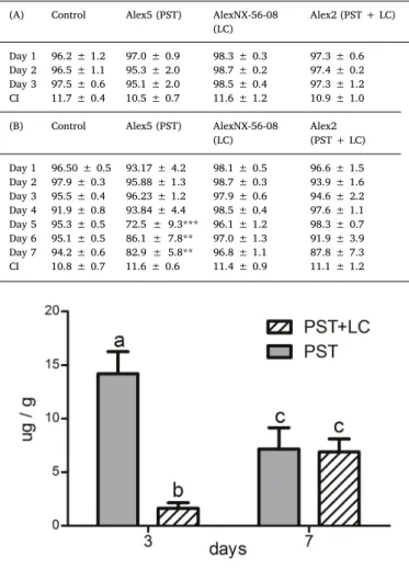
Cryptophyte R. salina bioassays showed that Alex2 (PST + LC) cultures used for the 3 days feeding experiment, displayed around 3 times more lytic activity than AlexNX-57-08 (LC) (EC5094 cells per mL, CI 92 to 97, vs. EC50313 cells per mL, CI 293 to 334); while for the 7 days experiment, lytic activity was around two times higher for the Alex2 (PST + LC) than for the AlexNX-57-08 culture (LC) (EC50
233 cells per mL, CI 222 to 246, vs. EC50556 cells per mL, CI 518 to 597). Alex5 (PST) culture supernatant had no detectable lytic activity (Fig. 1A and B) comparable to theN. salinacontrol (data not shown).
3.2. General mussel conditions
Two-way ANOVA was significant for diet and time interaction (p< 0.001). M. edulis filtered between 95 and 98.5% of the total Alexandriumcells provided daily during the 3 day experiment, and no significant differences were observed when compared to the control (Table 2A). During the 7 day experiment, feeding activity was about 20–30% lower in mussels of the Alex5 (PST) treatment than in the rest
of the treatments, showing significant differences at 5 (p< 0.001), 6 (p< 0.01) and 7 days (p< 0.01) (Table 2B). The observation fre- quency of algal cells attached to fecal pellets was less than 2% for all Alexandriumand control treatment containers.
After 3 days of feeding, 3% of mussels from the Alex5 (PST) treat- ment were paralyzed (no mantle retraction); while 3% and 6% of mussels from AlexNX-56-08 (LC) the treatment were paralyzed after 3 and 7 days, respectively. Alex 2 (PST + LC) diet caused paralysis in 6%
and 12% of the mussels after 3 and 7 days, respectively. The condition index was not affected by diet on day 3 nor on day 7 (Table 2A, B).
Mortality of 3% was registered in mussels of the Alex5 (PST) treatment after 7 days of feeding. No paralysis signals or mortalities were ob- served in control treatments.
3.3. PST accumulation in digestive gland
No PST were detected in digestive glands from control and AlexNX- 56-08 (LC) treatments. Two-way ANOVA was significant for diet and time interaction (p < 0.001) in mussels from Alex5 (PST) and Alex2 (PST + LC). After 3 days, the total amount of PST in digestive gland (μg/g) was 15 fold higher in mussels from Alex5 (PST) than in those from Alex2 (PST + LC) treatment (p< 0.001). After 7 days, toxin amount decreased to the half in Alex5 (PST)-treated groups (p< 0.05) and increased 3-fold in Alex2 (PST + LC) (p< 0.05), compared with 3-day values. No significant differences were observed between treat- ments after 7 days of exposure. Results are shown inFig. 2.
3.4. Cytotoxicity
After 3 days, lysosomal membrane stability was 40% lower in he- mocytes of mussels of the Alex5 (PST), AlexNX-56-08 (LC) and Alex2 (PST + LC) treatments, than in those of the control (p< 0.05, for all comparisons) (Fig. 3A). After 7 days, the effect of Alex5 (PST) and AlexNX-56-08 (LC) treatments was unchanged (p< 0.05, compared to the control) but a stronger effect was observed in the Alex2 (PST + LC) treatment, which resulted in 60% less membrane stability (p< 0.001), in comparison to the control (Fig. 3B).
Percentage of viable hemocytes in control treatments were 96%
after 3 days and 90% after 7 days. For graphical clarity, the results obtained forAlexandriumtreated groups were expressed as percentage of the control values (100%). After 3 and 7 days, hemocyte viability reached a maximum of 20% lower values in mussels from Alex5 (PST) (p < 0.05; p < 0.001), AlexNX-56-08 (LC) (p < 0.01; p < 0.001) and Alex2 (PST + LC) treatments (p < 0.001; p < 0.001), than those of the corresponding controls (Fig. 4A and B). No significant differences between treatments were observed: Alex5 (PST), AlexNX-56-08 (LC) and Alex2 (PST + LC) within the same experimental period or between experimental periods (3 vs. 7 days).
3.5. Immune function
The number of circulating hemocytes was lower in mussels fed for 3 days with Alex5 (PST) and AlexNX-56-08 (LC) cells, than in control and Alex2 (PST + LC) treatments (p< 0.01 for all comparisons) (Fig. 5A).
Table 1
PST amounts (pg/cell) measured inAlexandrium catenellacells: Alex5 (PST) and Alex2 (PST + LC), used for feeding mussels during 3 and 7 day experiments. Results are expressed as mean ± SEM.
Strain Feeding Toxin (pg/cell)
STX NEO GTX1/4 GTX2/3 C1/C2 Total
Alex 5 (PST) 3 days 3.84 ± 0.05 15.09 ± 0.01 24.61 ± 0.01 2.12 ± 0.01 33.90 ± 0.02 79.56
7 days 2.63 ± 0.31 14.33 ± 0.01 23.82 ± 0.28 0.93 ± 0.01 30.72 ± 0.08 72.42
Alex 2 (PST + LC) 3 days 2.35 ± 0.28 6.10 ± 0.01 1.32 ± 0.24 0.18 ± 0.01 6.30 ± 0.10 16.25
7 days 2.02 ± 0.62 6.97 ± 0.02 2.22 ± 0.35 0.22 ± 0.01 6.63 ± 0.06 18.25
After 7 days, no significant differences were observed among treat- ments (Fig. 5B).
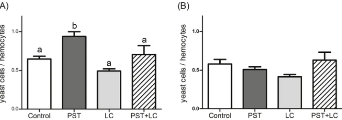
Phagocytic activity of hemocytes was higher in mussels fed for 3 days with Alex5 (PST) cells than in mussels from control (p< 0.01), AlexNX-56-08 (LC) (p < 0.01) and Alex2 (PST + LC) (p < 0.05) treatments (Fig. 6A). After 7 days, no significant differences were ob- served among treatments (Fig. 6B).
3.6. ROS production
Intracellular ROS production was lower in mussels fed 3 days with Alex5 (PST), AlexNX-56-08 (LC) and Alex2 (PST + LC) cells, than in mussels from control (p< 0.05, for all comparisons), whereas no dif- ferences were observed among treatments after 7 days (Fig. 7A and B).
Extracellular ROS levels were lower in mussel hemolymph after 3 days of the Alex2 (PST + LC) treatment than in controls (p< 0.05), and this effect was significantly higher in mussels fed with only Alex5 (PST) (p< 0.01) or only AlexNX-56-08 (LC) cells (p< 0.01,Fig. 7C). After 7 days, extracellular ROS were significantly lower in mussels of the Alex2 (PST + LC) treatment than in controls and Alex5 (PST) treatments (p< 0.05, for both comparisons) (Fig. 7D).
4. Discussion
All of the observed harmful effects that Alexandrium exerts on shellfish can be attributed to at least one out of two different compound classes produced separately or as a combination in different strains: PSP toxins (PST) and lytic compounds (LC). To better separate the effects of both compound classes, and to understand combined or synergistic effects on the mussels, we used three well-characterized strains of Alexandrium and investigated sublethal markers of functional dis- turbance, using whole animals and their hemocyte cells as indicators.
4.1. Effects of individual compound classes
Theeffect that was exclusively attributable to PSTwas the en- hanced hemocyte phagocytic activity after three exposure days to the most toxic cells (Alex 5) and caused a reduction of feeding activity (cell absorption) upon prolonged exposure (> 5 days). This would explain why injection of A. catenella STX extract induced the expression of phagocytosis-related genes in M. chilensis hemocytes [23]. Reduced feeding rate is the main mechanism through whichfiltering organisms avoid harmful diets [30]. Particularly,M. chilensisdisplays small and temporal reductions of ingestion rates when PST-producerA.catenella is included in the diet [8,9]. In our work, the supply of a highly toxic mono-diet may have caused a reduction offiltering activity [1] in Alex5 (PST) treated mussels afterfive days of feeding. Recovery ofM. edulis phagocytic activity after seven feeding days can hence be linked to decreased toxin uptake from reduced ingestion. As shown earlier for oysters fed withA. catenella[22], altered feeding activity leads to a decrease of PST content in digestive gland ofM. edulisafter seven days of feeding with Alex5 (PST). Contrarily to the effects observed in scallops [29] and oysters [48], the reduction in feeding activity is not shown to be related to LC exposure inM. edulis.
Paralysis was not clearly relatable to either compounds class. The negligible response of mussels to neurotoxic effects of PST exposure reflects their unusual tolerance to feed on PST producing microalgae [1]. The bacterial biofilm of the digestive tract ofM. eduliswas shown to reduce up to 90% of PST toxicity of Alexandrium spp. (a strain Fig. 1.Whole cell cryptophyteRhodomonas salinabioassay showing lytic activity in whole cultures ofAlexandriumspp.: Alex2 (PST + LC), Alex5 (PST), AlexNX-56- 08 (LC), used for feeding mussels during 3 (A) and 7 (B) day experiments, n = 3.
Table 2
Percentage of cells ofNannochloropsis salina(Control) andAlexandriumspp.:
Alex5 (PST), AlexNX-56-08 (LC), Alex2 (PST + LC),filtered byMytilus edulis during 3 (A) and 7 day (B) experimental feeding. CI: condition index. Results are expressed as mean ± SEM.*p < 0.05, **p < 0.01 and ***p< 0.01 between Alex5 (PST) and the rest of the treatments, within the same day, n = 6.
(A) Control Alex5 (PST) AlexNX-56-08
(LC)
Alex2 (PST + LC)
Day 1 96.2 ± 1.2 97.0 ± 0.9 98.3 ± 0.3 97.3 ± 0.6 Day 2 96.5 ± 1.1 95.3 ± 2.0 98.7 ± 0.2 97.4 ± 0.2 Day 3 97.5 ± 0.6 95.1 ± 2.0 98.5 ± 0.4 97.3 ± 1.2
CI 11.7 ± 0.4 10.5 ± 0.7 11.6 ± 1.2 10.9 ± 1.0
(B) Control Alex5 (PST) AlexNX-56-08
(LC)
Alex2 (PST + LC) Day 1 96.50 ± 0.5 93.17 ± 4.2 98.1 ± 0.5 96.6 ± 1.5 Day 2 97.9 ± 0.3 95.88 ± 1.3 98.7 ± 0.3 93.9 ± 1.6 Day 3 95.5 ± 0.4 96.23 ± 1.2 97.9 ± 0.6 94.6 ± 2.2 Day 4 91.9 ± 0.8 93.84 ± 4.4 98.5 ± 0.4 97.6 ± 1.1 Day 5 95.3 ± 0.5 72.5 ± 9.3*** 96.1 ± 1.2 98.3 ± 0.7 Day 6 95.1 ± 0.5 86.1 ± 7.8** 97.0 ± 1.3 91.9 ± 3.9 Day 7 94.2 ± 0.6 82.9 ± 5.8** 96.8 ± 1.1 87.8 ± 7.3
CI 10.8 ± 0.7 11.6 ± 0.6 11.4 ± 0.9 11.1 ± 1.2
Fig. 2.Total PST accumulation in digestive gland ofMytilus edulisfed with Alexandrium catenellastrains: Alex5 (PST) and Alex2 (PST + LC), during 3 and 7 days of experimentation. Different characters denote significant differences among treatments (p < 0.05), n = 6.
reported as“A. tamarense”but of unknown molecular identity) [49]. In scallops, for instance enzymatic biotransformation and accumulation in labial palps and digestive glands may contribute to block the passage of PST to gill, mantle and adductor muscle [7]. Mortality events are not frequently reported in Mytilusspp. fed with PST-producer algae [8,9, among others]. Only one mussel out of 30 died after seven days of feeding with Alex5 (PST), which renders difficult to attribute mortality to PST in this case. Movement/feeding impairment and no mortality were observed in scallops [29] and oysters [48] exposed to the BC
producer A. minutum. To our knowledge, such effects have not been previously evaluated inM. edulisexposed toAlexandriumBC producing strains.
Hemocytes of mussels exposed to the strain with high PST content showed neither diminished hemocyte viability, nor was lysosomal membrane destabilization exaggerated over the other treatment groups.
An oxidative burst response (enhanced intracellular and/or extra- cellular ROS) was not clearly evident, although the Alex 5 (PST) ex- posed mussels had the highest ROS levels after seven days of exposure, Fig. 3.Lysosomal membrane stability in hemocytes of Mytilus edulis fed with Nannochloropsis salina (control) and Alexandrium spp. strains: Alex5 (PST), AlexNX-56-08 (LC), Alex2 (PST + LC), during 3 (A) and 7 day (B) experiments. OD:
optical density. Different characters denote significant differences among treatments (p< 0.05), n = 6.
Fig. 4.Hemocyte viability inMytilus edulis fed with Nannochloropsis salina (control 100%) andAlexandriumspp. strains: Alex5 (PST), AlexNX-56-08 (LC) and Alex2 (PST + LC), during 3 (A) and 7 day (B) experiments. Asterisks denote significant differences between each treatment and the control treatments (*p < 0.05;
**p < 0.01; ***p< 0.001), n = 6.
Fig. 5.Total number of hemocytes in hemolymph ofMytilus edulisfed withNannochloropsis salina(control) andAlexandriumspp. strains: Alex5 (PST), AlexNX-56-08 (LC) and Alex2 (PST + LC), during 3 (A) and 7 day (B) experiments. Different characters denote significant differences among treatments (p< 0.05), n = 6.
Fig. 6.Hemocyte phagocytic activity ofMytilus edulisfed withNannochloropsis salina(control) andAlexandriumspp. strains: Alex5 (PST), AlexNX-56-08 (LC) and Alex2 (PST + LC), during 3 (A) and 7 day (B) experiments. Different characters denote significant differences among treatments (p< 0.05), n = 6.
but the effect did not reach statistical significance.
Cytotoxic effects of LCsproduced byAlexandriumspp. are clearly based on their direct disruptive effect on membranes, increasing per- meability and leading to cell death, as was previously shown for various species of photosynthetic and heterotrophic protists [24,32] and in rat neuroendocrine PC12 cells [50]. Interestingly, increased lysosomal de- stabilization in our experiments after seven days of feeding with Alex2 (PST + LC) cells is likely attributable to a time and dose dependent response to LC. Direct damage of circulating hemocyte membranes by LC would not be expected duringin vivoexposures, since there is no evidence of lytic damage on internal organs of aquatic organisms ex- posed to Alexandrium spp. LC-producer strains [51]. Alternatively, physiological stress produced as a consequence of gill/intestine tissue damage, lipid oxidation in digestive gland, mantle and muscle, and possibly respiratory impairment [29,52] may be causing indirect sub- lethal damage to hemocytes, with early manifestation in the form of lysosome destabilization [53]. However, enhanced lysosomal labiali- zation in Alex2 (PST + LC) treated mussels was not accompanied by increased hemocyte mortality. This could indicate that the harmful effects are not strong enough to increase cell death, and/or that he- mocytes display some kind of adaptive mechanism to this stressful conditions.
4.2. Shared and combined effects of PST and LC on circulating hemocytes After three days, the total number of circulating hemocytes was affected by feeding on algal strains exclusively producingeitherPSTor LC. This suggests cellular diapedesis and tissue accumulation as in- flammatory response to toxic cell encapsulation or PST detoxification [54]. Specific mechanisms involving increased toxin-lipofuscin complex accumulation in lysosomes of hemocytes has been proposed as me- chanism for PST elimination in the intestinal lumen ofM. edulisafter feeding onAlexandrium(a strain reported as“A. fundyense”but of un- known molecular identity) [55]. In this context, Borcier et al. [29]
suggested that direct contact with bioactive compound producing A.
minutum cells leads to hemocyte infiltration of affected tissues. It is therefore interesting to note that in our experiment both toxins when applied in the diet as a combination had the reverse effect and rather stabilized the number of circulating hemocytes at control level, coun- teracting the reducing effect of the single substance application. The
same recovery response was observed after seven days for all mussels treated separately with PST or LC producer algae. As suggested by Galimany et al. [55] and Haberkorn et al. [4], these results could in- dicate increased hemocyte production and stocking in hemolymph under strong toxicity exposure (dose/time) to support tissue infiltration for defense against toxic algae.
Intracellular ROS production and consequently the extracellular ROS levels ofM. edulishemocytes were reduced following three days of exposure to all three Alexandrium strains. Undiminished phagocytic activity indicates that an“oxidative burst”is not always associated with phagocytosis in bivalves [18,54], and that ROS production might be curtailed by the chemical action of both types of toxins. Haberkorn et al. [4] speculated that reduced ROS production in hemocytes of oysters fed withA. minutumcould be caused by high PST accumulation, which exceeded the“tolerance”of these bivalves, affecting their phy- siological and metabolic activities directly and profoundly. However, theA. minutumstrain (AM89BM) used by Haberkorn et al. [4] was later on described to produce also extracellular BC [28]. Mello et al. [21]
suggested that both PST and allelopathic/lytic compounds produced by this algae reduce ROS production in oyster hemocytes. Inhibition of ROS production in hemocytes of clams exposed to hemolytic super- natant ofKarenia selliformiswas also related to physiological impair- ment by cellular damage [54]. The fact that both intracellular ROS production and extracellular ROS levels were back to control values after seven days of feeding indicates physiological recovery and re- activation of the normal immune response, in spite of continued ex- posure to separate toxins. Wooton et al. [56] suggested that high basal levels of phagocytosis, cellular enzyme activity and superoxide gen- eration inM. edulishemocytes might explain its considerable resilience to adverse environmental conditions, compared to other bivalve spe- cies. However, strong suppression of extracellular ROS levels were observed after seven days of feeding with Alex2 (PST + LC), which would indeed represent immune impairment upon exposure to the toxin combination.
5. Conclusion
Both compound classes caused paralysis and affected hemocyte in- tegrity and function. However, we showed that increased phagocytosis and impairment of feeding activity are clearly associated to PST-specific Fig. 7.Fluorescence units (FU) indicating intracellular ROS production (A,B) and ex- tracellular ROS levels (C,D) in hemocytes of Mytilus edulisfed withNannochloropsis salina (control) and Alexandrium spp. strains:
Alex5 (PST), AlexNX-56-08 (LC) and Alex2 (PST + LC), during 3 (A,C) and 7 day (B,D) experiments. Different characters denote significant differences among treatments (p< 0.05), n = 6.
effects, when mussels are exposedin vivoto harmful algae; while ly- sosomal membrane labilization seems to be a LC effect, albeit not ex- clusively. In addition, our results support earlierfindings describing the astonishing resilience ofM. edulisto prolonged exposure to PST and/or LC producing Alexandrium spp. Bivalves displayed adaptive fitness traits in maintaining stable hemocyte viability, restoring phagocytic activity, and limiting intracellular ROS production upon feeding under environmentally relevant HAB conditions (cell concentrations and ex- posure duration). Knowledge gaps remain with respect to dose-depen- dent PST and LC effects on hemocyte cells.
Acknowledgments
This work was supported by a short-term grant from German Academic Exchange Service to VB (N° 91612818). Authors would like to acknowledge Stefanie Meyer for technical support.
References
[1] V.M. Bricelj, S.E. Shumway, Reviews infisheries science paralytic shellfish toxins in bivalve Molluscs : occurrence, transfer kinetics, and biotransformation paralytic shellfish toxins in bivalve Molluscs : occurrence, transfer kinetics, and bio- transformation, Rev. Fish. Sci. 6 (1998) 315–383,https://doi.org/10.1080/
10641269891314294.
[2] M. Wiese, P.M. D'Agostino, T.K. Mihali, M.C. Moffitt, B.A. Neilan, Neurotoxic al- kaloids: saxitoxin and its analogs, Mar. Drugs 8 (2010) 2185–2211,https://doi.org/
10.3390/md8072185.
[3] S.E. Ford, V.M. Bricelj, C. Lambert, C. Paillard, Deleterious effects of a nonPST bioactive compound(s) fromAlexandrium tamarenseon bivalve hemocytes, Mar.
Biol. 154 (2008) 241–253,https://doi.org/10.1007/s00227-008-0917-z.
[4] H. Haberkorn, C. Lambert, N. Le Goic, M. Guéguen, J. Moal, E. Palacios, P. Lassus, P. Soudant, Effects ofAlexandrium minutumexposure upon physiological and he- matological variables of diploid and triploid oysters,Crassostrea giga, Aquat.
Toxicol. 97 (2010) 96–108,https://doi.org/10.1016/j.aquatox.2009.12.006.
[5] H. Hégaret, K.B. Brokordt, C.F. Gaymer, K.B. Lohrmann, C. García, D. Varela, Effects of the toxic dinoflagellateAlexandrium catenellaon histopathogical and escape re- sponses of the Northern scallopArgopecten purpuratus, Harmful Algae 18 (2012) 74–83,https://doi.org/10.1016/j.hal.2012.04.006.
[6] S.M. Etheridge, Paralytic shellfish poisoning: seafood safety and human health perspectives, Toxicon 56 (2010) 108–122,https://doi.org/10.1016/j.toxicon.2009.
12.013.
[7] N.A. Estrada, N. Lagos, C. García, A.N. Maeda-Martínez, F. Ascencio, Effects of the toxic dinoflagellateGymnodinium catenatumon uptake and fate of paralytic shellfish poisons in the Pacific giant lions-paw scallopNodipecten subnodosus, Mar. Biol. 151 (2007) 1205–1214,https://doi.org/10.1007/s00227-006-0568-x.
[8] M.J. Fernández-Reiriz, J.M. Navarro, A.M. Contreras, U. Labarta, Trophic interac- tions between the toxic dinoflagellateAlexandrium catenellaandMytilus chilensis:
feeding and digestive behaviour to long-term exposure, Aquat. Toxicol. 87 (2008) 245–251,https://doi.org/10.1016/j.aquatox.2008.02.011.
[9] J.M. Navarro, A.M. Contreras, An integrative response byMytilus chilensisto the toxic dinoflagellateAlexandrium catenella, Mar. Biol. (2010) 1967–1974,https://
doi.org/10.1007/s00227-010-1465-x.
[10] W. Medhioub, P. Lassus, P. Truquet, M. Bardouil, Z. Amzil, V. Sechet, M. Sibat, P. Soudant, Spirolide uptake and detoxification byCrassostrea gigasexposed to the toxic dinoflagellateAlexandrium ostenfeldii, Aquaculture 358–359 (2012) 108–115, https://doi.org/10.1016/j.aquaculture.2012.06.023.
[11] B. Krock, C.G. Seguel, A.D. Cembella, Toxin profile ofAlexandrium catenellafrom the Chilean coast as determined by liquid chromatography withfluorescence de- tection and liquid chromatography coupled with tandem mass spectrometry, Harmful Algae 6 (5) (2007) 734–744,https://doi.org/10.1016/j.hal.2007.02.005.
[12] V.M. Bricelj, L. Connell, K. Konoki, S.P. MacQuarrie, T. Scheuer, W.A. Catterall, V.L. Trainer, Sodium channel mutation leading to saxitoxin resistance in clams increases risk of PSP, Nature 434 (2005) 763–767,https://doi.org/10.1038/
nature03415.
[13] H. Hégaret, G.H. Wikfors, P. Soudant, C. Lambert, S.E. Shumway, J.B. Bérard, P. Lassus, Toxic dinoflagellates (Alexandrium fundyenseandA. catenella) have minimal apparent effects on oyster hemocytes, Mar. Biol. 152 (2007) 441–447, https://doi.org/10.1007/s00227-007-0703-3.
[14] V.M. Bricelj, S.E. Ford, C. Lambert, A. Barbou, C. Paillard, Effects of toxic Alexandrium tamarenseon behavior, hemocyte responses and development of brown ring disease in Manila clams, Mar. Ecol. Prog. Ser. 430 (2011) 35–48,https://doi.
org/10.3354/meps09111.
[15] L. Basti, S. Nagai, J. Go, S. Okano, K. Nagai, R. Watanabe, T. Suzuki, Y. Tanaka, Differential inimical effects ofAlexandrium spp. andKarenia spp. on cleavage, hatching, and two larval stages of Japanese pearl oysterPinctada fucata martensii, Harmful Algae 43 (2015) 1–12,https://doi.org/10.1016/j.hal.2014.12.004.
[16] H. Haberkorn, C. Lambert, N.L. Goïc, J. Moal, M. Suquet, M. Guéguen, I. Sunila, P. Soudant, Effects ofAlexandrium minutumexposure on nutrition-related processes and reproductive output in oystersCrassostrea gigas, Harmful Algae 9 (2010) 427–439,https://doi.org/10.1016/j.hal.2010.01.003.
[17] T. Yan, M. Zhou, M. Fu, Y. Wang, R. Yu, J. Li, Inhibition of egg hatching success and larvae survival of the scallop,Chlamys farreri, associated to exposure of cells and cell fragments of the dinoflagellateAlexandrium tamarense, Toxicon 39 (2001) 1239–1244.
[18] L. Donaghy, C. Lambert, K.-S. Choi, P. Soudant, Hemocytes of the carpet shell clam (Ruditapes decussatus) and the Manila clam (Ruditapes philippinarum): current knowledge and future prospects, Aquaculture 297 (2009) 10–24,https://doi.org/
10.1016/j.aquaculture.2009.09.003.
[19] E.E. Philipp, S. Lipinski, J. Rast, P. Rosenstiel, Immune defense of marine in- vertebrates: the role of reactive oxygen and nitrogen species, Oxidative stress in aquatic ecosystems (2012) 236–246,https://doi.org/10.1002/9781444345988.
ch17.
[20] H. Hégaret, P.M. da Silva, G.H. Wikfors, C. Lambert, T. De Bettignies, S.E. Shumway, P. Soudant, Hemocyte responses of Manila clams,Ruditapes phi- lippinarum, with varying parasite,Perkinsus olseni, severity to toxic-algal exposures, Aquat. Toxicol. 84 (2007) 469–479,https://doi.org/10.1016/j.aquatox.2007.07.
007.
[21] D.F. Mello, P.M. da Silva, M.A. Barracco, P. Soudant, H. Hégaret, Effects of the dinoflagellateAlexandrium minutumand its toxin (saxitoxin) on the functional ac- tivity and gene expression ofCrassostrea gigashemocytes, Harmful Algae 26 (2013) 45–51,https://doi.org/10.1016/j.hal.2013.03.003.
[22] M. Lassudrie, P. Soudant, J.L. Nicolas, P. Miner, J. Le Grand, C. Lambert, N. Le Goïc, H. Hégaret, C. Fabioux, Exposure to the toxic dinoflagellateAlexandrium catenella modulates juvenile oysterCrassostrea gigashemocyte variables subjected to dif- ferent biotic conditions, Fish Shellfish Immunol. 51 (2016) 104–115,https://doi.
org/10.1016/j.fsi.2016.02.017.
[23] G. Núñez-Acuña, A.E. Aballay, H. Hégaret, A.P. Astuya, C. Gallardo-Escárate, Transcriptional responses ofMytilus chilensisexposedin vivoto saxitoxin (STX), J.
Molluscan Stud. 79 (2013) 323–331,https://doi.org/10.1093/mollus/eyt030.
[24] U. Tillmann, U. John, Toxic effects ofAlexandrium spp. on heterotrophic dino- flagellates: an allelochemical defence mechanism independent of PSP-toxin content, Mar. Ecol. Prog. Ser. 230 (2002) 47–58,https://doi.org/10.3354/meps230047.
[25] H. Ma, B. Krock, U. Tillmann, A. Muck, N. Wielsch, A. Svatoš, A. Cembella, Isolation of activity and partial characterization of large non-proteinaceous lytic allelo- chemicals produced by the marine dinoflagellateAlexandrium tamarense, Harmful Algae 11 (2011) 65–72,https://doi.org/10.1016/j.hal.2011.07.004.
[26] Y. Matsuyama, H. Usuki, T. Uchida, Y. Kotani, Effects of harmful algae on the early planktonic larvae of the oyster,Crassostrea gigas, in: G.M. Hallegraeff,
S.I. Blackburn, C.J. Olch, R.J. Lewis (Eds.), Harmful Algal Blooms, Proceedings of the Ninth International Conference on Harmful Algal Blooms 2000, UNESCO, France, 2001, pp. 411–414.
[27] G. Arzul, M. Seguel, L. Guzman, E. Erard-Le Denn, Comparison of allelopathic properties in three toxicAlexandriumspecies, J. Exp. Mar. Biol. Ecol. 232 (1999) 285–295,https://doi.org/10.1016/S0022-0981(98)00120-8.
[28] A. Lelong, H. Haberkorn, N. Le Goc, H. Hégaret, P. Soudant, A new insight into allelopathic effects ofAlexandrium minutumon photosynthesis and respiration of the diatom chaetoceros neogracile revealed by photosynthetic-performance analysis andflow cytometry, Microb. Ecol. 62 (2011) 919–930,https://doi.org/10.1007/
s00248-011-9889-5.
[29] E. Borcier, R. Morvezen, P. Boudry, P. Miner, G. Charrier, J. Laroche, H. Hegaret, Effects of bioactive extracellular compounds and paralytic shellfish toxins produced byAlexandrium minutumon growth and behaviour of juvenile great scallopsPecten maximus, Aquat. Toxicol. 184 (2017) 142–154,https://doi.org/10.1016/j.aquatox.
2017.01.009.
[30] J.H. Landsberg, The effects of harmful algal blooms on aquatic organisms, Rev. Fish.
Sci. (2002) 113–390,https://doi.org/10.1080/20026491051695.
[31] U. John, R.W. Litaker, M. Montresor, S. Murray, M.L. Brosnahan, D.M. Anderson, Formal revision of theAlexandrium tamarensespecies complex (Dinophyceae) tax- onmomy: the introduction offive species with emphasis on molecular-based (rDNA) classification, Protist 165 (2014) 779–804.
[32] U. Tillmann, T.L. Alpermann, R.C. da Purificação, B. Krock, A. Cembella, Intra- population clonal variability in allelochemical potency of the toxigenic dino- flagellateAlexandrium tamarense, Harmful Algae 8 (2009) 759–769,https://doi.
org/10.1016/j.hal.2009.03.005.
[33] I. Okomus, N. Bascinar, M. Ozkan, The effects of phytoplankton concentration, size of mussel and water temperature on feed consumption andfiltration rate of the Mediterranean mussel (Mytilus galloprovincialisLmk), Turk. J. Zool. 26 (2002) 167–172.
[34] M. Diener, K. Erler, S. Hiller, B. Christian, B. Luckas, Determination of paralytic shellfish poisoning (PSP) toxins in dietary supplements by application of a new HPLC/FD method, Eur. Food Res. Technol. 224 (2) (2006) 147–151.
[35] H. Ma, B. Krock, U. Tillmann, A. Cembella, Preliminary characterization of extra- cellular allelochemicals of the toxic marine dinoflagellateAlexandrium tamarense using aRhodomonas salinabioassay, Mar. Drugs 7 (2009) 497–522,https://doi.org/
10.3390/md7040497.
[36] V.A. Bianchi, J.M. Castro, I. Rocchetta, F. Bieczynski, C.M. Luquet, Health status and bioremediation capacity of wild freshwater mussels (Diplodon chilensis) exposed to sewage water pollution in a glacial Patagonian lake, Fish Shellfish Immunol. 37 (2014) 268–277,https://doi.org/10.1016/j.fsi.2014.02.013.
[37] M. Starr, J.H. Himmelman, J.-C. Therriault, Direct coupling of marine invertebrate spawning with phytoplankton blooms, Science 247 (4946) (1990) 1071–1074, https://doi.org/10.1126/science.247.4946.1071.
[38] C. Kittner, H.U. Riisgård, Marine Effect of temperature onfiltration rate in the mussel Mytilus edulis: no evidence for temperature compensation, Mar. Ecol. Prog.
Ser. 305 (2005) 147–152,https://doi.org/10.3354/Meps305147.
[39] J. Widdows, The effects of temperature on the metabolism and activity ofMytilus
edulis, Neth. J. Sea Res. 7 (1973) 387–398,https://doi.org/10.1016/0077-7579(73) 90060-4.
[40] H.G. Gudfinnsson, A. Eydal, K. Gunnarsson, K. Gudmundsson, K. Valsdóttir, Monitoring of toxic phytoplankton in three Icelandic fjords, ICES CM 12 (2010) 1–18.
[41] B. Luckas, K. Erler, B. Krock, Analysis of marine biotoxins using LC-MS/MS, chapter 18: 277–297, in: D. Stengel, S. Connan (Eds.), Natural Products from Marine Algae, Humana Press, Springer, New York, Heidelberg, Dordrecht, London, 2015ISBN:
978-1-4939-2683-1.
[42] A. Novas, R. Barcia, J.I. Ramos-Martínez, After the Prestige oil spill modifications in NO production and other parameters related to the immune response were detected in hemocytes ofMytilus galloprovincialis, Aquat. Toxicol. 85 (2007) 285–290, https://doi.org/10.1016/j.aquatox.2007.09.011.
[43] J. Coles, S. Farley, R. Pipe, Alteration of the immune response of the common marine musselMytilus edulisresulting from exposure to cadmium, Dis. Aquat. Org.
22 (1995) 59–65,https://doi.org/10.3354/dao022059.
[44] M. Bradford, Bradford Protein Assay Protocol, (2006), pp. 1–3,https://doi.org/10.
1016/0165-022X(93)90047-ROctober.
[45] F.M. Akaishi, S.D. St-Jean, F. Bishay, J. Clarke, I. da S. Rabitto, C.A. de O. Ribeiro, Immunological responses, histopathologicalfinding and disease resistance of blue mussel (Mytilus edulis) exposed to treated and untreated municipal wastewater, Aquat. Toxicol. (2007),https://doi.org/10.1016/j.aquatox.2007.01.008.
[46] R.P. Kuchel, D.A. Raftos, S. Nair, Immunosuppressive effects of environmental stressors on immunological function inPinctada imbricata, Fish Shellfish Immunol.
29 (6) (2010) 930–936,https://doi.org/10.1016/j.fsi.2010.07.033.
[47] B. Moss, B. Allam, Fluorometric measurement of oxidative burst in lobster hemo- cytes and inhibiting effect of pathogenic bacteria and hypoxia, J. Shellfish Res. 25 (2006) 1051–1057.
[48] J. Castrec, P. Soudant, L. Payton, D. Tran, P. Miner, C. Lambert, N.L. Goïc, A. Huvet, V. Quillien, F. Boullot, Z. Amzil, H. Hégaret, C. Fabioux, Bioactive extracellular compounds produced by the dinoflagellateAlexandrium minutumare highly
detrimental for oysters, Aquat. Toxicol. 199 (2018) 188–198,https://doi.org/10.
1016/j.aquatox.2018.03.034.
[49] C.J. Donovan, J.C. Ku, M.A. Quilliam, T.A. Gill, Bacterial degradation of paralytic shellfish toxins, Toxicon 52 (2008) 91–100,https://doi.org/10.1016/j.toxicon.
2008.05.005.
[50] H. Ma, B. Krock, U. Tillmann, U. Bickmeyer, M. Graeve, A. Cembella, Mode of action of membrane-disruptive lytic compounds from the marine dinoflagellate Alexandrium tamarense, Toxicon 58 (2011) 247–258,https://doi.org/10.1016/j.
toxicon.2011.06.004.
[51] J.H. Landsberg, The effects of harmful algal blooms on aquatic organisms, Rev. Fish.
Sci. 10 (2) (2002) 113–390,https://doi.org/10.1080/20026491051695.
[52] L. Basti, T. Oda, K. Nagai, S. Nagai, Role of cytotoxicity and hemolysis in the de- leterious effects ofAlexandrium affineandA. catenellaon early-life development of the Japanese pearl oyster,Pinctada fucata martensii, J. Shellfish Res. 34 (2015) 608.
[53] A. Viarengo, D. Lowe, C. Bolognesi, E. Fabbri, A. Koehler, The use of biomarkers in biomonitoring: a 2-tier approach assessing the level of pollutant-induced stress syndrome in sentinel organisms, Comp. Biochem. Physiol. C Toxicol. Pharmacol.
146 (2007) 281–300,https://doi.org/10.1016/j.cbpc.2007.04.011.
[54] H. Hégaret, P.M. Da Silva, G.H. Wikfors, H. Haberkorn, S.E. Shumway, P. Soudant, In vitro interactions between several species of harmful algae and haemocytes of bivalve mollusks, Cell Biol. Toxicol. 27 (2011) 249–266,https://doi.org/10.1007/
s10565-011-9186-6.
[55] E. Galimany, I. Sunila, H. Hégaret, M. Ramón, G.H. Wikfors, Experimental exposure of the blue mussel (Mytilus edulis, L.) to the toxic dinoflagellateAlexandrium fun- dyense: histopathology, immune responses, and recovery, Harmful Algae 7 (2008) 702–711,https://doi.org/10.1016/j.hal.2008.02.006.
[56] E.C. Wootton, E.A. Dyrynda, N.A. Ratcliffe, Bivalve immunity: comparisons be- tween the marine mussel (Mytilus edulis), the edible cockle (Cerastoderma edule) and the razor-shell (Ensis siliqua), Fish Shellfish Immunol. 15 (2003) 195–210,https://
doi.org/10.1016/S1050-4648(02)00161-4.