Genotype-phenotype relations in patient-derived point mutations in collagen II
Inaugural-Dissertation zur Erlangung des Doktorgrades
der Mathematisch-Naturwissenschaftlichen Fakultät der Universität zu Köln
vorgelegt von
Chakkalakal Anandan Salin aus Trichur, India
im September 2005
Berichterstatter: Herr Prof. Dr. Mats Paulsson Herr Prof. Dr. Helmut W. Klein Tag der mündlichen Prüfung: 2. Dezember 2005
TO MY PARENTS
TABLE OF CONTENTS
Abstract
Zusammenfassung Abbreviations
1 Introduction 1
1.1 Collagen 1
1.2 Classification of collagens 1 1.3 Genes and nomenclature 2 1.4 Structure and stability of the collagen triple helix 3 1.5 Biosynthesis of collagen 4
1.6 Collagen II 9
1.7 Chondrodysplasias 9 1.8 Achondrogenesis type II and hypochondrogenesis (OMIM 200610) 10 1.9 Spondyloepiphyseal dysplasia and congenital spondyloepiphyseal
dysplasia (OMIM 183900) 11 1.10 Stickler syndrome (OMIM 108300) 12 1.11 Osteoarthritis 14 1.12 Animal models for chondrodysplasia due to collagen II mutations 15 1.13 Arginine to cysteine mutations 16 1.14 Aims of the thesis 17 2 Materials and methods 18
2.1 Materials 18
2.1.1 Enzymes for molecular biology 18 2.1.2 Materials for cell culture 18 2.1.3 Plasmid and adenoviral vectors 18 2.1.3.1 pBluescript KS+ (pBS KS+) phagemid 18 2.1.3.2 pSL1180 superlinker phagemid 19 2.1.3.3 Eukaryotic expression vector pCEP-Pu 20 2.1.3.4 Adenoviral vector pGS66 and shuttle vector pGS70 20 2.2 Molecular biology methods 22 2.2.1 Plasmid DNA isolation from E. coli 22
2.2.2 Pure MIDI plasmid DNA isolation 22 2.2.3 DNA agarose gel electrophoresis 22 2.2.4 Elution of DNA fragments from agarose gels 23 2.2.5 Measurement of DNA and RNA concentrations 23 2.2.6 Restriction digestion of DNA 23 2.2.7 Dephosphorylation of 5´ends of linearized vectors 23 2.2.8 Ligation reaction 24 2.2.9 Polymerase chain reaction (PCR) 24 2.2.10 Collagen II cDNA and patient derived mutations 25 2.2.11 Subcloning of collagen II cDNA 25 2.2.12 Site directed mutagenesis 26 2.2.13 Removal of the collagen II signal peptide 29 2.2.14 Screening for recombinants and sequencing 29 2.2.15 Eukaryotic expression vector 29 2.2.16 RNA isolation from cultured HT1080 cells 30 2.2.17 Reverse transcription 30 2.3 Cell biology methods 30 2.3.1 Cell culture and maintenance of 293 EBNA and HT1080 cells 30 2.3.2 Transfection 293 EBNA and HT1080 cells with FuGene6 31 2.3.3 Isolation and culture of bovine chondrocytes 31 2.3.4 Passaging of cell lines 31 2.3.5 Freezing cells for storage 31 2.3.6 Thawing of frozen cells 32 2.3.7 Cell counting 32 2.3.8 Immunofluorescence staining 32 2.3.9 Image analysis 33 2.3.10 Comet assay or single cell gel electrophoresis (SCGE) 33 2.3.11 Nick labelling 34 2.3.12 Transduction of chondrocytes and transgene expression 35 2.3.13 Cell toxicity 35 2.4 Biochemical methods 35 2.4.1 Harvesting of supernatants from 293 EBNA cells 35 2.4.2 Isolation and purification of collagens from the supernatants 35
2.4.4 SDS polyacrylamide gel electrophoresis (SDS PAGE) 36 2.4.5 Coomassie blue staining of SDS-polyacrylamide gels 37 2.4.6 Silver staining of gels 37 2.4.7 Transfer of proteins to nitrocellulose membranes 38 2.4.8 Immunodetection of membrane bound proteins 38 2.4.9 Trypsin digestion of purified collagen II proteins 39 2.4.10 Measurement of circular dichroism spectra and melting curves 39 2.4.11 Negative staining of collagens for electron microscopy 40 2.4.12 Inhibition of proteolytic processing of collagen II using specific inhibitors 40 2.4.13 Mass spectrometry of purified collagen II variants 40
3 Results 41
3.1 Subcloning of collagen II cDNA 41 3.2 Cloning of collagen II cDNA into pSL1180 vector 42 3.3 Cloning of collagen II cDNA without signal peptide into the mammalian
expression vector pCEP-Pu 42 3.4 Transient expression of collagen II proteins in 293 EBNA cells 43 3.5 Purification of recombinantly expressed collagen II 44 3.6 Coomassie staining of purified collagen II variants 45 3.7 Immunoblot of purified collagen II variants 45 3.8 Trypsin digestion of different collagen II variants 46 3.9 Circular dichroism of purified collagen II proteins 47 3.10 Melting curves of purified recombinant collagen II proteins 47 3.11 Mass spectrometric analysis of purified collagen II proteins 49 3.12 Negative electron microscopy of purified collagen II proteins 50 3.13 HT 1080 cells transiently transfected with collagen II constructs 52 3.14 Effect of the MMP inhibitor GM6001 on HT1080 cells expressing collagen II 52 3.15 Effect of proteasome inhibitor MG132 on processing of R789C 53 3.17 ER stress response in collagen expressing HT1080 cells 54 3.18 Immunofluorescence and light microscopy of HT1080 cells
expressing collagen II variants 55 3.19 Detection of DNA cleaved due to apoptosis by nick labelling 61 3.20 Single cell gel electrophoresis (comet assay) of HT1080 cells
transfected with collagen II variants 62
3.21 Comparison of the efficiency and toxicity of first and third generation
adenoviral vectors 63
4 Discussion 65
4.1 Effect of point mutations on protein expression and secretion 65 4.2 Impact of arginine to cysteine mutations on the structure and
stability of collagen II 68 4.3 Impact of collagen II mutations on protein trafficking 71 4.4 Fate of cells expressing R740C and R789C collagens 73 4.5 Consequences of intracellular retention of collagen II for the
assembly of extracellular matrix 75
4.6 Conclusion 76
5 References 78
Erklärung
Acknowledgements Lebenslauf
Appendix
Abstract
Abstract
Collagen II is the major collagen present in the extracellular matrix of cartilage, in addition it is found in the vitreous of the eye and it is also detected during early embryogenesis. Due to the complexity of the biosynthesis, assembly and secretion, collagen II is highly susceptible to mutations leading to disease states which are broadly classified as chondrodysplasias. Most of these mutations are substitutions of glycine in the Gly-X-Y repeats in the triple helical domain resulting in destabilization of the helix. Point mutations leading to arginine to cysteine substitution are interesting since they occur at either X or Y position and cause two different diseases termed Stickler syndrome and congenital spondyloepiphyseal dysplasia (SEDC) in association with osteoarthritis.
The present study aimed at determining the consequences of arginine to cysteine substitutions in either X or the Y position in the Gly-X-Y repeats and at either the N- or the C- terminus of the triple helix. The impact of these mutations on protein trafficking, secretion and cell survival was also analyzed.
Biochemical studies revealed great similarities between R75C, R134C and R704C collagens and the wild type molecules with the exception that electron micrographs of R75C collagen displayed kinks in the structure. R740C and R789C collagens accumulated in the cells and the R789C protein migrated faster on SDS gels. The R740C and R789C proteins were also susceptible to protease digestion and circular dichroism spectra were altered and showed lower Tm values than other collagen II variants. Due to the altered structure, R789C protein was more susceptible to MMP cleavage in the vicinity of the mutation causing the truncation of the protein.
Additionally, electron micrographs revealed only scarce and thin filamentous structures in preparations of R740C and R789C protein. The biochemical results indicate that the R740C and R789C proteins have unstable triple helices and that this affects the overall protein structure.
Protein trafficking was monitored in HT1080 cells expressing the collagen II variants.
Intracellular retention in the ER due to misfolding of the R740C and R789C proteins triggered an ER stress response including splicing of XBP-1 and induction and binding of BiP. Continuous accumulation of misfolded proteins in the ER caused apoptosis of the R740C and R789C expressing cells.
Substitution of arginine to cysteine in the X or Y position towards the C-terminus of the triple helix caused pronounced instability of the triple helix with a deleterious effect on the cells, while R704C and more N-terminal mutations did not cause any significant changes irrespective of being in the X or the Y position.
The different severities of patient phenotypes are due to a combination of structural factors, which may be synergistically augmented by genetic modifiers, additional unknown mutations and environmental factors.
Zusammenfassung
Collagen II ist der Hauptbestandteil der extrazellulären Matrix des Knorpels. Außerdem wird Collagen II im Glaskörper des Auges exprimiert und konnte während der frühen embryonalen Entwicklung nachgewiesen werden. Collagen II ist durch seine komplexe Biosynthese, Assemblierung und Sekretion besonders anfällig für Mutationen, die im Menschen zu Krankheiten führen, die allgemein unter dem Begriff Chondrodysplasien zusammengefasst werden. Bei den meisten dieser Mutationen handelt es sich um den Austausch eines Glycins in den Gly-X-Y Triplets der tripelhelikalen Domäne, die zu einer Destabilisierung dieser Helix führen. Punktmutationen, die zu einem Austausch von Arginin zu Cystein führen, kommen sowohl in der X- als auch Y-Position vor und führen interessanterweise zu zwei unterschiedlichen Krankheitsbildern, dem Stickler Syndrom bzw. der congenitalen Spondyloepiphysiären Dysplasie verbunden mit Osteoarthrose.
In der vorliegenden Arbeit wurden Auswirkungen von Substitutionen eines Arginins zu einem Cystein sowohl in der X- als auch in der Y-Position bzw. im N- oder C-terminalen Bereich der Tripelhelix untersucht. Außerdem wurden Effekte dieser Mutationen auf den intrazellulären Proteintransport, die Sekretion und die Zellvitalität analysiert.
Biochemische Untersuchungen ergaben keinerlei wesentliche Unterschiede zwischen den Mutanten R75C, R134C und R704C im Vergleich mit dem Wildtyp Protein mit Ausnahme der elektronenmikroskopischen Aufnahmen, in denen die Mutation R75C zu charakteristischen Knicken in der Struktur des Collagens führte. Die Proteine R740C und R789C akkumulierten intrazellulär, und das Collagen R789C zeigte veränderte Laufeigenschaften im SDS Gel. Die Proteine R740C und R789C waren außerdem empfindlicher gegenüber Proteasen, und die Analyse mittels CD Spektroskopie ergab untypische Spektren und erniedrigte Schmelztemperaturen. Die durch die veränderte Struktur erhöhte Empfindlichkeit des Proteins R789C gegenüber dem Abbau durch MMPs in unmittelbarer Nähe der Mutation führte zu einer Verkürzung des Proteins durch Proteolyse.
In elektronenmikroskopischen Untersuchungen der Proteine R740C und R789C konnten nur kurze filamentöse Strukturen nachgewiesen werden. Diese Ergebnisse führen zu dem Schluss, dass die Proteine R740C und R789C lediglich instabile Tripelhelices ausbilden und die Mutationen so die Gesamtstruktur des Proteins beeinflussen.
Der intrazelluläre Transport der mutierten Proteine wurde in transfizierten HT1080 Zellen untersucht. Die fehlgefalteten Proteine R740C und R789C wurden im endoplasmatischen Retikulum (ER) zurückgehalten und lösten eine ER Stressantwort aus, die mit einem Splicing von XBP-1 und einer erhöhten Induktion und Bindung des Chaperons BiP einhergeht. Die andauernde Akkumulation dieser fehlgefalteten Proteine im ER führte schließlich zur Auslösung der Apopotse.
Der Austausch von Arginin zu Cystein im C-terminalen Bereich der Tripelhelix führte sowohl in der X- als auch der Y-Position zur Instabilität der Tripelhelix mit schädlichen Auswirkungen auf die exprimierenden Zellen. Im Gegensatz hierzu resultierten bereits die Mutation R704C und weiter N-terminal gelegene Mutationen in keinerlei signifikanten Veränderungen, unabhängig davon, ob sie in der X- bzw. Y-Position vorlagen.
Die in Patienten beschriebenen, sehr unterschiedlich ausgeprägten Krankheitsbilder sind wahrscheinlich durch eine Kombination sich synergistisch verstärkender struktureller und genetischer Faktoren sowie unbekannter Mutationen und Umwelteinflüsse zu erklären.
Abbreviations
Abbreviations
AA Acrylamide
AP Alkaline phosphatase APS Ammonium persulphate BCA Bicinchoninic acid
BiP Immunoglobulin heavy chain binding protein BSA Bovine serum albumin
BM-40 Basement membrane protein, molecular weight 40 kD CD Circular Dichroism
COMP Cartilage oligomeric matrix protein DEPC Diethylpyrocarbonate
DMEM Dulbecco’s minimum essential medium DMSO Dimethylsulfoxide
dNTP Deoxyribonucleotide triphosphate DTT 1,4-dithiothreitol
EBNA Ebstein barr virus nuclear antigen EDTA Ethylenediaminetetraacetic acid EK Enterokinase
ER Endoplasmic reticulum FBS Fetal bovine serum
FITC Fluorescein-5-isothiocyanate GFP Green fluorescence protein
His Histidine
HRP Horse radish peroxidase IgG Immunoglobulin G
kb Kilobase pairs kD Kilodalton
MALDI-TOF-MS Matrix assisted laser desorption time of flight MED Multiple epiphyseal dysplasia
MMP Matrix metalloproteinase MOI Moieties of infection mRNA Messenger ribonucleic acid NEM N-Ethylmaleimide
NP-40 Nonylphenylpolyethyleneglycol NTA Nitriloacetic acid
OD Optical density
OMIM Online mendelian inheritance in man PDI Protein disulfide isomerase PMSF Phenylmethylsulphonylfluoride PAGE Polyacrylamide gel electrophoresis
SC Pepsin extracted collagen from bovine nasal cartilage (Sigma) SCGE Single cell gel electrophoresis
SDS Sodium dodecyl sulphate
SEDC Spondyloepiphyseal dysplasi congenita TAE Tris acetate EDTA
XBP X-box DNA binding protein
Introduction
1 Introduction
1.1 Collagen
Collagens form a family of proteins that constitute the major structural components of the ECM, representing approximately one third of the body protein in man. The general definition of a collagen is “a protein consisting of three identical or related polypeptide chains, which are folded into at least one triple-helical domain and assemble into supramolecular aggregates mostly in the extracellular space”. They are found in almost all tissues of the body, and are particular abundant in bone, skin, tendon, ligaments, cartilage and vessel walls (Myllyharju & Kivirikko 2004).
1.2 Classification of collagens
Collagens are characterized by the presence of triple-helical domains, which are either composed of three identical polypeptide chains (homotrimers) or two to three genetically distinct chains (heterotrimers). The collagen types are designated with Roman numerals in the order of their discovery, and the chains found in each collagen type are identified with Arabic numerals (Kivirikko 1993, Pihlajaniemi & Rehn 1995, Prockop & Kivirikko 1995, Myllyharju &
Kivirikko 2004). Collagens are broadly classified into two groups on the basis of their primary structure, physicochemical properties and macromolecular assembly:
1. Fibrillar collagens (over 70% of total collagens; e.g. type I, II, III, V, XI), possessing a large, uninterrupted triple-helical domain capable of fibril formation. Their fibrils create the structural frameworks for many tissues.
2. The non-fibril-forming collagens, made up by basement membrane collagens (type IV and VII), transmembrane collagens (type XIII and XVII), fibril-associated collagens with interrupted triple helices (FACIT; type IX, XII, XIV, XVI and XIX), multiplexin collagens (type XV and XVIII), short-chain collagens (type VI, VIII and X) form a more heterogeneous group. A distinct feature of non-fibril-forming collagens is the presence of one or more non collagenous interruptions within the collagenous sequence. Members of this group form filaments, sheet-like structures, network-like elements and anchoring fibrils and can associate with collagen fibrils and membranes in a highly tissue-specific manner in variety of organs.
Fig 1.1 summarizes the classification within the collagen superfamily, the known collagen supramolecular assemblies and molecular and functional properties.
Fig 1.1 Members of the collagen superfamily and their known supramolecular assemblies. The collagen superfamily can be divided into nine families on the basis of the supramolecular assemblies and other features of its members: (a) fibril-forming collagens; (b) fibril-associated collagens with interrupted triple helices (FACITs), located at the surface of fibrils, and structurally related collagens; (c) collagens forming hexagonal networks; (d) the family of type IV collagens located in basement membranes; (e) type VI collagen, which forms beaded filaments; (f) type VII collagen, which forms anchoring fibrils at basement membranes; (g) collagens with transmembrane domains; and (h) the family of type XV and XVIII collagens. The supramolecular assemblies of families (g) and (h) are unknown and are therefore not shown (Myllyharju & Kivirikko 2004).
1.3 Genes and nomenclature
The genes encoding the fibril-forming collagens I-III show structural similarity but vary in size from 18 to 44 kb (Prockop & Kivirikko 1995, Yamada et al. 1980). They consist of 51-54 exons with the major triple-helical domain being encoded by 44 exons. The similarity in gene structure
Introduction the C-propeptides are encoded by four exons. The gene loci for the members of the collagen family have been given names beginning with COL in a human context and col in animals, followed by an Arabic numeral denoting the collagen. For example, COL1A1 is the gene locus for the human proα1(I) chain, and COL1A2 for the proα2(I) chain.
All the exons encoding the triple-helical domain are of sizes that are multiples of 9 bp (e.g. 45, 54, 99, 108 and 162 bp, coding for 5, 6, 11, 12 and 18 Gly-X-Y triplets, respectively), the most common size being 54 bp. It has been suggested that the ancestral gene for the fibril-forming collagens must have evolved by amplification of a 54 bp unit embedded into intron sequences (Yamada et al. 1980). All these exons start with a codon for glycine and end with a complete codon for an amino acid in the Y position.
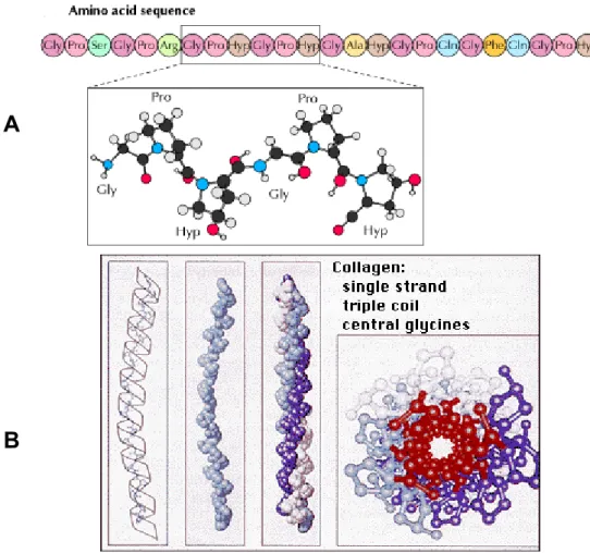
1.4 Structure and stability of the collagen triple helix
In collagens three α chains are wound around each other into a right-handed triple helix. In some collagens all three chains in the molecule are identical and form homotrimers, while other types contain two or even three different chains leading to heterotrimeric molecules (Engel & Prockop 1991, Hulmes 1992). At the amino acid level the collagenous or triple helical domains contain of repeats consisting of three amino acids (Fig 1.2A). The presence of glycine as every third amino acid in the repeating Gly-X-Y sequence is essential because a larger amino acid would not fit in the center of the triple helix where the three chains come together (Fig 1.2B). Proline is frequently in the X position of the Gly-X-Y sequence and 4-hydroxyproline is frequently in the Y position. These two amino acids limit the rotation of the polypeptide chains. The triple helix is further stabilized by hydrogen bonds and water bridges, many of which require the presence of 4-hydroxyproline. Stabilization of the triple helix by glycines has been studied using collagen- like peptides (Bella et al. 1994, 1995). In addition, many glycine substitutions in the collagen triple helix have been identified in various heritable connective tissue disorders (Kivirikko 1993, Dalgleish 1997, 1998). These glycine substitutions result in a small local untwisting of the triple helix and reduce its thermal stability (Bella et al. 1994), suggesting that similar conformational changes may occur in the case of glycine substitutions at other positions. It is also thought that glycine substitutions may reduce the rate of folding of the triple helix, as a Gly to Cys substitution within the collagen domain of type I collagen prolongs the time needed to reach the triple-helical state (Raghunath et al. 1994).
Fig 1.2 Arrangement of amino acids in the Gly-X-Y repeat in the triple helical part of a fibrillar collagen. (A) Characteristic Gly-X-Y repeat with the orientation of glycine. (B) The central fit of glycine in the structure of triple helix with all other amino acid being more peripheral (Cooper & Hausman 2004, Nelson & Cox 2005).
1.5 Biosynthesis of collagen
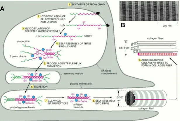
Collagen biosynthesis and assembly follows the normal pathway of a secretory protein. Fig 1.3 represents a schematic illustration of the intracellular and extracellular events during collagen biosynthesis, which are briefly described below.
1. Synthesis: Collagen chains are synthesized as longer precursors called procollagens. The growing polypeptide chains are co-translationally transported into the lumen of the rough endoplasmic reticulum (ER). In the ER, the procollagen chain undergoes a series of post- translational modifications, in particular hydroxylation and glycosylation.
2. Hydroxylation: Specific proline and lysine residues in the propeptide are hydroxylated by membrane-bound hydroxylases. These hydroxyproline residues are essential for the stability of triple helical molecules since underhydroxylated procollagens are not stable at normal body
A
B
Introduction temperature. Lysyl hydroxylase and proline hydroxylase catalyses the hydroxylation of lysine and proline residues in the procollagen chains. The hydroxylysine residues serve as attachment sites for carbohydrate units and are also needed for the formation of intermolecular cross-links during the collagen fibril formation. Ascorbate is a cofactor required for hydroxylase activity and a deficiency may lead to triple helix instability.
Fig 1.3 Intracellular and extracellular events during collagen biosynthesis and fibrillogenesis. (A) Procollagen synthesis, posttranslational modification, secretion and fibril assembly. (B) Electron micrograph of a negatively stained collagen fibril reveals its typical striated appearance (Alberts et al. 1994).
3. Glycosylation: Procollagens are further glycosylated in the ER and in the Golgi complex.
Galactose and glucose residues are added to hydroxylysine residues and longer oligosaccharides are transferred to certain asparagine residues in the C-terminal propeptide.
4. Procollagen assembly: N- and C-propeptides first fold and intrachain disulphide bonds are formed within them (Bächinger et al. 1981, Doege & Fessler 1986). Three pro α chains then associate through non-covalent interactions between the folded C-propeptides followed by formation of interchain disulphide bonds catalyzed by protein disulphide isomerase (PDI) (Olsen et al. 1976, Forster & Freedman 1984, Noiva & Lennarz 1992). The formation of a triple-helical procollagen molecule begins by association of the C-propeptides of the three pro α chains
B A
B
(Bächinger et al. 1981, Bulleid et al. 1996) and the triple helix is formed in a zipper-like manner proceeding from the C-terminal region towards the N-terminus (Engel & Prockop 1991).
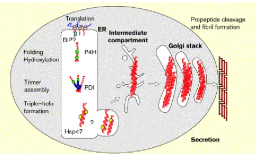
5. Quality control: During the entire process molecular chaperones play an important role in regulating the correct folding and assembly of proteins (Fig 1.4). Chaperones bind to hydrophobic regions of unfolded proteins and prevent aggregation while the polypeptides are synthesized (Gething & Sambrook 1992, Becker & Craig 1994, Hartl et al. 1994). They also serve as folding quality controllers, preventing the secretion of misfolded proteins and promoting their degradation. BiP is a 78 kD protein located within the lumen of the ER and plays a general role in protein folding and oligomeric assembly (Haas & Wabl 1983). In certain osteogenesis imperfecta patients, mutant type I collagen remains bound to BiP (Chessler & Byers 1992). PDI also acts as a molecular chaperone during the assembly of procollagen chains (Wilson et al.
1998). PDI interacts specifically with the propeptides of monomeric type I procollagen chains and prevents their premature assembly or aggregation (Wilson et al. 1998).
Fig 1.4 Interactions of ER chaperones during procollagen assembly and secretion. Procollagen interacts with a variety of ER resident proteins during folding and assembly within the ER. The carboxy-terminal propeptide may interact transiently with BiP (light blue) before it folds correctly and assembles covalently with two other pro-α- chains to form a trimer. Protein disulfide isomerase (PDI; blue) catalyzes this latter event. Proline hydroxylases (P4H; green) modifies selected proline and lysine residues in the triple helix-forming domains. These hydroxylated residues form hydrogen bonds that stabilize the helix once it forms. This triple helical form of procollagen is the preferred substrate of Hsp47 (yellow) (Hendershot & Bulleid 2000).
Hsp47 is an endoplasmic reticulum (ER)-resident molecular chaperone that is specific for collagen and plays a role in collagen maturation (Nagata & Hosokawa 1996, Nagata 2003). After
Introduction cis-Golgior ER-Golgi intermediate compartment, where it dissociates fromprocollagen in a pH- dependent manner (Saga et al. 1987, Satohet al. 1996).
6. Secretion: The mechanism responsible for transporting assembled procollagen molecules from the ER to the Golgi complex is poorly understood. Various studies have shown that triple- helical procollagen molecules form large electron-dense aggregates in a cis-Golgi compartment of fibroblasts and that these aggregates then move across the Golgi stacks without leaving the lumen of the Golgi cisternae (Bonfanti et al. 1998). Progressive maturation of the Golgi cisternae appears to be responsible for the transport through the Golgi complex (Mironov et al. 1997). If triple helix formation is prevented and random coil polypeptide chains accumulate within the cisternae of the rough endoplasmic reticulum, they are degraded in part or secreted at a delayed rate (Kivirikko et al. 1992). Detailed investigations using video-electron microscopy, serial- section 3D reconstruction and electron tomographyhave shown that procollagens are transported insaccular structures formed directly from protruding portionsof the ER membrane (Mironov et al. 2003).
7. Procollagen processing: In the extracellular space the N- and C-propeptides of procollagens are cleaved by specific proteinases before the molecules start to self-assemble into fibrils (Prockop & Hulmes 1994, Canty & Kadler 2005) (Fig 1.5). These procollagen proteinases are endopeptidases and require a divalent cation such as Ca2+ for maximal activity (Hojima et al.
1989, Prockop et al. 1998). Propeptides are cleaved by two enzymes, procollagen N proteinase and procollagen C proteinase, which cleave N- and C-propeptides respectively (Prockop et al.
1998, Kivirikko 1995) Procollagen C proteinase activity (Hojima et al. 1985) is exerted by members of thetolloid family of zinc metalloproteinases [bone morphogeneticprotein 1 (BMP- 1), mammalian tolloid (mTLD) and tolloid like1 (TLL-1); (Hartigan et al. 2003, Kessler et al.
1996, Liet al. 1996, Scott et al. 1999)] and N-proteinase activityis provided by members of the ADAMTS (metalloproteinase with thrombospondin motifs) family: ADAMTS-2, ADAMTS-3 and ADAMTS-14 (Colige et al. 1997, 2002, Fernandes et al. 2001).
8. Fibril assembly: Fibril formation starts after removal of theglobular N and C propeptides from procollagen by the procollagen N- and C-proteinases (Leung et al. 1979) (Fig 1.5).
Cleavage of the C-propeptides reduces the solubility of the protein and the collagen molecules self-assemble into fibrils (Kadler et al. 1987, Prockop & Hulmes 1994, Canty & Kadler 2005).
After assembly of the collagen molecules into fibrils, covalent cross-links are formed and these provide the fibrils with their tensile strength and mechanical stability. The crosslinks are formed
from lysine and hydroxylysine derived aldehydes that are synthesized in a reaction catalyzed by lysyl oxidase (Prockop & Kivirikko 1995), which causes the oxidative deamination of the ε- amino group in certain lysine and hydroxylysine residues (Kagan & Trackman 1991). The crosslinks formed from a hydroxylysine derived aldehyde are more stable than those formed from a lysine derived aldehyde (Prockop & Kivirikko 1995). A characteristic feature of the fibril forming collagens is that they form highly ordered, quarter-staggered, 67 nm banded fibrils, i.e.
adjacent molecules overlap by a distance of 67 nm or a multiple of this, with a 40 nm gap between the ends of the continuous non-overlapping molecules (Prockop & Kivirikko 1995, Bella et al. 1994). Collagen fibril diameters range from 20 to 500 nm, depending on the tissue and age.
Fig 1.5 Overview of the steps involved in the production of collagen fibrils by fibroblasts. Procollagen chains are synthesized in the endoplasmic reticulum (ER), are brought together by interactions between the C-propeptides and fold to form a rod-like triple-helical domain flanked by globular N- and C-propeptides. Removal of the N- and C-propeptides from fully folded procollagen only occurs after transport of procollagen across the Golgi stacks and results in collagen molecules that are then able to assemble into fibrils. Covalent crosslinks occur within and between triple-helical collagen molecules in fibrils (Canty & Kadler 2005).
Introduction 1.6 Collagen II
Collagen II is the major cartilage collagen, but is also expressed in the vitreous of the eye and is detected during early embryogenesis in the cranial mesenchyme and sclerotome of the somites.
Collagen II transcripts are further present in the axial and appendicular skeleton, in nonchondrogenic tissues such as notochord, neural retina, cornea and conjunctival epithelia and in the sclera of the developing eye (Brewton & Mayne 1992). Unexpectedly, localisation of type II collagen mRNA was also reported in the proliferative ventricular cells of the forebrain and midbrain of embryos and in the cervical spinal cord (Cheah et al. 1991). The gene that codes for the polypeptide chains of homotrimeric collagen II consists of 54 exons and is about 31 kb in size (Ala-Kokko & Prockop 1990, Ala-Kokko et al. 1995). Its chromosomal location is 12q13.11-q13.12 (Takahashi et al. 1990). The sequence coding for the N-terminal propeptide contains an alternatively spliced exon that codes for a 69-amino acid cysteine-rich domain (Ryan
& Sandell 1990). Collagen II molecules including (IIA) or excluding this domain (IIB) have distinct distributions in various stages of chondrogenesis, type IIA predominating in the prechondrogenic mesenchyme and differentiating chondrocytes, and type IIB in differentiated chondrocytes (Nah & Upholt 1991, Sandell et al. 1991). The collagen II molecules in the fibril overlap with each other by a distance of about a quarter of their length, thus forming a banded fibril. The molecules are covalently crosslinked between the triple helical domain and the N- or C-terminal telopeptides of adjacent collagen II molecules. Collagen II has a high capacity to withstand forces exerted. Indeed, it has a tensile strength comparable to steel and thereby strongly influences the biomechanical properties of cartilage (Vikkula et al. 1994). Due to the complexity of the folding and assembly, collagen II structure is susceptible to mutations leading to disease states that are collectively termed chondrodysplasias and possess a wide range of well- characterised clinical phenotypes (Vikkula et al. 1994, Mundlos & Olsen 1997, Myllyharju &
Kivirikko 2001).
1.7 Chondrodysplasias
Most chondrodysplasias are due to point mutations in the collagen II gene. Most of these result in the substitution of glycine in the Gly-X-Y repeats and thereby the stability of the triple helix is decreased. Mutations in the X or Y position in the peptide chain are less frequent. In addition, nonsense mutations occur which lead to premature termination of the chain. Deletions have also been reported, leading to severe pathological conditions (Vikkula et al. 1994, Mundlos & Olsen
1997, Myllyharju & Kivirikko 2001). Table 1.1 summarizes the known mutations and the resulting phenotype. A brief description of each phenotype is given below.
Table 1.1 Types of chondrodysplasia caused by point mutations and the resulting amino acid exchanges in collagen II. Numbers indicate the position in the triple helical domain.
Achondrogenesis II Hypochondrogenesis SEDC Stickler syndrome Gly253Asp
Gly310Asp Gly313Ser Gly517Val Gly571Ala Gly571Asp Gly580Arg Gly595Arg Gly694Glu Gly748Asp Gly769Ser Gly781Ser Gly817Val
Gly313Ser Gly517Val Gly571Ala Gly604Ala Gly691Arg Gly805Ser Gly853Glu Gly913Cys Gly988Arg Thr1190Asn
Arg 75 Cys Arg789Cys Gly973Arg Gly997Ser
Arg9Term Gly 67Asp Arg365Cys Leu467Phe Gly506Term Arg519Cys Arg585Term Arg704Cys Arg732Term
SEDC, Spondylo Epiphyseal Dysplasia Congenita; Term, Termination Mutations in bold are used in the present study
1.8 Achondrogenesis type II and hypochondrogenesis (OMIM 200610) Achondrogenesis type II and hypochondrogenesis were considered two separate disorders (Langer et al. 1969, Saldino 1971, Maroteaux et al. 1983). After individuals with an intermediate phenotype have been characterized they are defined as different degrees of severity of the same disorder (Borochowitz et al. 1986). Infants die perinatally or within the first weeks of life. The disorder is characterized by a short, barrel-shaped trunk, very short extremities, large head, soft cranium, flat face and hydropic appearance. Radiographically, the affected infants show varying degrees of underossification of the axial skeleton. Histological and electron microscopic examinations reveal hypercellular epiphyseal cartilage with poorly organized or absent growth plate and diminished extracellular matrix that contains thick, irregular collagen fibrils together with large chondrocytes with a dilated rough endoplasmic reticulum. Biochemical studies of
Introduction hyaline cartilage from these infants have indicated abnormal and diminished collagen II (Eyre et al. 1986, Godfrey et al. 1988, Godfrey & Hollister 1988, Spranger et al. 1994.). The genetic defect leading to this pathology was found in the COL2A1 gene (Vissing et al. 1989). Table 1.1 lists various mutations leading to achondrogenesis II. All of them involve the replacement of glycine by a bulkier amino acid in the triple helical region of the 1(II) chain.
1.9 Spondyloepiphyseal dysplasia and congenital spondyloepiphyseal dysplasia (SEDC, OMIM 183900)
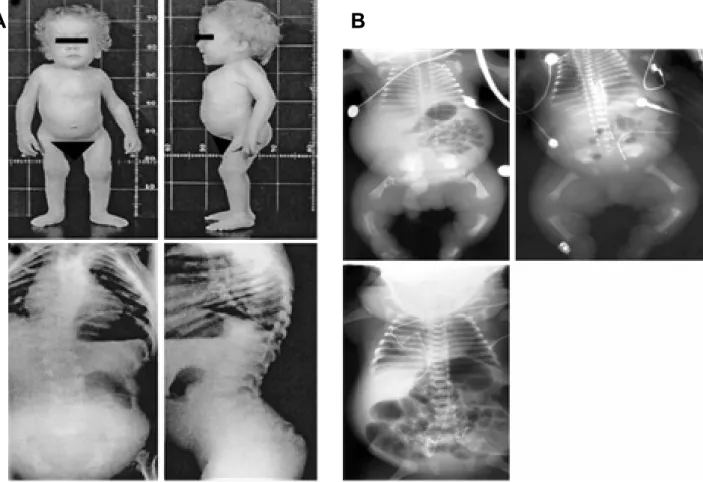
Spondyloepiphyseal dysplasia is characterized by short trunk, short extremities, barrel-shaped chest, kyphosis, severe myopia, retinal detachments, cleft palate and clubfoot (Fig 1.6A).
Radiographs (Fig 1.6B) show defects in ossification of the spine and primarily proximal extremities (Mortier et al. 2000).
Fig 1.6 Phenotype of SEDC patients. (A) Disproportionate short stature with short trunk, enlarged joints, high forehead, frontal bossing, pectus carinatum deformity and radiological findings of anteroposterior and lateral spine radiographs shows: thoracic scoliosis, accentuated lumbar lordosis, flattened and dysplastic vertebral bodies (Sobetzko et al 2000). (B) Anteroposterior radiographs of patients representing a severe phenotype characterised by short tubular bones with metaphyseal irregularities, minimal ossification of vertebral bodies in the thoracic spine, hypoplastic iliac wings with flat acetabular roofs, short ribs along with shortened tubular bones, flattened but normally ossified vertebral bodies, normal acetabular roofs, and a less narrow chest (Mortier et al 2000).
A B
Histological studies show defects resembling those in achondrogenesis type II- hypochondrogenesis and biochemical examination indicates abnormalities in collagen II (Murray et al. 1989). SED refers to a heterogeneous group of disorders that affect the spine and epiphyses, and as the symptoms resemble those of achondrogenesis type II-hypochondrogenesis, SED can be considered to represent the mild end of the spectrum comprising achondrogenesis type II-hypochondrogenesis and SED phenotypes (Spranger et al. 1994). Most cases of SED show autosomal dominant inheritance, but an autosomal recessive form of inheritance has also been suggested in some cases (Harrod et al. 1984). Lee et al. (1989) identified a deletion of an entire exon in the COL2A1 gene, leading to a lack of 36 amino acids in the triple helical region of the chain, and subsequently numerous mutations, including insertions, mutations causing aberrant RNA splicing, glycine mutations and a cysteine for arginine substitution, have been characterized (Vikkula et al. 1994, Kuivaniemi et al. 1997). However, some investigations have excluded the COL2A1 gene as a locus for SED in certain families, indicating that there must also be some other locus or loci for the disorder (Wordsworth et al. 1988, Anderson et al. 1990).
SEDC is of autosomal dominant inheritance and affects the vertebrae, juxtatruncal epiphyses and leads to a lack of ossification of pubis, distal femoral and proximal tibial epiphyses, talus and calcaneues, flattening of vertebral bodies, short limbs, cleft palate, mental retardation, myopia etc. (Gunthard et al. 1995). Previous studies indicate that the processing and assembly of collagen is disrupted but the exact mechanism of pathogenesis is yet to be unraveled.
1.10 Stickler syndrome (OMIM 108300)
Stickler syndrome, originally called hereditary progressive arthroophthalmopathy, is an autosomal dominantly inherited disorder and is caused by mutations in collagen II or collagen XI. The α 3 (XI) chain is the IIB splicing variant product of the COL2A1 gene (Wu & Eyre, 1995). Phenotypically, it is characterised by progressive myopia and vitreoretinal degeneration resulting in retinal detachment and blindness. Midfacial hypoplasia and micrognathia are often accompanied by cleft palate or a lesser degree of clefting, and a sensorineural hearing defect is common (Fig 1.7). The skeletal manifestations include juvenile progressive arthropathy, irregularity of the vertebral bodies and hypermobile joints. Skeletal growth is usually normal.
Mild epiphyseal dysplasia and overtubulation of long bones are seen in radiological examination (Temple 1989, Snead & Yates 1999). Linkage studies have shown that the COL2A1 gene is a locus for Stickler syndrome (Francomano et al. 1987, Knowlton et al. 1989). Altogether nine mutations have been reported to date, all of them leading to premature termination of translation
Introduction (Ahmad et al. 1991, Brown et al. 1992, Ahmad et al. 1993, Ritvaniemi et al. 1993, Brown et al.
1995a, Ahmad et al. 1995, Williams et al. 1996). The COL2A1 gene has also been excluded in several families with Stickler syndrome (Knowlton et al. 1989, Vintiner et al. 1991, Bonaventure et al. 1992).
Fig 1.7 Clinical phenotypes of patients with Stickler syndrome. (A) Probands with mild nasal root hypolasia, midfacial hypoplasia and slender digits. (B) Vitreous phenotypes displaying membraneous congenital vitreous anomaly, beaded appearance of vitreous and a fibrillar vitreous anomaly in comparison with normal vitreous (Richards et al. 2000, 2002a, 2002b).
B A
Stickler families with congenital vitreous anomaly (Stickler syndrome type 1) have linkage to the COL2A1 gene, while in those with congenitally defective vitreous gel architecture but no congenital vitreous anomaly (Stickler syndrome type 2) linkage is excluded (Snead et al.1994).
Subsequently, Richards et al. (1996) demonstrated linkage to the COL11A1 gene in a family with Stickler syndrome type 2, and traced it to a missense mutation converting glycine to valine in the N-terminal part of the triple helix of the α 1(XI) chain. In addition, Wilkin et al. (1998) have suggested still another locus for Stickler syndrome apart from the genes coding for collagens II and XI. It has also been reported that mutations in collagen II in the X position cause haploinsufficiency and that leads to reduced amount of collagen, resulting in characteristic membranous congenital anomaly in the vitreous (Snead et al. 1994, Snead and Yates 1999, Richards et al. 2000).
1.11 Osteoarthritis (OMIM 165720)
Osteoarthritis (OA) is the most common articular disorder, characterized by joint pain and tenderness, stiffness, crepitus and limitation of motion. Radiographs of patients indicate joint- space narrowing, osteophytes, subchondral bone sclerosis and subchondral cyst formation. In severe cases, deformity of the bone ends is also seen. OA is classified into primary (idiopathic) or secondary subsets, in primary cases there is no known predisposing factor, while in secondary cases factors such as trauma, infection, some other joint disorder or a metabolic disease is indicated. Further classification divides OA into localized, in which only certain joints are affected, and generalized subsets. Even though OA is often considered an consequence of ageing, female sex, obesity, occupational load and heavy sports exertion are risk factors (Altman 1995, Creamer & Hochberg 1997). The suggestion of genetic factors underlying OA in certain families was made several decades ago (Stecher et al. 1953, Kellgren et al. 1963). Palotie et al (1989) demonstrated a linkage to the COL2A1 gene in a family with primary OA. Knowlton et al. (1990) made a similar finding in a family with precocious OA and associated mild chondrodysplasia and Ala-Kokko et al. (1990) subsequently demonstrated a mutation in this family. The mutation converted arginine to cysteine, an amino acid not normally found in the triple helical domains of collagens, in the middle of the triple helix of the collagen II molecule.
Altogether five families with this same mutation have been reported to date, and three of them have been shown to have a common ancestor (Bleasel et al. 1995, Kuivaniemi et al. 1997, Bleasel et al. 1998). In addition, a mutation converting arginine to cysteine at position 75, near the N-terminus of the triple helix, has been reported in three families with OA associated with
Introduction mutations in other non collagenous proteins; e.g. a missense mutation in the epidermal growth factor-like domain in matrilin-3 leading to OA in several families. (Stefansson et al. 2003).
Kizawa et al. (2005) identified polymorphism in asporin that suppressed TGF-β-mediated expression of the genes coding for aggrecan (AGC1) and type II collagen (COL2A1) accompanied with reduced proteoglycan accumulation leading to osteoarthritis. Similar forms of osteoarthritis were reported to be due to functional variants within the secreted frizzled-related protein 3 gene (Loughlin et al. 2004).
1.12 Animal models for chondrodysplasias due to collagen II mutations
Transgenic mice carrying a partially deleted human COL2A1 gene developed the phenotype of a chondrodysplasia with dwarfism, short and thick limbs, short snout, cranial bulge, cleft palate, and delayed mineralization of bone (Vandenberg et al. 1991). In cultured chondrocytes from transgenic mice, the minigene was expressed as shortened pro-α-1(II) chains that were disulfide- linked to normal mouse type II collagen chains. Therefore, the phenotype was probably explained by depletion of endogenous mouse type II procollagen through the phenomenon of procollagen suicide. Transgenic mice harboring a glycine-to-cysteine mutation at residue 85 of the triple helical domain of mouse type II collagen displayed severe chondrodysplasia with short limbs and trunk, craniofacial deformities, and cleft palate (Garofalo et al. 1991). Electron microscopic analysis showed a pronounced decrease in the number of typical thin cartilage collagen fibrils, distention of the rough endoplasmic reticulum of chondrocytes, and the presence of abnormally large banded collagen fibril bundles. Garofalo et al. (1991) postulated that the abnormally thick collagen bundles were related to a defect in crosslinking.
A transgenic mouse model of SEDC carrying collagen II transgene with an R789C mutation, in combination with a murine Col2a1 promoter directing the gene expression to cartilage, displayed overall short stature, had shorter limbs with disorganized growth plates, a short nose, cleft palate, and died at birth (Gaiser et al. 2002). The cellular organization of the cartilage architecture was profoundly disturbed in the transgenic growth plate, although the overall polarity was maintained. There were fewerstacks of flattened chondrocytes in the proliferative zone of the transgenic mouse growth plates. In addition to the abnormal cartilaginous tissue, the perichondrium was thicker and electron microscopy revealed a marked reduction of collagen fibrils in cartilage matrix from transgenic compared with that of wild-type mice. Increased distension of rough endoplasmic reticulum was observed in these chondrocytes. Using cell culture experiments and molecular modeling, Gaiser et al. (2002) suggested that this Y-position
mutation acts in a dominant-negative manner, resulting in destabilization of collagen molecules during assembly and reduction in the number of fibrils formed.
1.13 Arginine to cysteine mutations
The majority of chondrodysplasia-causing missense mutations in COL2A1 are substitutions of obligatory glycine residues in the triple helical domain. Only a few non-glycine missense mutations have been reported and among these, the arginine to cysteine substitutions predominate. In addition to the above R75C and R789C substitution (Table 1.1), recently two additional mutations (R365C and R1076C) were found in unrelated probands (Hoornaert et al.
2005).
Most of these chondrodysplasias are due to point mutations in the collagen II gene, altering the amino acid sequence in a way which destabilizes the triple helix. In addition, nonsense mutations have been detected which lead to premature termination of the chain. Deletions have also been reported leading to severe pathological conditions.
Point mutations leading to a change from arginine to cysteine are interesting since different mutations of this kind cause two well characterized clinical phenotypes, i.e Stickler syndrome and congenital spondyloepiphyseal dysplasia (SEDC) and osteoarthritis-associated SED. It is being speculated that Stickler syndrome can be caused by amino acid substitutions in the X position of Gly-X-Y repeats (Richards et al 2000) and substitutions in the Y position rather lead to SEDC. One of the notions derived from previous studies on mutations in collagen I is that the mutant molecules are intracellularly degraded leading to a reduction in the pool of extracellular collagen molecules available for the formation of correct collagen fibrils. Another hypothesis is that some of the mutant collagens are incorporated into collagen fibrils, thereby affecting their biomechanical characteristics (Royce & Steinmann 2002).
Introduction
1.14 Aims of the thesis
The mechanisms by which point mutation in collagen II cause damage to the structure of connective tissues are not fully understood, and some of the proposed pathways leading from a mutant genotype to a phenotype are controversial.
The aims of this thesis were therefore:
1. To investigate the effects of arginine to cysteine mutations in collagen II and to determine how these may lead to different clinical phenotypes.
2. To analyze the biochemical consequences of arginine to cysteine substitutions found in patients with Stickler syndrome and SEDC.
3. To determine the importance of the position of point mutations within the triple helical domain (towards the N- or C-terminal of the triple helix) with respect to triple helical integrity and overall collagen structure.
4. To elucidate the effects of these point mutations on protein trafficking, secretion and cell survival.
2 Materials and methods
2.1 Materials
Standard chemicals and enzymes were if not otherwise mentioned purchased from Merck (Darmstadt), Sigma (Taufkirchen), Invitrogen (Karlsruhe), Biozym (Oldendorf), Roche (Mannheim) or New England Biolabs (Schwalbach).
2.1.1 Enzymes for molecular biology Calf intestinal alkaline phosphatase (NEB)
M-MLV reverse transcriptase (Promega) Restriction endonucleases (NEB)
Ribonuclease A (Sigma) T4 DNA ligase (NEB)
Taq DNA polymerase (Roche)
2.1.2 Materials for cell culture
Dulbecco’s Modified Eagle Medium (DMEM-F12), Invitrogen Non-essential Amino Acids (NEA), Invitrogen
L-Glutamine (200 mM), Invitrogen Penicillin/Steptomycin, Invitrogen
Fetal Bovine Serum (FBS), Biochrom KG, Berlin
2.1.3 Plasmid and adenoviral vectors
2.1.3.1. pBluescript KS+ (pBS KS+) phagemid
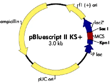
The phagemid vector pBluescript KS+ (pBS KS+) was used for subcloning and was the basic vector used for site directed mutagenesis. Fig. 2.1 shows the map of pBS KS+ vector. The complete manual and sequence information for this vector are available from Stratagene at http://www.stratagene.com/manuals/212205.pdf.
Material and methods
Fig. 2.1 Map of the cloning vector pBS KS+. It consists of an f1 and PUC origin of replication (pUC ori), an ampicillin resistance ORF and a multiple cloning site (MCS) flanked by restriction sites for Kpn I and Sac I.
2.1.3.2. pSL1180 superlinker phagemid
Fig.2.2 Map of the superlinker phagemid pSL1180. It consists of an ampicillin resistance (Ampr) ORF, an M13 origin of replication and a multiple cloning site. The multiple cloning site and restriction sites are shown in detail.
The phagemid vector pSL1180 was used for subcloning of collagen II cDNA after site directed mutagenesis and to remove the endogenous signal peptide. Fig. 2.2 shows the map of pSL1180 vector with details of multiple cloning sites. A complete description and sequence informations from Amersham Biosciences can be found at http://www4.amershambiosciences.com/- aptrix/upp01077.nsf/Content/Products? OpenDocument&parentid=41175&moduleid=41179.
2.1.3.3 Eukaryotic expression vector pCEP-Pu
The mammalian expression vector pCEP-Pu is a derivative of the pCEP-3 vector (Invitrogen) now containing a BM-40 signal peptide and a puromycin resistance (Kohfeldt et al 1997). In addition an enterokinase cleavage site was introduced between the sequence coding for the his6- myc tag and the multiple cloning site (Wuttke et al 2001). This vector was used for expression of all collagen II variants.
Fig.2.3: Map of the eukaryotic expression vector pCEP-Pu (Wuttke et al 2001). This vector contains the signal peptide from BM-40 (BM-40 SP), a his6-myc-tag, the site for protease digestion by enterokinase (EK) and multiple cloning site between the CMV promoter and the SV 40 polyadenylation signal (pA).
2.1.3.4. Adenoviral vector pGS66 and shuttle vector pGS70
The adenoviral vector pGS66 and the shuttle vector pGS70 were kindly provided by G.
Schiedner and S. Kochanek from the Center for Molecular Medicine, University of Cologne, Germany (Schiedner et al. 2000). The adenoviral vectors were used for transfection of primary chondrocytes.
BM-40 SP- his6-myc-EK-collagen II cDNA
Material and methods
Collagen II cDNA from pCEP-Pu and pGS70 vector was digested with Hind III and Bam HI.
After separation by electrophoresis the collagen II cDNA was purified using a gel purification kit. The linearised vector pGS70 was dephosphorylated and purified. Equimolar amounts of vector and insert were used for ligation.
Fig.2.4: Map of adenoviral vectors used for cloning collagen II cDNA. (A) Map of the first generation adenoviral vector pGS66 with an ampicillin resistance (ampr) ORF, an adenoviral internal terminal repeats and a Pac I site for cloning the insert. (B) Map of pGS70 vector with a multiple cloning site for use with different enzymes, a SV40 origin of replication and antibiotic resistance markers for kanamycin (Kanr) and neomycin (Neor).
Collagen II cDNA was released from the vector pGS70 by digesting with the restriction enzyme Pac I. The vector pGS66 was also digested with Pac I and dephosphorylated to avoid self
ligation. Equimolar amounts of vector and insert were used for ligation. 10 µl ligation mixture was used for transformation and colonies obtained were screened for the insert by restriction digestion of the plasmid.
2.2 Molecular biology methods
2.2.1 Plasmid DNA isolation from E. coli
Plasmid DNA was prepared from small scale bacterial cultures. Bacteria were lysed by treatment with a solution containing 1% SDS and 0.5 M NaOH. SDS denatures bacterial proteins and NaOH denatures chromosomal and plasmid DNA. The mixture was neutralized with potassium acetate, causing the plasmid DNA to reanneal rapidly. Most of the chromosomal DNA and bacterial proteins remain in the precipitate, as does SDS forming a complex with the potassium, and are removed by centrifugation. The reannealed plasmid DNA from the supernatant was concentrated by ethanol precipitation.
2.2.2 Pure MIDI plasmid DNA isolation
Highly pure plasmid preparations were purified on either a small or a large scale using kits from Macherey-Nagel (Nucleobond AX kit for small scale plasmid preparations) or Qiagen (Qiagen Midi and Maxi Prep kit for large scale plasmid preparations). Briefly, overnight cultures of bacteria, grown in the presence of an antibiotic marker for the plasmid DNA, were pelleted and the cells were lysed by alkali. The supernantant containing plasmid DNA was passed over silica columns whereby the DNA was bound to the matrix and the impurities and digested RNA were washed off. The plasmid DNA was eluted and concentrated by ethanol precipitation. Highly pure plasmid DNA obtained was washed with 70% ethanol to remove salts and further reconstituted in TE buffer. This plasmid DNA was used for site directed mutagenesis, PCR, sequencing and transfection of eukaryotic cells. The protocols were as described by the manufacturer.
2.2.3 DNA agarose gel electrophoresis 10X DNA loading buffer
40% w/v sucrose 0.5% w/v SDS
0.25% w/v bromophenol blue in TE buffer pH 7.4
50X Tris acetate buffer (1000 ml) 242.2 g Tris
57.5 ml acetic acid 100 ml of 0.5 M EDTA pH 8.0, adjusted with NaOH