Analysis of the functional consequences of p97 mutations leading to the
multisystem disorder IBMPFD
INAUGURAL-DISSERTATION zur
Erlangung des Doktorgrades
der Mathematisch-Naturwissenschaftlichen Fakultät der Universität zu Köln
vorgelegt von
Christian Ulrich Hübbers aus Köln
Druck: Copy-Star GmbH Köln
Referees/Berichterstatter: Prof. Dr. Angelika A. Noegel Prof. Dr. Thomas Langer
Date of oral examination: 21.11.2007
(Tag der mündlichen Prüfung)
The present research work was carried out under the supervision of Prof. Dr. Angelika A. Noegel and Dr. Christoph S. Clemen, in the Institute of Biochemistry I, Medical Faculty, University of Cologne (Cologne, Germany) from November 2004 to November 2007.
Diese Arbeit wurde von November 2004 bis November 2007 am
Biochemischen Institut I der Medizinischen Fakultät der Universität zu Köln
Contents
1. ACKNOWLEDGEMENT... 7
2. INTRODUCTION... 9
2.1. F
UNCTIONAL ROLE OF P97
AND ITS COFACTORS... 9
2.2. F
UNCTION OF P97
IN THEERAD
PATHWAY... 11
2.3. T
HE ALTERNATIVE DEGRADATION SYSTEM AUTOPHAGY IS FUNCTIONALLY COUPLED TO THEUPS ... 12
2.4. C
HARACTERIZATION OF THE DISEASEIBMPFD ... 13
2.5. C
LINICAL OBSERVATIONS IN PATIENTS INVOLVED IN THIS STUDY... 14
2.6. S
CIENTIFIC QUESTIONS AND WORKING PLAN... 17
3. RESULTS ... 18
3.1. M
ORPHOLOGICAL,
ULTRASTRUCTURAL AND BIOCHEMICAL ANALYSIS OF THE SKELETAL MUSCLE AND CARDIAC PATHOLOGY... 18
3.1.1. Skeletal muscle pathology... 18
3.1.2. Cardiac pathology ... 22
3.1.3. p97 protein expression in IBMPFD muscle ... 22
3.1.4. Analysis of normal and IBMPFD primary human myoblasts ... 24
3.2. G
ENERATION AND CHARACTERIZATION OF WT AND MUTANT P97
CONSTRUCTS... 26
3.2.1. Immunofluorecence analysis of p97 overexpressing cells ... 27
3.2.2. 1D- and 2D SDS-PAGE followed by immunoblotting of p97 overexpressing cells and primary myoblasts ... 33
3.2.3. Cell proliferation assay... 36
3.3.
P97
RESPONSE TO CELLULAR STRESS... 37
3.3.1. Use of various cell stressors to induce p97 aggregates ... 37
3.3.2. Time course of aggregate formation and clearance ... 38
3.4. S
TUDY OF PROTEASOME ANDERAD
ACTIVITIES... 40
3.4.1. Proteasomal activity ... 40
3.4.2. Defects in ERAD activity in the presence of mutated p97... 42
3.4.3. Fluorescence protease protection assay analysis of ER and Golgi morphology ... 46
3.4.4. Fluorescence protease protection assay of autohagosomes ... 51
3.5. F
ILTER ASSAY COMBINED WITHGC/MS
ANALYSIS OF PUTATIVE LIGANDS BINDING TO RECOMBINANT P97 ... 54
3.6. G
ENERATION OF AR155C-
P97
MOUSE KNOCK-
IN TARGETING VECTOR... 55
4. DISCUSSION ... 58
4.1. C
LINICAL PHENOTYPE... 58
4.2. A
NALYSIS OF THE P97
PROTEIN EXPRESSION AND LOCALIZATION... 59
4.3. S
TRUCTURAL ANALYSIS OF WILD TYPE P97
VERSUSR93
ANDR155
MUTANT P97 ... 60
4.3.1. In silico screening of small ligand binding ... 64
4.4. A
NALYSIS OF PROTEASOME ANDERAD
ACTIVITY... 66
4.5. R
OLE OF P97
IN THE REGULATORYERAD
PATHWAY... 67
4.6. O
UTLOOK... 68
5.1.2. Transfection methods ... 71
5.1.3. Generation of growth curves ... 72
5.1.4. Cell stress experiments ... 72
5.2. I
MAGING... 73
5.2.1. Indirect immunofluorescence and imaging of living cells... 73
5.2.2. Fluorescence protein protection (FPP) assay ... 74
5.3. G
EL ELECTROPHORESIS ANDW
ESTERN BLOTTING... 75
5.4.
P97-
CDNA,
SITE-
DIRECTED MUTAGENESIS AND PLASMIDS: ... 76
5.5. G
ENERATION OF MOUSE TARGETING VECTOR... 77
5.6. H
ISTOLOGICAL ANALYSIS... 78
5.7. A
NTIBODIES... 78
5.8. P
ROTEIN PURIFICATION... 80
5.9. F
ILTER ASSAY... 81
5.10. P
ROTEASOME ASSAY... 81
5.11. S
TATISTICAL ANALYSIS... 83
6. REFERENCES... 84
7. ABBREVIATIONS ... 98
8. ABSTRACT ... 99
9. ZUSAMMENFASSUNG ... 100
10. ERKLÄRUNG... 101
11. CURRICULUM VITAE / LEBENSLAUF ... 102
Acknowledgement
1. Acknowledgement
This work would never have been accomplished without the helpfulness, willingness to share knowledge and support of many people.
I would like to thank them:
At first I have to thank my supervisor Dr. Christoph Clemen for his guidance throughout the course of this investigation and for always having an open eye and ear when discussing my work and writings.
I am deeply grateful to Prof. Angelika A. Noegel for offering me the opportunity to perform my PhD work in her lab and for all the thoughts and helpful suggestions.
I also owe my thanks to Prof. Rolf Schröder for supporting and discussing my work.
I thank Prof. Andreas Hofmann (Brisbane, Australia), Dr. Anett Böddrich (Berlin) and Prof. Franz-Georg Hanisch (Cologne) for collaboration.
My special thanks also go to Maria who always gave me excellent technical support and introduced me to the established methods in this lab.
I thank Berthold for fire fighting and for the help in all cell culture matters.
I have to thank Budi and Gudrun for giving me plenty of rope in all computer
matters, Dörte for her help in administration issues, Bärbel, Brigitte and Sonja for
preparing all this little common things that made laboratory work so easy and
effective, and Martina, Rolf and Rosi for technical support in special issues.
Thanks also to the present and former members of Lab 12: André, Charles-Peter, Karthik, Markus, Raphael, Soraya and Vasily. Laboratory work was never boring with you.
All the present and former people I spent my time during the work in this institute, Eva-Maria, Jessica, Georgia, Carola, Marion, Francisco, Wenshu, Ria, Yvonne, Verena, Hafida, Sascha, Åsa, Akis, Hua, Mary, Subhanjan, Anne, Tanja, Deen, Vivek, Surayya, Rashmi, Jianbo, Rui, Marcel, Ludwig, Monika, Julia, Kristina, Claudia, Anke, Kira, Andrea, Maarten, Thomas, Dominic and Prof. Stefan Höning.
Thank you for discussing my work, all the birthday cakes and other things in everyday’s live that made my time interesting in scientific and personal matters.
.
My parents I have to thank for all the support during my studies and in all other circumstances!
Steffi, I thank you for all the power and support. Your love means everything to me!
Christian
Introduction
2. Introduction
2.1. Functional role of p97 and its cofactors
Mammalian p97 is an ubiquitously expressed member of the type II AAA (ATPase associated with various activities) ATPase family. Highly conserved orthologs are e.g. expressed in the archaebacterium Sulfobus acidocaldarius (SAV), Saccharomyces cerevisiae (CDC48), Dictyostelium discoideum (cdcD), Arabidopsis thaliana (AtCDC48), and Drosophila melanogaster (TER94) thus demonstrating that p97 is an essential and evolutionary highly conserved cellular protein (Moir et al., 1982; Feiler et al., 1995; Confalonieri, et al. 1994; Pinter et al., 1998;
Bakthavatsalam et al., 2007). The unfavourable name VCP (for valosin containing protein) was initially given to p97 after trying to purify the hormone peptide valosin but turned out to be an artefact (Koller and Brownstein, Nature 1987). p97 has a tripartite structure comprising an N-terminal domain (CDC 48) involved in ubiquitin binding, and two central D1- and D2-domains, which provide ATP binding and hydrolysis (DeLaBarre and Brünger, 2003). p97 assembles into functional hexamers with a central cylinder formed by the D-domains surrounded by the N-domains. The D1-domain is mainly responsible for p97 hexamerization while the D2-domain provides the major ATPase activity to p97 that enables it to function as a molecular chaperone interacting with a diverse group of adaptors to perform specific cellular functions (Zhang et al., 2000).
p97 has been associated with a wide variety of essential cellular processes
comprising nuclear envelope reconstruction, the cell cycle, postmitotic Golgi
proteasome protein degradation system and the endoplasmic reticulum associated protein degradation (ERAD) pathway (Kondo et al., 1997; Rabouille et al., 1998;
Meyer et al., 2000; Hetzer et al., 2001; Ye et al., 2001; Rabinovich et al., 2002; Ye et al., 2004; Lilley and Ploegh, 2005).
Most, if not all of these functions seem to be directly linked to p97’s ability to bind (multi-)ubiquitinated proteins and to segregate them from their binding partners, or to extract them from protein complexes (Rape et al., 2001; Meyer et al., 2002). The association of p97 with substrates is still not fully understood (Jentsch and Rumpf, 2007). In vitro binding studies indicate a direct interaction of ubiquitinated substrates with the N-domain of p97 (Dai and Li, 2001; Rape et al., 2001). However, the more common mechanism seems to be the indirect binding of p97’s N-terminal domain to ubiquitinated substrates mediated by cofactors (Rape et al., 2001; Meyer et al. 2002;
Hartmann-Petersen et al., 2004; Schuberth et al., 2004; Richly et al., 2005).
Apart from these ‘segregase’ functions, p97 seems to influence the degree of
ubiquitination of the bound substrates (Hartmann-Petersen et al., 2004; Richly et al.,
2005). Hence, so-called ‘substrate-processing cofactors’ together with p97 can either
promote polyubiquitination, inhibit polyubiquitination or deubiquitinate
(multi-)ubiquitinated substrates (Koegl et al., 1999; Uchiyama et al., 2002; Burnett et
al., 2003; Wang et al., 2004; Richly et al., 2005; Rumpf and Jentsch, 2006).
Introduction
2.2. Function of p97 in the ERAD pathway
The ubiquitin-proteasome system (UPS) is a major degradation system for short- lived proteins (Hershko and Ciechanover, 1998). Proteins to be degraded are labeled with ubiquitin. Ubiquitin is conjugated through its carboxy terminus usually to ε-amino groups of lysine residues. This conjugation typically involves three types of
enzyme: E1 (ubiquitin-activating enzyme) hydrolyses ATP and forms a thioester- linked conjugate between itself and ubiquitin; E2 (ubiquitin-conjugating enzyme) receives ubiquitin from E1 and forms a similar thioester intermediate with ubiquitin;
and E3 (ubiquitin ligase) binds both E2 and the substrate, and transfers the ubiquitin to the substrate. The ubiquitin itself can form a substrate for further rounds of ubiquitination, resulting in the formation of a polyubiquitin chain. Chains of four or more ubiquitin molecules (linked by lysines at residue 48) appear to form a recognition signal that allows substrates to be degraded by the 26S proteasome complex (Richly et al., 2005; Rubinsztein, 2006). The degradation is thus specifically targeted to selected proteins. Prompt removal of these proteins is critical to the precise and timely regulation of intracellular signaling involved in multiple cellular processes.
The endoplasmic reticulum-associated degradation (ERAD) pathway degrades initially endoplasmic reticulum (ER) localized protein substrates through the ubiquitin-proteasome system (Bonifacino and Weissman, 1998; Hampton, 2002).
The ER is an intracellular membranous structure that performs such important
functions as protein post-translational modifications, protein folding and
membrane proteins as well as normal ER-resistant proteins for regulatory purposes.
As the ubiquitin-proteasome system is absent from the ER lumen, these proteins have to be retrotranslocated to the cytosol or extracted from the ER membrane. p97 together with the cofactors Ufd1 and Npl4 is required for most ERAD substrates in an unclear mechanism which retrotranslocates or extracts or segregates them from a proposed retrotranslocation channel (Bays et al., 2001; Ye et al., 2001; Jarosch et al., 2002; Rabinovich et al., 2002; Braun et al., 2002; Elkabetz et al., 2004). Further general and/or specialized components like Ufd2, the homologues Rad23/Dsk2 and Ubx2 are involved in ERAD function (Medicherla et al., 2004; Schuberth et al., 2004; Richly et al., 2005)
2.3. The alternative degradation system autophagy is functionally coupled to the UPS
Macroautophagy (referred as autophagy hereafter) is the second major intracellular
degradation system. Unlike the UPS, autophagy is mainly responsible for the
degradation of long-lived proteins and other cellular contents (Levine and Klionsky,
2004; Lum et al., 2005). Autophagy starts with the formation of double-membrane-
bounded structures known as autophagosomes, or autophagic vacuoles (AVs). These
fuse with lysosomes to form autophagolysosomes, and their contents are then
Introduction
functionally coupled and suppression of UPS activates autophagy (Brodsky and Scott, 2007; Ding et al., 2007; Pandey et al., 2007).
2.4. Characterization of the disease IBMPFD
Autosomal dominant inclusion body myopathy (IBM) associated with Paget disease of the bone (PDB) and frontotemporal dementia (FTD), or IBMPFD (OMIM 605382), is a late-onset human multisystem disorder caused by mutations of the p97 protein on chromosome 9p13-p12 (Watts et al., 2004; Haubenberger et al., 2005;
Schröder et al., 2005). Apart from R191Q and A232E mutations, which reside in the N-D1-linker region and D1-domain, respectively, all other pathogenic mutations described so far are located in exons coding for the CDC48 domain of the p97 protein (Watts et al., 2004; Haubenberger et al., 2005; Schröder et al., 2005).
Fig. 1. Domain structure of the p97 protein:
CDC48 domain composed of double ψ barrel (amino acids 25–106, orange) and the
four-stranded β barrel (amino acids 112–186, cyan), connected by a short linker
region (amino acids 107-111, green). The CDC48 domain connects the
D1-AAA-ATPase domain (amino acids 208-459, blue) by a linker region (amino
acids 187-208, yellow). Linker region L2 (dark green), second AAA-ATPase domain
(amino acids 481-761, D2, dark blue) and C-domain (amino acids 762-806, grey)
are indicated. Mutations detected in our three German IBMPFD patients affect
A further pathogenic link of p97 to protein degradation pathways is highlighted by the observation that p97-positive protein aggregates have been documented in skeletal muscle and in neurons of the central nervous system of IBMPFD patients (Watts et al., 2004; Schröder et al., 2005). In neurons, these p97-positive inclusions are exclusively present in the nucleus, whereas in skeletal muscle only cytoplasmic p97-positive aggregates have been reported. However, p97-positive aggregates are not specific for IBMPFD and have been documented in a wide variety of neurodegenerative disorders comprising Parkinson’s disease, Lewy Body disease, Huntington’s disease, amyotrophic lateral sclerosis, and spinocerebellar ataxia type III (SCAIII; Machado-Joseph disease) (Hirabayashi et al., 2001; Mizuno et al., 2003; Nan et al., 2005). p97 directly interacts with ataxin-3, the protein mutated in SCAIII, and recent in vivo studies using Drosophila demonstrated that p97 selectively modulates aggregation and neurotoxicity induced by pathogenic ataxin-3 (Böddrich et al., 2006).
2.5. Clinical observations in patients involved in this study
Clinical data and biopsy material from three German patients suffering from
IBMPFD are included in this study (Schröder et al., 2005; Hübbers et al., Brain
2007). Clinical investigation of patients and probes derived from these patients was
Introduction
weakness of the lumbar trunk was noticed. Repeated neuropsychiatric evaluation showed evidence of progressive personality changes and cognitive decline due to frontotemporal brain dysfunction. Axial computed tomography revealed Paget-like bone changes in the right hip. Mutation analysis revealed a novel heterozygous nucleotide substitution from arginin to cystein in exon 3 (p.Arg93Cys) of the p97 gene (GenBank AC004472; Hübbers et al., 2007).
Patient II was a 62-year-old female patient harbouring of a progressive proximal muscle weakness and frontal and temporal brain atrophy leading to a severely demented patient with a flaccid, predominantly proximal tetraparesis. The mother of the reported patient, her mother’s brother as well as one of his children suffered from similar medical conditions. She died of pneumonia and cardiac failure in 1998.
Autopsy at that time showed severe generalized wasting of her skeletal muscles, but no signs of Paget’s disease of the bone. The total heart weight was 480 g; left ventricular and right ventricular wall thickness was 1.7 and 0.7 cm, respectively.
Neurons exhibited nuclear inclusions containing p97- and ubiquitin-containing material. A heterozygous mutation in exon 5 leading to an amino acid substitution from arginine to cystein in codon 155 was identified (p.Arg155Cys; Schröder et al., 2005). Additionally, mutations of the desmin and αB-crystallin genes were ruled out by direct sequence analysis in this patient.
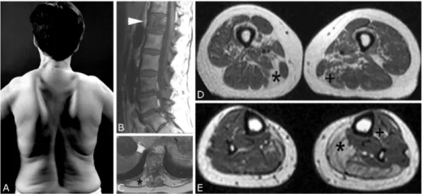
Patient III is a 54-year-old female. She gave a 30-year history of slowly progressive
muscle weakness and atrophy predominantly affecting her shoulder girdle, trunk and
distal leg muscles (Fig. 2A). Paget’s disease of the bone confined to the first lumbar
vertebra (Fig. 2B). A detailed neuropsychological evaluation suggested mild
weakness and atrophy of her scapular fixator muscles (deltoid, rhomboid, supra- and infraspinatus) and trunk extensors. In addition, she had slight to moderate muscle weakness of her finger extensor, hip flexor and distal leg muscles. Whole-body MRI demonstrated widespread muscular involvement with pronounced signal changes in her erector spinae, hamstring and calf muscles (Fig. 2C, D and E). p97 mutation analysis of a DNA sample revealed a heterozygous nucleotide substitution causing an amino acid substitution in the same codon as in patient II from arginine to histidine (p.Arg155His).
Fig. 2. Clinical and MRI findings in Patient III.
(A) The marked scapular winging and lumbar lordosis may be noted. (B) The sagittal view of the lumbar spine reveals a stripy ossification of the first lumbar vertebra (arrowhead) due to Paget’s disease. T1-weighted TSE-sequence with 600/12 ms (TR/TE), 4 mm slice-thickness. (C) The cross-cut view at the level of the thoracic spine demonstrates a complete fatty replacement of the erector spinae muscles (*).
T1-weighted TSE-sequence with 450/17 ms (TR/TE), 5 mm slice-thickness. (D) The
Introduction
2.6. Scientific questions and working plan
In summary this study was initiated to answer questions regarding the pathogenesis of the IBMPFD disease, which was described recently. To date, information about the complex pathology of the IBMPFD disease and experimental data about the cell biology and biochemistry were limited. To illuminate mechanisms that are involved in the development of this late onset and ultimatively lethal disease, the following questions were addressed:
• What are the morphological and ultrastructural changes in the IBMPFD diseased striated muscle tissue?
• Does mutant p97 induce changes in its post-translational modification or subcellular localization?
• Can a cell culture model of the IBMPFD disease be established and do p97 overexpressing cells exibit a phenotype similar to that of IBMPFD diseased tissue? Is a cell culture model capable to reproduce the formation of pathological protein aggregates?
• Are known pathways in which p97 is involved impaired by the p97 mutations? Are ERAD and proteasomal degradation activities affected?
• What are the consequences of p97 mutations on the molecule structure and which functional conclusions can be drawn out of it?
To address these questions, a variety of histological, molecular and cell biological,
and in silico investigations were performed.
3. Results
3.1. Morphological, ultrastructural and biochemical analysis of the skeletal muscle and cardiac pathology
3.1.1. Skeletal muscle pathology
Morphological evaluation of a vastus lateralis and tibial anterior biopsy from Patient I showed severe degenerative changes consisting of increased fibre size variation, atrophy of both fibre types, presence of terminal atrophic and angulated fibres, hypertrophic type-1 fibres, degenerating and a few regenerating fibres,
‘myopathic grouping’ as well as marked fatty replacement of muscle fibres and
broadening of connective tissue (Fig. 3A). A diagnostic muscle biopsy taken from
the biceps brachii muscle of Patient II displayed the classical myopathological
picture of an IBM with an abundance of rimmed vacuoles (Fig. 3B). In contrast, the
biopsy from the vastus lateralis muscle in Patient III showed only mild and
unspecific myopathological changes consisting of type I fibre predominance,
atrophic and hypertrophic fibres (Fig. 3C). In addition, few de- and regenerating
fibres could be demonstrated. It is noteworthy that in biopsies from Patient I and III
only a few fibres with rimmed vacuoles could be detected. None of the three reported
cases showed inflammatory infiltrates.
Results
Fig. 3. Morphological analysis of IBMPFD muscle.
(A) Biopsy from Patient I revealed severe degenerative muscle changes. The marked fatty replacement of muscle fibres, broadening of connective tissue, rounding and atrophy of muscle fibres and hypertrophic fibres may be noted. The arrow denotes a hypertrophic fibre with a central rimmed vacuole. (B) Biopsy from Patient II showed the classical picture of an IBM with an abundance of rimmed vacuoles. (C) Biopsy from Patient III showed only mild and unspecific myopathological changes. Fibres containing rimmed vacuoles are marked by arrows. [Haematoxylin and Eosin staining; bars: (A) 100 µm, (B) 50 µm, (C) 60 µm].
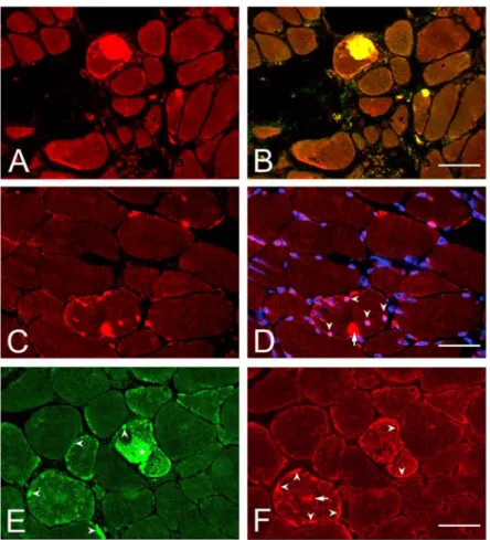
Double-immunofluorescence analysis of skeletal muscle from Patient III revealed a
small number of fibres (<5%) containing cytoplasmic foci of p97- and ubiquitin-
positive protein aggregates (data not shown), whereas the corresponding analysis of
the muscle biopsy from Patient II showed a high number of fibres (30-40%) with
single or multiple p97- and ubiquitin-positive cytoplasmic inclusions (Fig. 4A and
B). In addition, double-staining with p97 antibody and DAPI
(4’,6’-diamidino-2-phenylindoledihydrochloride) revealed multiple fibres with
p97-positive nuclear inclusions (Fig. 4C and D). Further analysis revealed multiple
fibres displaying subsarcolemmal and cytoplasmic areas with increased αB-crystallin
(Fig. 4E) and desmin labeling (Fig. 4F).
Fig. 4. Indirect immunofluorescence analysis of IBMPFD muscle from Patient II.
(A) p97 labeling of cytoplasmic aggregates. (B) p97 and ubiquitin double-immunofluorescence labeling of cytoplasmic aggregates. (C) p97 labeling of cytoplasmic and nuclear aggregates. (D) p97 and DAPI labeling. The presence of cytoplasmic (red, arrow) and nuclear (pink, arrowheads) aggregates may be noted.
(E) Pathological αB-crystallin staining with positive labeling of a giant cytoplasmic (*) and multiple small subsarcolemmal aggregates (arrowheads). (F) Pathological desmin staining in two muscle fibres displaying increased subsarcolemmal (arrowheads) and cytoplasmic areas (arrow) with increased desmin immunolabeling.
Bars: (B) 70 µm, (D) 50 µm, (F) 40 µm.
A detailed ultrastructural analysis was performed on skeletal muscle from Patient II.
Results
(Fig. 5B). Multiple fibres also displayed areas with granulofilamentous material as seen in the group of myofibrillar myopathies (Fig. 5C). Immunogold EM showed a dense desmin-positive labeling of these areas (Fig. 5D).
Fig. 5. Ultrastructural analysis of skeletal muscle from Patient II.
(A) Filamentous nuclear inclusion (*); arrows indicate the nuclear membrane. (B)
Cytoplasmic area with loosely and densely packed filamentous material. (C) The
arrows denote an area with granulofilamentous material. (D) Immunogold electron
microscopy with the monoclonal anti-desmin antibody (mab-D33) and a secondary
antibody coupled to 10 nm gold particles showed a dense labeling of filamentous
aggregates. Bars: (A) 0.5 µm, (B) 0.6 µm, (C) 0.7 µm, (D): 0.25 µm.
3.1.2. Cardiac pathology
Post-mortem analysis of the heart of Patient II revealed a marked left ventricular dilatation and thickening of the left ventricular wall (Fig. 6A). Histopathological examination showed cellular hypertrophy of myocytes and in conjunction with multiple small parenchymal scars in both ventricles. Immunostaining of formalin- fixed and paraffin-embedded cardiac tissue revealed multiple cardiomyocytes displaying ubiquitin-positive cytoplasmic and single nuclear inclusions (Fig. 6B and C).
Fig. 6. Cardiac pathology in IBMPFD.
(A) Post-mortem image of the heart from Patient II displaying left ventricular dilatation (*) and thickening of the left ventricular wall (brace). (B and C) Ubiquitin immunostaining of cardiac muscle tissue. The presence of cytoplasmic (arrows) and intranuclear (arrowhead) ubiquitin-positive inclusions may be noted. (B and C) Alkaline phosphatase anti-alkaline phosphatase staining (APAP). Bars: (B) 50 µm, (C) 15 µm.
3.1.3. p97 protein expression in IBMPFD muscle
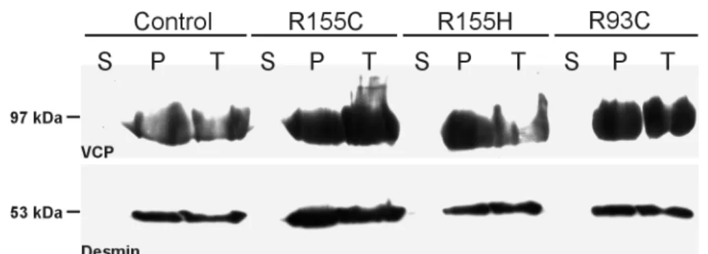
We performed 1D- and 2D-SDS-PAGE in conjunction with western blotting of total
Results
transient transfection experiments using p97-R155H-GFP and p97-R95G-GFP constructs (Weihl et al., 2006). To study the distribution of p97 between soluble and insoluble muscle protein fractions, we performed differential centrifugation of muscle tissue lysates. However, in IBMPFD and normal control muscle p97 was exclusively found in the pellet fraction of IBMPFD and normal control muscle (Fig.
7).
Fig. 7. p97 immunoblot analysis of normal and IBMPFD muscle after differential centrifugation.
Western blotting of equal amounts of total protein extracts (T), soluble (S), and pellet fractions (P) from normal (Control) and diseased (R155C, R155H, R93C) skeletal muscle after centrifugation at 100.000 x g. Desmin labeling (53 kDa) was used as an internal loading control. p97-immunoblotting detected a single band corresponding to a molecular weight of ~97 kDa in pellet and total protein fractions of all probes analysed.
p97 immunoblotting after 2D gel electrophoresis of total protein extracts from
normal human skeletal muscle revealed a prominent spot at pH 5.20. In addition, a
second spot with reduced signal intensity was detected at the position of pH 5.16,
which corresponds well with the calculated pI 5.14. A corresponding analysis of
diseased skeletal muscle (R155C) showed an identical pattern compared to the
normal human control muscle (Fig. 8).
Fig. 8. 2D gel electrophoresis followed by p97 indirect immunoblot analysis of total protein extracts from normal and diseased (R155) human skeletal muscle.
A mixture of both samples was loaded as internal control to confirm that the detected spots of normal and diseased proteins correspond to each other.
3.1.4. Analysis of normal and IBMPFD primary human myoblasts
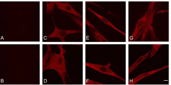
In order to study pathological protein aggregate formation in cultured cells, we analysed normal and IBMPFD (R155C-p97 mutant) primary human myoblasts.
Immunostaining using FK2 (Fig. 9C, D) and p97 antibodies (data not shown) revealed an identical reticular staining pattern in normal and IBMPFD myoblasts. In contrast to IBMPFD muscle, no pathological protein aggregate formation could be detected. Even treatment with clasto-lactacystin β-lactone (irreversible 20S proteasome inhibitor) or MG132 (reversible 26S proteasome inhibitor) did not induce protein aggregates (Fig. 9E – H).
SM WT SM R155C SM WT+R155C
pH 5.20
pH 5.16
Results
Fig. 9. Confocal immunofluorescence images of IBMPFD (A, C; E and G; R155C) and normal (B, D, F and H) primary human myoblasts.
Cells were stained with an antibody directed against poly-ubiquitin (FK2). (A and B) Controls lacking the primary antibody. (C and D) untreated cells, (E and F) MG132 treated cells, (G and H) Lactacystin treated cells. Note that both normal and IBMPFD myoblasts display an identical reticular FK2-staining pattern without any evidence of FK2-positive protein aggregates in treated and untreated cells; bar, 20 µm.
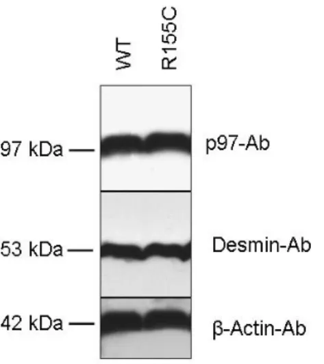
We also performed indirect immunoblotting after gel electrophoresis of untreated
total cell lysates from these primary myoblasts (Fig. 10). p97 staining (upper pannel)
showed equal signals for both mutated and control muscle, thus indicating that there
is no obvious influence of the mutation on the total amount of p97 protein
expression. Desmin (middle panel) was used to confirm the myoblast nature of the
primary cells and as an internal loading control, respectively. β-actin (lower panel)
was used as an additional internal loading control.
Fig. 10. Immunoblot analysis of total protein extracts from cultured normal and IBMPFD (R155C-p97 mutant) primary myoblsts.
Note the equal p97 signal intensities in samples from normal and IBMPFD myoblasts. Desmin was used to confirm the myoblasts nature of the primary cells and as an internal loading control, respectively. In addition, β -Actin was used as an internal loading control.
3.2. Generation and characterization of wt and mutant p97 constructs
We generated wild type and mutant (R93C, R155C, R155H) cDNA constructs to
study the effects of p97 protein mutants at the cellular level (Fig. 11). The following
transfection and transduction experiments were performed: (i) Wt- and
mutant-GFP-p97-FLAG and p97-FLAG-GFP were transiently and stably expressed
in HEK293 cells; (ii) wt- and mutant-p97-FLAG-GFP were transiently expressed in
C2F3 myoblasts; (iii) wt- and the R155C-p97-FLAG mutant were stably expressed in
C2F3 myoblasts.
Results
Fig. 11. Schematic representation of constructed p97 vectors.
Upper panel: Representation of the endogenous p97 protein. Panel 2 – 4: Full length p97 constructs for the expression of wt, R93C, R155C, and R155H proteins were constructed with an additional FLAG-Tag at the C-terminus (all constructs) and a GFP was fused to the C-terminus (2) or N-terminus (3) for fluorescence detection.
Lower panel: Partial p97 construct for the expression of the first 208 amino acids of wt, R155C and R155H protein representing the N-domain and the N-D1 linker were fused to a FLAG-tag.
3.2.1. Immunofluorecence analysis of p97 overexpressing cells
Transfected cells were analysed by life cell imaging and indirect immunofluorescence analysis after methanol or paraformaldehyde fixation.
Expression of either wt-p97-constructs in HEK293 cells resulted in an intense
labeling of the entire cytoplasm and, inconsistently, in a less intense nuclear signal of
the GFP fusion proteins (Fig. 12A-O and Fig. 13A-O). All N- or C-terminally tagged
full-length mutant p97 constructs showed the same localization as endogenous p97,
with no evidence of abnormal cytoplasmic protein aggregate formation in HEK293
and C2F3 cells, as it was presented earlier by Weihl et al. (2006). Transfection of
GFP alone yielded in a strong uniform labeling of both the cytoplasm and the nucleus (Fig. 12A, F, K and Fig. 13A, F, K).
To address the issue of protein aggregate formation in a more physiological setting, we performed stable transfections of HEK293 and C2F3 cells. Two months after the initial transfection, cells were analysed by life cell imaging. The localization of the three p97 mutants again was indistinguishable from wt-p97, with no evidence of protein aggregate formation (data not shown). Triton X-100 treatment before or after fixation of HEK293 cells did not unmask any protein aggregates (data not shown).
Additionally, we performed indirect immunofluorescence analysis of the transfected
HEK293 cells using antibodies directed against p97, FLAG and poly-ubiquitinated
proteins (FK2). Here, p97 (Fig. 12P-T and Fig. 13P-T) and FLAG (data not shown)
labeling showed a pattern analogous to N- or C-terminally GFP- or FLAG-tagged
wt- and mutant-p97 constructs in living and fixed cells. The FK2 antibody, a
sensitive marker for pathological aggregates containing poly-ubiquitinated proteins,
showed a diffuse cytoplasmic staining with occasional small foci displaying
accentuated FK2 immunolabeling in the cytoplasm and nucleus of non-transfected
(data not shown) as well as transfected (wt-, R93C-, R155H-, R155C-p97) HEK293
cells (data not shown).
Results
Fig. 12. Confocal images of HEK293 cells stably overexpressing wt- or mutant p97 protein fused to N-terminal GFP.
(A – E) Living cells, GFP signal; (F – J) Methanol fixation, GFP signal; (K – O)
Paraformaldehyde fixation, GFP signal; (P-T) Indirect immunofluorescence of
paraformaldehyde fixated cells, p97 staining. (A) GFP Vector control, (B) GFP-wt-
p97-FLAG, (C) GFP-R93C-p97-FLAG, (D) GFP-R155C-p97-FLAG, (E) GFP-
R155H-p97-FLAG. (F) GFP Vector control, (G) GFP-wt-p97-FLAG, (H) GFP-
R93C-p97-FLAG, (I) GFP-R155C-p97-FLAG, (J) GFP-R155H-p97-FLAG. (K) GFP
Vector control, (L) GFP-wt-p97-FLAG, (M) GFP-R93C-p97-FLAG, (N) GFP-
R155C-p97-FLAG, (O) GFP-R155H-p97-FLAG, (P) p97 staining control lacking
primary antibody, (Q) GFP-wt-p97-FLAG, (R) GFP-R93C-p97-FLAG, (S) GFP-
R155H-p97-FLAG, (T) GFP-R155C-p97-FLAG. Bar, 20 µm.
Fig. 13. Confocal images of HEK293 cells stably overexpressing wt- or mutant-p97 protein fused to a C-terminal GFP.
(A – E) Living cells, GFP signal; (F – J) Methanol fixation, GFP signal; (K – O)
Paraformaldehyde fixation, GFP signal; (P-T) Indirect immunofluorescence of
paraformaldehyde fixated cells, p97 staining. (A) GFP Vector control, (B) wt-p97-
FLAG-GFP, (C) R93C-p97-FLAG-GFP, (D) R155C-p97-FLAG-GFP, (E) R155H-
p97-FLAG-GFP, (F) GFP Vector control, (G) wt-p97-FLAG-GFP, (H) R93C-p97-
FLAG-GFP, (I) R155C-p97-FLAG-GFP, (J) R155H-p97-FLAG-GFP, (K) GFP
Vector control, (L) wt-p97-FLAG-GFP, (M) R93C-p97-FLAG-GFP, (N)R155C-p97-
FLAG-GFP, (O) R155H-p97-FLAG-GFP, (P) p97 staining control lacking primary
antibody, (Q) wt-p97-FLAG-GFP, (R) R93C-p97-FLAG-GFP, (S) R155H-p97-
FLAG-GFP, (T) R155C-p97-FLAG-GFP. Bar, 20 µm.
Results
In order to rule out effects of the GFP-tag, we retrovirally transduced C2F3 (a subclone of C2C12) myoblasts using wt-p97-FLAG and R155C-p97-FLAG expression constructs. Anti-FLAG- (Fig. 14C and D) and anti-p97-staining (data not shown) revealed an intense labeling of the entire cytoplasm and, inconsistently, a less intense nuclear signal. Transduced C2F3 cells differentiated into myotubes showed the same results (Fig. 14E and F). However, neither myoblasts nor up to 6-day-old myotubes showed any evidence of protein aggregates.
Fig. 14. Confocal immunofluorescence images of retrovirally transduced C2F3 myoblasts and myotubes overexpressing wt- or mutant-p97-FLAG protein.
Cells were stained with an antibody directed against the FLAG-epitope. Controls,
immunofluorescence images of untransduced cells (A) and of wt-p97-FLAG
expressing myoblasts lacking the primary antibody (B). Distribution of wt-p97-FLAG
in myoblasts (C) and differentiated myotubes (E). Localization of R155C-p97-FLAG
in myoblasts (D) and differentiated myotubes (F); bar, 20 µm.
The N-domain of p97 is reported to be crucial for cofactor-binding. To induce possible dominant negative-like effects, we also generated p97 plasmids for stable expression of the first 208 amino acids of the N-terminus of p97 (Fig. 11; wt, R155H and R155C) and analyzed HEK293 cells for possible differences in the localization of the wild type and mutant N-terminal peptides (Fig. 15). Indirect immunofluorescence analysis of paraformaldehyde fixated cells showed an intense labeling of the cytoplasm with no apparent differences between wt and mutant p97 and, especially, no hints for aggregate formation.
Fig. 15. Confocal immunofluorescence images of transfected HEK293 cells
overexpressing wt and mutant N-terminal peptide fragments of p97.
Results
3.2.2. 1D- and 2D SDS-PAGE followed by immunoblotting of p97 overexpressing cells and primary myoblasts
For further biochemical analysis of our transfected HEK293 cells, we performed immunoblotting of total protein extracts using p97, GFP, FLAG and FK2 antibodies (Fig. 16). p97 immunoblotting labelled the endogenous p97 protein as well as the GFP–p97 fusion protein. The GFP and FLAG antibodies exclusively detected the respective fusion proteins. Comparison of signal intensities indicated an endogenous p97 to wt-, R93C-, R155H and R155C–p97 fusion protein ratio of 3:1.
Fig. 16. (Page 34) Western blot analysis of HEK293 total protein extracts from non-transfected and transfected cells as indicated.
β -Actin was used as an internal loading control (lower panel). The ratio of 3:1 of
endogenous versus GFP-fusion proteins (upper panel) may be noted. Poly-ubiquitin
western blotting (FK2) revealed no significant differences in the pattern of
immunolabelled proteins in all samples analyzed. Western blotting against
FLAG-epitope and GFP-tag exclusively labelled transfected p97 proteins at
Immunoblotting after differential centrifugation of cell lysates showed that both the
endogenous p97 and wt-, R93C-, R155H-, R155C–p97 fusion proteins are almost
exclusively present in the soluble fraction (data not shown). To analyse whether
Results
myotubes showed an identical pattern to the one in normal and IBMPFD muscle (Fig. 17).
SM WT SM R155C SM WT+R155C C2F3 diff VCP-wt C2F3 diff VCP-R155C
pH 5.20 pH 5.16