J. Clin. Chem. Clin. Biochem.
Vol. 16, 1978, pp. 407-411
An Improved and Simple Micro-method of Sphingomyelinase Assay in Leukocytes and Urine
By D. Seidel, J. Klenke1), G. Fischer and H. Äfef Neurologische Universitäts-Klinik Hamburg
(Received December 19,1977/April 24, 1978)
Summary: A simple one-vial-method was developed for the quantitative determination of Sphingomyelinase activity in human leukocytes and urine, using [14C-inethyl] sphingomyelin. The measured activities of healthy control persons show a higher scatter in (n=50) urine (1.2 ± 0.5 nmol/h - ml urine) than in (n=9) leukocytes (2.15 ± 0.35 nmol/h - mg protein). Long term tests showed that the enzyme activities in urine can best be correlated to the 24-h-creatinine excretion. A distinct loss of enzyme activity was found in dialyzed urine starting at about the third day; this did not occur in undialyzed urine. The method also shows good reproducibility in micro-tests. It is therefore suitable for screening tests (urine of persons suffering fiomNiemann-Pick disease) and for the prenatal diagnosis of sphingo- myelinosis.
" For one out of two children with symptoms of sphingomyelinosis (hepatosplenomegaly, mental retardation, and neurological deterioration) the diagnosis was confirmed by morphological examination of tissues obtained by biopsy.
In both cases leukocytes and urine revealed normal Sphingomyelinase activity. These biochemical results in conjunct- ion with the clinical and morphological picture were indicative of type C Niemann-Pick disease.
Eine Mikromethode zur Bestimmung von Sphingomyelinase in Leukocyten und Urin
Zusammenfassung: Eine leicht durchführbare Methode zur quantitativen Bestimmung der Sphingomyelinaseaktivität in Leukocyten und Urin wird vorgestellt. Als Substrat dient das käufliche [14C-Methyl] Sphingomyelin. Die im Urin (n=50) gemessene Enzymaktivität gesunder Versuchspersonen weist mit l ,2 ± 0,5 nmol/h · ml Urin, verglichen mit den Werten für Leukocytenprotein (n=9) mit 2,15 ± 0,35 nmol/h - mg Protein, eine größere Streuung um den Mittel- wert auf.
In einem Langzeitversuch über drei Wochen konnte gezeigt werden, daß sich die täglich im Urin ermittelten Enzym- aktivitäten am engsten zur 24-Stunderi-Kreatininausscheidung korrelieren lassen. Am dialysierten Urin trat etwa ab dem dritten Tag ein deutlicher Enzymäktivitätsverlüst gegenüber undialysiertem Urin auf.
Wegen der Zuverlässigkeit der Methode gerade im Mikrobereich eignet sie sich für klinisch-diagnostische Zwecke (Screening-Untersuchungen und pränatale Diagnostik der Sphingomyelinosen).
Zwei Kinder wiesen etwa ab dem dritten Lebensjahr zunehmend Symptome (Hepatosplenomegalie, geistiger Ent- wicklungsrückstand und neurologische Störungen) auf, die an eine Lipidspeicherkrankheit denken ließen. Tatsäch- lich wurde in dem einen untersuchten Fall die Diagnose einer Sphingomyelinose lichtmikroskopisch und elektronen- optisch an Biopsiematerial gesichert. Die normale Sphingomyelinaseaktivität in Leukocyten und Urin erlaubte eine Zuordnung zum prognostisch relativ günstigeren Typ C der NiemanfrPick-Eikrankung.
Introduction ination. There are, however, aspects of the diseases, which so far can only be ascertained by use of radio- For the diagnosis of lyspsomal disorders, acid hydrolases activejy Celled, natural substrates. Several of these are measured, preferably in leukocytes, serum, cultured ^^^ substances> especially sphingolipids, can now be skin-fibroblasts and sometimes also in urine. The sources urchasedj ^ this leads to a simplification of the tests, of these enzymes in prenatal diagnosis are native or
cultured amnion cells and amniotic fluid. Either the hydrophobic part of the sphmgolipid molecule (e.g. by tritiation of the double-bond of sphingosine) (2), In most cases synthetic chromogeriic (1) and fluoro- Qr fte hydrophilic part (3> 4> 6> 7) can be labelled. The genie substrates are employed for the enzyme determ- ]atter type of labelling is found ^ one of the commerc- ') These studies are part of the thesis of/, Klenke (5). ially available sphingomyelins, where the 14C-marker is
408 Seidel, Klenke, Fischer and Pilzf: Sphingomyelinase assay in leukocytes and urine localized on the methyl-groups of the phosphorylcholine.
After the reaction has been accomplished, substrate (sphingomyelin) and the reaction product phosphoryl- choline can be separated by a simple jFbfcA-phase-distrib- ution (6-8). Since often only minimum amounts of enzyme are available, and because of radioactive hazards, a micro-method seems most suitable.
In our laboratory a one-vial-method for Sphingomyelinase determination with a good reproducibility was developed.
By means of this method Sphingomyelinase activity in leukocytes and urine of healthy control persons has been determined.
In addition, the urine of two normal persons was kept at room temperature, and the amount of Sphingomyelinase was measured daily for three weeks, and correlated to a possible reference parameter.
Material and Methods Control persons
For the assay of the Sphingomyelinase activity in leukocytes, blood samples were taken from healthy test persons (laboratory personnel) of different age and sex. The urine of two students (male 28 years, female 30 years) was collected daily over a period of three weeks, and used for determination of the enzyme excretion. The assay of the creatinine-clearance proved that both test persons had no renal defect.
Leukocyte preparation
Chemicals: 25 000 USP-E heparin (e.g. 5 ml Liquemin) are added to a solution of 25 g dextran (Sigma, Mr 200 000-275 000) and 3.5 g NaCl in 500 ml distilled water.
According to the method ofKampine (4), 5 ml venous blood were collected in a syringe prepared with heparin. The blood was added to a test tube, containing 1 ml of the above dextran solution, carefully mixed, and kept at room temperature for approx. 45-60 minutes. The overlay (plasma and leukocytes) was removed and transferred to a pre-wejghed centrifuge tube.
After centrifugation (10 min at 2700 rpm) the overlay was removed. The pellet contains leukocytes and some erythrocytes.
Red blood cells were removed by mixing the pellet with 5 ml ice-cold distilled water, followed by the addition of 5 ml 17 g/1 NaCl solution. After repeated centrifugation the remaining aqueous phase was discarded. The wet leukocytes were weighed, and the leukocyte pellet combined with 6 vol. of distilled water.
The mixture was then homogenized, followed by addition of 2 vol. of solvent (containing distilled water and 2 mmol/1 of the detergent taurodeoxycholate, TDC Sigma, in a ratio of 3 vol. + 1 vol). The protein content was determined by the method of Lowry (9).
Examination of native urine
10 ml of a 24 h urine sample were filtered (filter paper). The filtrate was adjusted to pH 5-6 with dilute acetic acid or sodium hydroxide solution and dialyzed against distilled water for 24 h at 4°C. The volume must be measured again after dialysis. The creatinine contents of serum and urine were determined with the Jaffe method (formation of a coloured complex with picric acid), using a test kit (Boehringer Mann- heim, Nr. 124 192).
Sphingomyelinase assay
370 kBq (ΙΟμΟί) of the commercially available sphingomyelin (Amersham—Buchler) (specific activity 2220 GBq/mol (60 mCi/mmol)), 14C-labelled in the methyl groups of the choline part of the molecule, was diluted with 34 mg pure, cold sphingomyelin (Mr 780) (Kochiight), and dissolved
in 50 ml benzene/ethanol 25 ml + 25 ml. The specific activity was then 8736 MBq/mol (228 MCi/mmol). For the Sphingo- myelinase assay 25 μΐ of the marked [14C)sphingqmyelin (23 nmol) were transferred to an Eppendorf reaction vial, and the solvent was evaporated. The residue was resuspended in
100 μΐ 2 g/l Triton X-100 in 0.1 mol/1 sodium acetate buffer pH 5.0, using ultrasonic treatment; then 50 μΐ solvent (contain- ing distilled water and 2 mmol/1 TDC in a ratio of 3 vol. + 1 vol.) and 50 μΐ enzyme solution were added. The total volume of the reaction mixture was 200 μΐ.
After incubation for 24 hours at 379C, the reaction was stopped by adding 800 μΐ chloroform/methanol 200 nil + 100 ml. Two phases in a ratio 1 vol. + 2 vol. were separated by centrifugation.
The aqueous upper phase contained the split phosphoryl [14C]
choline; the lower phase includes the unchanged [14C] sphingo- myelin. Aliquots (150 μΐ of the upper phase and 300 μΐ of the lower phase) are transferred to scintillation vials. The solvents were evaporated under infrared light with a stream of nitrogen.
After addition of 15 ml of scintillator solution (Unisolve 1, Koch-Light), the radioactivity was measured in a scintillation counter (Packard, model Tricarb No. 2425).
Analysis
upper phase counts/min
upper phase counts/min + lower phase counts/min Evaluation:
degraded fraction X 23 X 1000
= degraded fraction
24 X μg protein iri 50 μΐ leukocyte extract = nmol/h · mg protein degraded fraction X 23 X 20
24 = nrnpl/h · ml urine
Reference enzyme
α-glucosidase, as a reference enzyme with the fluorogenic substrate ^methylumbeUiferol-a-glucopyranoside (Sigma), was tested at the same time.
Results
For prior examination, increasing amounts of leukocyte protein were added to test the relation between the amount of hydrolyzed sphingomyelin and the amount of enzyme added. Even with small amounts of protein, the relation was nearly linear (fig. 1).
Batches of 100 μ% leukocyte protein were incubated for different periods of time under the conditions indicated above. Even for short incubation periods the relation was linear (fig. 2).
In addition batches of 25 μΐ leukocyte extract (equi- valent to ca. 40 Mg protein) were incubated with im creasing amounts of substrate (9—110 nmol sphingo- myelin). The amount of free phosphorylcholine was constant. Substrate is actually required in excess for the measurement of enzyme activity. A degradation less than 0.005 was obtained with routinely performed controls containing no, or heat-denaturated enzyme protein.
Sphingomyelinase activity (mean value) in leukocytes of 9 normal persons was 2.15 nmol/h · mg protein . (standard deviation s = 0.35, coefficient of variation CV = 16%), and (mean value x) 1.2 nmol/h - ml in
2.5
I 2.0
Ο.
c
"αί
§1.5cn
I
* 1.0
,52 t*
*0.5
ι
25 50
Leukocyte protein 75
Fig. l. Cleavage of C-sphingomyelin with various amounts of leukocyte protein. Ordinate: amount of 14C-sphingo- myelin hydrolyzed expressed as pmol/h. Abscissa:
amount of added protein [jug].
Further details see: Material and Methods.
urine (n = 50, s = 0.5, CV = 41.6%). The enzyme activ- ities measured in urine were notably more scattered.
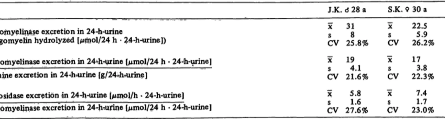
Therefore, the Sphingomyelinase activity in the urine of two healthy test persons was measured over a period of three weeks and related to the daily urine excretion, the daily excretion of creatinine, and α-glucosidase as reference enzyme for the 24-hour-urine. The mean value, standard deviation, and coefficient of variation were calculated from the daily determined relation coefficients (tab. 1).
Furthermore, urine samples were tested daily for a period of six days, to see whether storage of urine at room temperature causes a decrease of activity. It turned out that no significant decline of sphingomyelin-
t l h l 15
Fig. 2. Time-dependence of the cleavage of 14C-sphingomyelin.
Amount of C-sphingomyelin hydrolyzed expressed as nmol after addition of 100 μg leukocyte protein as a function of time.
Further details see: Material and Methods.
ase and α-glucosidase activity could be observed for the undialyzed urine. Yet, from the third day on, dialyzed urine samples showed a decrease to 30—0% of the initial enzyme activity.
Discussion
The common method for the determination of Sphingo- myelinase activity is based on the precipitation of marked undegraded sphingomyelin with concentrated trichloroacetic acid. Our method is based on the direct phase separation of degraded water-soluble phosphoryl- choline and water-insoluble sphingomyelin (6—8). This method is not only more sensitive, but it also has a good reproducibility of the degradation rates for the labelled substrate. Deviation of the degradation rates was not higher than 10% in simultaneous tests.
Our Sphingomyelinase activity in leukocytes is lower than the values reported by Kampine (4) evaluated with
Tab. 1. Sphingomyelinase activity, α-glucosidase activity and creatinine excretion in urine (estimated daily for three weeks).
sphingomyelin se excretion in 24-h-urine
(sphingomyelin hydrolyzed [μήιοΙ/24 h · 24-h-urine])
Sphingomyelinase excretion in 244i-urine [μηιρΙ/24 h · 24-h-urine]
creatinine excretion in 244v=urine [g/24-hrurine]
α-glucosidase excretion in 24-h-urine (μιηοΐ/h · 24-h-urine]
sphiiigoniyelinase excretion in 24-h^unne [μιηοΙ/24 h · 24-h-urine]
J.K. ό 28 a χ 31s 8 CV 25.8%
χ 19s 41 CV 21.6%
χ 5.8 s 16CV 27.6%
S.K.
χ
sCV
X
CV
X c
CV 9 30 a
22.55.9 26.2%
1730 22.3%
7.41 7 23.0%
410 Seidel, Klenke, Fischer and Pilzf: Sphingomyelinase assay in leukocytes and urine a different method (3.2-5.6 nmol/h · mg protein). Our
values, however, show a much lower scattering. They are in the range of leukocyte protein values reported by Gal et al. (2.2-3.7 nmol/h - mg protein) (1). However, the latter authors regard [14C]sphingomyelin as the pre- ferred substrate for sphingomyelinase assays in leuko- cyte preparations, owing to the poor degradation rate of their chromogenic substrate (2-hexadecanoylamino-4- nitrophenylphosphorylcholine) by leukocyte protein.
Problems of enzyme measurements in urine are well known (11). To our knowledge, there are no represent- ative investigations on sphingomyelinase activities in urine. The range of the values is higher for urine than for leukocytes. It has been shown that the meas- urable enzyme activities in the urine can be most directly correlated to the 24-hour creatinine excretion (11).
For screening of sphingomyelinase deficiency in urine the stability of the enzyme is of great importance, because the material is often sent through the post. In fact, there will be no important loss of enzyme activity.
α-glucosidase, as a reference enzyme, was measured every day in the urine of the two persons over a period of three weeks. This enzyme has a higher degree of scattering than sphingomyelinase. In spite of this fact
1—2 reference enzymes should be tested simultan- eously to eliminate interfering artefacts (bacterial influence, medication). Our finding, that within 6 days no significant decrease of the enzyme activity in the undialyzed urine was observed, is of great practical importance. However, the dialyzed urine always showed nearly a complete decrease of the measurable activity,
beginning at about the third day of dialysis. This may result from the loss of ions caused by dialysis.
Test results of numerous authors indicate that the actual enzyme deficiencies in vivo are more reliably reflected, if natural instead of synthetic substrates are used. In addition the employment of radioactively labelled natural substrate allows a microscale assay that requires only small amounts of enzyme. This may be important for pediatric examinations, family tests, or for prenatal diagnosis.
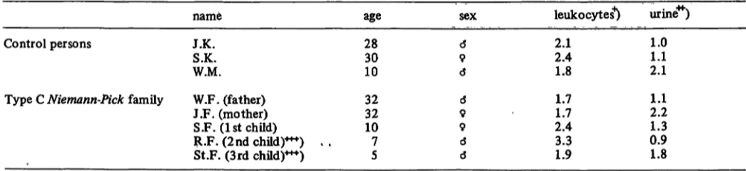
In a family (family F. from East-Frisia) two out of three children both starting with the third year of age showed mental retardation and neurological deterior- ation combined with hepatosplenomegaly. The light microscopic and electronic optical examination of bone marrow and rectal mucous membrane obtained by biopsy revealed a typical picture of sphingomyelin storage disease for the examined second child. Both children are still alive. They are now five and seven years old. However, no decrease in sphingomyelinase activity was found in leukocytes and urine for any member of the family (tab. 2). Based on the classific- ation of sphingomyelinoses according to biochemical and clinical criteria proposed by Frederickson et al.
(10), the mentioned cases can be related to type C Niemann-Pick disease. That type is the one with the mildest course of storage disease and therefore it has a relatively favourable prognosis.
Acknowledgment
The investigations were financially supported by Deutsche Forschungsgemeinschaft (SFB 33).
Tab. 2. Sphingomyelinase activity in human leukocytes and urine (Control persons and one family with Niemann-Pick disease).
Control persons
Type C Niemann-Pick family
name J.K.S.K.
W.M.
W.F. (father) J.F. (mother) S.F. (1st child)
R.F. (2nd child)***) . , St.F. (3rd child)***)
age 2830 10 3232 107 5
sex d9
d d9 9d d
leukocytes4) 2.12.4 1.8 1.71.7 2.43.3 1.9
urine4*") 1.01.1 2.1 2.21.1 0.91.3 1.8
+) sphingomyelin hydrolyzed [nmol/h · mg proteinj
; sphingomyelin hydrolyzed [nmol/h · ml dialyzed urine]
+H) Compared to the first child these two children were affected by hepatosplenomegaly, mental retardation, and neurological deterioration starting with the third year of age.
References
1. Gal, A. E., Brady, R. O., Hibbert, S. R. & Pentschev, P. G.
(1975), New Engl. J. Med., 293, 632-636.
2. Poulos, A. & Pollard, A. C. (1976), Clin. Chim. Acta 72, 327-335.
3. Brady, R. O., Kanfer, J. N., Mock, M. B. & Frederickson, D. S. (1966), Biochemistry 55, 366-369.
4. Kampine, J. P., Brady, R. O. & Kanfer, J. N. (1967), Science 755, 86-88.
5. Klenke, J. Eine vereinfachte SphingomyelinasebeStimmung in Leukozyten und Urin bei Normalpersonen und Patienten mit Niemann-Pick Erkrankung. Dissertationsthema; Med.
Fak. der Georg August-Univ. Göttingen.
6. Mraz, W., Fischer, G. & Jatzkewitz, H. (1976X FEBS-Letters 67,104-109.
7. Wenger, D. A., Sattler, M., Clark, C., Tanaka, H., Suzuki, K.
& Dawson, G. (1975), Science 188, 1310-1312.
8. Folch-Pi, J., Lees, M. & Sloane-Stanley, G. H. (1957), J.
Biol. Chem. 226,497-508.
9. Lowry, O. H., Rosebrough, N. J., Farr, A. L. & Randall, R. J.
(1951), J. Biol. Chem. 193, 265-280.
10. Frederickson, D. S. & Sloane, H. R. (1972), Sphingomyelin lipidoses: Niemann-Pick disease, in The Metabolie Basis of Inherited Diseases, third ed., (Stanbury, J. B., Wyngaarden, J. B. & Frederickson, D. S., eds.) New York, Me Graw-Hill, p. 783-807.
11. Raab, W. P., (1972), Clin. Chem. 18, 5-25 Dr. D. Seidel
Neurolog. Univ.-Klinik Martinistr. 52
D-2000 Hamburg 20
Federal Republic of Germany