• Keine Ergebnisse gefunden

Immunomodulation by NOD2/CARD15 and Vitamin D

N/A
N/A
Protected

Academic year: 2021

Aktie "Immunomodulation by NOD2/CARD15 and Vitamin D"

Copied!
134
0
0

Wird geladen.... (Jetzt Volltext ansehen)

Volltext

(1)

Immunomodulation by NOD2/CARD15 and Vitamin D 3

DISSERTATION ZUR ERLANGUNG DES

DOKTORGRADES DER NATURWISSENSCHAFTEN (DR. RER. NAT.) DER FAKULTÄT FÜR BIOLOGIE UND VORKLINISCHE MEDIZIN

DER UNIVERSITÄT REGENSBURG

vorgelegt von Carina Saraiva de Matos

aus Mortágua, Portugal

im Jahr 2017

(2)

The present work was carried out from November 2013 to October 2017 at the Clinic and Polyclinic of Internal Medicine III at the University Hospital Regensburg.

Die vorliegende Arbeit entstand im Zeitraum von November 2013 bis Oktober 2017 an der Klinik und Poliklinik für Innere Medizin III des Universitätsklinikums Regensburg.

Das Promotionsgesuch wurde eingereicht am:

27 Oktober 2017

Die Arbeit wurde angeleitet von:

Prof. Dr. Marina Kreutz

Unterschrift:

____________________________

(3)
(4)

To my parents

Aos meus pais

(5)

I

Table of Contents

LIST OF FIGURES ... IV LIST OF TABLES ... VII LIST OF ABBREVIATIONS ... VIII

1. INTRODUCTION ... 1

1.1. Hematopoietic stem cell transplantation ... 1

1.2. Graft-versus-host disease (GvHD) ... 1

1.2.1. Acute GvHD (aGvHD) ... 1

1.2.2. Chronic GvHD (cGvHD) ... 2

1.3. Role of innate immune cell responses in GvHD ... 3

1.3.1. Importance of antigen-presenting cells (APCs) in GvHD ... 3

1.3.1.1. Origin of dendritic cells (DCs) ... 4

1.3.1.2. Tolerogenic dendritic cells ... 5

1.3.2. The role of pattern-recognition receptors in GvHD ... 6

1.3.3. NOD2 status and GvHD ... 7

1.3.4. Gut microbiome and GvHD ... 8

1.4. Role of the adaptive immune system in GvHD ... 10

1.4.1. B cells in GvHD ...10

1.4.2. Regulatory T cells (Tregs) in GvHD ...11

1.5. Vitamin D ... 14

1.5.1. Vitamin D metabolism ...14

1.5.2. Vitamin D and immune function ...16

1.5.3. Vitamin D status and GvHD ...18

2. RESEARCH GOALS AND OBJECTIVES ... 20

3. MATERIAL ... 21

3.1. Equipment ... 21

3.2. Consumables ... 22

3.3. Media buffers and solutions ... 23

3.4. Enzymes, kits and reagents for molecular biology... 24

3.5. Chemicals for cell culture ... 25

3.6. Antibiotics ... 25

3.7. Oligonucleotides for qRT-PCR ... 25

3.8. Antibodies ... 26

3.8.1. Western blot antibodies ...26

(6)

II

3.8.2. Flow cytometry antibodies ...26

3.8.2.1. Anti-mouse antibodies ...26

3.8.3. Anti-human antibodies ...27

3.9. Mice ... 28

3.10. Databases and software ... 29

4. METHODS ... 30

4.1. Cell culture ... 30

4.1.1. Isolation and culture of human monocytes ...30

4.1.2. Macrophage culture ...31

4.1.3. Dendritic cell culture ...31

4.1.4. Mixed lymphocyte reaction ...31

4.1.5. Cytokine analysis from culture supernatant ...32

4.1.6. Enzymatic determination of lactate ...32

4.1.7. Analysis of the oxygen content in cultures ...32

4.1.8. Sorting of CD4

+

25

-

T cells ...33

4.1.9. Freezing and thawing of cells ...33

4.1.10. Cell counting ...33

4.2. Flow cytometry ... 33

4.2.1. Compensation ...34

4.2.2. Surface staining ...34

4.2.3. Intracellular staining ...34

4.2.4. Blood sample preparation for flow cytometry ...35

4.3. Molecular biological methods ... 35

4.3.1. RNA isolation ...35

4.3.2. Polymerase chain reaction ...35

4.3.3. DNA isolation ...37

4.4. Biochemical methods ... 38

4.4.1. Protein isolation ...38

4.4.2. SDS polyacrylamide gel electrophoresis ...40

4.4.3. Western blot analysis ...41

4.5. Mouse experiments ... 43

4.6. Microbiota analysis ... 44

4.7. Statistical analysis ... 44

5. RESULTS ... 45

5.1. Impact of NOD2 polymorphisms on the immune cell composition in

peripheral blood of healthy donors ... 45

(7)

III

5.1.1. Impact of NOD2

-/-

on immune cell composition in mice ...48

5.2. Impact of blood sampling and cell isolation method on the cell phenotype ... 49

5.2.1. CD16 downregulation: Importance of the recognized epitope ...51

5.2.2. Effect of platelets and TGF-β on human monocytes ...52

5.3. Impact of NOD2 on monocyte-derived macrophages ...59

5.4. Impact of NOD2 on dendritic cell differentiation cultures ... 61

5.4.1. Dendritic cell cultures of donors with NOD2 WT SNP8 and SNP12 and 13 61 5.4.2. Effect of the NOD2-ligand MDP on monocyte-derived dendritic cells ...64

5.4.2.2. Cytokine profile of Mo-derived DC versus MNC-derived DC ...66

5.4.2.3. Oxygen consumption and lactate production of Mo-derived DC cultures 67 5.5. Impact of vitamin D

3

on immune cells ... 70

5.5.1. Effect of vitamin D

3

on human monocytes ... 70

5.5.2. Effect of Vitamin D

3

on human T cells ...71

5.6. In vivo effects of vitamin D

3

... 75

5.6.1. Vitamin D

3

sufficient versus deficient mouse model...75

5.6.1.1. Impact of the serum 25(OH)D

3

level on immune cell composition ...77

6. DISCUSSION & PERSPECTIVES ... 83

6.1. Impact of the NOD2 status on immune cell composition in human blood ... 83

6.1.1. Role of NOD2 in myeloid cell differentiation ...84

6.1.1.1. NOD2 status and dendritic cell differentiation/maturation ...84

6.1.1.2. NOD2 status and macrophage differentiation ...85

6.1.2. Regulation of CD16 ...86

6.1.3. Role of NOD2 for monocyte activation ...86

6.1.3.1. Impact of NOD2 on NF-κB signalling ...87

6.1.4. Role of NOD2 ligands for cell activation and differentiation ...88

6.2. Role of vitamin D

3

on immune regulation ... 90

6.2.1. Effect of vitamin D

3

on human T cells ...91

6.2.2. In vivo effects of vitamin D

3

on mice ...93

6.3. Impact of vitamin D

3

on microbiota ... 95

6.4. Perspectives ... 96

7. SUMMARY ... 99

8. ZUSAMMENFASSUNG ... 101

9. REFERENCES ... 104

PUBLICATIONS ... 118

ACKNOWLEDGMENT ... 119

(8)

IV

List of Figures

Figure 1.1. The three phases of GvHD………...……...2

Figure 1.2. Structure of NOD2/CARD15………...…..…7

Figure 1.3. Development of nTregs and iTregs…………..……….11

Figure 1.4. Vitamin D metabolism……….………...15

Figure 1.5. Effects of 1,25(OH)

2

D

3

in various immune cells……….…….17

Figure 5.1. Impact of NOD2 SNP mutations on the blood cell composition of healthy donors………..………..…...46

Figure 5.2. Amount of CD4

+

, and CD4

+

CD25

+

and CD8 + T cells among living CD3+ cells in the blood of WT and donors with NOD2 variants……….….46

Figure 5.3. Amount of myeloid and plasmacytoid dendritic cells among living lineage negative HLA-DR

+

cells in the blood of WT, SNP8, 12 and 13 healthy donors…………47

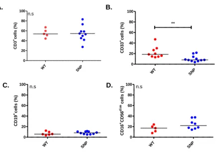
Figure 5.4. Amount of CD33+ cells among living leukocytes in the blood of WT, SNP8, 12 and 13 healthy donors ………...48

Figure 5.5. Impact of NOD2 expression on myeloid blood cells of WT and NOD2-/- mice...49

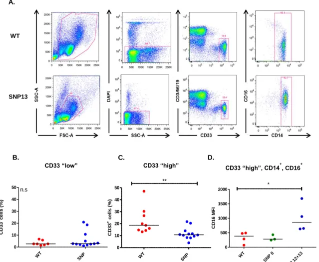
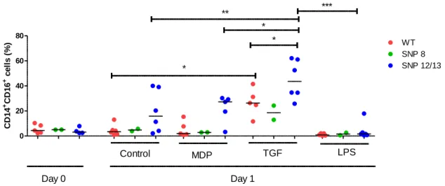
Figure 5.6. Median fluorescence intensity (MFI) of CD14+16+ monocytes among living CD33+ cells in the blood of WT, SNP8, 12 and 13 healthy donors……….……….50

Figure 5.7. Impact of the blood collection method on CD16 expression ………..………..51

Figure 5.8. Relative CD1 expression on monocyte using different antibody clones……52

Figure 5.9. Percentage of CD14

+

CD16

+

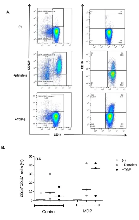
WT monocytes co-cultured with platelets or TGF-β………..…...53

Figure 5.10. Percentage of CD14

+

CD16

+

monocytes stimulated with or without MDP, TGF-β or LPS………..……….54

Figure 5.11. Percentage of CD14

+

monocytes stimulated with or without MDP, TGF-β or LPS……….…….……..55

Figure 5.12. Percentage of HLA-DR

+

monocytes stimulated with or without MDP, TGF-β

or LPS ……….……….……...55

(9)

V Figure 5.13. Cytokine secretion in monocytes stimulated with or without MDP, TGF-β and LPS or LPS+MDP……..……….…57 Figure 5.14. Effect of NOD2 mutations on iκBβ and phospho p38 expression……...………58 Figure 5.15. Expression of maturation markers in monocyte-derived macrophages from donors with NOD2 WT and NOD2 variants. ………...60 Figure 5.16. SNP13 donors displayed an impaired DC maturation……….…...…62 Figure 5.17. IL-10 production is similar in cultures from WT and NOD2 SNP donors……...62 Figure 5.18. SNP12 and 13 donors showed enhanced activation in classical DCs...63 Figure 5.19. IL-10 and IL-12 production is impaired in cultures from NOD2 SNP donors………...64 Figure 5.20. MDP is incapable to induce the differentiation of human monocyte-derived dendritic cells (Mo-DCs).………...65 Figure 5.21. Expression of tolerogenic DC markers after culture with LPS, MDP and 1,25(OH)

2

D

3

………...66 Figure 5.22. IL10 and IL-12 secretion dendritic cells generated under different protocols.

………..………...67 Figure 5.23. Oxygen consumption and lactate production of the Mo-derived DCs ……..68 Figure 5.24. Functional analysis of the generated DCs ………...69 Figure 5.25. Amount of FOXP3+ CD4+ cells after co-culturing with DCs matured either with MDP or LPS ……….70 Figure 5.26. NOD2 mRNA expression in human monocytes………...…………...71 Figure 5.27. Amount of FoxP3+ cells and cell proliferation in CD4+ T cell cultures.

……….……….………...72 Figure 5.28. . FOXP3 induction by 1,25(OH)2D3 and 25(OH)D3 in comparison to TGF-β

………..………...73 Figure 5.29. FOXP3 mRNA expression is increased upon TGF-β + 1,25 (OH)

2

D

3

treatment ………...……….………...73 Figure 5.30. Amount of 1,25(OH)

2

D

3

in the culture supernatant of human CD4+ T cells.

……….………....74

(10)

VI Figure 5.31. Cytokine profile of CD4

+

25

-

T cells stimulated with TGF-β, 25(OH)D

3

or 1,25(OH)

2

D

3.

………...75

Figure 5.32. Serum vitamin D3 levels of BALB/c mice and CD1 mice at the age of 10

weeks……….……….………..…...76

Figure 5.33. Immune cell composition of BALB/c and CD-1 mice ………..……….…...77

Figure 5.34. Analysis of immune cell subpopulations in the blood, spleen and bone

marrow of BALB/c and CD-1 mice ...………79

Figure 5.35. Analysis of immune cell subpopulations in the blood, spleen and bone

marrow of BALB/c and CD-1 mice………..……….80

Figure 5.36. Mice fed a vitamin D

3

deficient diet have higher microbiota

diversity………...…...…81

Figure 5.37. The ten most abundant bacterial genera in mice stool samples...82

(11)

VII

List of Tables

Table 1.1. Properties of both natural and induced regulatory T cells……….….12

Table 4.1. Elutriation parameters and cell types……….29

Table 4.2. Criteria for primer design……….……….35

Table 4.3. Reaction mix used in qRT-PCR……….……….36

Table 4.4. Program used for qRT-PCR……….………...………...……….36

Table 4.5. Buffer A composition……….………37

Table 4.6. Buffer B composition………...………..38

Table 4.7. Buffer C composition………...………...38

Table 4.8. SDS sample buffer composition……….……….38

Table 4.9. SDS-PAGE stock solutions………...………..39

Table 4.10. SDS-PAGE gel mix solutions……….……….………..39

Table 4.11. Required buffers and solutions for SDS gel preparation……….…...39

Table 4.12. Western blot buffers………...………41

Table 4.13. Composition of ECL solutions……….………..41

(12)

VIII

List of Abbreviations

aGvHD acute Graft versus Host Disease

ANOVA Analysis of variance

APCs Antigen presenting cells

APS Ammonium persulfate

BM Bone Marrow

CARD Caspase Recruitment Domain

cDNA complementary DNA

cGvHD chronic Graft- versus -Host Disease

CTLA4 Cytotoxic T-lymphocyte-associated Protein 4

DAPI 4',6-diamidino-2-phenylindole

DAMPs Danger associated molecular patterns

Def. Deficient

DNA Deoxyribonucleic acid

DCs Dendritic cells

DMSO Dimethyl sulfoxide

dNTPs 2'-deoxyribonucleosid-5'-triphosphate

dsDNA Double stranded DNA

ECL Enhanced chemiluminescence

ECP Extracorporeal Photophoresis

EDTA Ethylene diamine tetra acetic acid

ELISA Enzyme-linked immunosorbent assay

FCS Fetal calf serum

Foxp3 Forkhead Box Protein 3

GI Gastro intestinal

GM-CSF Granulocyte -monocyte colony-stimulating factor

GvHD Graft versus Host Disease

GvL Graft versus Leukemia

Gy Gray

HANK´s BSS Hank´s buffered saline solution

HLA Human Leukocyte Antigen

HRP Horse radish peroxidase

HSCs Hematopoietic Stem Cells

HSCT Hematopoietic Stem Cell Transplantation

IDO Indoleamine 2,3-dioxygenase

(13)

IX

IFNγ interferon gamma

Ig Immunoglobulin

IL Interleukin

iTregs induced Tregs

IκB Inhibitor of kappa B

KDa Kilodalton

LCs Langerhans cells

LPS Lipopolysaccharide

MALDI-TOF matrix-assisted laser desorption/ionization-time-of-flight

MDP Muramyl Dipeptide

MDSC Myeloid-derived suppressor cells

MHC Major Histocompatibility Complex

mRNA messenger RNA

NF-κB Nuclear factor κB

NK Natural Killer

NKT Natural Killer T cell

NOD2 Nucleotide oligomerization domain 2

nTregs natural Tregs

PAMP Pathogen associated molecular pattern

PBS Phosphate buffered saline

PBSC Peripheral Blood Stem Cells

PCR Polymerase chain reaction

PRR Pathogen recognition receptor

qRT-PCR Quantitative real-time PCR

RNA Ribonucleic acid

RT Room temperature

rRNA ribosomal RNA

s.e.m standard error of mean

SDS-PAGE Sodium dodecyl sulfate polyacrylamide gel electrophoresis

SNP Single nucleotide polymorphism

Suf. Sufficient

TBI Total Body Irradiation

TBS Tris buffer saline

TBST TBS + Tween 20

TCR T Cell Receptor

TGF Transformation Growth Factor

TLR Toll Like Receptor

(14)

X

TNF Tumor Necrosis Factor

Tregs Regulatory T cells

UV Ultraviolet

(15)

Introduction

1

1. Introduction

1.1. Hematopoietic stem cell transplantation

Hematopoietic stem cell transplantation was originally conceived more than 60 years ago as a therapeutic option for irradiation injury. Later, this procedure was adopted as a method for treating several disorders, namely cancers and autoimmune diseases [1].

There are different types of hematopoietic stem cell transplantation (HSCT): Autologous, where the patient´s own stem cells are transplanted; allogeneic, using stem cells from a donor and syngeneic, where cells from an identical twin are used [2].

One of the most common complications after allogeneic HSCT (aHSCT) is the development of Graft-versus-host disease (GvHD).

1.2. Graft-versus-host disease (GvHD)

GvHD is still the most common life-threatening complication associated with HSCT[3]. It was first observed in 1956 in a murine model, where Barnes et al. demonstrated that irradiated mice which were infused with allogeneic bone marrow and spleen cells, recovered from irradiation injury and marrow aplasia but additionally developed several complications like diarrhea, weight loss and skin changes, due to a “secondary disease”.

This condition was later recognized as Graft-versus-host disease [4]. GvHD can be considered both acute and chronic, depending on the timing of its occurrence.

1.2.1. Acute GvHD (aGvHD)

Acute GvHD generally occurs before day 100 after HSCT, but may also occur later. It is a reaction of donor immune cells against host tissues and affects the skin, liver, and gastrointestinal tract [5].

The pathophysiology of aGvHD is often described as a three-step process where the innate and adaptive immune system interact (Figure 1.1). In the first step, tissue damage occurs as a result of conditioning, releasing danger signals which induce pro- inflammatory cytokines such as tumor necrosis factor (TNF) and interleukin-1 (IL-1β).

This in turn leads to the activation of host antigen presenting cells (APCs). The activated APCs then stimulate donor T cells in the second step, also known as afferent phase.

These T cell responses depend on the disparity between donor and recipient e.g.

regarding human leucocyte antigen (HLA) or minor antigens.

In the last phase, or efferent phase, both innate and adaptive immune cells work

synergistically to exacerbate T cell induced inflammation. Microbial products like

lipopolysaccharide (LPS) leak through the damaged epithelium and continue to stimulate

(16)

Introduction

2 mononuclear cells (monocytes, macrophages) leading to the release of inflammatory cytokines. As a result, further destruction of epithelial cells occurs especially in the gastrointestinal (GI) tract [4, 6].

1.2.2. Chronic GvHD (cGvHD)

Chronic GVHD was initially defined as “late” GvHD syndrome that appears more than 100 days post-transplant, either as an extension of aGvHD (progressive onset GVHD), after a disease-free interval (quiescent cGvHD), or without preceding aGvHD (de novo cGvHD) [7].

The pathophysiology of cGvHD is poorly understood; therefore it remains one of the most significant complications of long-term survivors after aHSCT [8].

The absence of reliable animal models hampers the experimental study of this condition, although in the last decades some theories attempt to explain the pathophysiology of cGVHD: (1) thymic damage and the defective negative selection of T cells, (2) regulatory T cell deficiencies, (3) auto-antibody production by aberrant B cells, and (4) the formation of profibrotic lesions [9].

Figure 1.1. The three phases of aGvHD, according to Ferrara et al [6]. In phase I, chemotherapy or

radiotherapy as part of transplant conditioning causes host tissue damage and release of inflammatory cytokines

such as TNF, IL-1β, and IL-6, with resulting priming of host antigen-presenting cells (APCs). In phase II, host

APCs activate mature donor cells, which then proliferate and differentiate. Release of additional effector

molecules, such as TNF and IL-1β, mediates additional tissue damage. Lipopolysaccharide (LPS) that has leaked

through damaged intestinal mucosa triggers further TNF production. The TNF can damage the tissue directly by

inducing necrosis and apoptosis in the skin and gastrointestinal tract through either TNF receptors or the Fas

pathway. TNF plays a direct role in intestinal GVHD damage, which further amplifies the damage in the skin, liver,

and lung in a “cytokine storm.” The process culminates in death of host cells through CD8

+

cytotoxic T cell-

mediated apoptosis.

(17)

Introduction

3 1.3. Role of innate immune cell responses in GvHD

1.3.1. Importance of antigen-presenting cells (APCs) in GvHD

Many cell types, including both hematopoietic and non-hematopoietic cells (e.g.

endothelial cells), participate in the process of antigen presentation with distinct levels of efficiency. Hematopoietic APCs include B cells, monocytes, macrophages, as well as myeloid and plasmacytoid dendritic cells (pDCs). Besides antigen-presentation, APCs also are crucially involved in the production of (inflammatory) cytokines which regulate GvHD development [7].

Naïve B cells are absent from the skin and most mucosal sites, but instead they recirculate between blood and the secondary lymphoid organs. Here, they pick up antigens through specific B cell receptors for presentation on MHC class II and become competent APCs on interaction with CD40L

+

CD4

+

T cells [8].

Monocytes and macrophages are important players in inflammation. Their role includes phagocytosis, elimination of bacteria and restoration of tissue homeostasis [9, 10]. To fulfill their tasks, they can adopt different functional phenotypes dependent on the respective microenvironment [10]. Monocytes infiltrate into tissues where the polarizing stimuli to which they respond, result in macrophage or DC differentiation. Distinct macrophage and DC subsets have also been described in vitro [10-13]. Classical or M1- activated macrophages are anti-microbial, anti-tumorigenic and pro-inflammatory. This M1 phenotype plays a central role in chronic inflammation and autoimmunity. In contrast, alternative or M2 activation by IL-4 and/or IL-13 leads to differentiation of anti- inflammatory macrophages [10]. It is believed however, that these M1 and M2 subtypes represent extremes of a continuum of activation states found in vivo [7]. Monocytes and macrophages can also act as APCs and stimulate a secondary T cell response, but initiate a primary immune response in a weaker degree, which is the main function of the specialized APCs known as DCs. Importantly, DCs possess the capacity to pick up antigens in peripheral tissues and traffic to secondary lymphoid organs [14]. Therefore, DCs are largely effective at priming naïve T cells, although the extent to which they do so is greatly dependent on their origin and activation history [15].

Mouse studies have demonstrated that CD4

+

T cell-dependent (MHC-mismatched) aGvHD can be induced by either host or donor APCs [16-18]. Little is known about the role of DCs in cGvHD because of the lack of relevant mouse models. Both host and donor APCs have been implicated; skin cGvHD can be induced by either donor or host APCs, whereas donor APCs are dominant in intestinal cGvHD [19].

Generally, T cells have been the primary focus in the development of GvHD, but given

the important role of DCs in its pathogenesis, APCs also represent an important target.

(18)

Introduction

4 DCs could be manipulated using multiple approaches in vivo or in vitro, and the generation of tolerogenic DCs (tDCs) could be an interesting approach to regulate immunity and suppress GvHD [20].

1.3.1.1. Origin of dendritic cells (DCs)

DCs were first described in the mid-1970s by Ralph Steinman, who observed in the spleen a subpopulation of cells with a remarkable dendritic shape. It was soon appreciated that these cells existed in all lymphoid and most non-lymphoid tissues [21].

They are the most potent APC of the immune system and they are termed professional APC as a result of their exclusive ability to capture and to present antigens to naïve T cells.

They are part of the myeloid lineage of hematopoietic cells and arise from a precursor that can also differentiate into monocytes [22].

Although they share many common features, DCs are a heterogeneous cell population.

Various cytokines and transcription factors are known to be responsible for the development of DC subpopulations. Depending on the subpopulation and the maturation state of these cells, they are able to induce either a broad cytotoxic immune response or induce immune tolerance which is of great importance within the context of autoimmunity [15].

Epithelial DCs, named Langerhans cells (LCs) are present exclusively in epithelial tissues (epidermis, mucosa) and they express receptors for antigen uptake, e.g. Fcγ and Fcε, as well as CD1a, a molecule involved in the presentation of glycolipids to T cells.

Interstitial DCs, identified in the interstitium of most tissues, are characterized by the expression of CD68 and factor XIIIa, but lack LC markers. These cells display similarities with monocyte-derived DCs.

Monocyte-derived DCs can be generated in vitro in the presence of IL-4 and granulocyte- macrophage colony-stimulating factor (GM-CSF). Allegedly, in the course of inflammation in vivo, this type of differentiation occurs physiologically. Blood monocytes can also differentiate into DCs during transendothelial migration [23].

In the human blood, different DC subsets have been identified, namely myeloid dendritic cells (mDCs), characterized by the expression of HLA-DR and CD11c, and plasmacytoid dendritic cells (pDCs) which have a plasma cell-like morphology and lack CD11c expression [23].

Under normal (healthy) conditions, DCs exist in an immature or steady state and their main function is to maintain immune tolerance by keeping adaptive immune cells from attacking host cells that possess “self” antigens. However, if DCs encounter “non-self”

entities in the periphery, they opsonize them, process their antigens for cross-

(19)

Introduction

5 presentation, migrate to the lymph nodes, and prime naïve T cells for the respective antigen. iDCs (immature DCs) exhibit continuous endocytic activity and therefore continuously present “self” antigens to T cells. Nevertheless, T cells are not polarized toward an effector state but are rather polarized to facilitate tolerance or immunosuppression. Such immunotolerance is accomplished due to the complete lack of stimulatory signals provided by the iDC [24]. On the other hand, when DCs encounter pathogens, they switch to a mature state exhibiting strong phenotypic and functional stimulation. Mature DCs are characterized by the high expression of costimulatory molecules, namely CD80, CD83 and CD86. The simultaneous presence of phenotypic maturation ligands and the appropriate cytokine profile of the DC (e.g. high IL-12 secretion) helps to activate effector functions in interacting T cells, thus polarizing them for antigen-specific elimination of the “non-self” entity [24].

1.3.1.2. Tolerogenic dendritic cells

Tolerogenic DCs are characterized by the downregulation of different costimulatory molecules such as CD80 and CD86 as well as the upregulation of inhibitory receptors like PDL-1, PDL-2, ILT3, ILT4 and TIM3. It is believed that ILT3 and ILT4 directly inhibit proliferation of T cells and drive FOXP3

+

Treg expansion [25].

Tolerogenic DCs also exhibit a special cytokine profile. This type of DC produces higher amounts of IL-10 and reduced amounts of IL-12. As IL-12 is crucial for the induction of a Th1 response, tolerogenic DCs are more likely to induce the generation of Th2 cells or Tregs [26].

Indoleamine 2, 3-dioxygenase (IDO), an immune checkpoint enzyme is also expressed by tolerogenic DCs. IDO metabolizes tryptophan to kynurenine and its expression by DCs has been implicated in the inhibition of T cell proliferation and survival. Furthermore, it has been reported that IDO expression by DCs promotes the development of Tregs [27].

Tolerogenic DCs have the capability to induce long-lasting tolerance to transplanted allogeneic grafts and suppress the development of autoimmune diseases [28]. Hence, tolerogenic DCs represent a subtype of polarized DCs which inhibit immune activation.

Diverse factors have been reported to support the generation of tolerogenic DCs with low antigen-presenting capability. These factors belong to different categories, such as cytokines (e.g. IL-10 and TGF-), vitamins (e.g. vitamin D) and drugs (e.g.

dexamethasone or rapamycin) [29].

With their regulatory capacities tolerogenic DC seem to be a promising tool to mitigate

GvHD

(20)

Introduction

6 In fact, the suppression of DC function is under investigation in the prevention and treatment of human GvHD while ensuring the conservation of graft-versus-leukemia effects [19].

1.3.2. The role of pattern-recognition receptors in GvHD

Specific receptors on innate immune cells, called pattern recognition receptors (PRRs), sense pathogen-associated molecular patterns (PAMPs), such as LPS and damage- associated molecular patterns (DAMPs), like high mobility group box 1 (HMGB-1).

Danger signalling is transmitted through PRRs when they bind PAMPs and DAMPs.

Several signalling molecules, such as toll-like receptor (TLR), Nucleotide-binding oligomerization domain (NOD)-like receptor (NLR), and retinoic acid-inducible gene 1 (RIG-1) signalling are recognized.

TLRs are a family of PRRs and are expressed on a variety of cells derived from both hematopoietic and non-hematopoietic lineages [30]. There are several TLRs identified (TLR1-13) and they interact with different ligands. For example, LPS binds to TLR4 whereas flagellin binds to TLR5.

Another known PRR subtype refers to NLRs. These receptors are commonly expressed by immune cells and some non-immune cells. NOD1 and NOD2 are the most extensively investigated NLRs in GvHD. NOD1 and NOD2 recognize different kinds of peptidoglycan (PGN) fragments from the bacterial cell wall. NOD1 binds to diaminopimelate-containing N-acetyl glucosamine-N-acetylmuramic acid (GluNAc-MurNac) tripeptides from gram- negative bacterial PGN, whereas NOD2 binds to muramyl dipeptide (MDP) that is produced by all bacteria [30].

Some studies have shown that TLR4 mutations lead to the reduction of GvHD risk in mice and humans [12, 13]. In a study using TLR4-knockout mice (TLR4

-/-

) as bone marrow and splenocyte cell graft donors or recipients, the occurrence of GvHD symptoms and mortality were delayed compared to wild-type (TLR4

+/+

) mice [12]. It has also been reported that TLR4 expression is upregulated in patients with cutaneous cGvHD [14]. Nevertheless, there are also both mice and human studies showing no significant impact of TLR4 mutations in the outcome of HSCT [31].

In the clinical setting, single nucleotide polymorphisms (SNPs) of the gene encoding

NOD2 are associated with a higher GvHD incidence [32-34]. SNPs are defined as

mutations with an allele frequency of at least 1% in a given population and can have

modest biological effects [22]. NOD2 has been shown to have an inhibitory effect on

GvHD by suppressing the function of APCs and favouring the generation of regulatory T

cells (Tregs) [35].

(21)

Introduction

7 1.3.3. NOD2 status and GvHD

The NOD2/CARD15 gene encodes the NOD2 protein that functions as a sensor for MDP. It takes part in the regulation of inflammatory responses through intracellular pathways involving nuclear factor κB (NF-κB) and IκB kinase [36].

The NOD2 protein (Figure 2) contains a series of carboxy-terminal leucine-rich repeats (LRR), as well as a central nucleotide-binding domain (NBD; also known as NOD domain). In addition, the amino-terminal domain contains tandem CARDs, which interact with receptor interacting protein 2 (RIP2) upon NOD2 stimulation [37]. Activation of RIP2 leads to subsequent steps that ultimately result in activation attended by nuclear translocation of NF-B and transcription of its target genes [38].

The ligand sensing occurs at the LRR domain of NOD and the recognition of MDP is believed to trigger a conformational change in the protein, promoting a NBD-mediated oligomerization of NOD2 [39] .

Figure 1.2. Structure of NOD2/CARD15 gene. NOD2 contains tandem N-terminal CARDs that interact with the CARD of receptor-interacting protein 2 (RIP2). NOD2 contains a central nucleotide-binding domain (NBD), which binds ATP and mediates NOD oligomerization and carboxy-terminal leucine-rich repeats (LRRs which are important for ligand sensing. Adapted from [39].

NOD2 was identified several years ago and is one of the well-studied members of the NLR family. Initially, NOD2 expression was reported to be relatively tissue specific and restricted to APCs such as monocytes, macrophages, DCs, and certain epithelial cells [40]. More recently, it has been reported that basal levels of NOD2 expression are usually quite low in other cell types (myeloblastic cells, dental pulp) but can be induced by a variety of inflammatory signals such as LPS), TNF-, interferon (IFN)- and 1,25- dihydroxylvitamin D3 (1,25(OH)

2

D

3

) [40].

The association of SNPs of the NOD2 gene with a number of inflammatory pathologies, including Crohn´s disease (CD), and Blau syndrome, highlights its pivotal role in host- pathogen interactions and the inflammatory response [41]. More than 10 years ago, SNPs in the NOD2 gene locus have been associated with GvHD [33, 34]. Holler et al.

found an association between SNPs (alleles 8, 12, and 13) of the NOD2 gene in the

(22)

Introduction

8 donor or host and a higher incidence of GvHD, as well as an increased transplant-related mortality. These results were partially confirmed by Elmaagacli et al [32] who found a relation between NOD2 status and outcome after allogeneic stem cell transplantation only if both recipient and donor carried NOD2 SNPs. Although there is still no consensus regarding the role of the NOD2 status with regard to the outcome of HSCT [42, 43], MDP sensing by hematopoietic cells seems to be of crucial importance for the intestinal immune homeostasis [44] and it appears that NOD2 SNPs of patients receiving aHSCT influence post-transplant immunity, even though the underlying mechanism is still not clear [45].

1.3.4. Gut microbiome and GvHD

It is estimated that about one thousand microbial species live in symbiosis with the human gut [46]. These bacterial communities, collectively called microbiome, play an important role in the regulation of metabolic processes and also appear to be critical for the optimal function of the immune system [46]. Several studies have already demonstrated that particular species of microbiota can contribute to the induction of different immune cell types such as Foxp3

+

regulatory T cells, suggesting a close relationship between the microbiome and the immune system [47].

The gut microbiome is regulated by different mechanisms: On the one hand, the gastrointestinal epithelial barrier, in addition to its absorptive role, is involved in the regulation of microbial colonization and mucosal immune responses. On the other hand, specialized intestinal epithelial cells (IECs), such as the secretory goblet cells and Paneth cells, regulate bacterial species composition through the production of a mucous layer and the secretion of antimicrobial proteins, such as defensins. These antimicrobial peptides are produced by cells in the course of innate defence and serve as signals which initiate, mobilise, and amplify adaptive immune defences by the host. Defensins can be classified in two sub-families. Based on their tertiary structure, there are α- and β- defensins. While human α-defensins are mostly expressed by neutrophils, β-defensins are secreted by epithelial cells of the skin and mucosae. Besides their anti-microbial activity, accumulating data arose in the past decade indicating that defensins have extended functions in human patho(physio)logy. Indeed, defensins appear as modulators of the adaptive immune system and angiogenesis, key mediators of wound healing and determinant players in male fertility [48].

In early studies, mice transplanted in germ-free conditions or treated with antibiotics

developed significantly less GvHD, demonstrating a negative role for bacteria and the

microbiome in the GvHD-related mortality [49, 50]. Also, human studies using

(23)

Introduction

9 prophylactic gut decontamination demonstrated prevention in the development of aGvHD [51].

More recent studies indicate that the gut microbiome is altered after HSCT and the composition of the microbiome is important for transplantation outcome. Patients who develop GvHD have an altered microbiome, with loss of dominance of Clostridiales species replaced by a higher number of Lactobacillales or Enterobacteriales [47]. Results from a retrospective study showed that Clostridium difficile infection (CDI) is strongly associated with aGVHD and increased nonrelapse mortality in aHSCT patients [69].

Whether dysbiosis is just an epiphenomenon due to the use of antibiotics is still not clear but the loss of commensal bacteria correlates with GvHD severity and pathogenesis in human patients [52]. Furthermore, the importance of the microbiome in the setting of GvHD was demonstrated in animal studies [47], showing that the balance between specific gut bacteria plays a pivotal role in transplantation outcome. Namely, the expansion of Lactobacillales and loss of Clostridiales was found to be present in a GvHD mouse model [52].

The protective effect of commensal bacteria can be explained by the secretion of certain bacterial metabolites. In a study conducted by Reddy et al., the authors demonstrated that the bacterial metabolite butyrate is consistently decreased in the intestinal tissue after allogeneic stem cell transplantation. Butyrate is mainly produced by Clostridium, Eubacterium, and Butyrivibrio bacteria genera and it has been shown that butyrate improves intestinal epithelial barrier function, thereby mitigating GvHD [53]. Another possible mechanism connecting dysbiosis and GvHD is the ability of certain bacteria to promote T cell differentiation. For instances, Clostridiales bacteria have been reported to be upregulators of colonic regulatory T cells (Tregs) [54]. This may suggest that GvHD could deplete anti-inflammatory cell populations by reducing the abundance of Clostridiales.

In line with the hypothesis that the intestinal microbial balance is of the outmost importance for the transplantation outcome, fecal microbiota transplantation (FMT) could be an option for transplantation patients. FMT refers to infusion of a fecal suspension from a healthy individual into the GI tract of a patient to restore “healthy” intestinal microbiota and cure disease [55]. In recent years, FMT has emerged as an effective method for the treatment of CDI. However, there is risk that fecal suspension could contain harmful bacteria, viruses, and parasites. An alternative solution to this problem is to screen the beneficial bacteria from stool and infuse these bacteria as a cocktail into the gastrointestinal tract [56].

Although the importance of the gut microbiome in the regulation of the (intestinal)

immune system is indisputable, further studies are needed to clarify the relation between

(24)

Introduction

10 gut microbiome and complications after transplantation. These could pave the way for new therapeutic options for the prevention of GvHD.

1.4. Role of the adaptive immune system in GvHD

In GvHD pathophysiology, the interplay between innate and adaptive immune system is of utmost importance.

After the initial response of the innate immune system to microbial products and molecules released by cellular damage, the adaptive immune response is initiated.

Both donor CD4

+

and CD8

+

T cells have crucial roles in the pathophysiology of GvHD.

Therefore, the most effective approach for GvHD prevention still is the depletion or suppression of donor T cells [57].

However, recent animal studies suggest that B cells might also play an important role in the biology of GvHD [58]. Further evidence for the involvement of B cells in GvHD pathogenesis comes from reports of successful treatment of GvHD with B cell depletion therapies [59].

1.4.1. B cells in GvHD

Little is known about the role of B cells in the pathogenesis of aGvHD. In mice, B cell depletion results in a decreased incidence of aGvHD [58]. Further support for a role of B cells in aGvHD comes from several recent clinical observations: Encouraged by reports of the effectiveness of B cell depletion in the treatment of cGvHD, Kamble et al treated patients with aGvHD, who were refractory to multiple immunosuppressive drugs, with rituximab, a chimeric monoclonal antibody directed against the B cell-specific CD20 antigen. They reported that three patients who received rituximab showed a complete remission of aGvHD. Hence, B cell depletion might also be effective for the prevention of aGvHD. Rituximab administered as part of a myeloablative or nonmyeloablative conditioning regimen or given shortly before or after transplantation results in lower-than- expected rates of GvHD [60].

Even though these results indicate that B cells are involved in the development of GvHD, most likely due to their role as APCs, other reports show that host B cells can also have protective effects as B cell-deficient mice experienced more severe aGvHD than wild- type mice. In this experimental model IL-10 production by recipient B cells is responsible for the suppression of aGvHD [61].

The involvement of B cells in cGvHD seems to be more straightforward since both

preclinical and clinical data suggest an important role of B cell dysregulation in cGvHD

pathogenesis and treatment [35]. Post-transplant administration of rituximab appears to

reduce the rate of cGVHD in preliminary studies [62].

(25)

Introduction

11 1.4.2. Regulatory T cells (Tregs) in GvHD

Tregs are a subset of CD4

+

T cells with immunosuppressive activity. They are characterized by the expression of the IL-2R α-chain (CD25) and the transcription factor FOXP3 [63]. However, identification of human Tregs is problematic since these and other markers (CD25, CTLA-4, CD127, FOXP3) seem to represent general T cell activation markers [74].

Tregs are essential for the maintenance of self-tolerance, regulation of the peripheral T cell pool, and contribute to tolerance in solid organ transplantation and protection from GvHD lethality in bone marrow transplantation (BMT) models [64].

It is believed that there are two different subsets of CD4

+

Treg cells. The natural occurring Tregs (nTregs) develop in the thymus. During negative selection, nTreg cells upregulate FOXP3 during self-antigen recognition [65]. nTreg cells are responsible for maintaining immune homeostasis and tolerance to self-antigen by inhibiting self-reactive T cells in the periphery. The other subset of CD4

+

Tregs has been named “induced”

Tregs (iTregs). These cells are generated in the periphery when conventional T cells are activated in the presence of TGF-β and IL-2 [66] (Figure 1.3).

Figure 1.3. Development of nTregs and iTregs. Adapted from [66]. nTregs (top) differentiate from naïve

conventional T cells to Foxp3+ Tregs in the thymus. In the periphery, natural Tregs express a number of cell

surface markers, indicated in the box below the depiction of the natural Treg. However, none of these cell

surface markers are unique to Tregs as they are also found on activated conventional T cells. Natural Tregs

utilize the cytokines IL-10, IL-35 and TGFβ to exert their suppressive effects upon conventional T cells. TGFβ

and IL-2 have also been shown to be important to the maintenance and fidelity of the Treg signature. iTregs

(bottom) can be generated from conventional T cell precursors. Once in the periphery, naïve conventional T

cells can be induced to become Foxp3− Tr1 cells or Foxp3+ Th3 cells via IL-10 and/or TGFβ secreted by

APCs such as dendritic cells and macrophages. These induced Tregs share similar cell surface markers as

natural Tregs. Foxp3+ induced Tregs can accumulate in the gut through upregulation of CCR9 and α4β7 via

TGFβ and retinoic acid produced by CD103+ dendritic cells.

(26)

Introduction

12 Induced Tregs can be also generated in vitro by activating naïve T cells with the proper culture conditions. T cells activated with either antigen or anti-CD3/28 beads in the presence of TGF-β and IL-2 are capable to up-regulate Foxp3, and also display other features used to characterize regulatory T cells, such as a suppressive function.

The in vitro-generated iTregs are attractive in the clinical setting [67], since they can be generated in large numbers, facilitating the adoptive transfer of these cells into the recipients.

The two Treg subsets differ in their main antigen specificities, in the T-cell receptor signal strength and also in the co-stimulatory requirements needed for their generation (Table 1). Nonetheless, it is still not clear whether iTregs exist in vivo.

Table 1.2. Properties of both natural regulatory T cells (nTregs) and induced regulatory T cells (iTregs). Adapted from [68].

nTregs iTregs (TGF-β+ IL-2)

Principal antigen

specificities Self Foreign

Generation

IL-2 and TGFβ required No Yes

CD28-dependent Yes No

CTLA-4-dependent No Yes

Maintenance

IL-2 and TGFβ required Yes Yes

Stability

IL-6 can convert to Th17

cells Yes No

Can block other cells from

producing IL-17 No Yes

Suppression

IL-6 can block

suppressive activity Yes No

There is evidence that IL-6 is capable of converting nTregs to Th17 cells, while iTregs

induced by IL-2 and TGF-β are resistant to this cytokine and retain their suppressive

function at inflammatory sites. Thus, nTregs and iTregs may have different roles in the

adaptive immune response [68].

(27)

Introduction

13 Since GvHD is characterized by the loss of tolerance and the occurrence of autoimmune manifestations, it seems reasonable to assume that Tregs may have a critical role in the pathophysiology of GvHD [67]. In fact, animal studies have already demonstrated that a deficiency in Tregs contribute to both aGvHD and cGvHD [69, 70].

Given the critical role of Tregs in maintaining tolerance, several groups postulated that the adoptive transfer of Tregs should ameliorate GvHD [70, 71]. The first study was performed in mice by Taylor et al. [70]. In this study, the authors depleted both CD4

+

CD25

+

T cells from the donor and recipient before transplantation and observed that the depletion of this cell population was associated with worsening of GvHD. In contrast, the adoptive transfer of CD4

+

CD25

+

nTreg cells along with the marrow graft resulted in the amelioration of disease. Hoffmann and co-workers [72] demonstrated that freshly isolated CD4

+

CD25

+

T cells from unprimed animals can rescue recipients from lethal GvHD induced by CD4

+

CD25

T cells after allogeneic transplantation. That protection from lethality is partially dependent on IL-10 produced by transplanted CD4

+

CD25

+

T cells.

The role of Tregs in GvHD mitigation has also been demonstrated in humans: Rezvani and colleagues demonstrated that a high number of CD4

+

CD25

+

FOXP3

+

T cells in the donor graft were associated with a reduced risk of GvHD [73]. Also, they found that low nTreg counts early after aHSCT were associated with an increased risk of GvHD.

Since one of the challenges in nTreg therapy is the reduced frequency of these cells in peripheral blood, there have been several attempts in order to expand this population. It is possible to expand nTreg cells through in vivo-stimulation of nTreg proliferation or through ex vivo-expansion [74]

Based on the fact that nTregs express high levels of IL-2Rα, Kennedy-Nasser et al [75]

treated 16 pediatric patients with ultralow doses of IL-2 three times per week, starting from day 7 or 30 after transplantation for 12 weeks, and they observed an increase in CD4

+

CD25

+

FOXP3

+

Tregs from baseline. These cells were able to suppress alloreactive responses in vitro, indicating functionality.

The first clinical trial using Tregs to suppress acute/chronic GvHD in patients was

reported eight years ago. This “first-in-man-study” described the adoptive transfer of ex

vivo-expanded CD4

+

CD25

+

CD127

Tregs in one patient with cGvHD and another with

aGvHD after HSCT with an HLA-identical sibling donor [76] . The therapy resulted in

significant alleviation of the symptoms and reduction of pharmacologic

immunosuppression in the case of cGvHD, while in the case of grade IV aGvHD it only

transiently improved the condition. Since then, several other studies have been

conducted [77] including the use of umbilical cord blood as a source of Tregs for in vitro-

expansion [78].

(28)

Introduction

14 1.5. Vitamin D

The classical, hormonal actions of vitamin D are related with calcium and bone homeostasis. Vitamin D enhances absorption of calcium in the small intestine and stimulates osteoclast differentiation and calcium reabsorption from bone. Furthermore, Vitamin D promotes mineralization of the collagen matrix [79].

Over the last decade it has become evident that the role of Vitamin D in human health is not limited to bone homeostasis. Several studies have already suggested that Vitamin D deficiency is associated with the development of cardiovascular diseases, various types of cancer and autoimmune disorders, such as type 1 diabetes mellitus (T1D), multiple sclerosis (MS) and inflammatory bowel disease (IBD) [80].

With the finding that the Vitamin D receptor (VDR) and enzymes important in Vitamin D metabolism such as 1-α-hydroxylase (CYP27B1) are expressed in different cell types like epithelial cells, and cells of the immune system, the interest of studying the impact of this hormone raised, particularly in the field of human immunology.

1.5.1. Vitamin D metabolism

There are three possible sources of vitamin D: nutritional, UVB-dependent endogenous production and supplements.

In humans, the main source is through UVB exposure. Only a small part is derived from the diet. There are very few natural products that can be used as vitamin D source.

These include fatty fish such as salmon, sardines and cod liver oil [79].

Due to its dependence on UVB exposure, vitamin D levels can fluctuate depending on the season of the year, latitude, skin pigmentation and lifestyle [79].

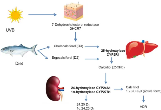
In the human skin, cholecalciferol is synthesized from 7-dihydrocholesterol by ultraviolet

irradiation (Figure 1.4).

(29)

Introduction

15

Figure 1.4. Vitamin D metabolism. Cholecalciferol is synthesized from 7-dihydrocholesterol by UV irradiation, or can be obtained in small amounts from the diet. In the liver, cholecalciferol is hydroxylated to calcidiol 25(OH)D

3

through the action of CYP2R1. Once in the kidney, 25(OH)D

3

is further converted into its active form, 1,25(OH)

2

D

3

.

Cholecalciferol is biologically inactive. It binds to transport proteins such as vitamin D binding proteins or albumin and enters the circulation. In the liver cholecalciferol is hydroxylated, a process catalysed by the enzymes CYPR1 and CYP27A1, resulting in the production of the inactive form 25-hydroxyvitamin D

3

(25(OH)D

3

), which represents the main circulating form of vitamin D. This form is also the most stable and is used as a parameter to define one person’s vitamin D status [79, 81].

In the kidney, 25(OH)D

3

is further converted to the biologically active form of vitamin D, calcitriol (1,25(OH)

2

D

3

) by the action of the enzyme 1-α-hydroxylase (CYP27B1).

Calcitriol levels are regulated in a renal negative feedback loop, including the inhibition of

CYP27B1 by high calcitriol levels and stimulation of CYP24A1 (24-hydroxylase), an

enzyme that metabolizes calcitriol to calcitroic acid, the inactive, water soluble form,

which is then excreted in the bile. Although the circulation levels of 1,25(OH)

2

D

3

levels

are mainly determined by renal CYP27B1, other cell types such as immune cells which

express this enzyme are also capable of converting 25(OH)D

3

into its biologically active

form. The production of 1,25(OH)

2

D

3

by macrophages and dendritic cells may lead to

high local concentrations of 1,25(OH)

2

D

3

which are assumed to be important for

immunomodulation [81].

(30)

Introduction

16 1.5.2. Vitamin D and immune function

Early evidence for the importance of vitamin D for the immune system dates from the 19

th

century. Cod liver oil was used as a treatment for infections such as consumption, a condition these days known as tuberculosis. Tuberculosis patients were also sent to sanatoriums, where therapy included sunlight exposure. It was believed that sun exposure directly kills Mycobacterium tuberculosis bacteria [79]. In the meantime we know that the effect of both, sunlight and cod liver oil, is due to vitamin D.

M. tuberculosis is a strong inducer of CYP27B1 and VDR in monocytes and macrophages after pathogen recognition by toll-like receptors. 1,25(OH)

2

D3 in turn can induce antimicrobial peptides such as cathelicidin and β-defensins [92] [79].

Human cathelicidin (LL37) is produced as a precursor protein called hCAP18, which upon cleavage releases LL37. Further processing can occur in the skin which results in the release of shorter peptides. hCAP18 is widely expressed in the immune system and also in a variety of epithelial cells. Beside tuberculosis, low 25(OH)D

3

serum levels have been associated with various infectious diseases, such as influenza, chronic obstructive pulmonary disease and allergic asthma and were found to be significantly lower in critically ill septic patients. It has also been shown that low 25(OH)D

3

serum levels correlates with decreased concentrations of antimicrobial peptides such as cathelicidin [82].

Taken together, these findings support the idea that the vitamin D status regulates antimicrobial protein levels that are critical for the control of infection.

Besides its effect on antimicrobial protein regulation, vitamin D also has other effects on the activation of immune cells [83]. In the hematopoietic system, VDR is expressed in various hematopoietic precursors as well as in monocytes, thymocytes and activated B and T cells [80]. Some studies with VDR-knock-out (VDR-KO) mice show that these animals produce normal number and proportion of blood cells, demonstrating that vitamin D signalling is not necessary for the differentiation of the hematopoietic cell repertoire [84]. Although the number of cells is not changed, it has been shown that there are changes in the cytokine profile in VDR-KO mice that may affect T-helper (Th) immune responses indicating that 1,25(OH)

2

D3 may regulate immune cell function [84].

Nevertheless there are studies stating that VDR also seems to be critical for the development of invariant natural killer T (iNKT) cells, a subset of T cells with a regulatory role in autoimmunity and infection [85]. Cantorna and co-workers have demonstrated that CD8

+

T cells and iNKT cells are targets for vitamin D [85]. VDR-KO mice have significant less iNKT cells, and the few iNKT cells present in the periphery are functionally defective.

Like iNKT cells, there are also less CD8αα/TCRαβ precursors in the thymus of VDR-KO

animals. CD8αα T cells are mainly present in the gut, where they have a role in

(31)

Introduction

17 preserving tolerance and suppress inflammation [85]. Taken together, these results show that VDR expression is compulsory for the development of both iNKT and CD8αα/TCRαβ cells.

Dendritic cells differentiated in the presence of vitamin D remain in an immature-like tolerogenic state. This can be accessed by the cytokine profile of these cells. Pro- inflammatory cytokines like IL-12 and TNF are reduced and anti-inflammatory cytokines like IL-10 are increased. Due to their maturation status, these tolerogenic DCs (tDCs) are less capable of stimulating T cell proliferation and cytokine production of pro- inflammatory T cells, whereas they induce the differentiation of regulatory T cells.

It was demonstrated that 1,25(OH)

2

D3 promotes the anti-inflammatory M2 phenotype of macrophages and is capable to induce the differentiation of monocytes into macrophages in vitro [86].

Although the effect of 1,25(OH)

2

D3 on B cells is still not completely understood, several reports suggest that vitamin D reduces the proliferation of B cells, induces their apoptosis and inhibits immunoglobulin class switching [87]. There is also evidence that vitamin D decreases antibody production [83].

For a long time it was believed that the effect of vitamin D on T cells was indirect and mediated exclusively by antigen-presenting cells. However, it has become clear now that upon activation, T cells upregulate the VDR, making this cell type another important target for vitamin D.

Figure 1.5. Effects of 1,25(OH)

2

D

3

on various immune cells. Adapted from [80].

(32)

Introduction

18 Vitamin D treatment has been shown to have an impact on different T cell subtypes., namely by inducing regulatory T cells [88]. When 1,25(OH)

2

D

3

is added to T cells, it inhibits the differentiation of pro-inflammatory Th1 cells by suppressing IFN-γ and favouring Th2 response. In VDR KO, the Th1 response is also impaired.

1.5.3. Vitamin D status and GvHD

In an optimal stem cell transplantation scenario, the donor immune system would destroy the malignant cells without harming the healthy recipient tissues. In order to prevent or treat GvHD, immune suppressive medication is required, which is crucial to (re)balance the immune response in patients.

Since vitamin D has already been evidenced as an important immunomodulatory hormone, this suggests a possible significance in the outcome of HSCT [77].

In fact, the relationship between vitamin D serum levels and GvHD outcome has already been investigated by a number of groups [89-92].

Kreutz et al. reported an analysis of 25(OH)D

3

and 1,25(OH)

2

D

3

serum levels in 48 patients undergoing allogeneic bone marrow transplantation (BMT) and found a decline in both metabolite levels in the course of transplantation [89].

In a different study, Glotzbecker and colleagues demonstrated that vitamin D deficiency before HSCT is associated with an increased risk of cGVHD [90].

VDR genes are polymorphic in the human population and their variants have also been a subject of investigation in patients undergoing HSCT [93-95].

Cho and co-workers hypothesized that polymorphisms of the VDR gene could affect clinical outcomes of allogeneic HSCT [96]. Three VDR gene polymorphisms (BsmI G>A, ApaI G>T, and TaqI T>C) were genotyped in 147 patients who underwent HLA-matched sibling aHSCT. Frequencies of infection, GvHD, overall survival (OS), and disease-free survival (DFS) were compared according to genotypes and haplotypes. In this study they found a correlation between polymorphisms at the TaqI cleavage site, where heterozygous patients had better DFS and OS than TT homozygote patients.

Also, they found that recipients having two copies of the “A” allele for the ApaI polymorphism had lower rates of aGvHD and infection. Homozygosity of the “A” allele is associated with higher VDR activity and in a study performed by Middleton et al. [95] a similar effect was reported: a solid trend toward reduced aGvHD in recipients with the AA genotype.

Bogunia-Kubik and co-workers also published a study regarding VDR polymorphisms and HSCT, but included an analysis of patients receiving cells from unrelated donors.

Here, they found an association between the FF genotype (associated with higher VDR

activity) and the outcome after HSCT. If both donor and recipient had the FF genotype,

(33)

Introduction

19 the recipient experienced higher risk of GvHD development. Interestingly, GvHD risk based on VDR polymorphisms depends on whether they are present in the donor or recipient.

So far, data suggest that both VDR and vitamin D may play a role in immune

reconstitution and immune surveillance in transplant patients. Prospective studies are

needed to investigate the effects of vitamin D supplementation, e.g. with 1,25(OH)

2

D3

analogues, on HSCT outcome [97]. In HSCT recipients, supplementation could promote

better immune function in the reconstituted immune system and help to prevent the

deleterious effects of GvHD.

(34)

Research goals and objectives

20

2. Research Goals and Objectives

GvHD is still the most common major life-threatening complication associated with allogeneic hematopoietic stem cell transplantation (aHSCT). Previous data demonstrate that single nucleotide polymorphisms of the NOD2/CARD15 receptor are associated with increased risk of GvHD. Furthermore, vitamin D has emerged as a central player in the immune system, with its deficiency being implicated in the pathogenesis of several diseases, including the development of GvHD.

The aims of this dissertation were to clarify the role of NOD2 status for the regulation of the immune response, namely by studying the immune cell composition in peripheral blood of healthy donors differing in their NOD2 status. Also, the effects of muramyl dipeptide (MDP), the natural ligand of NOD2, were studied in monocyte/MNC-derived dendritic cell cultures.

Moreover, the effect of vitamin D on human T cells with particular interest in the

generation of inducible regulatory T cells was investigated in vitro. In parallel, the in vivo-

effects of vitamin D levels were also studied, using an animal model of vitamin D

sufficiency versus deficiency to examine the impact of vitamin D in the immune cell

composition of blood, spleen and bone marrow of the mice. Additionally, the impact of

vitamin D levels on murine gut microbiota was analysed.

(35)

Material

21

3. Material

3.1. Equipment

Autoclave Technomara, Fernwald, Germany

Balance LP1200S Sartorius, Göttingen, Germany

Bioanalyzer 2100 Agilent Technologies, Böblingen, Germany

Biofuge fresco Heraeus, Osterode, Germany

CASY Cell Counter Innovatis/Roche, Basel, Switzerland Centrifuge Megafuge 3.0R Heraeus, Osterode, Germany Centrifuge Sigma 2 Sartorius, Göttingen, Germany Electrophoresis equipment Biometra, Göttingen, Germany Electrophoresis equipment (PFGE) Biostep, Jahnsdorf, Germany

ELISA plate reader MWG Biotech, Ebersberg, Germany EVOS Cell Imaging System Life Technologies, Carlsbad, CA, USA FACS Calibur flow cytometer BD Biosciences, Franklin Lakes, NJ, USA

Forceps Aesculap, Tuttlingen, Germany

Heat sealer Eppendorf, Hamburg, Germany

Hemocytometer Marienfeld, Lauda-Königshofen, Germany

Incubators Heraeus, Hanau, Germany

Laminar Flow Cabinet Clean Air Telstar, Woerden, The Netherlands LSR II flow cytometer Becton Dickinson, Heidelberg, Germany LSR Fortessa Becton Dickinson, Heidelberg, Germany

Microscopes Zeiss, Jena, Germany

Multifuge 3S-R Heraeus, Osterode, Germany

Multipipettor Multipette plus Eppendorf, Hamburg, Germany

NanoDrop 1000 Thermo Fisher Scientific, Schwerte, Germany PCR Thermocycler PTC-200 MJ-Research/Biometra, Oldendorf, Germany

pH Meter Knick, Berlin, Germany

pH Meter, portable Hanna Instruments, Kehl am Rhein, Germany

Picofuge Heraeus, Osterode, Germany

Abbildung

Figure  1.1.  The  three  phases  of  aGvHD,  according  to  Ferrara  et  al  [6].  In  phase  I,  chemotherapy  or  radiotherapy as part of transplant conditioning causes host tissue damage and release of inflammatory cytokines  such  as  TNF,  IL-1β,  an
Figure 1.3. Development of nTregs and iTregs. Adapted from [66]. nTregs (top) differentiate from naïve  conventional T cells to Foxp3+ Tregs in the thymus
Figure  1.4.  Vitamin  D  metabolism.  Cholecalciferol  is  synthesized  from  7-dihydrocholesterol  by  UV  irradiation, or can be obtained in small amounts from the diet
Figure 1.5. Effects of 1,25(OH) 2 D 3  on various immune cells. Adapted from [80].
+7

Referenzen

ÄHNLICHE DOKUMENTE

Dies sollte bei der Vit D Dosisanpassung im Falle einer Unterversorgung/ Mangel berücksichtig werden, damit es zu keiner unbeabsichtigten Überdosierung kommt.. Klinisch bewährt

Vitamin D spielt eine zentrale Rolle für die Knochengesund- heit und damit für die Osteoporose-Prophylaxe.. Auch das Immunsystem profitiert von ausreichend Vitamin D, denn es

This may lead to osteoporosis and rickets, and calcium is also involved in nerve function, mu- scle contraction and relaxation, digestion, blood clotting and blood

Laif ® 900 Balance darf nicht eingenommen werden bei bekannter Allergie gegenüber Johanniskraut oder einem der sonstigen Bestandteile?. Hinweise: Ausreichende Erfahrungen über

Eine aktuelle Untersuchung der Universität von Tel Aviv zeigt, dass bei Asthmapatienten mit einem Vi- tamin-D-Dezifit das Risiko, einen Asthmaanfall zu er- leiden, um 25 Prozent

Für die 75 nmol/l sprechen, dass erst ab diesem Zielwert eine Fraktur- und Sturzreduktion er- wartet werden kann (4, 5).. Diesen Ziel- spiegel zu erreichen, empfiehlt denn auch

Da unge - nügende Vitamin-D-Speicher aber für breite Bevölkerungsschichten ein Pro- blem seien, müsse hier die Botschaft lauten: «Geht regelmässig hinaus an die Sonne, holt euch

Zudem zeigt eine weitere Metaanalyse, die sich zurzeit im Druck befindet, dass Kalzium alleine einen neu- tralen Effekt auf nicht vertebrale Frakturen hat (40), ein Resul- tat,