Case studies on the genes
zerknüllt, decapentaplegic and short gastrulation in the beetle Tribolium illustrate
concepts in evolutionary developmental biology
Maurijn van der Zee aus Leiderdorp, Niederlande.
Köln, 2006
Berichterstatter: Prof. Dr. Siegfried Roth
Priv.-Doz. Dr. Wim G. M. Damen Vorsitzender der Prüfungskommission: Prof. Dr. Diethard Tautz
Tag der mündlichen Prüfung: 9. Juni 2006
Als Gregor Samsa eines Morgens aus unruhigen Träumen aufwachte, fand er sich in seinem Bett zu einem ungeheueren Ungeziefer verwandelt.
Er lag auf seinem panzeratig harten Rücken und sah, wenn er den Kopf ein wenig hob, seinen gewölbten, braunen, von bogenförmigen Versteifungen geteilten Bauch, auf dessen Höhe sich die Bettdecke, zum gänzlichen Niedergleiten bereit, kaum noch halten konnte.
Seine vielen, im Vergleich zu seinem sonstigen Umfang kläglich dünnen Beine
flimmerten ihm hilflos vor den Augen.
Franz Kafka: Die Verwandlung
Cover illustration: B. van der Zee (1917-1997)
CONTENTS
CHAPTER 1
General introduction 7
A history of Evo-Devo 7
Concepts in modern Evo-Devo 9
Aim of this thesis 12
CHAPTER 2
Distinct functions of the Tribolium zerknüllt genes in serosa specification and dorsal closure 15
2.1 SUMMARY 15
2.2 INTRODUCTION 16
2.3 MATERIALS AND METHODS 18
2.4 RESULTS 20
Expression of the zerknüllt genes in Tribolium 20
Functional analysis of the zerknüllt genes by parental RNAi 20 Loss of Tc-zen1 causes a transformation of presumptive serosa into germ rudiment 23 After loss of Tc-zen1 the amnion covers the yolk at the dorsal side of the embryo 29
Size regulation after loss of Tc-zen1 30
Tc-zen2 is required for dorsal closure 33
Simultaneous loss of Tc-zen1 and Tc-zen2 rescues the Tc-zen2 knock-down phenotype 37
2.5 DISCUSSION 38
Expansion of the germ rudiment 38
Size regulation 39
Dorsal closure 40
The function of the serosa 41
Hox3 and bicoid 42
2.6 ACKNOWLEDGMENTS 43
Contents CHAPTER 3
Sog/chordin is required for ventral-to-dorsal Dpp/BMP
transport and head formation in a short germ insect 45
3.1 SUMMARY 45
3.2 INTRODUCTION 46
3.3 MATERIALS AND METHODS 49
3.4 RESULTS 50
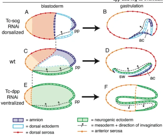
sog is expressed in a ventral domain of early Tribolium embryos, while dpp expression
initially lacks dorsoventral asmmetry 50
Tc-sog directs Dpp activity to the dorsal side 53
Loss of BMP signaling severely affects gastrulation 59
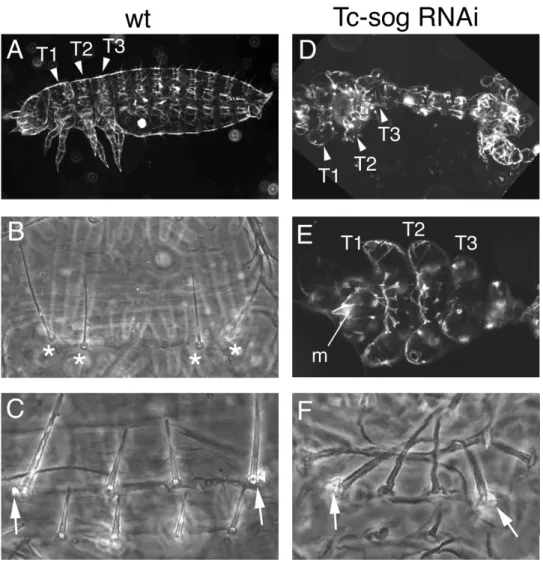
Tc-sog RNAi leads to a loss of the entire central nervous system and to a mirror image
duplication of dorsal cell fates 61
BMP signaling plays a role in head formation in Tribolium 70
3.5 DISCUSSION 71
The role of the dorsal Dpp transport by Sog 71
The origin of the double dorsal phenotype 75
Comparisons to vertebrates and the function of BMP signaling in head formation 76
3.6 ACNKOWLEDGMENTS 78
CHAPTER 4
General discussion 79
Modules 79
Gene duplication, redundancy and subfunctionalization 80
Co-option 82
Cis-regulatory evolution, gene networks and hubs 85
Concluding remarks 87
References 88
Acknowledgments/Danksagung 98
Abstract 99
Zusammenfassung 100
Talks and conferences 102
Erklärung 103
Lebenslauf 104
CHAPTER 1
General introduction
A history of Evo-Devo
Evolutionary developmental biology (Evo-Devo) aims to unveil how developmental processes and mechanisms become modified during evolution and how from these changes the past and present biodiversity arose (Baguna and Garcia-Fernandez, 2003). Evo-Devo is often called a “new science” or a
“revolution” (Carroll, 2005a); however, as soon as evolution entered scientific thinking, development was intimately associated with it. In the eighteenth century, scientists left the static view of Aristotle that all organisms are unchangeable and can be ordered from simple to complex in a fixed “Chain of Being”. Organisms were rather supposed to “march up” this Ladder of Being during evolution, as Lamarck later formulated it. This progressive view forced the static embryological theory of that time – preformationalism – to change. Preformationalism teaches that development merely makes visible the organism, which is already present in all its details in the egg (or sperm). And in this miniature organism in the egg, even smaller, similar miniatures of its own progeny are already present, and so on.
There was no space for evolutionary change. The preformationalist Bonnet (1720- 1793) was the first to construct a synthesis, paraphrased by Gould, (Gould, 1977):
“Why, after all, should the succession of encapsulated generations in the ovaries of a primal Eve all bear the same form. We might open the Russian doll in this primal ovary and find only ten dolls of identical form; inside the tenth we might discover a vastly superior creature, and after ten similar boxes another being of still more perfect design.”
In the nineteenth century, the idea of unchanging species and preformationalism were left for good. A new theory joining development and evolution became popular – recapitulation – with Haeckel as its later and most fanatic proponent. He formulated the biogenetic law: ontogeny is a recapitulation of phylogeny. That means: during its own embryological development, a higher organism quickly passes through the adult stages of its paleontological ancestors.
Haeckel thought that new, higher organisms arise by the addition of development at the end of the current development, a process called terminal addition. Of
Chapter 1
course, in order to prevent that development would finally become endless in the highest organisms, the ancestral development was shortened, or “condensed”
(Gould, 1977). Although there is some superficial truth in Haeckels observations, the statement that an early stage in human development literally is, for example, an adult fish, is too dogmatic.
Von Baer (1792-1876) put the resemblances of embryonic stages of mammals with, for example, adult fish in a better perspective. He observed that development proceeds from the most general to the most specialized. If one goes back in embryological development of a vertebrate, one will find the general form of vertebrates. A mammalian embryo never passes through a real fish state, but fish are simply less different from their early embryos. Von Baer notes (Von Baer, 1828):
“It is only because the least developed animals are but little removed from the embryonic condition, that they retain a certain similarity with the embryos of higher animal forms.”
For Darwin (1809-1882) this was inspiring. He realized that similarity of embryos reveals a common ancestry. Paraphrased by Gould (1977):
“The gill slits of the human fetus represent no ancestral adult fish: we see no repetition of adult states, no recapitulation. Yet adult fish, as primitive ancestors, have departed least from this embryological condition of all vertebrates.”
Hence, embryology can give important clues about phylogeny and can help to determine true homologies. I will call this the first principle connecting development and evolution. Despite this important finding, developmental biology lost its importance for evolution in the beginning of the twentieth century.
Haeckels student Roux encouraged embryologists to leave the seashore and go into the laboratory, thus announcing the start of experimental embryology in 1894.
In the same year, Bateson claimed that the embryological method has failed when it came to the mechanisms of evolution (Gilbert, 2003). Famous experimental embryologists like Spemann entered the scene. Furthermore, the formulation of the Modern Synthesis (around 1940) providing a cause for evolution by integrating genetics and evolutionary biology, made evolutionary biology go its own way.
However, there have always been scientists trying to bridge development and evolution. Garstang (1922), for example, claimed: “Ontogeny does not recapitulate phylogeny; it creates it.”, or De Beer (1954), who stressed the importance of heterochrony (both in: Gilbert, 2003). After the formulation of the Modern Synthesis, there were even scientists who tried to integrate genetics, evolutionary and developmental biology. Most notably Goldschmidt, Waddington
and Schmalhausen. Goldschmidt speculated that mutations in developmentally important genes could cause big changes in the body plan (‘hopeful monsters’), enabling macroevolution. Waddington plead for the study of the process that turns the genotype into a phenotype (= development), and developed important modern concepts, like developmental constraint, canalization and genetic assimilation, the latter originally proposed by Baldwin. Canalization and genetic assimilation were independently discovered by Schmalhausen (Gilbert, 2003).
Concepts in modern Evo-Devo
Canalization is the property of developmental pathways to produce standard phenotypes despite mild genetic or environmental perturbations. Genetic assimilation is the phenomenon that an environmentally induced reaction can easily come under genetic control, if this is advantageous. Both can be illustrated by sex determination in reptiles. In some reptiles, sex determination is temperature dependent. Canalization achieves that an individual does not have an intermediate sex, when developed at an intermediate temperature, but that an individual is either male or female. Genetic assimilation effectuated that sex determination came under genetic control in some reptiles (Kirschner and Gerhart, 2005).
In 1977, Stephen Jay Gould wrote the important book ‘Ontogeny and Phylogeny’, rediscovering the classical evolutionary embryologists. The book is mainly about heterochrony. Heterochrony assumes that developmental processes are more or less dissociated and can be independently accelerated or retarded. The results can be paedomorphosis (the new adults look more like the embryo of the ancestor) or recapitulation (the new embryos look like adults of the ancestor).
Paedomorphosis can be caused by progenesis (acceleration of maturation with regard to somatic development) or by neoteny (retardation of shape/somatic development with regard to maturation). Recapitulation can be caused by hypermorphosis (retardation of maturation with respect to somatic development) or by acceleration (acceleration of shape/somatic development with respect to developmental stage). Some people call this book about developmental mechanisms of evolutionary change the conception (Gilbert, 2003), or even the birth of Evo-Devo (Hall, 2003).
However, the biggest impetus for modern Evo-Devo was provided by the integration of genetics and molecular biology into embryology. The discovery of Hox genes, and the screen for developmental genes in Drosophila by Christiane Nüsslein-Volhard and Eric Wieschaus made clearer how genes direct development. Although Ernst Mayr warned that “the search for homologous genes is quite futile except in very close relatives”, it was soon discovered that many
Chapter 1
developmental genes (for example Hox genes) are evolutionary conserved among phyla (McGinnis et al., 1984). The comparison of developmental genes between species became the new discipline of Evo-Devo (Carroll, 2005a).
The main idea is that mutations in developmental genes cause a change in development (“developmental reprogramming”; Arthur, 2004) and lead to evolutionary novelty. By comparing genes among organisms with known phylogenetic relationships, those changes can be traced and evolutionary scenarios can be inferred. I will call this the second principle connecting development and evolution. An example is the discovery of a mutation in the abdominal Hox protein Ultrabithorax (Ubx) in higher insects. This mutated Ubx protein represses Distal-less expression. Thus, abdominal legs are repressed in higher insects whereas other arthropods, which do not have this mutation, bear abdominal legs (Galant and Carroll, 2002; Hittinger et al., 2005; Ronshaugen et al., 2002). The evolutionary scenario would be that higher insects acquired this mutation and lost the abdominal legs.
However, the Ubx mutation did probably not happen in a ‘hopeful monster’ way. Ubx is partially redundant with the Hox gene Abdominal-A which also represses abdominal legs (Hittinger et al., 2005; Tour et al., 2005). And, although ectopic Ubx can repress thoracic legs, removal of the mutation does not simply lead to abdominal legs (Hittinger et al., 2005; Tour et al., 2005).
Evolutionary novelty is hardly ever reducable to a mutation in a protein. Changes in proteins are usually deleterious. Gene duplication (Ohno, 1970) can circumvent this problem. After duplication, one copy of the gene could continue to carry out the ancestral function, while the other copy can acquire mutations and adopt a new function.
Another way to achieve morphological novelty without affecting protein functionality, is changing cis-regulatory sequences (Carroll, 2005a; Carroll, 2005b). By the creation of new binding sites for transcription factors in cis- regulatory sequences of genes, those genes or gene duplicates can be recruited to perform their task - or a new task - in novel tissues or at novel moments in development. This process can be called co-option (Raff, 1996). A beautiful example is the co-option of NADPH:quinone oxidoreductase as the lens protein ζ- crystallin in guinea pigs (Lee et al., 1994). Co-option can occur at the morphological level as well when existing structures become recruited for a new function, for example the reptilian jaw articulation elements, which became the bones of the middle ear in mammals. Thus, nature works with “what is already there”, rather than inventing completely new things. Evolution is tinkering, as Jacob formulated it (Jacob, 1977).
This tinkering requires that the “tinkering units” can be dissociated and are not highly connected. Therefore, life must have a modular organization. The separable modules can be used again and again in different combinations by nature (Raff, 1996). Modularity and dissociation stay at the basis of many Evo-Devo concepts. Heterochrony, for example, can only occur when developmental processes can be independently regulated. Regulatory sequences of genes also have a modular organization; the ζ-crystallin of guinea pigs, for example, has a lens specific and an enzyme specific promoter. An important way to achieve modularity in the spatial dimension is segmentation. This divides the animal body into independent modules, of which for example appendages can be independently modified, without affecting other appendages. This is also called compartmentation (Kirschner and Gerhart, 2005).
The word compartmentation can also be used in describing gene networks.
Genes can be considered nodes and their regulatory connections can be considered links in a network (Barabasi, 2002). It appears that gene networks have a modular organization too, with high connectivity within, but low connectivity among the subnetworks, or compartments (Niehrs, 2004). The high connectivity within a compartment is caused by only a few genes with lots of links:“hubs” (Somogyi et al., 2004). The way nodes are connected gives the network certain properties (Barabasi, 2002). By changes in cis-regulatory sequences, the connections in a network and thus the properties of the network change. By recruiting a hub, the whole subnetwork is co-opted. Network growth by gene duplication will enhance the formation of hubs, because a duplicating gene is probably connected to a hub.
Since duplicated genes will initially retain their ancestral connections, the hub has received an extra link by the duplication event (Barabasi, 2002).
If modules are not well dissociated, evolution becomes constrained. If a gene is for example involved in two processes, favorable mutations for one process might be disadvantageous for the other process. Thus, pleiotropy can keep genes at a suboptimal level. Gene duplication can sometimes circumvent this problem, since gene duplication can be followed by subfunctionalization: both copies specialize in a specific function of the ancestral gene (Force et al., 1999;
Lynch et al., 2001; Piatigorsky and Wistow, 1991). However, pleiotropy and highly connected modules form developmental constraints.
A beautiful example of a developmental constraint is the conserved number of cervical vertebrae in mammals, caused by the pleiotropic effects of the Hox genes determining this number (Galis, 1999). Especially during the phylotypic stage, modules seem to be highly connected and evolution highly
Chapter 1
constrained (Galis and Metz, 2001). Beacuse embryology constrains certain changes and facilitates the reuse of already present modules (Kirschner and Gerhart, 2005), embryos ‘steer’ evolution (Arthur, 2004). Of course, random mutations are always required, but the chance for some type of changes to happen is bigger than for other types. I will call this the third principle connecting development and evolution. Although this is only recently more appreciated (Arthur, 2004; Kirschner and Gerhart, 2005; Schlosser and Wagner, 2004) it has already intelligently described by Rupert Riedl in 1975 (Riedl, 1975), whom I herewith boldly declare the Father of Evo-Devo.
Summarizing, there are three important principles that connect embryology to evolution. First, as the classical comparative embryologists already discovered, embryology supplies important clues about phylogenic relationships and about true homologies. Second, by comparing developmental genes among organisms with known phylogenetic relationships, changes that caused developmental reprogramming can be traced and evolutionary scenarios can be inferred. Third, embryos can steer evolution by constraining certain types of changes and by facilitating other types.
Aim of this thesis
In this thesis, the function of developmental genes in the red flour beetle Tribolium castaneum is compared to the function of their homologues in other animals, like the fruitfly Drosophila melanogaster. The phylogeny of insects is well known (Whiting, 2004). It appears that Drosophila belongs to a derived group of Diptera. The development of Drosophila displays a lot of derived characters: the anterior segments are specified by the maternal gene bicoid, pattern formation largely takes place in the syncytial blastoderm, all segments are present in the blastoderm and no growth zone is present, the legs develop from imaginal discs and the larval head is folded inward. Tribolium represents a more ancestral state with regard to these characters and its genes probably retained their ancestral function. By comparing those gene functions to the function of their homologues in Drosophila, I attempt to reconstruct what evolutionary changes occurred in the lineage leading to Drosophila.
The thesis consists of two case studies. In the first one (chapter 2), the function of the two zerknüllt genes in Tribolium is investigated. One of the biggest morphological differences between Tribolium and Drosophila embryos is the architecture of their blastoderm and their extraembryonic membranes. Nearly the whole Drosophila blastoderm will give rise to the embryo proper and only a small
part will give rise to a very reduced extraembryonic membrane. In contrast, a big part of the Tribolium blastoderm is occupied by two extraembryonic membranes which later play a role in important morphological processes like dorsal closure. In this respect, most insects develop like Tribolium (Roth, 2004). Drosophila zerknüllt has been shown to specify the extraembryonic membrane. One of the questions I hope to answer by studying zerknüllt function in Tribolium is how Drosophila could reduce its extraembryonic membranes so much without affecting important morphogenetic processes like dorsal closure.
In the second case study (chapter 3), the functions of decapentapegic (dpp) and short gastrulation (sog) are examined. The vertebrate homologues of these genes are called BMP and chordin respectively. BMPs belong to a well known family of ligands, the TGFβ family. BMPs play a role in a lot of processes, but also in dorsoventral patterning in vertebrates and in Drosophila. Sog and chordin are BMP inhibitors. In vertebrates, BMP has to be inhibited anteriorly to form the head. This seems not to be the case in Drosophila. Since the only functional study of Dpp and Sog in arthropods has been carried out in Drosophila, it is still unclear whether BMPs play a role in dorsoventral patterning in all arthropods, and if Sog really does not have a role in head formation in other arthropods. By investigating the function of Sog and Dpp in Tribolium, I investigate another arthropod and hope to resolve those uncertainties.
In chapter 4, the conclusions and results of the case studies are discussed in a bigger perspective: I explore to what extent the introduced concepts of Evo- Devo apply to them. It turns out that the comparative studies of developmental genes in Tribolium and their evolutionary implications illustrate many concepts in evolutionary developmental biology.
CHAPTER 2
Distinct functions of the Tribolium zerknüllt genes in serosa specification and dorsal closure
2.1 SUMMARY
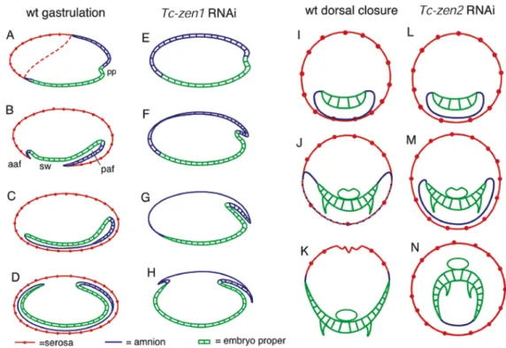
Background: In the long germ insect Drosophila a single extraembryonic membrane, the amnioserosa, covers the embryo at the dorsal side. In ancestral short germ insects an inner membrane, the amnion, covers the embryo ventrally and an outer membrane, the serosa, completely surrounds the embryo. An early differentiation step partitions the uniform blastoderm into the anterior-dorsal serosa and the posterior-ventral germ rudiment giving rise to amnion and embryo proper. In Drosophila amnioserosa formation depends on the dorsoventral patterning gene zerknüllt (zen), a derived Hox3 gene.
Results: The short germ beetle Tribolium castaneum possesses two zen homologues, Tc-zen1 and Tc-zen2. Tc-zen1 acts early and specifies the serosa. The loss of the serosa after Tc-zen1 RNAi is compensated by an expansion of the entire germ rudiment towards anterior. Instead of the serosa, the amnion covers the embryo at the dorsal side and later size regulation normalizes the early fate shifts, revealing a high degree of plasticity of short germ development. Tc-zen2 acts later and is required for the amnion and serosa fusion, necessary for dorsal closure.
After Tc-zen2 RNAi, the amnion and serosa stay apart and the embryo closes ventrally, assuming a completely everted (inside-out) topology.
Conclusions: In Tribolium, the duplication of the zen genes was accompanied by subfunctionalization. One of the paralogues, Tc-zen1, acts as an early anterior-posterior patterning gene by specifying the serosa. In absence of the serosa, Tribolium embryogenesis acquires features of long germ development with a single extraembryonic membrane. I discuss implications for the evolution of insect development including the origin of the zen-derived anterior determinant bicoid.
Chapter 2
2.2 INTRODUCTION
One of the evolutionary innovations that probably contributed to the unparalleled success of the insects is the formation of two protecting extraembryonic membranes: the amnion and the serosa (Zeh et al., 1989). These membranes are absent from all other arthropods (Machida and Ando, 1998; Roth, 2004). The prominent distinction between cells of the presumptive serosa and cells of the germ rudiment, giving rise to the amnion and the embryo proper, is the first differentiation step that occurs within the uniform blastoderm. This stage is called the “differentiated blastoderm” (Roth, 2004). Remarkably, this stage is absent in a small group of insects, the higher dipterans, to which Drosophila belongs (Schmidt-Ott, 2000; Schwalm, 1988). In Drosophila, the germ rudiment occupies the entire blastoderm. A single extraembryonic membrane, the amnioserosa, develops from a dorsal stripe of the blastoderm and covers the yolk at the dorsal side of the egg during further development.
The red flour beetle Tribolium castaneum may be a representative of all other insects, given that it still possesses an amnion and a serosa and the differentiated blastoderm stage. At this stage, the prospective serosa is visible as a dorsally tilted anterior cap of flattening cells. During gastrulation, the serosa begins to cover the embryo proper at the posterior pole, forming the posterior amniotic fold, together with the amnion. The same occurs to a lesser extent at the anterior pole, forming the anterior amniotic fold. When the crests of the amniotic folds meet, a round serosal window is formed. Finally, the serosal window closes, forming a continuous outer membrane, the serosa and an inner membrane, the amnion (Handel et al., 2000) (see also Fig. 9A-D for schematic representation).
In Drosophila, amnioserosa formation requires the gene zerknüllt (zen) which encodes a homeobox transcription factor (Rushlow and Levine, 1990) and is a target gene of maternal and zygotic DV morphogen gradients. During early blastoderm stages, the maternal NF-κB/Dorsal protein gradient represses zen in the ventral half of the embryo and thus confines its transcription to a broad dorsal domain (Rushlow et al., 1987b). During gastrulation, a zygotic BMP gradient with peak levels along the dorsal midline activates zen in a 5 cell-wide dorsal expression domain, comprising the cells that give rise to the amnioserosa (Raftery and Sutherland, 2003). In zen mutants, the amnioserosa is lost and is replaced by dorsal ectoderm. zen maps to the Antennapedia complex (ANT-C) between deformed and proboscipedia (Wakimoto et al., 1984). This region harbours two closely linked transcription units, zen and z2, with identical expression patterns (Rushlow et al., 1987a). Since zen alone could rescue a deletion covering both zen
and z2, it was suggested that z2 is dispensible (Rushlow et al., 1987a). This was later confirmed by producing a deletion specific for z2 (Pultz et al., 1988).
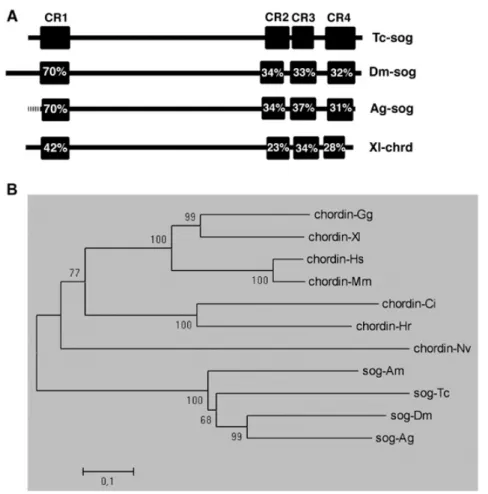
zen homologues have also been found in Tribolium castaneum and the grasshopper Schistocerca gregaria. Schistocerca-zen is expressed in the serosa and late amnion, while Tribolium-zen is expressed in the serosa (Dearden et al., 2000; Falciani et al., 1996). Later sequencing of the Tribolium ANT-C revealed two zen homologues: Tc-zen1, which corresponds to the homologue cloned by Falciani et al. (Falciani et al., 1996), and Tc-zen2 (Brown et al., 2002). Notably, the duplication of zen in Drosophila and Tribolium has happened independently, as inferred from phylogenetic analysis (Brown et al., 2002) and the absence of z2 in Drosophila pseudoobscura (Randazzo et al., 1993). So far, expression of Tc- zen2 has not been investigated. The expression of Schistocerca-zen and Tribolium- zen1 suggests a conserved function for zen in the patterning or differentiation of extraembryonic tissues in all insects. However, until now no functional study has been conducted except in Drosophila.
In this chapter, I show that after duplication, the zen genes of Tribolium acquired partially different expression patterns and diverged completely with regard to their function. RNAi with Tc-zen2 results in incorrect dorsal closure, generating completely everted (inside-out) larvae. RNAi with Tc-zen1 leads to the complete loss of the anteriormost cell fate, the serosa, and a compensating expansion of the more posterior germ rudiment. The fact that Tc-zen1 is involved in early specification of an anterior structure might have been a favourable condition for the evolution of the zen-derived anterior determinant bicoid which specifies head and thorax in higher dipterans (St Johnston and Nusslein-Volhard, 1992). Despite absence of the serosa after Tc-zen1 RNAi, size regulation generates normal larvae which exhibit perfect dorsal closure. This developmental plasticity sheds light on the evolution of long germ insects with a single extraembryonic membrane (the amnioserosa).
Chapter 2
2.3 MATERIALS AND METHODS
Stock maintenance
Beetles were kept on wheat flour (Diamant extra, type 405) supplemented with dried bakers yeast in plastic boxes at 30°C. For egg collection, beetles were placed for one to three days on instant flour (Diamant instant, type 405). Beetles were sieved out with a 710 µm maze sieve and eggs collected with a 300 µm maze sieve (Retsch). All food was kept at least one night at -20°C and all sieves were kept at 60°C in order to prevent infections.
Embryo fixation
Embryos were dechorionized with a hypochlorite solution and fixed for 30 minutes in 4 ml PEMS (0.1 M PIPES, 2mM MgSO4, 1mM EGTA, pH 6.9), 5 ml heptane and 1ml 20% formaldehyde. Subsequently, the water phase was removed and embryos were devitellinized by a methanol shock and stored in methanol at -20°C. Older embryos, difficult to devitellinize by a methanol shock, were devitellinized with forceps and needle.
Cloning of Tc-zen2
RNA was islolated by a trizol (Invitrogen) extraction and cDNA was made using the Cloned AMV First Strand Synthesis Kit (Invitrogen). Tc-zen2 was amplified from this cDNA with the forward primer CCATTCTCGGGGC TTTTCATAG and the reverse primer ACAATTCTTCCCTTGGTAATACTG at an annealing temperature of 60°C and cloned into TOPO vector (Invitrogen).
Synthesis of dsRNA and in situ probes
dsRNA was synthesized with the MEGAscript RNAi kit of Ambion according to the manufacturers protocol. DIG-labeled in situ probes were made with the MAXIscript T7/T3 Kit (Ambion), but with DIG RNA Labeling mix (Roche). Preferentially linearized plasmids were used as template instead of PCR products. lacZ dsRNA: A fragment including the lacZ gene and a T7 site was amplified from the TOPO II vector with the forward primer AGCGCCC AATACGCAAACCG and the reverse primer CACACCCGCCGCGCTTAATG at an annealing temperature of 65°C and cloned into the TOPO 2.1 vector (Invitrogen). Fragments with two opposing T7 sites were excised with PvuII, purified (Purification Kit, Amersham) and used as template for dsRNA synthesis.
Parental RNAi
Female pupae were collected shortly before hatching and the dorsal side of the terminal segment was fixed on a microscope slide with Fixogum (Marabu).
Approximately 0.2 µl of a 0.5-1.0µg/µl dsRNA solution was ventrally injected between the third and fourth abdominal segment (see Fig. 2 for total amounts).
After 5 days, wild type male beetles were added and offspring was collected and analyzed. (Bucher et al., 2002)
In situ hybridizations and immunostainings
In situ hybridizations were performed as described (Tautz and Pfeifle, 1989), but without proteinase K treatment. In situ stained embryos were counterstained with DAPI. Immunostainings were essentially carried out as described in (Roth et al., 1989). The Engrailed antibody 4D9 was used 1:5 and the anti-phospho-histon3 antibody was used 1:1000 in PBST.
Cuticle preparation
Eggs were transferred to a 96 well plate. After 5 days, hatching rates were counted and a few drops of a 9:1 lactic acid:ethanol mixture were added to every well. After one night at 60°C, cuticles were studied under a darkfield binocular or mounted for light microscopy.
TUNEL assays
Assays were essentially performed as described in (Prpic, 2004b).
However, 0.1 % Tween-20 was added to the TdT and DNAse I buffer.
Preparation of cross sections for confocal microscopy
Embryos in their vitelline membrane were cut with a razor blade into 5 or 6 slices in PBST, treated with RNAse and washed in PBST. After refixing 10 minutes in 4% formaldehyde in PBST, sections were washed with PBST, blocked for one hour in 1% bovine serum albumin and 3% normal goat serum in PBST and subsequently incubated with undiluted anti-phospho-tyrosine antibody overnight at 4°C. After washing with PBST and 30 minutes blocking, sections were incubated for two hours with Alexa 555 anti-mouse antibody (Molecular Probes, 1:400) and YOYO-1 (Molecular Probes, 1:25.000) in PBST at room temperature. After washing with PBST, sections were embedded in vectashield (Vector Laboratories) and examined under a confocal microscope.
Chapter 2
2.4 RESULTS
Expression of the zerknüllt genes in Tribolium
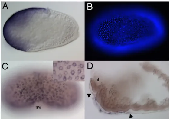
I first asked whether the duplication of the zen genes in the lineage leading to Tribolium led to differences in the expression pattern of the two paralogues. Tc- zen2 is expressed similar to Tc-zen1 (Falciani et al., 1996) in the presumptive serosa at the anterior pole of blastoderm embryos (Fig. 1A). The flat cells of the presumptive serosa are easily recognised by the wider spaced nuclei (Fig. 1B).
During and after gastrulation, when the serosa covers the embryo, Tc-zen2 continues to be expressed in the serosa, like Tc-zen1 (Fig. 1C). As for Tc-zen1, no expression was detected in the embryo proper or in the early amnion. Whereas Tc- zen1 expression remains restricted to the serosa, Tc-zen2 shows a new expression domain in the late amnion (Fig. 1D). Tc-zen2 transcripts can be detected in the flattened amniotic cells which cover the posterior head and the anterior thorax region (Fig. 1D). This expression is initiated shortly before the amnion and serosa begin to fuse (see below).
Functional analysis of the zerknüllt genes by parental RNAi
To investigate the function of the Tribolium zen genes, I performed knock- down experiments using the parental RNAi technique As a negative control, female pupae were injected with lacZ dsRNA. Aside from 13% unfertilized or undeveloped eggs, all developing young embryos from these mothers were indistinguishable from wild type (Fig. 2, penultimate bar). After injection of 0.1 µg Tc-zen1 or Tc-zen2 dsRNA, similar frequencies of unfertilised or undeveloped embryos were observed (Fig. 2). The developing embryos however, displayed distinct, specific phenotypes which are described in the next sections. Control in situ hybridizations confirmed the absence of Tc-zen1 or Tc-zen2 transcripts after the respective dsRNA injection and the absence of both transcripts after the combined injections. Furthermore, I monitored the interdependence of the paralogues. Tc-zen2 does not control Tc-zen1 expression, since normal Tc-zen1 expression was detected after Tc-zen2 RNAi. In contrast, the early expression of Tc-zen2 is dependent on Tc-zen1, since no early Tc-zen2 transcripts were found after Tc-zen1 RNAi. However, late amniotic expression of Tc-zen2 could be detected after Tc-zen1 RNAi, demonstrating that this late expression is independent of Tc-zen1 and that a non-specific knock down of Tc-zen2 transcripts by Tc-zen1 dsRNA injections can be excluded.
Fig. 1. Expression of Tc-zen2
All embryos show in situ hybridisations for Tc-zen2. (A) Optical midsection of a differentiated blastoderm embryo. Tc-zen2 is expressed in a tilted anterior cap. (B) Fluorescence image of the surface of the embryo shown in A. DAPI staining visualizes the nuclei. Tc-zen2 is expressed in the wider spaced nuclei of the presumptive serosa. (C) Serosal window stage. Tc-zen2 is expressed in the serosa, which completely covers the embryo. Inset: serosal surface at larger magnification. Tc-zen2 transcripts are found in the cytoplasm surrounding the nuclei of the stretched-out serosal cells. (D) Extended germband stage. Tc-zen2 is expressed in the amnion, covering the thoracic and posterior head region. The serosa has been removed. Arrowheads indicate the limits of the expression. sw= serosal window, hl = head lobe.
Chapter 2
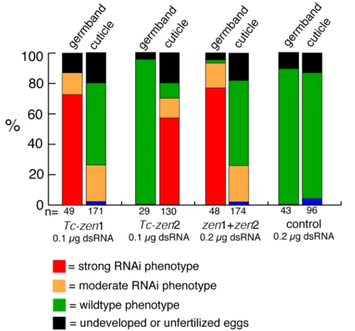
Fig. 2. Phenotypic frequencies for parental RNAi with the two Tc-zen homologues.
Frequencies of phenotypes after Tc-zen1 RNAi, Tc-zen2 RNAi, combined RNAi and a control injection with LacZ dsRNA. Every first bar represents the germband stages analyzed (12-18h). Every second bar represents the cuticles analyzed (>96h). Strong RNAi phenotypes are shown in red, moderate RNAi phenotypes in orange, wildtype phenotypes in green, unfertilized or undeveloped eggs in black and non-specific effects in blue. Tc- zen1 RNAi has an early effect, the abnormal phenotype is later restored (compare first and second bar). Tc-zen2 RNAi has a late effect (compare third and fourth bar). The combined RNAi is similar to Tc-zen1 RNAi. Short description of the categories: Germ band extension phenotypes after Tc-zen1 RNAi: Strong phenotype = big head, no serosa.
Moderate phenotype = normal appearing head, no serosa. Cuticle phenotypes after Tc-zen1 RNAi: Moderate phenotype = unhatched, strongly curved larvae. Cuticle phenotypes after Tc-zen2 RNAi: Strong phenotype = completely everted. Moderate phenotype = partially everted. Combined injection: see Tc-zen1 RNAi. Note that after Tc-zen1 RNAi, all differentiated blastoderm embryos (6-9h) lack the serosa and have an expanded germ rudiment (see text). The first bar, however shows a slightly later stage (12-18h), in which 71% displays dramatic consequences of this phenotype (red), but 14% are already partially recovered (orange).
Loss of Tc-zen1 causes a transformation of presumptive serosa into germ rudiment
In wild type embryos at the differentiated blastoderm stage, a clear distinction arises between the wider spaced serosal cells and the more densely spaced cells of the germ rudiment (Fig. 1B). This difference is even more pronounced at the onset of gastrulation, marked by the formation of the primitive pit at the posterior pole (Fig. 3A). At this stage the serosal nuclei become larger than those of the germ rudiment since they undergo polyploidisation. After Tc- zen1 RNAi, the embryos never partition the blastoderm into regions with distinct nuclear spacing (Fig. 2 and Fig. 3G). The nuclear density throughout the embryo resembles that of the germ rudiment. Thus, high nuclear densities are also found in the anterior third of the embryo, where the serosa would form normally. At the anterior tip a slight decrease in nuclear density is observed (Fig. 3G, 4J). However, even here the nuclei are more densely packed than in the presumptive serosa of a wild type embryo at the corresponding stage. Furthermore, in this region the nuclear size does not increase as in serosal nuclei and there is no sharp boundary separating this region from the remaining cells.
The high nuclear density throughout the embryo can be explained by assuming that Tc-zen1 RNAi causes a loss of the serosal cell fate. Almost all cells of the blastoderm appear to adopt the fate of germ rudiment. The slight decrease in nuclear density at the anterior pole could be due to an incomplete knock-down of Tc-zen1 after dsRNA injection. However, the later development of Tc-zen1 RNAi embryos described below suggests another explanation. To test the assumption that the germ rudiment expands at the expense of the serosa after Tc-zen1 RNAi, I analysed the expression of five marker genes.
Tc-twist served as a ventral marker. In wild type at the uniform blastoderm stage, Tc-twist is weakly expressed in a ventral stripe along the entire AP axis.
Shortly before primitive pit formation however, Tc-twist is excluded from the emerging serosa and is strongly upregulated in the germ rudmiment (Fig. 4A,B;
(Handel et al., 2005; Sommer and Tautz, 1994). In Tc-zen1 RNAi embryos, I observed this strong upregulation along the entire AP axis (Fig. 4I,J). The expanded stripe of strong twi expression includes the slightly wider spaced cells at the anterior tip, demonstrating that they do not behave like serosal cells. The change in twi expression suggests that the whole ventral side of the embryo adopted the cell fate of the germ rudiment.
Chapter 2
Fig. 3. The development of embryos after parental Tc-zen1 RNAi
(A-F) Wild type embryos, lateral views, unless otherwise indicated. (G-L) Embryos of the corresponding age after Tc-zen1 RNAi. (A-E, G-K) DAPI stainings. (D-F, J-L) engrailed antibody stainings. (A) Onset of gastrulation (primitive pit stage). The nuclei of the serosa are larger and wider spaced than those of the germ rudiment. (B) Serosal window stage.
The amnion and the serosa grow over the head and abdomen of the embryo proper. (C) Extending germ band stage. After closure of the serosal window, the amnion and the serosa cover the embryo as continuous membranes. The amnion (a) is well visible. The serosa has been removed. (D) Extended germ band stage. Amnion and serosa have been removed. (E) Ventral view of the third engrailed stripe of an extending germ band embryo.
(F) Retracting germ band stage. (G) After Tc-zen1 RNAi the distinction between the widely spaced nuclei of the serosa and the densely spaced nuclei of the germ rudiment is absent. All nuclei are densely spaced. (H) During gastrulation, cells condense slowly to the ventral side. No serosal window is visible. (I) More cells than in wildtype contribute to the head, which develops slower. The posterior amniotic fold forms. (J) The cells of the big head express Engrailed. The En stripes are laterally expanded and further apart in the anteroposterior dimension. Development of the head is delayed with regard to development of the abdomen. (K) Ventral view of the third engrailed stripe of an extending germband embryo. The stripe consists of 1.5 times more cells, which are less densely spaced. (L) At the retracted germband stage, Tc-zen1 RNAi embryos look rather normal. Arrowheads point at the double extraembryonic membrane, covering the abdomen of embryo. a = amnion, paf = posterior amniotic fold, pp = primitive pit, sw = serosal window.
Fig. 4. Expansion of the germ rudiment after Tc-zen1 RNAi.
(A-G) Wild type differentiated blastoderm stages. (A-F) and early gastrulation (G,H).
Lateral views unless indicated otherwise. (I-P) Corresponding stages after Tc-zen1 RNAi.
(B, D, F, H) and (J, L, N, P) show DAPI counterstainings of the embyos shown above. (A, B) Ventral view. Tc-twist is expressed in the germ rudiment, but not in the flattened cells of the serosa characterized by wider spaced nuclei. (C,D) Tc006A12 expression in the anterior germ rudiment, just posterior of the serosa. Onset of expression in the primitive pit allows accurate staging of the embryos. (D, E) Tc-evenskipped at the onset of the differentiated blastoderm stage. (G, H) Tc-hairy expression at the onset of gastrulation. (I, J) Ventral view. Tc-twist upregulation has expanded to the anterior tip. Some slightly wider spaced nuclei are present at the anterior tip. However, they express twi and are therefore not of serosal origin. (K,L) The anterior domain of Tc006A12 expression shows a dramatic expansion towards anterior. The more posterior domain also seems to be expanded. (M, N) Tc-eve expression at the primitive pit stage. Both the posterior cap and the stripe are expanded towards anterior and are enlarged. No serosal nuclei are present.
(O, P) All three Tc-hairy stripes expanded towards dorsal and anterior, and shifted anteriorly. The effect is diminished for the more posterior stripes.
Chapter 2
To investigate how the AP coordinates of the germ rudiment change after Tc-zen1 RNAi, the expression of the novel gene Tc006A12 (Fig. 4C,D;
GENBANK accession number CB335374 (Savard, 2004)) was analyzed. In wild type embryos at the primitive pit stage, Tc006A12 is expressed in a wedge-shaped domain, immediately posterior the serosa (Fig. 4C,D). The onset of expression of an additional posterior Tc006A12 domain (Fig. 4C,D) allows accurate staging of Tc-zen1 RNAi embryos in comparison to wildtype. In Tc-zen1 RNAi embryos, the anterior Tc006A12 domain expands and is shifted towards the anterior pole, covering the area, which harbours the serosa in wild type (Fig. 4K,L). Since the expansion is more pronounced at the dorsal in comparison to the ventral side, the domain becomes more symmetrical along the DV axis. These data suggest that the serosa is replaced by enlarged anterior regions of the germ rudiment.
To investigate if also more posterior fates of the germ rudiment expand, the pair-rule genes Tc-eve (Brown et al., 1997) and Tc-hairy (Sommer and Tautz, 1993) were used as markers. In wildtype, Tc-eve is expressed in a broad posterior cap and in one anterior stripe at the very onset of primitive pit formation (Fig. 4E, F). At this stage, both domains are expanded and shifted anteriorly after Tc-zen1 RNAi (Fig. 4M,N). The same was observed for the expression of Tc-hairy. Thus, all cell fates of the germ rudiment appear to be expanded towards anterior, including the more posterior ones. Furthermore, the Tc-eve stainings allowed me to assess when Tc-zen1 starts to act. At the early uniform blastoderm stage, Tc-eve is expressed in a posterior cap, with its anterior border in the middle of the blastoderm. At this stage, no dramatic differences between wildtype and Tc-zen1 RNAi embryos could be detected, suggesting that the fateshifts towards anterior do not take place until shortly before the differentiated blastoderm stage.
At the onset of gastrulation, Tc-eve and Tc-hairy are expressed in three stripes, restricted to the condensing germ rudiment (Fig. 4G,H for Tc-hairy). Tc- zen1 RNAi causes an expansion and a shift of all stripes towards anterior. This effect is most pronounced for the anterior stripe and decreases towards posterior (Fig. 4O,P for Tc-hairy). Furthermore, Tc-hairy is expressed along the entire DV axis in Tc-zen1 RNAi embryos. The same was observed for Tc-eve. However, at this stage the expansion of the serosa has lead to a considerable ventral condensation of the germ rudiment in wild-type embryos. The absence of this process is probably the main reason why the Tc-hairy stripes show a dramatic dorsal expansion after Tc-zen1 RNAi (see discussion).
In summary, these data demonstrate that the earliest overt cell differentiation step of the Tribolium embryo, the differentiation into serosal and germ rudiment cells, depends on Tc-zen1. The absence of serosal cells after knock-
down of Tc-zen1 is compensated by an anterior expansion of the entire germ rudiment, including the more posterior fates. In absence of the serosa, both the posterior and ventral condensation of the germ rudiment are blocked.
Concomitantly, the expression domains of early embryonic patterning genes cover a larger area of the embryo surface. This leads to a situation resembling long-germ development.
After loss of Tc-zen1 the amnion covers the yolk at the dorsal side of the embryo
In gastrulating wild type embryos, the germ rudiment condenses to the ventral side and the amnion and serosa start to cover the abdomen by formation of the posterior amniotic fold (Fig. 3B, see also Fig. 9 for schematic representation).
After formation of the fifth engrailed (en) stripe, the amnion and the serosa have completely covered the embryo proper (Fig. 3C shows only the amnion). During later stages of Tc-zen1 RNAi embryos, cells also condense towards the ventral side and an increasing number of wider spaced nuclei becomes visible at the anterior and dorsal side (Fig. 3H, I). However, this process is delayed in comparison to wild type, and no sharp boundary forms between the area of the wider spaced nuclei and the developing germ band. In addition, the abdomen starts to be covered by a double layer of extraembryonic tissue (Fig. 3I), which resembles the posterior amniotic fold of wild type embryos. However, this process is much delayed and a serosal window never forms. The embryo will never be completely covered by extraembryonic cell layers, even when 17 en stripes are present (Fig.
3J).
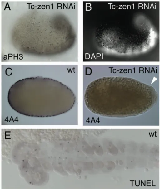
The appearance of widely spaced nuclei and a double layer of extraembryonic tissue seem to indicate the presence of serosal cells. Three lines of evidence, however, strongly suggest that all extraembryonic tissue in these embryos is of amniotic origin. First, cell divisions could still be detected by anti- phosphohiston3 (αPH3) staining among the wider spaced nuclei (Fig. 5A, B).
Serosal cells never show αPH3 staining since they stop dividing prior to gastrulation and later become polyploid. In contrast, amniotic cells maintain mitotic activity even after they become flattened and widely spaced during wild type development (Handel et al., 2005). Second, the serosal marker Tc004A04 ((Savard, 2004); GENBANK accession number CB335138) is not expressed in these flattened cells (Fig. 5C, D). Third and most importantly, these cells express the amniotic marker gene Tc-pannier (pnr) (Fig. 6 and (Berns, 2001)). In wild type at the primitive pit stage Tc-pnr expression is excluded form the serosa. Tc-pnr
Chapter 2
starts to be expressed in a dorsal stripe of the germ rudiment which extends into the posterior amniotic fold (Fig. 6A). Thus, early Tc-pnr expression occurs presumably in the amniotic anlagen. In accordance with this assumption, during germ band extension Tc-pnr becomes highly expressed in the amniotic cells which have folded over the embryo at the ventral side (Fig. 6D). In Tc-zen1 RNAi embryos all wider spaced, stretched-out cells that emerge anterior and dorsal to the condensing embryonic anlagen express Tc-pnr (Fig. 6B, C). The expression level increases during further development and the border towards the condensing embryonic anlagen sharpens (Fig. 6E, F). Tc-pnr is also expressed in the double layer of cells, partially covering the abdomen after Tc-zen1 RNAi (Fig. 6E). Since no stretched-out cells exist after Tc-zen1 RNAi, which lack Tc-pnr expression, I conclude that all serosal cells are lost and all stretched-out cells are amniotic. The flattening of these cells occurs with the same time course as that of wild type amniotic cells, i.e. much later and more continuous than the flattening of the serosal cells. This may explain the delayed condensation of the embryonic anlagen after Tc-zen1 RNAi (Fig. 3). The slightly wider spaced nuclei at the anterior tip of early Tc-zen1 RNAi embryos might be the first sign of amnion formation (Fig. 3G;
Fig. 4J).
Size regulation after loss of Tc-zen1
The “big head” phenotype is the most striking consequence of a loss of Tc- zen1 and is clearly visible throughout germ band elongation (Fig. 3I, J). The head and thorax region of the embryo is enlarged in comparison to wild type (Fig. 3C, D, I, J). The development of the head and thorax is also considerably delayed, as indicated by a retarded formation of the head lobes and segmental grooves in this area. The abdominal segments, however, look normal, probably because they emerge from a growth zone and are unaffected by fate shifts in the blastoderm.
To get a quantitative measure for these changes, I analysed the size of the Engrailed (En) stripes (Fig. 3E, K). The third En stripe consisted of 1.5 times as many cells in the lateral dimension compared to the wild type stripe (n=4). The total area occupied by this stripe is even larger than 1.5 times the area of a wild type stripe. Hence, not only does the anterior of the embryo consist of more cells, those cells are also less densely packed than in wild type. The latter may be the consequence of a slower condensation of head and thoracic cells.
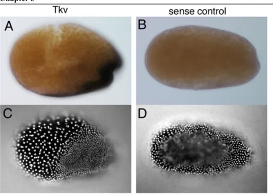
Fig. 5. αPH3, A4A staining and TUNEL assay
(A) Extending germband (big head) after Tc-zen1 RNAi. Black dots are staining for antiphosphohiston3 and indicate cell divisions. Ectopic divisions are detected in the serosa- like wider spaced nuclei, not belonging to the germ rudiment, demonstrating that they are amniotic. (B) DAPI image at a slightly different focal plane of the same embryo as in (A).
(C) Wildpype staining of the serosal marker Tc004A04. (D) After Tc-zen1 RNAi, no 4A4 staining could be detected, even in the double extraembryonic memebrane (white arrowhead). (E) TUNEL assay in wild type. Black dots reveal cell death. At the extending germband stage, irregular and sparse cell death was detected. After Tc-zen1 RNAi, no difference could be detected within the germ rudiment (not shown).
Chapter 2
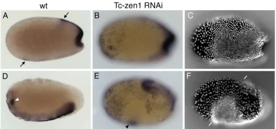
Fig. 6. The amnion covers the embryo at the dorsal side of Tc-zen1 RNAi embryos.
(A,D) Wild type embryos. (B, E) Embryos after Tc-zen1 RNAi. (C, F) DAPI counterstainings of embryos shown in (B, E). (A) Primitive pit stage. Tc-pnr is expressed dorsally, in the presumptive amnion and is absent in the flattened cells of the serosa. On the surface a faint rim of expression can be observed, just posterior to the serosa (arrows, out of focus in lateral regions). (B, C) Tc-zen1 RNAi embryo at a later stage than (A). At this stage cells start to flatten at the anterior pole as seen form the wider spaced nuclei (C).
These cells all express Tc-pnr. (D) Extending germ band stage. All cells of the amnion express Tc-pnr. The arrowhead points to a small group of cells within the head region, expressing Tc-pnr. (E, F) After Tc-zen1 RNAi all flattened cells express Tc-pnr. The arrowhead points at the small group of cells within the head region, expressing Tc-pnr. The white lines in (F) demarcate the anterior and posterior end of the embryo proper.