A Theoretical Study of the Tryptophan Synthase Enzyme Reaction Network
Dissertation
zur Erlangung des akademischen Grades doctor rerum naturalium
(Dr. rer. nat.) im Fach Chemie
Spezialisierung: Physikalische und theoretische Chemie Eingereicht an der
Mathematisch-Naturwissenschaftlichen Fakult¨at der Humboldt-Universit¨at
zu Berlin von
Dimitri Loutchko
Pr¨asidentin der Humboldt-Universit¨at zu Berlin Prof. Dr.-Ing. Dr. Sabine Kunst
Dekan der Mathematisch-Naturwissenschaftlichen Fakult¨at Prof. Dr. Elmar Kulke
1. Gutachter: Prof. Dr. Gerhard Ertl 2. Gutachter: Prof. Dr. Klaus Rademann 3. Gutachter: Prof. Dr. Yannick de Decker Tag der m¨undlichen Pr¨ufung: 09.07.2018
ii
Abstract iii
Abstract
The channeling enzyme tryptophan synthase provides a paradigmatic example of a chemical nanomachine. It catalyzes the biosynthesis of tryptophan from serine and indole glycerol phos- phate. As a single macromolecule, it possesses two distinct catalytic subunits and implements 13 different elementary reaction steps. A complex pattern of allosteric regulation is involved in its operation. The catalytic activity in a subunit is enhanced or inhibited depending on the state of the other subunit. The gates controlling arrival and release of the ligands can become open or closed depending on the chemical states. The intermediate product indole is directly channeled within the protein from one subunit to another, so that it is never released into the solution around it.
In this thesis, the first single-molecule kinetic model of the enzyme is proposed and analyzed.
All its transition rate constants are extracted from available experimental data, and thus, no fitting parameters are employed. Numerical simulations reveal strong correlations in the states of the active centers and the emergent synchronization of intramolecular processes in tryptophan synthase. Moreover, the effects of allosteric interactions are studied using modified in silico models with permanent and without any allosteric activations. The unmodified model of the native enzyme with transient activations significantly outperforms both modified models in terms of mean turnover times. An explanation is derived from the comparison of turnover time distributions showing a desynchronization of the two subunits in the modified models leading to cycles with long turnover times.
Thermodynamic data is used to calculate the rate constant for the reverse indole channeling, which has not been observed in experiments thus far. Using the fully reversible single-molecule model, the stochastic thermodynamics of the enzyme is examined. The Gibbs energy landscape of the internal molecular states is determined and the production of entropy and its flow within the enzyme are analyzed. The current methods describing information exchange in bipartite sys- tems are extended to arbitrary Markov networks and applied to the kinetic model of tryptophan synthase. They allow the characterization of the information exchange between the subunits resulting from allosteric cross-regulations and channeling.
The last part of this work is focused on chemical reaction networks of metabolites and en- zymes. Algebraic semigroup models are constructed based on a formalism that emphasizes the catalytic function of reactants within the network. These models admit a notion of successive and simultaneous functions not only of individual enzymes, but of any subnetwork. This in- cludes the catalytic function of the whole reaction network on itself. The function is then used to decide whether the network is self-sustaining and a natural discrete dynamics is utilized to iden- tify the maximal self-sustaining subnetwork. Then, a correspondence between coarse-graining procedures and semigroup congruences respecting the functional structure is established. A family of congruences that leads to a rather unusual coarse-graining is constructed: The net- work is covered with local patches in a way that the local information on the network is fully retained, but the environment of each patch is no longer resolved. Whereas classical coarse- graining procedures would fix a particular local patch and delete detailed information about the environment, the algebraic approach keeps the structure of all local patches and allows the interaction of functions within distinct patches.
iv Zusammenfassung
Zusammenfassung
Das Enzym Tryptophan Synthase ist ein ausgezeichnetes Beispiel einer molekularen Fabrik auf der Nanoskala. Es katalysiert die Biosynthese der essentiellen Aminos¨aure Tryptophan aus Serin und Indol-glycerolphosphat. Der katalytische Zyklus des Molek¨uls beinhaltet mindestens 13 El- ementarreaktionen, die in den katalytischen Zentren seiner zwei Untereinheiten stattfinden. Die Katalyse beruht zudem auf zahlreichen allosterischen Wechselwirkungen sowie der ¨Ubertragung des Intermediats Indol durch einen intramolekularen Tunnel.
In dieser Arbeit wird das erste kinetische Modell eines einzelnen Tryptophan Synthase Molek¨uls konstruiert und analysiert. S¨amtliche Reaktionskonstanten sind aus der Literatur bekannt, wo-durch das Modell keine freien Parameter enth¨alt. Numerische Simulationen zeigen starke Korrelationen zwischen den Zust¨anden der Katalysezentren sowie die Ausbildung von Synchronisation zwischen den intramolekularen Prozessen im Enzym. Des Weiteren werden die Effekte der allosterischen Wechselwirkungen durch den Einsatz von Modifikationen des Modells in silico, welche die Wechselwirkungen vollst¨andig unterdr¨ucken bzw. permanent aktivieren, un- tersucht. Es zeigt sich, dass das native Enzym eine erhelblich gr¨oßere Reaktionsgeschwindigkeit aufweist als beide Modifikationen. Durch eine Analyse der Histogramme der Umsatzzeiten einzelner Zyklen l¨asst sich diese Beobachtung auf eine selten auftretende Desynchronisation der Katalysezyklen in den Untereinheiten, welche zu sehr langen Umsatzzeiten f¨uhrt, zur¨uckf¨uhren.
Die thermodynamischen Eigenschaften des Modells werden mithilfe der stochastischen Ther- modynamik untersucht. Zun¨achst wird die experimentell unzug¨angliche Reaktionskonstante f¨ur die R¨uck¨ubertragung des Indols aus thermodynamischen Messdaten rekonstuiert. Die freie En- thalphie aller chemischen Zust¨ande des Molek¨uls, die Entropieproduktion sowie der Entropiefluss werden berechnet. Methoden, die den Informationsaustausch in bipartiten Markovnetzwerken charakterisieren, werden auf beliebige Markovnetzwerke verallgemeinert. Ihre Anwendung auf das kinetische Modell der Tryptophan Synthase f¨uhrt zu einer Charakterisierung des Informa- tionsaustauschs zwischen den Untereinheiten des Enzyms.
Der abschließende Teil der Arbeit befasst sich mit chemischen Reaktionsnetzwerken von Metaboliten und Enzymen. Ausgehend von einem Formalismus, der die katalytische Funktion von Reaktanten des Netzwerks hervorhebt, werden algebraische Modelle konstruiert. Es handelt sich dabei um Halbgruppen, welche aufeinanderfolgende und simultane katalytische Funktio- nen von Enzymen und von Unternetzwerken erfassen. Die Funktion des Netzwerkes auf sich selbst wird genutzt, um hinreichende und notwendige Bedingungen f¨ur seine Selbsterhaltung zu formulieren. Die Definition einer nat¨urlichen Dynamik auf den Netzwerken erlaubt auch die Bestimmung des maximalen selbsterhaltenden Unternetzwerkes. Anschließend werden die algebraischen Modelle dazu genutzt, um eine Korrespondenz zwischen Halbgruppenkongruen- zen und Skalen¨uberg¨angen auf den Reaktionsnetzwerken herzustellen. Insbesondere wird eine Art von Kongruenzen er¨ortert, welche dem Ausspuren der globalen Struktur des Netzwerkes unter vollst¨andiger Beibehaltung seiner lokalen Komponenten entspicht. W¨ahrend klassische Techniken eine bestimmte lokale Komponente fixieren und s¨amtliche Informationen ¨uber ihre Umgebung ausspuren, sind bei dem algebraischen Verfahren alle lokalen Komponenten zugleich sichtbar und eine Verkn¨upfung von Funktionen aus verschiedenen Komponenten ist problemlos m¨oglich.
Contents
Abstract iii
Acknowledgement xi
Introduction 1
1 Investigated System and Applied Methods 7
1.1 The Tryptophan Synthase Enzyme . . . 7
1.1.1 Structural Features . . . 8
1.1.2 Kinetics of Tryptophan Synthase . . . 15
1.2 Protein Models and Protein Kinetics . . . 17
1.3 Stochastic Thermodynamics . . . 20
1.3.1 Stochastic Thermodynamics of Chemical Systems . . . 21
1.3.2 Information Thermodynamics . . . 25
2 Markov Network Model 29 2.1 Previous Kinetic Models . . . 29
2.2 Kinetic Data . . . 31
2.3 Construction of the Single-Molecule Model . . . 34
2.4 Kinetic Markov Network Model . . . 41
2.5 Simulation Results . . . 42
2.6 Discussion . . . 46
3 Stochastic Thermodynamics of Tryptophan Synthase 49 3.1 Preliminaries . . . 49
3.2 Reverse Rate of Indole Channeling . . . 52
3.3 The Energy Landscape . . . 54
3.4 Entropy Production and Flow . . . 55
3.5 Discussion . . . 59
4 Information Exchange in Bipartite Systems 61 4.1 General Formalism . . . 62
4.2 Information Exchange in Tryptophan Synthase . . . 66
4.3 Discussion . . . 69
5 Semigroup Models for Reaction Networks 71 5.1 Motivation . . . 72
5.1.1 Self-Sustaining Reaction Networks . . . 72
vi Table of Contents
5.1.2 Coarse-Graining via Congruences . . . 76
5.2 Semigroup Models of CRS . . . 82
5.3 Semigroup Models of CRS with Food Set . . . 90
5.4 Dynamics on a Semigroup Model . . . 93
5.5 Identification of RAF Subnetworks . . . 95
5.6 Algebraic Coarse-Graining . . . 100
5.6.1 Existence of Congruences on Semigroup Models . . . 101
5.6.2 Constructions of Congruences . . . 104
5.7 Discussion . . . 112
A Forces and Fluxes in Phenomenological Thermodynamics 115 B Results of Numerical Simulations 119 B.1 Numerical Results under Experimental Substrate Concentrations . . . 119
B.2 Numerical Results under Physiological Substrate Concentrations . . . 120
List of Figures
1.1 Structure of tryptophan synthase . . . 8
1.2 α-Site reaction cycle . . . 9
1.3 β-Site reaction cycle . . . 10
1.4 Conformational rearrangements in the α-subunit . . . 11
1.5 Hydrogen bonding network in the β-subunit . . . 12
1.6 Allosteric interactions at the interface of α- and β-subunits . . . 13
1.7 Comparison of open and closed conformations. . . 15
1.8 Allosteric interactions . . . 16
1.9 Schematic operation of tryptophan synthase . . . 17
2.1 Kinetic model by Anderson et al. . . 30
2.2 Reaction rate constants . . . 36
2.3 Complete state space of combined states . . . 38
2.4 Reduced state space of combined states . . . 39
2.5 Transitions on reduced network of combined states . . . 40
2.6 The kinetic Markov network model . . . 42
2.7 Simulation data . . . 43
2.8 Joint probabilities p(a, b) . . . 44
2.9 Intramolecular correlations c(a, b) . . . 45
2.10 Joint probabilitiesp(a, b) of modified models . . . 46
2.11 Histogram of turnover times for a hypothetical enzyme with permanent activations . . . 47
3.1 Fully reversible kinetic Markov network model . . . 50
3.2 Energy landscape of tryptophan synthase . . . 54
3.3 Entropy production in the nonequilibrium steady-state . . . 57
3.4 Entropy export in the nonequilibrium steady-state . . . 58
4.1 Correlations i(a, b) . . . 67
4.2 Rates of change of mutual information . . . 68
5.1 Example of a catalytic reaction system . . . 75
5.2 Lattice of congruences on Z . . . 78
5.3 Biological motivation for quotient structures . . . 82
5.4 Examples of semigroup models of a simple CRS . . . 85
5.5 Example of a representation of a function as a tree . . . 89
5.6 Non-unicity of support function . . . 90
5.7 Example of semigroup models with food set . . . 91
viii List of Figures
5.8 Example of a CRS with possible oscillatory dynamics . . . 94
5.9 Illustration of corollary 5.3.7 and proposition 5.4.8 . . . 96
5.10 Conversion of catalytic reaction systems to chemical reaction networks . . 99
5.11 Cycle decomposition . . . 100
5.12 Reactions with substrates solely from ¯F lead to constant functions . . . 102
5.13 Nilpotency of ΦXF for SF without constant functions . . . 103
5.14 A CRS without nonzero constant functions and non-nilpotent semigroup model SF . . . 104
5.15 Example of metabolic pathways . . . 106
5.16 The partially ordered set M0 and the join semilattice M∗ . . . 108
5.17 Illustration of the impossibility to extendB to SF . . . 109
5.18 Example of functions φ, ψ∈ SF with comp(φ) +comp(ψ)< comp(φ◦ψ) . 110 5.19 Illustration of coarse-graining of the environment viaRn . . . 112
List of Tables
2.1 Kinetic data for the α-reaction . . . 32
2.2 Kinetic data for the β-reaction . . . 34
2.3 Enumeration of chemical states of α- and β-subunits . . . 37
2.4 Binding rate constants for a typical experimental situation . . . 40
3.1 Binding rate constants under physiological conditions . . . 51
5.1 The operation ◦in S{a,b} . . . 92
5.2 The operation + in S{a,b} . . . 92
5.3 ΦXF(∅) and XF∗ for the networks shown in figure 5.9 . . . 96
B.1 Joint probability distributionp(a, b) under experimental conditions . . . . 119
B.2 Marginal probabilitiesp(a) and p(b) . . . 119
B.3 Joint probability distributionp(a, b) for simulation setup without activations120 B.4 Joint probability distribution p(a, b) for simulation setup with permanent activations . . . 120
B.5 Turnover times for varied simulation conditions . . . 120
B.6 Joint probability distributionp(a, b) under physiological conditions . . . 120
x List of Tables
Acknowledgement
First and foremost, I would like to express my deep gratitude to my thesis advisors Prof. Gerhard Ertl and Prof. Alexander S. Mikhailov.
Prof. Mikhailov has spent many hours of his valuable time on teaching me how to write scientific texts, prepare talks and posters and of course on interesting discussions. He is a true artist when it comes to breaking down complicated phenomena to their core and constructing elegant models that precisely capture the essence of the phenomenon without any unnecessary distractive features. Watching his process was an invaluable experience for me. Prof. Mikhailov pushed me when it was necessary, but also gave me the freedom to wander off and explore on my own for months. Throughout my time as a student in his group, he was always there to help, to discuss and to guide.
Prof. Ertl had a magical way of guidance. Each and every conversation with him gave me a huge motivational boost to proceed with the project we had discussed. He gave me an enormous freedom to pursue my ideas, but also set wise boundaries based on his experience. I am especially thankful to him for the consistent support of my studies in mathematics parallel to the thesis work. Through this support I could acquire the technical skills desperately needed for the work as a theorist. To me, Prof. Ertl is great example of a successful scientist and, at the same time, a great role model concerning human behavior. He takes great care of this staff and is as concerned about their well-being at least as much as about their scientific success.
I am thankful to Maximilian G¨orgner with whom I had an exciting time at the FHI learning stochastic thermodynamics together spiced up with discussions about science, philosophy and life in general. I have very much enjoyed the time spent in Brussels with Didier Gonze working on the model of tryptophan synthase. I am grateful for the invitation of Holger Flechsig to spend a month in Hiroshima filled with interesting discussions about science and many other things. I would also like to thank Prof. Hisao Hayakawa for his hospitality in Kyoto, where I had all the time and freedom to think about the possibility of algebraic models in biology. With gratitude I think about Amartya Sarkar, Holger Flechsig, Jeff Noel and all other members of the complex systems group who created a pleasant working atmosphere. I am much obliged to Prof. Klaus Rademann, who supported my thesis work at the FHI.
I am deeply thankful to my parents and my brother, who accepted that I spent much of my spare time and weekends to work on the thesis rather than joining them for social events. Yet, they have always been there for me when I needed help or advice. Not only for the last four years, but for my entire life my parents have always been giving and caring without ever asking for a return. I would like to dedicate chapter 5 of this work to them.
xii
Introduction
Historically, the understanding of biological systems has been successively improved by the interplay of system reduction and integration of the reduced pieces. The reduction was, in most cases, enabled through the refinement of experimental techniques and the re- sulting possibility to observe smaller constituents of the system such as organs, cells, cell organelles, protein complexes, metabolites and ultimately the structure of DNA. Such constituents form a hierarchy resulting from the inclusion of smaller parts into larger structures. For example, cell organelles are included in cells and cells are included in organs. The goal of integration is concerned with the reconstruction of the properties of a particular structure from the properties of its lower level constituents. Each time new lower level structures have been discovered, the scientific community has spent much effort on formulating theories that achieve the integration of the newly found structures.
However, until the advent of molecular biology, for the lower level structures such as cells and cell organelles general interaction laws could not be formulated. Only phe- nomenological models adjusted to the respective experimental situation with experimen- tally determined parameters were available. Molecular biology, for the first time, allowed to envision that precise statements about biological systems could be made based onfirst principles. After all, the exact physical laws governing the structure and dynamics of molecules had been discovered in the early 20th century. The experimental accessibility - and thus the potential knowledge - of all molecular parts of an organism marks an im- portant milestone in the biological science: it is the completion of the reduction program and the end of a conceptual dichotomy between reductive and integrative ways of thought.
Meanwhile, the integration of lower level structures is far from being completed and is a main driving force in the life sciences. It is a recurring theme in numerous publications, where complex behavior is explained in terms of interactions of simpler lower-level con- stituents. The integrative branch of molecular biology is now known as systems biology.
It seeks to combine high-throughput data on the numbers, interactions and even time- evolution of metabolites, proteins, lipids, mRNA and DNA in a cell in order to develop detailed in silico models of the whole cell.
A remarkable success of systems biology is the identification of the molecular mech- anisms controlling the circadian rhythm, awarded the Nobel Prize in Physiology and Medicine in 2017. In gene knockout experiments, Benzer and Konopka were able to iden- tify a single gene (named period) whose knockout disrupted the circadian rhythm in fruit flies. Later, Hall, Rosbash and Young could show that the protein encoded by the gene (also called period) inhibits the transcription of its own gene and thereby forms a feed- back loop. The period protein is degraded through the influence of sunlight and therefore
2
its concentration fluctuates in a 24-hour rhythm driven by the day and night cycle: The concentration increases during the night (up to some threshold value controlled by the feedback loop) and decreases during the day (again to some threshold when degradation rate and synthesis rate cancel each other). In meticulous experimental work, the No- bel laureates were able to identify other genes and proteins that stabilize the regulatory network and control the entry of period into the nucleus. One of the many fascinating aspects about this work is the successful integration of simple chemical reactions governed by standard rate equations of a set of chemicals into a reaction network that has a signifi- cant influence on all the levels of organization within the organism: The circadian rhythm affects chemical characteristics such as hormone levels and metabolism, physical charac- teristics such as body temperature and blood pressure and even medicinal characteristics such as the desire for sleep, coordination, reaction times and mood. This means that the processes influenced by the reaction network based on period span a large interval of time and length scales emerging from the small time and length scales of individual chemical reactions involved in the network. In this regard, it is interesting to note that the circa- dian rhythm within each organism is controlled by a physical process on an astronomical scale, namely the earth’s rotation with respect to the sun.
The connection of processes on different time and length scales is becoming an in- creasingly important theme in the life sciences: While in the example discussed above, the connection between the period reaction network and higher-scale properties has not been made quantitative, there have been remarkable achievements in constructing quan- titative in silico multiscale models. An outstanding example is a series of models of the human heart constructed by Noble et al. [1, 2]. Such models include functionally impor- tant genes, proteins, metabolites and many details on ion channels at the molecular level.
These are included in models of all the main types of cardiac myocytes, which in turn are used in three-dimensional reconstructions of the whole organ as an elastic object paying attention to fiber orientation, sheet structure and the heart nervous system. Using such advanced models, many pathological states of the heart could be reproduced based on changes in the protein composition, drug interactions, or mutations of the ion channels.
Moreover, it was possible to study the influence of the heart contraction on the electrical state of the heart, giving unexpected results on the connection to changes in cell volume.
Along the same lines, arrhythmic behavior was successfully reproduced from models of the metabolic and electrophysiological processes following energy deprivation.
The period reaction network governing the circadian rhythm and the multiscale heart models each represent a major theme in system biological thought: At the molecular level, models of reaction networks of metabolites (called the metabolome), interaction networks of proteins (proteome), gene regulatory networks (genome) and mRNA expression levels are being integrated to determine mechanisms and regulatory motives within such net- works. Such models are based on large amounts of quantitative and qualitative data using high-throughput techniques that simultaneously monitor the cellular concentrations of a large number of different chemical species. Modern techniques even allow time-resolved data to be obtained. However, such approaches are inherently weak at capturing the emergence of and interactions with larger structures within an organism. In the example of the heart model, membranes, cells and the three-dimensional structure of the heart were not deduced from the respective molecular interaction networks, but added “by
3
hand”. Moreover, not the full reaction and interaction networks of molecules were taken into account, but only those important for the higher-scale processes under consideration.
This approach to systems biology is more an “artful crafting” of suitable models and less a “black-box” approach based on a fixed set of rules. Indeed, many prominent scientists such as Sydney Brenner [3], Dennis Noble [4] and Laurent Nottale [5] hold the opinion that there is no preferred scale of causation in nature and that neither the genetic code nor the molecular interaction network of organisms therefore contain a sufficient description of the organism.
Such problems are already present at one of the “lower levels” of organization, within individual proteins and their interactions networks. Does the understanding of individ- ual proteins provide deeper understanding of protein-protein interactions? How is the catalytic mechanism of a protein in diluted solutions in vitro or in silico related to its function in vivo? How important is the role of protein complexes when integrating high- throughput data without any a priori information on such complexes?
Proteins can be thought as the executive power of the cell. They carry out almost all functions in the living cell that involve manipulation and modification of the chem- ical and physical constitution of the cell or its environment. Enzymes catalyze most of the chemical reactions inside the cell. Through kinetic control they enable metabolic re- actions to take place in a controlled manner at appropriate rates. Moreover, key steps such as transcription, splicing and translation are carried out by large complexes. Motor proteins transport cargo in the cytoplasm or through the cell membrane and perform the various mechanical motions such as bacterial flagellar locomotion or muscle contraction in higher organisms. Proteins are crucial for the control of cellular processes. In particular, they are involved in the responses to external stimuli through signaling networks: Recep- tor proteins at the cell surface detect stimuli (e.g. from nutrients, poisons or hormones, but also mechanical stress) and initiate a cascaded response. Therein, several messenger proteins from a network reminiscent of a calculatory circuit including feedback control and amplification mechanisms. The circuit either directly initiates a response or leads to changes in protein biosynthesis through appropriate transcription factors.
All the processes just described heavily rely on the interaction between proteins - either within complexes or networks. A well-known example of an enzyme complex is the ribosome, consisting of the small and large subunits, ribosomal RNA and a variety of additional ribosomal proteins. Even larger structures are focal adhesions with over 50 proteins [6] or the spliceosome including over 200 proteins [7]. These complexes are sufficiently stable and the components well enough known that they can be studied in vitro or can already been observed in vivo using classical optical methods. However, the exact composition of these complexes varies dynamically in the living cell. For example, the number of proteins making up the spliceosome is known to vary by up to 60 between different functional states [8]. Such observations on large and well-known complexes seem to be just the tip of the iceberg concerning the role of enzyme complexes within living organisms. There is a growing volume of evidence suggesting that many biochemical re- actions within a cell are catalyzed by multi-enzyme complexes with poorly understood and highly dynamic higher order structure [9, 10, 11, 12, 13, 14, 15]. These complexes can implement entire metabolic pathways or significant parts of them. Within a complex,
4
intermediate products can be directly channeled [10, 11] to other enzymes for further processing, resembling the operation of an industrial conveyor belt. Moreover, different enzymes in a complex are usually coupled through allosteric regulatory loops [15]. Be- cause of product channeling and multiple allosteric interactions, a complex can operate in a synchronous manner, exhibiting strong correlations in the turnover cycles of involved enzymes. Experimental investigations of multi-enzyme complexes encounter difficulties because the complexes are often transient and only exist in vivo [12].
An interesting class of enzymes are channeling enzymes [16, 17] (see also review [18]).
They are similar in their properties to multi-enzyme complexes, but, in contrast, are smaller and have a well defined structure. A prototypical example of a channeling en- zyme is tryptophan synthase [19] (introduced in detail in section 1.1). It catalyzes the biosynthesis of the essential amino acid tryptophan from serine and indole glycerol phos- phate (IGP). This enzyme is employed by all bacteria, plants, fungi, but not by higher organisms and thus, can be a target for the development of antibiotics [20]. Its substrate IGP is scarce inside the cell and, therefore, high catalytic efficiency is required. Further- more, an intermediate product (indole) of the synthesis reaction is hydrophobic and can easily escape through the cell membrane. Therefore, its release into the cytoplasm must be avoided. Nature has found an elegant solution for these constraints. The entire syn- thesis encompassing 13 elementary reaction steps is performed within the enzyme with two different catalytic centers and the intermediate indole is channeled within the protein from one center to another. Thus, tryptophan synthase is a model for larger and more difficult to access protein complexes.
In chapter 2, asingle-molecule model of tryptophan synthase is constructed. It takes into account correlations between the states of the two catalytic centers arising through substrate channeling and mutual allosteric regulation. The stochastic model is formulated in terms of a Markov network. Because of the extensive experimental data available, all relevant microscopic rate constants in the model could be directly deduced from the data, so that no fitting parameters have been employed. Numerical simulations yield direct evi- dence of the presence of strong correlations and intramolecular synchronization of chemical processes in tryptophan synthase. They also allow to analyze the role of allosteric regu- lations in raising the catalytic efficiency of this enzyme. This work has been published in [21].
In chapter 3, the constructed Markov transition network is studied using the theory of stochastic thermodynamics for the operation of a single enzyme. Thereby, additional calorimetric data is used to determine the rate constant for reverse channeling that has not been experimentally observed. The energy landscape is constructed and an analysis of the entropy production and entropy flow within the enzyme in the nonequilibrium state corresponding to physiological conditions is performed.
Chapter 4 is focused on the information theoretic aspects of allosteric interactions between the two enzyme subunits and on the information effects of channeling events.
Recently, a theory of information transfer in bipartite Markov networks has been con- structed [22, 23, 24]. Bipartite Markov networks are networks whose state space can be factored as a product space A×B of two subsystems A and B such that all transitions
5
change either the state of the A-subsystem or of the B-subsystem, but not both at the same time. The Markov network models of allosteric proteins have exactly this structure:
The A-subsystem is the catalytic site and the B-subsystem is the allosteric site. A cat- alyzed reaction changes only the A-state and the binding or unbinding of some allosteric effector changes only theB-state. The allosteric interaction entails an effect of theB-state on the catalytic rates of the A-subsystem. This effect is made quantitative in the theory of information thermodynamics and, thus, it is straightforward to apply the theory to allosteric proteins. However, when mass transfer between the subsystem A and B takes place, there is no longer a bipartite structure, because substances leaving one subsystem immediately arrive in the other subsystem. In such cases, application of the theory is not straightforward, but it can be extended. This is done in section 4.1. As an illustration, the extended theory is applied to tryptophan synthase, which has both allosteric interactions between its two subunits and mass transfer due to indole channeling. The work presented in chapters 3 and 4 has been published in [25].
Chapter 5 takes a more general perspective on chemical reaction networks. The re- action networks are modeled by finite and discrete state spaces as in the case of the tryptophan synthase model. However, the states correspond to sets of metabolites and not to individual states of a single enzyme. As described in the first paragraphs of this introduction, high-throughput techniques generate large amounts of data on particular levels of organization, in particular, on reaction networks of metabolites, interaction net- works of proteins and genetic regulatory networks. The connection between this data and the hierarchical organization of biological systems across many scales is an omnipresent theme in modern systems biology, which has fascinated this author ever since he became aware of it. The methods in chapter 5 are a non-standard approach to establish such connections. Focusing on reaction networks of metabolites and the respective catalysts, algebraic procedures of coarse-graining are proposed as a natural tool to switch between multiple scales. In this regard, the joint and subsequent functions of single catalysts and of subnetworks on the reaction network are defined in sections 5.2-5.4. The set of the func- tions of all subnetworks forms a semigroup under composition. It is then demonstrated that such semigroups can be used to identify self-sustaining subnetworks (section 5.5).
Finally, biologically meaningful congruences and the resulting coarse-graining procedures are defined and discussed (section 5.6).
6
Chapter 1
Investigated System and Applied Methods
This chapter introduces the tryptophan synthase enzyme as the main system under in- vestigation in this thesis and the methods used to study it. In section 1.1, details on the structure and function of the enzyme are given. In section 1.2, approaches to model protein kinetics are discussed. Section 1.3 introduces the material on stochastic and in- formation thermodynamics used in this thesis.
1.1 The Tryptophan Synthase Enzyme
The enzyme tryptophan synthase catalyzes the last two steps in the formation of L- tryptophan (in the following: tryptophan) from indole glycerol phosphate (IGP) and L-serine (in the following: serine). It is present only as a dimeric α2β2 bienzyme complex with linearαββα alignment of the subunits. Theα-subunit catalyzes the formation of in- dole and glyceraldehyde-3-phosphate (G3P) from IGP. Indole is then transferred through a 25 ˚A-long tunnel to the β-subunit, where it reacts with serine to form tryptophan (fig- ure 1.1). To prevent loss or accumulation of the metabolite indole, the reactivity of both subunits is tightly coupled by allosteric interactions. Binding of both substrates IGP and serine triggers the closing of the α andβ-subunits and thereby significantly enhances the rate of indole formation. Only after indole channeling to the β-site and reaction with serine is completed the subunits are opened and the product tryptophan and G3P released.
Tryptophan synthase has been extensively studied since 1946, when first indications for the biosynthesis of tryptophan from serine were given by Gunsalus [26]. Already in 1958 it was discovered that IGP and serine react to form tryptophan without releasing indole into the solution [27]. Since 1970, kinetic and structural studies performed by the groups of Michael F. Dunn (University of California, Riverside) and Ilme Schlichting (Max Planck Institute for Medical Research, Heidelberg) have created a vast amount of insights and data on tryptophan synthase. By the late 1990s the most important intermediates in the enzyme’s cycle have been spectroscopically characterized and the reaction mecha- nism could be formulated. Since then research was focused on the understanding of the regulatory pathways synchronizing the α- and β-reactions.
A growing number of X-ray crystallographic structures of the wild-type enzyme and
8 1. Investigated System and Applied Methods
mutants thereof with naturally appearing and model ligands has aided to identify the domains and residues responsible for catalysis and allosteric regulation [28]. In addition, several kinetic studies involving mutant enzymes and isotopically labeled substrates have been conducted to identify the rate determining reaction steps and the residues involved therein. In 2007, a further milestone was set by determining the X-ray crystallographic structure of tryptophan synthase in its closed and catalytically active conformation [29].
The historical development of research on tryptophan synthase and the interconnection of experimental results and their implications are reviewed in [19]. Articles that focus on structural [30] and kinetic [31] properties of tryptophan synthase are also available.
Higher organisms obtain the essential amino acid tryptophan through their diet, while bacteria, yeasts and molds have a tightly controlled mechanism for its synthesis regulated by the tryptophan operon. Hence, the elucidation of the mechanisms governing the en- zyme’s behavior is of interest in areas related to the medicine of infectious disease, plant defense and herbicide design.
Figure 1.1: Structure of tryptophan synthase with its characteristic elements. The tunnel for indole channeling is represented by the dashed line. The COMM domain (orange) serves for allosteric information transfer between the subsites and prevents the escape of substances at the β-site in the closed conformation. The loops αL2 and αL6 (red) confer the allosteric communication at the α-site. In the closed conformation they pre- vent substrate exchange of the α-site with the enzyme environment. A ball and stick representation is used for the PLP cofactor at the β-site. PDB code: 2J9X.
1.1.1 Structural Features
The α-reaction
At the α-site of tryptophan synthase, indole-3-glycerol phosphate (IGP) is converted to indole and glycerol-3-phosphate (G3P) (figure 1.2). From X-ray crystallographic studies it is known that the α-subunit exists in at least two conformations termed as open and
1.1 The Tryptophan Synthase Enzyme 9
closed states [32, 28]. The open state has a low catalytic activity on IGP cleavage and is structurally characterized by a disordered αL6 loop consisting of the residues α179-α193, which becomes ordered in the closed conformation and prevents the escape of indole into solution [33, 34, 32]. Concerning the reaction mechanism for aldolytic cleavage of IGP, two alternatives have to be taken into account. The first is a series of proton transfers involving αGlu49 and αAsp60 as acid-base catalysts and the second is a concerted one- step reaction. Considering the hydrophobic microenvironment of the active site, the latter mechanism seems to be more likely [35, 36, 29]. Using a specific α-site ligand, transition state analogues supporting the hypothesis of a concerted mechanism could be synthesized and analyzed crystallographically [37, 38].
Figure 1.2: Transformation of IGP to indole and G3P catalyzed by the α-site of trypto- phan synthase
The β-reaction
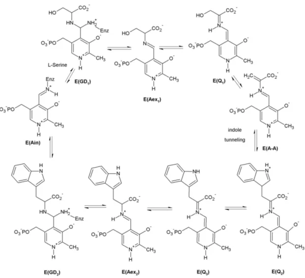
The β-subunit catalyzes the conversion of indole and serine to tryptophan (figure 1.3).
In the initial state E(Ain), the cofactor pyridoxal phosphate (PLP) is bound to βLys87.
It constitutes the main catalytic site for the complex reaction cycle by binding of the substrates through their amino groups as aldimines, germinal diamines and quinolines.
So far, nine intermediates have been characterized by UV/Vis spectroscopy, X-ray crys- tallography and by reaction and comparison with substrate analogues [39, 40, 41]. The β-reaction is commonly divided in two stages. In stage I, the aminoacrylate E(A-A) is formed from the internal aldimine E(Ain) with serine with the germinal diamine E(GD1), the external aldimine E(Aex1) and the quinoline E(Q1) appearing as intermediate states.
In stage II, E(A-A) reacts with indole to give tryptophan and return to the enzyme’s initial state E(Ain) via two quinolines E(Q2) and E(Q3), an external aldimine E(Aex2) and a germinal diamine E(GD2). As the first step of this stage, indole is channeled from the α-site to react with E(A-A). Like the α-subunit, the β-subunit can adopt at least two different conformations - an activated state with a closed conformation and an in- active open state. The catalytic cycles of the α- and β-sites are synchronized through a mechanism wherein conversion of E(Aex1) to E(A-A), via E(Q1), activates the α-site, whereas conversion of E(Q3) to E(Aex2) brings it back to the inactive open conformation [41, 42, 43]. In order to accommodate many different intermediates and thereby achieve reasonable reaction rates, the β-catalytic site possesses a certain structural flexibility, which is modulated by a monovalent cation (MVC) cofactor [44, 45, 46].
Mechanisms of Intersite Communication
Three levels of events comprise the allosteric communication in tryptophan synthase.
These consist of loop motions at the α-site (loop αL2 with residues α53 to α60 and loop
10 1. Investigated System and Applied Methods
Figure 1.3: Theβ-reaction cycle catalyzed by tryptophan synthase. Serine reacts with the internal aldimine E(Ain) and is transformed to aminoacrylate E(A-A) under elimination of water. E(A-A) incorporates indole to yield the geminal diamine E(GD2) via several intermediates, which releases tryptophan and returns to the initial state E(Ain).
1.1 The Tryptophan Synthase Enzyme 11
αL6 with residues α179 to α193), motions of single residues extending over the bienzyme complex and motion of the COMM domain (residues β102 to β189). These movements are correlated, but the extent of concertion has yet to be established. The known com- munication mechanisms will be described in the above given order.
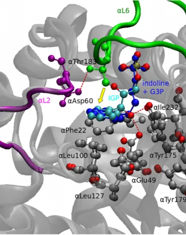
Figure 1.4: Conformational rearrangements in the α-subunit. The structures of an indoline-G3P adduct (dark gray, PDB code: 1QOP) with the IGP complex (light gray, PDB code: 2RHG) are compared. When the enzyme switches to the closed conforma- tion, the loop αL6 (green) moves towards the substrate IGP. In the process, αThr183 gets pulled by αAsp60 through hydrogen bridge formation and pushes the substrate (yel- low arrow). At this moment, IGP is able to interact with αGlu49 and αTyr175, which confer the concerted catalytic cleavage of IGP to G3P and indole. The residues αPhe22, αLeu100, αLeu127 and αIle232 form a suitable binding pocket for the product indole.
The figure was rendered with VMD and modified with Inkscape.
12 1. Investigated System and Applied Methods
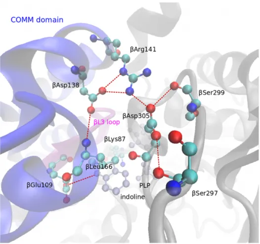
Figure 1.5: Hydrogen bonding network in the indoline derivative of the enzyme state E(Q2/3) (PDB code: 3CEP). When the enzyme adopts its closed conformation accom- panied by the release of water at the reactive site, the residue βAsp305 rotates towards βArg141, which in turn moves 4 ˚A towards βAsp305. βGlu109 moves towards the sub- strate and forms a hydrogen bond with the indoline ring. The bonding network serves to stabilize certain intermediates in the closed conformation and is thought to prevent mass exchange with the environment. Hydrogen bonds are represented by dashed red lines.
The figure was rendered with VMD and modified with Inkscape.
By using α-site ligand derivatives, it was possible to show that during the transition from the open to the closed conformation the loop αL2 moves towards αL6 and a crucial hydrogen bond is established between αThr183 on αL6 and αAsp60 on αL2 [29, 47].
αAsp60 then is orients so that it can stabilize charge developing during indole formation [37, 38] (figure 1.2). The residueαGlu49 is as well involved in proton transfer from C’-OH leading to the formation of indole via a push-pull mechanism. By X-ray crystallographic structures it has been shown to adopt two conformations: an inactive state with αGlu49 pointing away from the substrate [39] and the active conformation oriented towards the indole C’-OH group [48, 49]. This is assumed to be the most important interaction at the α-site for allosteric communication [30]. The structural details are shown in figure 1.4.
1.1 The Tryptophan Synthase Enzyme 13
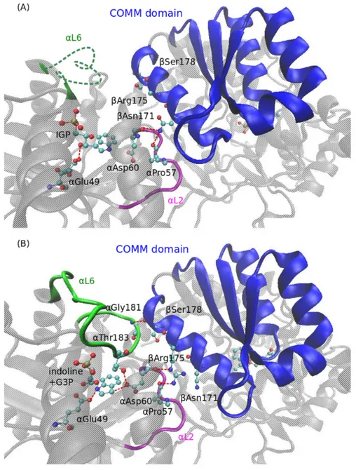
Figure 1.6: Comparison of the open and closed forms of tryptophan synthase at the interface between the α- and β-subsites. Red dashed lines denote hydrogen bonds. (A) Structure of the open state (IGP|Ain) (PDB code: 1QOQ). The open form is characterized by a disorderedαL6 loop (green) and interactions of theαL2 loop with the COMM domain (blue) via hydrogen bond formation fromαPro57 andαAsp60 to βAsn171. IGP is bound toαGlu49. (B)Structure of the indoline derivative of the closed state (G3P+indole|Q2/3) (PDB code: 3CEP) The αL6 loop is now ordered and αThr183 interacts with αAsp60 and the substrate (compare with figure 1.4). In addition, αGlu181 forms a hydrogen bond to βSer178 on the COMM domain. The αL2 loop is closer to the substrate than in the open conformation thereby enabling interactions between αAsp60 and IGP. The COMM domain is displaced by one turn thus placing βArg175 in contact to αPro57. The figure was rendered with VMD and modified with Inkscape.
At the β-site, the β loop (residues β109 to β115) on the COMM domain confers a highly specific binding site for the substrate’s and intermediates’ carboxylate groups.
14 1. Investigated System and Applied Methods
Surprisingly, the main conformational changes occur elsewhere: In the open E(Aex1) structure, the carboxylate group of βAsp305 binds to the hydroxyl group of the serine moiety, thereby stabilizes the E(Aex1) intermediate and prevents dehydroxylation by the acid-base catalytic βGly109 and βLys87 residues. Switching to the closed conformation leads to a movement of βArg141 by approximately 4 ˚A towards βAsp305. At this stage, the hydrogen bridge between βAsp305 and serine is broken and βAsp305 rotates about 100◦ [19]. This leads to an extended hydrogen bonding network between the residues βArg141, βAsp305, βSer297, βSer299, βAsp138, and βLeu166 [50, 28, 38, 48, 51, 52]
(figures 1.6 and 1.5).
The mobile domain, which has been termed the COMM domain [50], consisting of the residues β102 to β189 is the key element in synchronization of α- and β-reactions. Its position defines the closed and open states of theβ-subunit and couples to loopsαL2 and αL6. In its open state theβ-site is freely accessible from solution [39] while in the closed state the COMM domain moves towards the PLP cofactor closing the site and establishing interactions with other parts of the enzyme [38]. Within the COMM domain the helix βH6 is the main hub for intersite allosteric communication. In the open state, the residue βAsn171 on βH6 interacts with αAsp60 on αL2, which is part of the α-catalytic center [30]. When adopting the closed conformation, βArg175 interacts with αAsp60 and also αPro57. Moreover, hydrogen bridges are formed betweenβSer178 on βH6 and αGly181 on αL6 [53, 54] (figure 1.7).
The Monovalent Cation (MVC) Cofactor
In 1995, the group of Peracchi discovered that the tryptophan synthase enzyme utilizes a monovalent cation (MVC) cofactor [55]. It is bound to six carbonyl groups belonging to the residues βVal231, βGly232, βGly268, βLeu304, βPhe306, and βSer308, which form a loop around the cofactor [19]. The binding site is positioned 8 ˚A away from the β catalytic center [56]. Without the presence of the MVC cofactor, both the catalysis at the β-subsite and the allosteric communication are impaired. Removing the cofactor renders the aminoacrylate E(A-A) essentially unreactive towards indole [57]. Interestingly, the exact choice of the MVC species is rather robust towards size and charge density: Na+, K+, NH+4, Rb+ and Cs+ can serve as MVC cofactors [58, 44] and surprisingly also the large guanidinium ion [46, 59]. While the mechanistic influence of the cofactor on the allosteric communication has not yet been clearly worked out, modulation of theβreaction center has been clarified by analysis of crystal structures with different MVC cofactors.
While the Cs+-bound enzyme E(Cs+) exhibits a binding pocket suited for indole and derivatives thereof, the pocket is too small in the Na+-bound form E(Na+)[42, 60, 56].
Consistently, the form E(Cs+) favors the closed conformation and allows indole channeling and incorporation at theβ-site and the form E(Na+) favors the open conformation, where the formation of indole is kinetically hindered and thus a binding pocket for indole is not needed. In conclusion, the MVC cofactor is able to modulate the enzyme activity and to discriminate between the open and closed conformations. This is supported by the fact that for different cofactors different steady-state distributions of the respective enzymatic species have been measured [45, 61].
1.1 The Tryptophan Synthase Enzyme 15
Figure 1.7: Superposition of the structures of open (PDB code: 1KFK) and closed (PDB code: 2J9X) conformations of tryptophan synthase. The COMM domain performs an extensive tilting motion, whereas the rest of the β-subunit does not change detectably.
The α-subunit undergoes slight conformational changes. The figure was rendered with VMD and modified with Inkscape.
1.1.2 Kinetics of Tryptophan Synthase
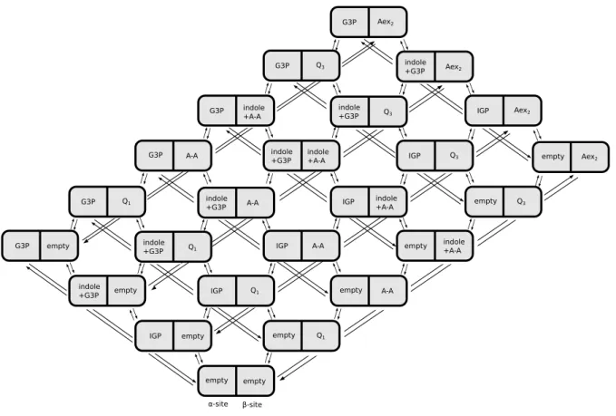
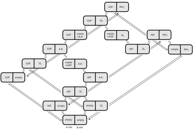
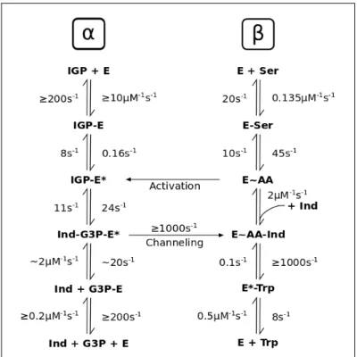
The reaction cycle involving all known enzyme states is shown in figure 1.8 (with the labels from figures 1.2 and 1.3). Each subunit is represented by a chain and mutual regulations are indicated by colored arrows. The following allosteric interactions are highlighted in the literature [19, 31]
1. The state α-IGP has an activating effect on the formation of β-A-A: the reaction rate increases 9.7-fold. This result was obtained by Ngo et al. by using α-site ligands (ASL) that closely resemble the structure of IGP, but cannot be cleaved.
The equilibrium distribution of the predominant β-species β-Aex1 and β-A-A was then analyzed for the native enzyme with and without different ASL [29].
2. β-A-A in turn activates the formation ofα-indole + G3P: the reaction rate increases 27.7-fold. This result was obtained by Brzovic et al. with similar methods as used by Ngoet al.. By binding serine analogues that could formβ-A-A, but did not react further to the β-site, the rate of IGP cleavage could be measured and compared to rates with bound serine analogues that could not form β-A-A [42].
3. α-indole + G3P can only form when the enzyme is in the closed state. Therefore the β-site has to be in one of the following states: E(Q1), E(A-A), E(Q2) or E(Q3) in order to enable the formation of α-indole + G3P.
4. In the closed conformation, the uptake and release of substrates and products is not possible. For the actual mechanism of the tryptophan synthase enzyme it has been
16 1. Investigated System and Applied Methods
suggested that the states E(Q1), E(A-A), E(Q2) and E(Q3) can exist in the open conformation [62]. Therefore, for these chemical states, two different conformational states - open and closed - have to be distinguished. In the former case, mass exchange with the environment is possible.
5. As discussed in section 1.1.1, the conversion IGP→G3P + indole most likely takes place as a concerted one-step reaction and no intermediate steps have to be taken into account.
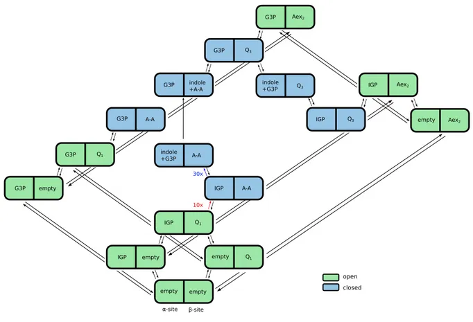
open
open/closed (depending on α-site ligand) closed
α-site β-site
Ain L-Ser GD1 Aex1 Q1 A-A Q2 Q3 Aex2 GD2 L-Trp Ain
empty IGP IGP indole
+G3P G3P G3P empty
Figure 1.8: Allosteric interactions between the two subunits. The transitions empty IGP and G3Pempty (magenta) in theα-site are blocked (i.e., the gate in theα-subunit is closed) in the states A-A, A-A + indole and Q3 of the β-site. The transitions IGP indole+G3P (light and dark blue) in the α-site are blocked in the states empty, Q1, Aex2 of the β-site. The rate of the transition IGP → indole+G3P (light blue) in the α-site is enhanced by a factor of 27.7 in the state A-A of the β-site. The transitions Q1 A-A and Q3 Aex2 (green) in the β-site are blocked in the state empty of the α-site.
The transition Q1 →A-A (light green) in the β-site is enhanced by a factor of 9.7 in the state IGP of the α-site. The changes indole+G3P G3P and A-A indole+A-A (red) corresponding to indole channeling from the α- to the β-site occur simultaneously and represent a single stochastic transition.
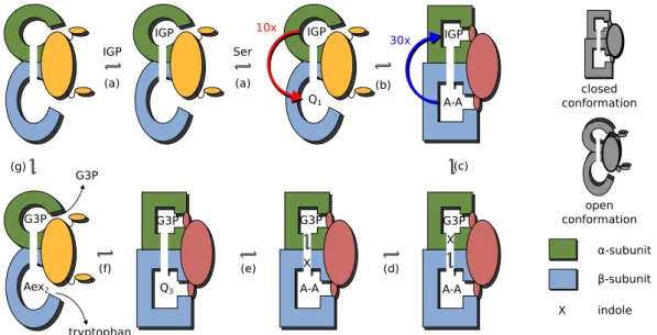
A simplified scheme of the catalytic cycle of tryptophan synthase with several omitted states is displayed in figure 1.9. Here, the α-subunit is shown in green and theβ-subunit in blue. The chemical states have the same notations as in figure 1.8. The catalytic cycle begins with the enzyme in the state where both sites are empty and the gates are open.
Then, the substrate IGP binds to the α-subunit and serine to the β-subunit, where it is quickly converted to the serine quinoline intermediate Q1. IGP activates the formation of the α-aminoacrylate A-A and the enzyme adopts the closed conformation, as schemat- ically shown in figure 1.9b. In the state (IGP,A-A) where both gates are closed, A-A activates the cleavage of IGP to produce G3P and indole. Indole is then channeled to the β-site where it reacts with A-A to give the tryptophan quinoline intermediate Q3 that is converted to tryptophan (Aex2 is the external aldimine of tryptophan in theβ-subunit).
In the state (G3P,Aex2) the gates open and the products tryptophan and G3P are re- leased. Thus the enzyme returns to the initial conformation (empty,empty) and is ready to start the next cycle.
The kinetic rates for all transitions are given in section 2.2.
1.2 Protein Models and Protein Kinetics 17
IGP
IGP IGP
X Q3
tryptophan
Ser
(b)
(c)
G3P X
A-A G3P
A-A G3P
Aex2
G3P
Q1
(d) (e)
(f) (g)
α-subunit β-subunit indole X
10x 30x
(a) (a)
IGP
A-A
G3P
open conformation
closed conformation
Figure 1.9: Schematic operation of tryptophan synthase. Operation of the machine: Once substrates are bound (a) at both catalytic sites, IGP activates (b) the formation of A- A and the enzyme adopts the closed conformation. A-A activates (c) the cleavage of IGP and indole is channeled (d) to the β-site where it reacts (e) with A-A to give Q3. Q3 undergoes (f) further transformations that return of the enzyme to the open conformation where tryptophan and G3P are released (g).
1.2 Protein Models and Protein Kinetics
There are several methods to model the structure, dynamics and kinetics of proteins.
Such methods include quantum mechanics (QM), all-atom molecular mechanics (MM) or molecular dynamics (MD), hybrid QM/MM approaches, coarse-grained structural mod- els such a Go models and elastic network models and phenomenological models with strongly reduced state spaces such as discrete Markov chains for chemical reactions, low- dimensional continuous parametrizations for conformational dynamics or a combination of both. The methods differ in the phenomena they are able to describe and in the time scales they are able to address. The most fundamental level is the description of a protein as a quantum mechanical system providing the full information on its electronic structure.
All-atom molecular dynamics (MD) models contain full information on the coordinates of the nuclei, but take into account the electronic interactions via ad hoc potentials between groups of nuclei. In phenomenological models, qualitative or quantitative experimental data on the protein under consideration governs the choice of the variables in the model.
Often the state space in such models is substantially reduced in comparison to MD models as many conformational and chemical states are not resolved, but treated as combined coarse-grained states. The time and length scales of the phenomena under investigation and the available experimental information determine the choice of the modeling approach.
Electronic processes in proteins take place on time scales of picoseconds, they are quantum chemical phenomena and have been modeled accordingly [63]. Examples of bio- logically relevant quantum mechanical processes are photon absorption in light harvesting complexes, substrate binding, proton and electron tunneling and chemical reactions cat- alyzed by enzymes. The light harvesting complexes photosystem I (PS I) and photosystem
18 1. Investigated System and Applied Methods
II (PS II) play a main role in the transformation of the energy from absorbed photons into chemical energy and thus have been studied extensively. The absorption spectra of chlorophyll complexes in PS I and their dependence on the complex geometry have been determined by semiempirical methods [64]. Recently, the absorption spectrum of PS II was determined withab initiomethods [65]. Moreover, in the case of PS II, the pathway of electron absorption could be modeled. It involves 6 cofactors coupled to 4 charge-transfer states. The characteristic time scales were obtained as well [66]. The [CaMn3(III)Mn(II)]
cofactor of PS II catalyzes the splitting of water and production of oxygen; the mech- anism of the reaction is still a topic of debate. The magnetic and electronic properties of the complex were calculated paving the way to a better understanding of the reaction mechanism [67]. In general, the electronic structure of metal cluster cofactors is impor- tant for the understanding of many biochemical processes, yet difficult to access. Another example are iron-sulfur clusters present in various classes of enzymes. Recently, it became possible to performab initiocalculations of the energy landscape of [2Fe-2S] and [4Fe-4S]
clusters without any fitting parameters [68]. Quantum chemical models have also been employed to determine binding energies of CO, NO and O2 to heme molecules [69]. The study revealed a change in the magnetic structure of the Fe(II) center upon NO binding as compared to CO and O2 ligands. Proton tunneling [70, 71] and electron tunneling [72, 73, 74, 75] pathways have been determined. Free energy barriers of chemical reac- tions in solution are accessible via quantum chemical methods [76]. There have also been attempts to model the dynamics of whole proteins using density functional theory [77, 78].
However, generally it is not possible to reach time scales relevant for the conforma- tional dynamics of proteins with using quantum chemical models. A popular approach to retain the accurate description of electronic processes provided by quantum mechanics and to simultaneously study the conformational dynamics of a protein is the hybrid quantum mechanics/molecular mechanics (QM/MM) approach [79, 80, 81]. Thereby, the chemical reaction center is modeled as a quantum chemical system and the protein backbone by MM methods. For example, a QM/MM hybrid approach allowed to model the catalytic reaction of cAMP-dependent protein kinase [82]. The residues in the catalytic pocket responsible for a substantial reduction of the activation energy as well as residues that keep the substrates in an appropriate conformation were identified. As another example, a QM/MM model enabled the identification of a critical arginine residue in the catalytic mechanism of citrate synthase and allowed to study the interplay of conformational dy- namics involving the arginine residue and catalytic activity [83]. Similarly, the coupling of vibrational excitations and catalytic activity in human purine nucleoside phosphory- lase [84] and the interplay of conformational and electronic states in cytochrome C450 oxidation [85] could successfully be modeled. Hybrid methods also allow to determine acidity constants, redox potentials and solvation free energies of proteins using ab initio calculations [86].
The QM/MM hybrid methods can successfully take into account small-scale conforma- tional motions at the catalytic site, but are not capable of reproducing domain motions in proteins as they take place on time scales of micro- to milliseconds. In many cases, insights into protein function can be gained without quantum chemical descriptions, but purely from the conformational dynamics of the protein [87]. All-atom molecular dynamics (MD) simulations trace the motions of all protein and solvent atoms using phenomenologically
1.2 Protein Models and Protein Kinetics 19
adjusted force fields. MD simulations played a major role in the determination of the catalytic mechanism of F1-ATPase. After the determination of the protein structures of the main chemical and conformational states of the catalytic cycle by protein crys- tallography, MD simulations have been used to interpolate between the structures in a biologically meaningful way and thereby provided a dynamical model of the functioning of F1-ATPase [88, 89]. Moreover, the ATP binding affinities in the different conformational states of the F1-ATPase β-subunits were determined using MD and an analysis of the thermodynamics of the simulated trajectories. This provided the solution to a dispute concerning the reaction mechanism [90]. Another example of the success of MD is the insight into the activity of Src tyrosine kinases, whose activated forms are known to be oncogenes [91]. Src kinases posses a catalytic domain, an SH2 peptide binding domain at the N-terminus of the catalytic domain and an SH3 binding domain at the C-terminus. In the inactive state, the SH2 and SH3 domains are tightly bound and block the entrance to the catalytic center [92]. Using MD simulations, it was possible to clarify the activation mechanism of the kinase: The catalytic domain possesses an activation segment that in- duces rearrangements in the SH2 domain and thereby weakens the SH2/SH3-interactions through long-range allosteric interactions. This leads to an increased accessibility of the catalytic center [93, 94].
The time scales accessible with molecular dynamics simulations are typically on the order of nanoseconds [95]. Using specifically designed computer architectures, a 1 mil- lisecond trajectory was calculated for small proteins [96], breaking the previous record of a 10 microsecond trajectory [97] by a 100-fold. Yet, even such state of the art simula- tion techniques cannot reach the time scales of protein folding or large domain motions in molecular machines which often take place on the order of milliseconds and seconds [98, 99, 100]. To model such phenomena, coarse-grained molecular dynamics methods are available [101]. Thereby, groups of atoms, whole amino acid residues or even pro- tein domains are grouped together to single particles and the dynamics is determined by potentials between such coarse-grained particles. The potentials can be introduced ad hoc, derived from all-atom potentials [102, 103], from statistical analysis of protein struc- ture data [104] or adjusted to the native structure of the protein (Go models) [105, 106].
Coarse-grained molecular dynamics leads to a 103-fold [107] to 107-fold [108] speedup in computation time as compared to all-atom MD. A particularly attractive field for the application of structure-based models is protein folding [109]. Such models were used to generate a large amount of folding trajectories for different proteins allowing a statistical analysis of the folding pathways and generating new deep insights into the process of protein folding [110, 111, 112]. Protein dynamics around the native state can be studied, for example, with elastic network models [113, 114]. In these computationally very effi- cient models, all amino acid residues are replaced by single point particles and particles within a given cutoff range interact through harmonic potentials. Using such models, it was possible to simulate the whole catalytic cycle of HCV helicase [115], to study the al- losteric interactions in myosin-V [116] and even to simulate global ribosome motions [117].
If the full structure of a protein is not available, it is possible to construct a state space from kinetic measurements and other experimental insights and to determine the transition rate constants between the states experimentally. The state space can con- sist of different chemical and conformational states [118]. The chemical state space is
20 1. Investigated System and Applied Methods
usually finite and discrete corresponding to the space of chemical intermediates occur- ring in the catalytic cycle. If the conformational motions are faster than the chemical reactions, then the conformational states can be absorbed into the chemical states yield- ing a discrete state Markov model. For example, the motor protein kinesin has been modeled in [119] as a Markovian process on a discrete state space determined by the chemical states of both legs. Hereby, each leg can adopt three different states (empty, ADP-bound and ATP-bound) resulting in nine different states. If the conformational motions are slower than the chemical reaction, the conformational motions are described by a drift process on a low-dimensional manifold given by collective coordinates. An example is a model of F1-ATPase, where the rotatory motion is characterized by a con- tinuous coordinate and the chemical states of the protein are discrete corresponding to the bound ligands (empty, ADP-bound and ATP-bound) [120]. Other phenomenological models for F1-ATPase [121, 122], kinesin [123, 119], myosin V , [124], dynein [125] and flagellar motors [126] have been constructed. Any protein model with discrete chemi- cal states and Markovian transitions between them is a phenomenological model in this sense. Phenomenological models are well suited to study global aspects of proteins such as thermodynamic efficiency or the mechanochemical coupling in protein motors [127].
In principle, the modeling approaches with higher temporal and spatial resolution can be converted to models with lower resolution via coarse-graining. Thereby, certain sub- spaces of the state space are lumped together into coarse-grained states. If the dynamics within the coarse-grained states is much faster than the transitions between them, i.e.
there is a separation of time scales, then a Markovian dynamics on the full state space transforms into a Markovian dynamics on the coarse-grained state space. For example, applying the Born-Oppenheimer approximation to a quantum mechanical description of a protein and integrating out the electronic degrees of freedom leads to a molecular dy- namics model. Replacing the centers of mass of certain domains in the MD model and integrating out the fast atomic motions within such domains leads to coarse-grained mod- els. The transformed dynamics is necessarily stochastic as the exact position within the coarse-grained states cannot be traced and the transitions between coarse-grained states occur at random with some given transition probability rates in discrete spaces or as a diffusive processes in continuous spaces. Even at the quantum mechanical level there are already sources of stochasticity in the dynamics due to the uncertainty relation. The stochasticity introduced through coarse-graining is, however, fundamentally different from quantum mechanical uncertainty, because it is not forced a priori by natural law.
1.3 Stochastic Thermodynamics
Classically, thermodynamics is applicable only to large systems with macroscopic state variables such as temperature, internal energy and entropy. The changes of the state variables are deterministic and can be associated with the quantities of work, heat and entropy production. In order for the variables to be well-defined, their fluctuations are required to be negligibly small.
Microscopic systems such as single proteins and mesoscopic systems such as reaction networks with low numbers of reactants are subject to large stochastic fluctuations and thus the classical theory of thermodynamics is not applicable to these systems. However,
![Table 2.1: α-Reaction: kinetic rate constants. The results from [169] were obtained using KINSIM](https://thumb-eu.123doks.com/thumbv2/1library_info/5574215.1690071/44.892.103.804.176.505/table-reaction-kinetic-constants-results-obtained-using-kinsim.webp)