assemblies and their role in laminopathies
INAUGURAL-DISSERTATION zur
Erlangung des Doktorgrades
der Mathematisch-Naturwissenschaftlichen Fakultät der Universität zu Köln
vorgelegt von Liu Yang
aus
Runan, Henan, P. R. China Köln, 2013
Referees/Berichterstatter: Prof. Dr. Angelika A. Noegel Prof. Dr. Arnd Baumann Prüfungsvorsitzender: Prof. Dr. Siegfried Roth
Date of oral examination: 25.06.2013 Tag der mündlichen Prüfung
The present research work was carried out under the supervision of Prof. Dr. Angelika A.
Noegel and Dr. Sascha Nuemann, in the Institute of Biochemistry I, Medical Faculty, University of Cologne, Cologne, Germany from October 2009 to March 2013.
Diese Arbeit wurde von Oktober 2009 bis März 2013 am Institut für Biochemie I der Medizinischen Fakultät der Universität zu Köln unter der Leitung von Prof. Dr. Angelika A.
Noegel und Dr. Sascha Nuemann durchgeführt.
I
LINC (Linker of nucleoskeleton and cytoskeleton) complexes connect the nucleoskeleton to the cytoskeleton by interactions among LINC complex proteins and their interactions to proteins in the nucleus and the cytosol. Nesprins (nuclear envelope spectrin repeat proteins) are core components of the LINC complex, together with their interaction partners along the nuclear envelope, such as lamins that form meshwork along inner nuclear membrane, they play critical roles in regulating many cellular functions including the maintenance of the structural integrity of nucleus and cytoskeleton or proper signal transduction across the NE.
Mutations in LINC complex or associated proteins, especially in lamin A/C, result in a variety of human diseases such as muscular dystrophies, neuropathies, lipodystrophy and progeria, that are collectively known as laminopathies.
Here we narrowed down the binding site between nesprin-2 and lamin A to aa 403-425 in lamin A and aa 6146-6347 in nesprin-2. Additionally, laminopathy causing mutations in or near the binding sites of both proteins were analyzed in this study. Currently only mutations in LMNA encoding lamin A/C have been described along the interaction sites. The lamin A mutations R401C (1201 C>T), G411D (1232G>A), G413C (1237G>T), V415I (1243G>A), R419C (1255T>C), L421P (1261T>C), R427G (1279C>G) and Q432X (1294C>T) were analyzed in this study. All lamin A mutations analyzed here lie in a loop between the central alpha helical rod and the C-terminal globular tail and are thus accessible for interaction. They modulate the interaction with nesprin-2 in a range from increasing to decreasing. The wide range of variations reflects the wide range of diseases caused by the mutations analyzed here, indicating that altered biochemical properties among distinct LINC complex components contribute to the formation of distinct laminopathies with different phenotypes. The most notable mutation was the lamin A mutation Q432X that formed aggregates along the NE and altered LINC complex protein assemblies by the sequestration of nesprin-2, lamin B1, emerin or lamin A/C. Additionally Q432X aggregates altered chromatin structure and transcription factor arrangements.
II
LINC (Linker of nucleoskeleton and cytoskeleton)Komplexe verbinden das Zellkerngerüst mit dem Zytoskelett durch Interaktionen von LINC Komplex Komponenten und weiteren Interaktionen mit Proteinen des Zellkerns und des Zytoplasmas. Nesprine (nuclear envelope spectrin repeat proteins) sind Kernkomponenten des LINC Komplexes, die zusammen mit ihren Interaktionspartnern an der Kernhülle, wie den Laminen, die entlang der inneren Kernmembran ein Geflecht bilden, essentielle Funktionen in der Regulation verschiedener zellulärer Prozesse wie die Aufrechterhaltung der strukturellen Integrität des Zellkerns, des Zytoskeletts oder einer geregelten Signaltransduktion entlang die Zellkernhülle, übernehmen. Mutationen in Komponenten des LINC Komplexes oder in assoziierten Proteinen, insbesondere in Lamin A/C, führen zu einer Vielzahl von menschlichen Krankheiten wie Muskeldystrophien, Neuropathien, Lipodystrophien oder Progerie, die als Laminopathien zusammengefasst werden.
Wir haben hier die Bindestellen zwischen nesprin-2 und lamin A auf die Aminosäuresequenzen 403-425 in lamin A und 6146-6347 in Nesprin-2 eingeengt. Zusätzlich wurden in der vorgelegten Arbeit Laminopathien verursachende Mutationen in bzw. nahe der Bindestellen beider Proteine untersucht. Bisher wurden in den Interaktionsbereichen beider Proteine lediglich Mutationen in LMNA, welches für Lamin A/C kodiert, beschrieben.
Die Lamin A Mutationen R401C (1201 C>T), G411D (1232G>A), G413C (1237G>T), V415I (1243G>A), R419C (1255T>C), L421P (1261T>C), R427G (1279C>G) und Q432X (1294C>T) wurden in dieser Studie untersucht. Jede dieser Mutationen befindet sich in in einer Schleifenstruktur, die zwischen der alpha helikalen, zentralen Domäne und der C-terminalen globulären Domäne von Lamin A/C liegt, was sie zugängig für Interaktionen macht. Die Ergebnisse zeigen, dass alle hier untersuchten Lamin A Mutationen die Interaktion zu Nesprin-2 modulieren, in einem Spektrum das von einer verstärkten bis hin zu einer abgeschwächten Interaktion reicht. Dieses große Spektrum an Variationen in den Bindungseigenschaften beider Proteine, spiegelt das große Spektrum an Krankheiten wieder, die durch die einzelnen Mutationen verursacht werden. Dies gibt einen Hinweis darauf, dass veränderte biochemische Eigenschaften zwischen LINC Komplex Komponenten zur Ausbildung spezifischer Phänotypen beitragen. Die bemerkenswerteste Mutation war die
III
akkumulieren. Zusätzlich konnten entlang der Q432X Aggregate Veränderungen in der Chromatin-Struktur und in der Verteilung von Transkriptionsfaktoren beobachtet werden.
1
Summary ... I Zusammenfassung ... II
1 Introduction ... 3
1.1 Nuclear envelope ... 3
1.2 NE proteins ... 4
1.3 The LINC complex in human diseases... 14
1.4 Aim of the study ... 18
2 Results ... 19
2.1 Narrowing down of binding sites between lamin A and nesprin-2 ... 19
2.2 Study on mutations in the binding sites of nesprin-2 and lamin A ... 25
2.3 Study on mutated SUN proteins in Emery-Dreifuss muscular dystrophy/Charcot-Marie- Tooth syndrome or Duchenne muscular dystrophy patients fibroblasts ... 50
2.4 Analysis the distribution of nesprin-2 and lamin A in ageing nesprin-2 knockout mice 54 3 Discussion ... 55
3.1 The binding sites between lamin A and nesprin-2 ... 55
3.2 Laminopathies causing lamin A mutations lie in or near the binding site of nesprin-2 to lamin A ... 56
3.3 Mutations in LMNA modulate binding affinities of lamin A for nesprin-2 ... 58
3.4 Mutated GFP lamin A proteins localize along the NE and Q432X proteins additionally form aggregates ... 60
3.5 LMNA mutation Q432X affect the localizations of several NE proteins ... 61
3.6 LMNA mutations do not alter NPC assemblies along the NE ... 63
3.7 The lamin A mutation Q432X causes alterations in the chromatin organization and SREBP1/ZNF239 misplacement ... 63
3.8 Study on mutated SUN proteins in Emery-Dreifuss muscular dystrophy/Charcot-Marie- Tooth syndrome or Duchenne muscular dystrophy patients’ fibroblasts ... 66
4 Materials and Methods ... 69
4.1 Plasmid constructs ... 69
4.2 Cell culture and transfections ... 72
4.3 In vitro pull-down Assays ... 73
4.4 Immunofluorescence and microscopy ... 76
2
4.7 Sequence alignment and lamin A fragment structure prediction ... 78 References... 79 Appendix ... 90
3
1 Introduction
1.1 Nuclear envelope
As the largest organelle found in eukaryotic cells, the nucleus contains most of the genetic material that is organized as chromatin consisting of DNA and associated proteins.
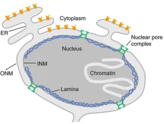
A main structural characteristic of the nucleus is the nuclear envelope (NE), which is a double lipid bilayer that encloses the genetic material in eukaryotic cells (Figure 1.1). It serves as a physical barrier, separating nucleoplasmic components from the cytosol. The double lipid bilayers that together from the NE are referred to as inner nuclear membrane (INM) or outer nuclear membrane (ONM) that are joined together at the nuclear pore complexes (NPC). NPCs are large macromolecular proteins assemblies responsible for the selective exchange between the nucleoplasm and the cytoplasm that prevents the exchange of materials not destined to cross the NE (Schirmer EC and Gerace L, 2002; Hertzer MW, 2010).
Figure 1.1 The nuclear envelope encloses the genetic material in eukaryotes.
The nuclear envelope is composed of an inner and an outer nuclear membrane (INM, ONM). A network of proteins termed the nuclear lamina underlies the inner membrane. Nuclear pore complexes bridge this system and regulate nucleo-cytoplasmic exchange of macromolecules. Additionally, the ONM continuous into the endoplasmic reticulum (ER). The genetic material is organized as chromatin (Schirmer EC and Gerace L, 2002).
4
Electron microscopy technique revealed that ONM and INM are continuous and the ONM additionally extends into the endoplasmic reticulum (ER). A variety of techniques were applied during the last decades to reveal the complex and unique structures of the NE membrane system and their associated proteins. Despite the membrane continuity between ONM, INM and ER, recent evidence demonstrates the presence of particular proteins assemblies (Watson ML, 1955;Hetzer MW, et al., 2005; Hetzer MW, 2010).
1.2 NE proteins
Along the NE one can observe the formation of distinct protein networks. In the following parts structures and functions of NE protein components will be described.
1.2.1 Nuclear pore complexes
NPCs are formed at sites where the inner and outer membranes of the nuclear envelope are joined, and are constructed of multiple copies of approximate 30 different proteins named nucleoporins (Nups). NPCs consist of three functional regions: First, the NPC scaffold and central transport channel, which is embedded in the plane of the nuclear envelope; second, peripheral structures, nuclear ring/nuclear basket; third, cytoplasmic ring/cytoplasmic filaments, which extend to the cytoplasmic or nucleoplasmic end of the NPCs.
The primary function of NPCs is that they regulate molecular trafficking between cytoplasm and nucleus that is crucial for maintaining the unique composition of both compartments.
NPCs allow the free transport of ions and small molecules without specific transport partner.
Large molecules like m-RNA or proteins with a molecular weight of more than 40~60 kDa are transported between nucleoplasm and cytoplasm by binding to nuclear transport receptors (Strambio-De-Castillia C, et al., 2010; Chow KH, et al., 2012; Raices M and D’Angelo MA, 2012).
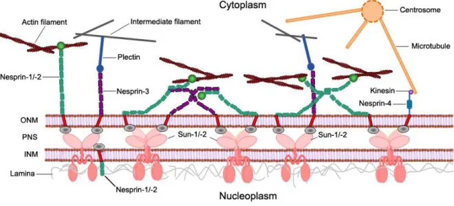
5 1.2.2 The LINC Complex
Along nuclear membranes, proteins assemble to form large protein complexes. At the INM, lamins form a meshwork that stabilizes nuclear morphology. NPCs are integrated into the NE and allow the passive and selective transport across the nuclear membranes. SUN proteins, lamins and nesprins, which will be described later in detail, are part of large protein complexes that traverse the NE to connect the nuclear interior to the cytoplasm, known as the LINC complex (linker of nucleoskeleton and cytoskeleton). At the heart of the LINC complex are the SUN (Sad1 and UNC-84) proteins and the nesprins (nuclear envelope spectrin repeat proteins). The KASH (Klarsicht, ANC1 and SYNE1 homology) domain of the nesprins interacts with the SUN domain of the SUN proteins in the perinuclear space (PNS) between the INM and the ONM (Figure 1.2).
Figure 1.2 Typical structure of the LINC complex. The LINC complex is composed of two major parts, ONM KASH domain proteins and INM SUN domain proteins. KASH domain proteins are anchored in the ONM by the interaction with the SUN domain of the SUN proteins in the perinuclear space (PNS) between ONM and INM.
Together they form a physical connection between nucleoplasm and cytoplasm. On the nucleoplasmic side, LINC complex components interact with the lamina and INM-associated proteins such as emerin. At the cytoplasmatic side, nesprins directly or indirectly connect the NE to cytoskeletal components, F-actin, microtubules and intermediate filaments (Taranum S, et al., 2012).
At the nucleoplasmic side LINC complex components interact with lamina proteins or INM- associated protein such as emerin or chromosomes. On the cytoplasmic side they directly or
6
indirectly bind to cytoskeletal components like F-actin, microtubules or the intermediate filament network (Starr DA and Fischer JA, 2005; Zhang Q, et al., 2005; Crisp M, et al., 2006;
Tzur YB, et al., 2006; Méjat A and Misteli T, 2010; Starr DA and Fridolfsson HN, 2010; ).
1.2.3 Nesprins
Nesprins also known as SYNE (synaptic nuclear envelope protein), ENAPTIN and NUANCE (nucleus and actin connecting element) belong to a group of proteins that are primarily found along both, the INM and the ONM. Nesprins are spectrin repeat (SR)-containing type II transmembrane proteins with evolutionarily conserved orthologous in lower eukaryotes such as in Dictyostelium discoideum (interaptin), C. elegans (ANC-1) and Drosophila (MSP- 300) (Rivero F, et al., 1998; Apel ED, et al., 2000; Zhang Q, et al., 2001; Starr DA and Han M, 2002; Zhen YY, et al., 2002; Padmakumar VC, et al., 2004; Yu J, et al., 2006).
Four different nesprins have been described in mammals until now (nesprin-1, -2, -3 and -4).
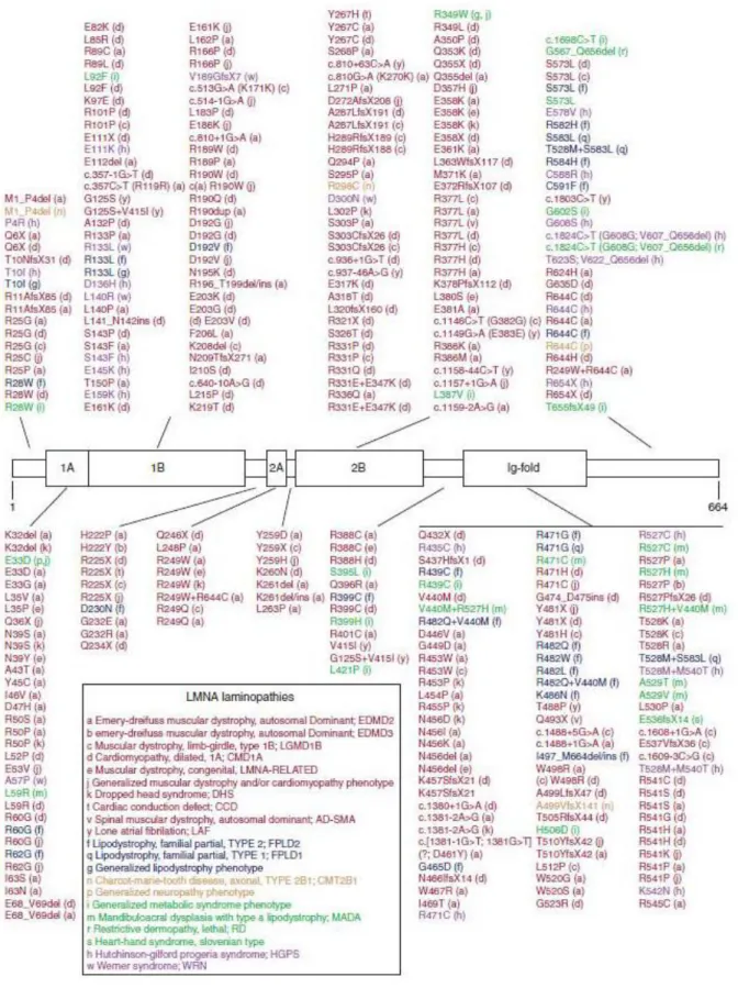
Each is encoded by a single gene. A multitude of isoforms has been described so far differing in size and domain composition which result from differential splicing and initiation of transcription. The largest nesprin isoforms are nesprin-1/2-giant with molecular weights of 1014/796 kilodalton (kDa) respectively (Figure 1.3).
Nesprin-1/2 giant contain N-terminal actin-binding domains (ABD) that interact with actin filaments. Nesprin-3 binds to plectin, which in turn binds to intermediate filaments. Nesprin- 2/4 bind to kinesin-1 and in this way they connect the nucleus to the microtubule network.
At the C-terminus of almost all nesprin isoforms, there is a highly conserved KASH domain that contains a transmembrane region. Nesprins are anchored along the ONM or INM through their KASH domain that interacts with the SUN domain of the SUN proteins in the lumen between the INM and ONM (Zhang Q, et al., 2001; Wilhelmsen K, et al., 2005; Roux KJ, et al., 2009; Gob E, et al., 2010; Noegel AA and Neumann S, 2011; Taranum S, et al., 2012)
7
Figure 1.3 Summary of mammalian nesprins and their isoforms.
Four different nesprins have been described in mammals (nesprin-1, -2, -3 and -4). Each is encoded by a single gene that gives rise to several isoforms. Main structural components of the nesprins are paired calponin homology (CH) domains, spectrin repeats (SRs) and a KASH (Klarsicht, ANC1 and SYNE1 homology) – transmembrane domain (Taken and combined from Rajgor D, et al., 2012 and Mellad JA, et al., 2011).
8
Nesprins play an important role in maintaining nuclear architecture. A loss of nesprin-2 for example results in misshapen nuclei or nuclear blebbing. Furthermore nesprins have been shown to play roles in cellular processes like muscle development, cell proliferation, nuclear positioning and anchorage. Wound healing studies in nesprin-2 giant deficient mice showed that a loss of nesprin-2 giant affects wound healing particularly at later stages during fibroblast differentiation and keratinocyte proliferation leading to delayed wound closure. A loss of nesprin-1 and nesprin-2 furthermore shows those both mediate centrosome positioning and migration and are essential for ciliogenesis by remodeling of the actin cytoskeleton, indicating important roles of the nesprins during cellular and developmental processes. Analysis of nesprins during human muscle development revealed isoform- and tissue-specific roles for nesprins in nuclear positioning. Nesprin-1 knockout studies show that nuclear positioning and anchorage are dysfunctional in skeletal muscle from knockout mice. (Luke Y, et al., 2008; Dawe HR, et al., 2009; Warren DT, et al., 2010; Randles KN, et al., 2010; Zhang J, et al., 2010; Mellad JA, et al., 2011).
Most of the functions described before refer to structural roles of the nesprins. However nesprins are also involved in regulating signaling events across the NE or inside the nucleus.
At the NE nesprin-2 interacts with a- and β-catenin and with emerin that has an impact on Wnt signaling. Inside the nucleus, nesprin-2 acts as a scaffolding protein for nuclear extracellular signal-regulated kinases 1 and 2 (ERK1/2, also known as MAPK1/2). A loss of nesprin-2 leads to sustained ERK1/2 activation and increased cell proliferation (Neumann S, et al., 2010; Warren DT, et al., 2010; Rashmi RN, et al., 2011; Yu J, et al., 2011)
1.2.4 SUN proteins
SUN proteins are type-II transmembrane proteins of the INM with an N-terminus facing the nucleoplasm, and a C-terminal conserved SUN domain localizing between INM and ONM.
The eponymous SUN domain is a conversed ~200 amino acid residues spanning C-terminal motif that follows a trans-membrane domain and a coiled coil segment that reaches into the PNS. SUN proteins contain at least one transmembrane domain (Figure 1.4). The name of the SUN domain is derived from homologous sequences that have originally been described in
9
Schizosaccharomyces pombe Sad1 and Caenorhabditis elegans UNC-84 proteins. Later these sequences have additionally been found in mammalian proteins.
Five SUN proteins have been identified until now: SUN1 (UNC-84A), SUN2 (UNC-84B), SUN3, SUN4 (SPAG4), and SUN5 (SPAG4L). To date, SUN domain proteins have been described in several species, such as mus musculus, Caenorhabditis elegans, Drosophila melanogaster (Klaroid and Giacomo), Schizosaccharomyces pombe (Sad1) and Saccharomyces cerevisiae (Mps3). SUN domain proteins also been reported in plants such as Arabidopsis thaliana (Shao X, et al., 1999; Tzur YB, et al., 2006; Hiraoka Y and Dernburg AF, 2009; Graumann K, et al., 2010; Jiang XZ, et al., 2010; Rothballer A, et al., 2013).
SUN proteins play important roles in a wide range of cellular processes. SUN1 and SUN2 double knockout mice die shortly after birth due to a breath system defect, suggesting that SUN proteins are essential. SUN1 specifically associates with telomeres during meiosis.
Studies on SUN1-deficient mice indicate that disruption of SUN1 in mice prevents telomere attachment to the nuclear envelope and meiosis. As the critical processes of maintaining genomic stability, DNA damage response and DNA repair were described to be regulated by SUN proteins according their interactions with DNA-dependent protein kinase, a protein known to function in DNA repair. Based on the SUN1 and SUN2 double knockout mice research, embryonic fibroblasts displayed premature proliferation arrest in S phase of cell cycle, increased apoptosis and DNA damage, and decreased perinuclear heterochromatin.
Mps3, a SUN protein found in yeast, has been described to play a role in the repair of DNA double strand breaks, indicating that SUN proteins have a role in maintaining genome stability. Studies on LMNA null or progeroid LMNA∆9 mutant mice described SUN1 abnormally accumulated at the Golgi. Furthermore, it has been described that high levels of SUN proteins at the NE in lamin A mutant cells result in toxicity through a hyperactivity of the DNA damage response (Ding X, et al., 2007; Oza P, et al., 2009; Chen CY, et al., 2012; Lei K, et al., 2012; Star DA, 2012).
10 Figure 1.4 Basic structures of SUN proteins.
Main structural characteristics shared by SUN proteins are the SUN domain (orange), and at least one trans- membrane region (red) that mediates the anchorage to the NE. The SUN domain localizes in the lumen between the INM and ONM, where it interacts with the KASH domain of the nesprins (Rothballer A et al., 2013).
1.2.5 Cellular functions of the LINC complex
As the core components of the LINC complex, KASH domain proteins nesprins are anchored along both ONM and INM through the interaction of the KASH domain with the SUN domain
11
of the SUN proteins in the PNS. Based on the localization between cytoplasm and nucleoplasm LINC complex components interact with proteins from both compartments.
INM nesprins and SUN proteins interact with NE proteins like emerin and lamin A/C. ONM nesprins directly or indirectly interact with all cytoskeletal systems of a cell.
As a linker of nucleoskeleton and cytoskeleton, the LINC complex plays an important role in many crucial cellular processes. The LINC complex and LINC complex-associated proteins form an essential connection between the cytoskeleton and the nucleoskeleton that is critical for intracellular force transmission and transfer of mechanical induced signals across the NE. Additionally, studies on the nesprin-4 and SUN1 KO mice revealed that mice lacking either nesprin-4 or SUN1 show hair cell defects and hearing impairments, that demonstrated the LINC complex is essential for hearing (Razafsky D, et al., 2009; Méjat A and Misteli T, 2010; Lombardi ML, et al., 2011; Horn HF, et al., 2013).
1.2.6 Nuclear lamins
Lamins are type V intermediate filaments proteins that form a meshwork that lines the nucleoplasmic surface of the INM.
They are grouped into A- and B-type lamins. A-type lamins include lamin A, A∆10, C and C2 that are encoded by LMNA and formed by alternative splicing and posttranslational modifications. Lamin A and C are the major components of the nuclear lamina. B-type lamins are encoded by LMNB1 (lamin B1) and LMNB2 (lamin B2 and B3) (Worman HJ and Bonne G, 2007).
1.2.5.1 Lamins assemble to form a meshwork along the INM
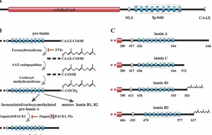
Basic structural aspects of mammalian lamins are the tripartite organization into an N- terminal head domain, a central α-helical rod domain and a C-terminal globular tail domain.
The C-terminal domain contains a structural Ig-fold and a nuclear localization signal (NLS), and in most cases contains a carboxy-terminal CAAX box (except lamin C). After four post-
12
translational processing steps for pre-lamin A, the farnesylation of a carboxyl-terminal cysteine, release of the last three amino acids –AAX of the protein, methylation of the farnesylcysteine, and the endoproteolytic release of the carboxyl-terminal 15 amino acids of the protein, the mature lamin A protein is generated (Figure 1.5).
Figure 1.5 Schemes summarizing structures and processing of lamins.
(A) Main structural components of pre-lamin A are the globular N-terminal head domain, a central α-helical rod domain and a C-terminal globular tail domain with a carboxy-terminal CAAX motive.
(B) Summary of post-translational processing of the pre-lamins A, B1 and B2. After four post-translational processing steps namely farnesylation of a carboxyl-terminal cysteine, release of the last three amino acids of the protein, methylation of the farnesylcysteine, and the endoproteolytic release of the carboxyl-terminal 15 amino acids of the protein, the mature lamin A protein is generated. In lamin B1 and B2 maturation ends with the methylation of the C-terminal cysteine and does not include the cleavage of the C-terminal 15 residues.
(C) C-terminal structures of mature lamins. The indicated amino acid positions are: start of the tail domain, first residue of the NLS, Ig-fold and most C-terminal residue of mature lamins (Dechat, et al., 2008).
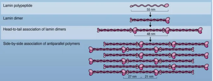
For the formation of higher order structures (Figure 1.6), initially two lamin polypeptides (about 55nm) assemble to form parallel head to tail dimer, which is driven by coiled-coil formation to a two stranded α-helical coiled-coil structure (around 48 nm from tail to tail).
13
Dimers then assemble into head-to-tail polar polymers, which require an overlapping interaction between the head and tail domains. Higher order structure is achieved by anti- parallel, side-by-side association of polymers, the proto-filaments that underlay the NE (Stuurman N, et al., 1998; Prokocimer M, et al., 2009; Ho CY and Lammerding J., 2012).
Figure 1.6 Assembly of lamins into filaments.
Initially two lamin monomers assemble along their central alpha helical rod domain to form a parallel head to tail dimer. Following, lamin dimers assemble into head to tail polar polymers based on overlapping interactions between head and tail domains. Polymers then laterally assemble anti-parallel into non-polar filaments that form a meshwork underlying the inner surface of the INM (Ho CY and Lammerding J., 2012).
1.2.5.2 Functions of lamins
Nuclear lamins were identified more than 30 years ago. At the early stage of research on lamins, they were described as nuclear matrix components, fulfilling structural functions such as maintaining shape and mechanical stability of the nucleus. Along with research progress, it became increasingly evident that lamina proteins are involved in basic cellular processes such as maintaining nuclear shape and mechanical stability, as well as regulating gene expression, signaling pathways and functional chromatin organization. Lamin functions have additionally been shown to play a role in mitosis, DNA replication and repair and transcription (Dechat T, et al., 2010; Dittmer TA and Misteli T, 2011; Gerace L and Huber MD, 2012; Worman HJ, 2012).
14 Figure 1.7 Interaction partners of A- and B-type lamins.
The scheme summarizes interaction partners that have been identified for A- and B-type lamins so far including their interaction sites in the lamin proteins. The interaction partners reflect the wide range of lamin functions along the NE and in organizing chromosomal structures as well as regulating gene expression. The majority of interactions have been shown for A-type lamins (Ho CY and Lammerding J., 2012).
The wide range of cellular functions in which lamins have been shown to play a role, is reflected by the large and constantly increasing number of interaction partners that have been described, especial for A-type lamins (Figure 1.7). The currently described interaction partners include nuclear membrane associated proteins such as nesprin-2 and emerin.
Lamins interact with chromosomal structures through interactions with histones or direct binding to DNA. They additionally interact with transcriptional regulators like sterol regulatory element-binding protein 1 (SREBP1) and ZNF239 (zinc finger transcription factor proteins, also known as MOK2) (Wilson KL and Foisner R, 2010; Ho CY and Lammerding J., 2012).
1.3 The LINC complex in human diseases
15
Several hundred mutations in NE proteins have been described that can result in a variety of human diseases such as Hutchinson-Gilford progeria syndrome (HGPS), Emery-Dreifuss muscular dystrophy (EDMD), Dunnigan-type familial partial lipodystrophy (FPLD), and cardiomyopathy that are collectively known as nuclear envelopathies. Due to the large number of mutations in the LMNA, the gene encoding for A-type lamins, these diseases are also known as laminopathies (Table 1.1).
Human diseases Associated mutated gene References
Emery-Dreifuss muscular dystrophy Cerebellar ataxia
Arthrogryposis
SYNE, encoding nesprins Zhang Q, et al., 2007 Gros-Louis F, et al., 2007 Attali R, et al., 2009 Duchenne muscular dystrophy
Emery-Dreifuss muscular dystrophy Charcot-Marie-Tooth syndrome
SUN, encoding SUN domain proteins
Taranum S., et al., 2012
Emery-Dreifuss muscular dystrophy EMD, encoding emerin Bione S, et al., 1994 Bione S, et al., 1995 Hutchinson-Gilford progeria
Emery-Dreifuss muscular dystrophy Familial partial lipodystrophy Dilated cardiomyopathy Charcot-Marie-Tooth disease Mandibuloacral dysplasia
LMNA, encoding A-type lamins
DeBusk FL, 1972 Wessely R, et al., 2005 Garg A, 2004
Olson TM and Keating MT, 1996 Bouhouche A, et al., 1999 Garavelli L., et al., 2009
Table 1.1 LINC complexes proteins and human diseases.
The table summarizes examples of mutations in NE genes and the resulting diseases. Most of them belong to a group of human diseases collectively known as laminopathies.
The first identified LINC complex component associated with human diseases was emerin. As a component of the NE, emerin interacts with several proteins at the inner and outer surface of the NE. These proteins are involved in regulating the activity of certain genes, controlling the cell cycle and maintaining the structure and stability of the nucleus. Emerin and related proteins also play a role in assembling the nucleus during cell division. Mutations in EMD, encoding for emerin, lead to an X-linked form of Emery-Dreifuss muscular dystrophy (XL- EDMD). More than 400 different mutations (Figure 1.8) have been identified in LMNA so far (www.umd.be/LMNA/). Only a few mutations have been identified in B-type lamins.
16
Mutations in nesprin-1/2 and SUN proteins have also been implicated in the formation of laminopathies like EDMD. Mutations in nesprin-1 and -2 additionally result in disorders which are not characterized by NE defects such as autosomal recessive cerebellar ataxia.
Along with research insight, more and more genes encoding NE components have been associated with human diseases (Bione S, et al., 1994; Cao H, et al., 2000; Vergnes L, et al., 2004;Méjat A and Misteli T, 2010; Worman HJ, et al., 2010; Meinke P, et al., 2011; Lombardi ML, et al., 2011).
To date, at least 15 different diseases have been linked to mutations in NE proteins like lamins, emerin, nesprins and SUN proteins. However, due to partially overlapping clinical manifestations the exact number of disease phenotypes may differ. Laminopathies range from skeletal muscle dystrophies to cardiac defects, metabolic diseases, diseases affecting the nervous system or diseases associated with early aging. Most laminopathies have a postnatal onset and are progressively developing during childhood or adolescence.
Currently, there is no cure for laminopathies and treatment is symptomatic and supportive, without treatment some laminopathies may lead to early death. It is still not clear how mutations in NE proteins result in phenotypically different laminopathies with tissue-specific pathologies, especially when considering that most NE proteins such as lamins, are expressed in almost all differentiated somatic cell types. Currently, three mutually non- exclusive hypotheses have been postulated. The first one is the structural hypothesis that focusses on the role of NE proteins in maintaining nuclear integrity. Mutations in NE proteins lead to structural weakness, and decreased ability of nuclei to maintain mechanotransduction along LINC complexes or to resist high mechanical strain. The second hypothesis is the gene-expression hypothesis, which refers to the role of NE proteins like nesprins or lamins in regulating gene expression. The third hypothesis, refers to the pathological impact of accumulated mutated NE proteins and their resulting cell toxic functions (Lammerding J, et al., 2004; Capanni C, et al., 2005; Columbaro M, et al., 2005;
Markiewicz E, et al., 2006; Kandert S, et al., 2007; Furukawa K, et al., 2009; Brosig M, et al., 2010; Dubinska-Magiera M, et al., 2012; Simon DN and Wilson KL., 2013).
17
Figure 1.8 Summary of laminopathy causing mutations in human lamin A .
The scheme summarizes mutations in lamin A and the resulting laminopathies are classified with a colour code.
Mutations shown in red affect skeletal or cardiac muscles. Mutations shown in blue lead to lipodystrophies, brown refers to neuropathies, green to systemic laminopathies and mutations in purple belong to premature aging disorders (Dittmer T and Misteli T, 2011).
18
1.4 Aim of the study
LINC complexes connect the nucleoskeleton to the cytoskeleton by interactions among LINC complex proteins and their interactions to proteins in the nucleus and the cytosol. LINC complexes reside along the interface between cytoplasm and nucleoplasm and they are involved in regulating many cellular functions including maintenance of the structural integrity of nucleus and cytoskeleton or proper signal transduction across the NE. Mutations in LINC complex components have been described in a wide range of human diseases that include muscle diseases, metabolic syndromes neuropathies or premature ageing diseases that are collectively known as laminopathies. For this reason it is of particular importance to have a detailed knowledge about LINC complex interactions.
The aim of my thesis is to further characterize interactions among LINC complex components and to study the pathological role of distinct mutations in these proteins. Nesprins interact with the INM proteins lamin A/C. The binding site between nesprin-1, -2 and lamin A/C have been mapped near their C-termini (Mislow JM, et al., 2002; Libotte T, et al., 2005). One aim of my project is to further narrow down the binding site between nesprin-2 and lamin A.
Additionally we want to find out how laminopathy causing mutations in or near the binding sites of both proteins affect their interaction, which will help to answer the question how different mutations in one gene lead to the formation of phenotypically different diseases.
To substantiate our knowledge about LINC complex protein assemblies I additionally characterize the novel interaction of SUN proteins to the microtubule network under normal or laminopathic conditions. The studies on SUN proteins include the characterization of the SUN1 mutation Q93P and T33A in SUN2 and their impact on NE protein assemblies.
Laminopathies include early ageing diseases like HGPS that resembles aspects of ageing at an early age, for this reason the expression of lamin proteins in the context of ageing nesprin-2 giant knockout (KO) mice will be analyzed.
19
2 Results
2.1 Narrowing down of binding sites between lamin A and nesprin-2
As a component of the LINC complex, nesprins interact with INM proteins, like lamin A/C.
The corresponding binding sites have been mapped near the C-terminus of nesprin-1 and nesprin-2 (Libotte T, et al., 2005).
In order to get a better understanding of the interaction between both proteins, we further narrowed down the binding sites between lamin A and nesprin-2 by using His-Tag and GST Pull down assays.
A
20 B
Figure 2.1 Lamin A and nesprin-2 constructs used for His-tag pull down.
(A) Lamin A constructs used in this study. The plasmids we used cover almost the entire sequence of lamin A.
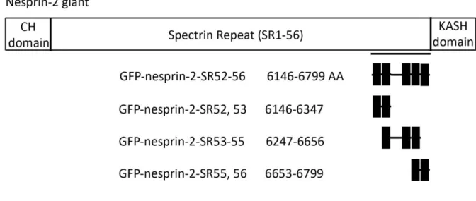
(B) Nesprin-2 constructs used in this study. On top nesprin-2 giant is shown. The C-terminal part that is known to interact with lamin A is underlined. The nesprin-2 constructs we used for our binding studies include five C- terminal spectrin repeats of nesprin-2 giant (SR52-56).
2.1.1 Identification of the lamin A amino acids that mediate the binding to nesprin-2
First we narrowed down the interaction site of nesprin-2-SR52-56 to lamin A. For this His-tag pull downs were performed by using pPET-TEV-LA (1-263), pPET-TEV-LA (264-402), pPET- TEV-LA (345-425) and Pet24d His-TEV- (436-548) lamin A expression constructs. Proteins were coupled to Ni-NTA beads, incubated with lysates of COS7 cells expressing GFP-nesprin- 2-SR52-56, and analyzed by western blot. The nesprin-2-SR plasmids used in this study are GST-nesprin-2-SR52-56, -SR52, 53, -SR53-55 and –SR55, 56. In western blots we obtained signals for GFP-nesprin-2-SR52-56 in the LA 345-425 fraction (Figure 2.2 A). From this we conclude that the interaction site of nesprin-2-SR52-56 to lamin A lies within the lamin A amino acid sequence from 345 to 425.
By taking a closer look at the lamin A constructs we used in the in vitro binding studies, one can see that there is an overlap between lamin A constructs LA 264-402 and LA 345-425.
21
Since we did not detect GFP-nesprin-2-SR52-56 together with LA 264-402, the binding site for lamin A to nesprin-2 must lie within lamin A amino acid 403-425 (Figure 2.2 B).
A
B
Figure 2.2 Amino acids 345-425 of human lamin A interact with GFP-nesprin2-SR52-56.
(A) His-tag pull down experiment with His tagged lamin A proteins and GFP-tagged nesprin-2-SR52-56. GFP- nesprin-2-SR52-56 precipitates only with LA 345-425. We did not get any signals from the other lamin A polypeptides (LA1-263, LA 264-402 and LA 436-548). That suggests nesprin-2 interacts with lamin A through the amino acids 345-425. WB: western blot, P: pellet, SPN: supernatant.
(B) Lamin A polypeptides 264-402 and 345-425 share an overlapping sequence. Since GFP-nesprin-2-SR52-56 precipitates with LA 345-425, but not with LA 264-402, the binding site for lamin A to nesprin-2 can be narrowed down to lamin A amino acid 403-425 (highlight in red).
2.1.2 Identification of nesprin-2 giant amino acids that mediate the binding to lamin A
22
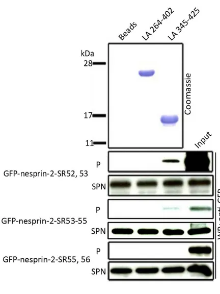
Next we performed His-tag pull down experiments to narrow down the binding site in nesprin-2 to lamin A. We used the lamin proteins LA 345-425 as positive controls, since we identified the binding site of GFP-nesprin-2-SR52-56 to these lamin A amino acids (Figure 2.2). LA 264-402 was used as a negative control, because this protein fragment does not interact with GFP-nesprin-2-SR52-56 (Figure 2.3B). His-tagged lamin A proteins that were bound to Ni-NTA beads were incubated with lysates of COS7 cells expressing GFP-nesprin-2- SR52, 53, -SR53-55, -SR55, 56 proteins, respectively (Figure 2.3 A).
In western blots we detected signals for GFP-nesprin-2-SR52, 53 and GFP-nesprin-2-SR53-55.
We did not obtain signals for GFP-nesprin-2-SR55, 56. The nesprin-2 constructs -SR52, 53 and -SR53-55 we used in the in vitro binding studies share an overlapping sequence. We got signals for both polypeptides in the western blot, however the signal for nesprin-2-SR53-55 was always very weak compare to -SR52, 53, indicating the interaction site of nesprin-2 to lamin A resides within the SRs 52 and 53 (Figure 2.3 B).
23 A
B
Figure 2.3 Nesprin-2-SR52, 53 and -SR53-55 interact with LA 345-425.
(A) His-tag pull downs were performed with lamin A proteins LA264-402, LA 345-425 that were bound to beads and the GFP nesprin-2 fusion polypeptide nesprin-2-SR52-56, nesprin-2-SR52, 53, -SR53-55 and –SR55, 56. We got signals for nesprin-2-SR52, 53 and –SR53-55.
24
(B) Nesprin-2-SR52, 53 and -SR53-55 share an overlapping sequence andGFP immunoblotting signals for -SR53- 55 were always much weaker compared to -SR52, 53. We concluded the binding site of nesprin-2 to lamin A lies within nesprin-2-SR52, 53, aa 6146-6347 (underline in red).
2.1.3 Nesprin-2-SR53 is sufficient to mediate the interaction to lamin A
In pull down experiments we found an interactions between lamin A aa 345-425 and GFP- nesprin-2-SR52, 53, and GFP-nesprin-2-SR53-55 (Figure 2.3). Since the signal for GFP- nesprin-2-SR52, 53 was always much stronger compared to GFP-nesprin-2-SR53-55 we concluded that the nesprin-2 interaction to lamin A mainly depends on SRs 52 and 53. To answer the question which one of these SRs is sufficient for the interaction, GST-pull down experiments were performed in which GFP-LA 403-425 and GST-nesprin-2-SR52, -SR53, and - SR52, 53 proteins were used. GST-nesprin-2-SR proteins coupled to beads were incubated with lysates of COS7 cells expressing GFP-LA 403-425. By western blot analysis, we obtained signals for GST-nesprin-2-SR53 and -SR52, 53. The signal for nesprin-2-SR53 was always much weaker compared to the signal of nesprin-2-SR52, 53, however this SR is sufficient to precipitate GFP-LA 403-425 (Figure 2.4).
Figure 2.4 GFP-nesprin-2-SR53 is sufficient to mediate the interaction to LA 403-425.
The GST-pull down experiment was performed by using lamin A proteins (GFP-LA 403-425) and GST-nesprin-2- SR proteins (-SR52, -SR53, and -SR52, 53). GFP-LA 403-425 was precipitated by nesprin-2-SR53 and -SR52, 53.
Nesprin-2-SR53 is sufficient for mediating the interaction between lamin A 402-425, but the signal was much
25
weaker compared to the longer fragments, indicating that additional amino acids are necessary for strengthening the interaction between both proteins.
2.2 Study on mutations in the binding sites of nesprin-2 and lamin A
2.2.1 Laminopathy causing mutations reside in the nesprin-2 binding site of lamin A
Currently there is accumulating evidence that lamins and further LINC complex or NE proteins play roles in the formation of laminopathies. For this reason we wanted to analyze if mutations which have been described in laminopathies that lie within or near the binding sites of lamin A and nesprin-2 affect the interaction between both proteins which might contribute to the formation of these diseases.
Many mutations in lamin A have been identified until now. Most are summarized on the website http://www.umd.be/LMNA/. Several mutations have been reported in or near lamin A aa 403-425, the binding site to nesprin-2, namely R401C (1201 C>T), G411D (1232G>A), G413C (1237G>T), V415I (1243G>A), R419C (1255T>C), L421P (1261T>C), R427G (1279C>G) and Q432X (1294C>T). These mutations are characterized by large differences in their clinical manifestation that include lipodystrophies or metabolic syndromes for G411D, R419C, L421P and skeletal or cardiac muscle dystrophies in case of R401C, G413C, V415I, R427G and Q432X (Vytopil M, et al., 2001; Haque WA, et al., 2003; Decaudain A, et al., 2007; Brauch KM, et al., 2009; Møller DV, et al., 2009; Dutour A, et al., 2011). No mutations in nesprin-2 have been described so far that might affect the interaction to lamin A. For this reason in the following we focused on characterizing the described mutations afore in LMNA.
Above all, we aim to analyze if changes in the binding ability between nesprin-2 and lamin A contribute to the formation of laminopathies.
2.2.2 Generation of mutated full length or short GFP lamin A constructs
The first mutation we found in the binding site between lamin A and nesprin-2 was the mutation L421P. cDNAs encoding WT and mutated lamin A amino acid 403-425, that were
26
shown to interact with nesprin-2 (Figure 2.2) were cloned into GFP vectors and confirmed by DNA-sequencing (Figure 2.5 A) and western blot analysis (Figure 2.5 B). The predicted molecular weight of GFP-LA 403-425 is approximate 30 kDa and thus differs only slightly from GFP that has a molecular weight of approximate 27 kDa (Figure 2.5 B).
A
B
Figure 2.5 Sequencing results and western blot analysis of GFP-LA 403-425 WT and -L421P.
(A) cDNAs encoding WT or L421P mutated lamin A amino acids 403-425 were cloned into GFP vectors and confirmed by DNA sequencing. WT and mutated nucleotides are highlighted by arrows.
(B) Western blot analysis of GFP-LA 403-425 fusion proteins. The proteins were transiently expressed in COS7 cells. Lysates were analyzed by SDS-PAGE followed by western blot with mAb K3-184-2 that confirmed the predicted molecular weights.
During our studies further mutations in lamin A have been described including [Arg401Cys (1201 C>T), Gly411Asp (1232G>A), Gly413Cys (1237G>T), Val415Ile (1243G>A), Arg419Cys (1255T>C), Leu421Pro (1262T>C), Ar427Gly (1279C>G), Gln432X (1294C>T)]. All mutations reside within a region of lamin A that is encoded by LMNA exon 7 and all are point mutations causing single nucleotide exchanges (Figure 2.6 A and B). Seven mutations result in amino acid changes whereas the mutation Q432X (1294C>T) causes the formation of a premature stop codon and thus a shorten lamin A (Figure 2.6 C and D). All LMNA mutations were
27
inserted into GFP full length lamin A by site directed mutagenesis. The success of mutagenesis was confirmed by DNA sequencing (Figure 2.6 B). Additionally all GFP plasmid were transiently expressed in COS7 cells and lysates of these cells were analyzed by western blot to confirm the molecular weight of each mutated protein. The predicted molecular weight for GFP WT lamin A and all mutations is 100 kDa. The truncated GFP-LA Q432X protein has a predicted molecular weight of 76 kDa. According to DNA sequencing and immunoblotting, all constructs are correct (Figure 2.6).
A
B
C
28 D
Figure 2.6 DNA Sequencing and western blot analysis of full length WT and mutated lamin A constructs.
(A) Nucleotides 1198 to 1296 of human LMNA are shown. Nucleotides encoding the amino acids that interact with nesprin-2 are underlined. LMNA mutations analyzed in the present study are highlighted below in red.
Similarities to the WT sequence are shown as dotted lines.
(B) LMNA mutations shown in (A) were inserted into plasmids encoding GFP tagged full length lamin A by side directed mutagenesis. Sequencing confirmed the success of the mutagenesis. Mutated nucleotides are highlighted by an arrow in each peak diagram.
(C) Aa residues 399 to 434 of WT lamin A are shown. The interaction site of lamin A to nesprin-2 is underlined.
Similarities to the WT sequence are shown as dotted lines, aa changes are indicated in red. The mutation Q432X leads to a stop codon (X).
(D) Western blot analysis of GFP lamin A fusion proteins. The proteins were transiently expressed in COS7 cells.
Lysates were analyzed by SDS-PAGE followed by western blot with mAb K3-184-2 that confirmed the predicted molecular weights.
2.2.3 The interaction site of lamin A to nesprin-2 is in a loop
The lamin A mutations that we analyzed here reside between aa 401 and 432 of human lamin A. In an alignment one can see highly conserved residues between human and Mus musculus lamin A along the binding site of lamin A to nesperin-2 (Figure 2.7 A). A main aim of the study is to characterize the impact of these LMNA mutations on the binding capacity to nesprin-2. For this we addressed the question in which structural region of lamin A aa 403-425 are located. Currently only parts of the three dimensional lamin A structure are available. Therefore we performed a structural prediction of human lamin A aa 351-490 that are part of the coil2B and C-terminal globular domain (Figure 2.7 A), by using the MULTICOM server and 1UFGA (C-terminal immunoglobulin like domain of mouse Lamin A), 1IFRA (globular tail of human Lamin A), and 2LLA (chain A of Mannose-6-phosphate/insulin-like
29
growth factor II receptor) structures from the protein data bank (PDB) as templates and pyMOL for presentation.
A
B
C Lamin A WT Mutate lamin A
30
Figure 2.7 The interaction site of lamin A to nesprin-2 is in a loop.
(A) An amino acid alignment was performed between human lamin A/C amino acids 351-490 and the corresponding sequence in human lamin B and Mus musculus lamin A. The interaction region of nesprin-2 to lamin A is highlighted in a green frame. Secondary structure elements are shown on top of the alignment and conserved residues are highlighted black, similar residues are framed.
(B) Three dimensional structure prediction of human lamin A aa 351-490. The three dimensional structure of human lamin A351-490 was predicted by using multiple PDB structures as templates (1UFGA, 1IFRA, and 2LLA) in the MULTICOM server. The interaction region of nesprin-2 to lamin A aa 403-425 is highlighted in green.
Lamin A mutations that lie in or near the binding site of nesprin-2 analyzed in this study are highlighted in red.
The prediction was performed by using pyMOL v1.3.
(C) Molecular surface properties of WT lamin A and laminopathies causing lamin A mutations lie in or near the binding site for nesprin-2, aa 403-425. The comparison of the surface between WT lamin A aa 403-425 and the mutate lamin A with the mutations analyzed in this study. Highly positive and negative charged residues are shown in blue and red, respectively. The positive charged groups that are lost due to mutations are highlighted with black arrowheads, the negative charged group lamin A mutation G411D is highlighted with red arrowhead.
The lamin A interaction site aa 403-425 to nesprin-2 lies in a loop (Figure 2.7 B) that is located between the α helical rod domain and the globular domain at the C-terminus. The amino acids should therefore be accessible for interactions. Interactions among proteins mainly base on hydrophobic or electrostatic interactions between amino acid residues. To analyze the impact of amino acid exchanges caused by mutations in lamin A, analyzed here on the electrostatic surface properties, we additionally generated a model predicting surface charges from which we can say that amino acid exchanges reach from charge neutrality to the generation of positively or negatively charged residues (Figure 2.7 C).
2.2.4 Both LA 403-425 and LA 403-425 L421P interact with nesprin-2
First we focused on characterizing the lamin A mutation L421P that lies within the binding site of lamin A to nesprin-2. We analyzed if this mutation has an effect on the interaction between nesprin-2 and lamin A by performing GST-pull down experiments using GST- nesprin-2-SR52, 53 and -SR54-56 polypeptides and lysates of COS7 cells expressing GFP-LA 403-425 wild type or LA 403-425 L421P, respectively.
31
In GST-pull down experiments, we observed an interaction between GST-nesprin-2-SR, - SR52, 53 and both WT GFP-LA 403-425 and GFP-LA 403-425 carrying the mutation L421P (Figure 2.8). That suggested that the lamin A mutation L421P does not inhibit the interaction with nesprin-2.
Figure 2.8 Both GFP-LA 403-425 WT and L421P interact with nesprin-2-SR52-56 and -SR52, 53.
GST-pull down experiments of WT GFP-LA 403-425 and 403-425 L421P together with GST-nesprin-2 proteins, show that nesprin-2-SR52-56 and -SR52, 53 interact with WT LA 403-425 and LA 403-425 L421P. The GFP western blot signal observed in precipitates of nesprin-2-SR52-56 polypeptides were always very weak compared to the signal in nesprin-2-SR52, 53 precipitates.
2.2.5 Mutations in LMNA modulate binding affinities of lamin A to nesprin-2
Figure 2.8 shows that the lamin A mutation L421P does not inhibit the interaction with nesprin-2. However, it remains to be determined if the binding affinity of lamin A to nesprin- 2 is increased, decreased or unaffected. To address that question, following series of GST pull down experiments were performed. Based on the interaction study between nesprin-2
32
and lamin A (Figure 2.8) we used GST tagged nesprin-2-SR52, 53 that interacts with lamin A and -SR55, 56 that does not shown an interaction to nesprin-2 together with lysates of COS7 cells expressing GFP full length WT or mutated lamin A proteins.
Interestingly mutated lamin A proteins precipitated in varying amounts with GST-nesprin-2- SR52, 53 (Figure 2.9 A, exemplified shown for lamin A mutations R419C, L421P, R427G and Q432X). Of special note are the lamin A mutations R401C and V415I that caused an enhanced binding, whereas of the mutation Q432X was precipitated less efficient by GST- nesprin-2-SR52, 53, indicating a reduced binding. All further mutations influenced the interaction moderately (summarized in Figure 2.9 B).
A
B
Figure 2.9 Mutations in LMNA modulate the interaction to nesprin-2.
-100 -80 -60 -40 -20 0 20 40 60 80
Binding intensity compared to WT nesprin-2 to lamin A interaction in %
33
(A) COS7 cells expressing WT or mutated GFP lamin A proteins were lysed and incubated with recombinant GST-nesprin-2-SR52, 53 proteins. The use of equal amounts of GST fusion protein was confirmed by a Coomassie stained SDS PAGE. GST-nesprin-2-SR55, 56 proteins were used as negative controls. After incubating beads bound GST-nesprin-2-SR fusion proteins with lysates of GFP lamin A expressing COS7 cells, the supernatants were collected and subjected to western blot analysis to confirm equal GFP protein amounts among the experiments.
(B) WT and mutant GFP-LA proteins show distinct binding properties to WT GST-nesprin-2-SR52, 53. The zero base line represents the 100 % binding strength between WT GST-nesprin-2-SR52, 53 and GFP-LA WT.
Deviations caused by distinct mutations in LMNA are given in percent each mutation was analyzed by four to seven independent experiments.
2.2.6 GFP-lamin A 403-425 WT and GFP-lamin A 403-425 L421P are distributed all over the nuclei
To study the impact of distinct mutations in lamin A on the localization of nesprin-2 and further NE or LINC complex components, we performed immunofluorescence studies on COS7 and HaCaT cells transfected with GFP-tagged lamin A, aa 403-425 that contain the binding site to nesprin-2 as WT or with the mutation L421P. GFP alone was used as a control.
It has been demonstrated that lamin A L421P fibroblasts have misshapen nuclei (Decaudain A, et al., 2007). GFP signals we observed for cells expressing GFP lamin LA 403-425 WT or LA 403-425 L421P showed similar distributions compared with cells expressing GFP only, all over and around the nucleus (Figure 2.10). Differences between the localization of endogenous lamin A along the NE and the accumulation of the lamin A 403-425 might be due to the small size of the fusion proteins. GFP-LA 403-425 WT carries only 23 amino acids in addition to GFP, which adds only 2 kDa to GFP.
34 A HaCaT
B COS7
Figure 2.10 In HaCaT and COS7 cells lamin A 403-425 WT and 403-425 L421P accumulate in the nucleus.
Nuclei of HaCaT (A) and COS7 cells (B) transfected with GFP-tagged LA 403-425 WT or LA 403-425 L421P are stained with DAPI. GFP-LA 403-425 or LA 403-425 L421P signals were observed throughout and around the transfected cells nuclei, especially inside the nucleus, similar to GFP alone.
Scale bar, 10μm.
GFP
GFP-LA 403-425 WT
GFP-LA 403-425 L421P
GFP
GFP-LA 403-425 WT
GFP-LA 403-425 L421P
35
2.2.7 Mutated full length lamin A proteins localize to the NE
Since we did not observe the characteristic NE localization for the short GFP-LA 403-425 WT or mutated L421P proteins in HaCaT cells (Figure 2.10), we continued our localization studies by transiently expressing WT or mutated full length lamin A in the cells. Interestingly all full length lamin A proteins localized along the NE in these cells undistinguishable from WT lamin A. The only exception was the truncation mutation Q432X that showed a more versatile distribution pattern. We found cells in which the protein localized along the NE like WT lamin A. However we found in the majority of cells aggregates formed by GFP-LA Q432X (Figure 2.11). These aggregates differed in size and amount.
Figure 2.11 Mutated full length lamin A proteins localize to the nuclear envelope.
HaCaT cells transiently transfected with plasmids encoding WT or mutated GFP-LA. DNA was stained with DAPI.
All the mutated lamin A proteins localized along the NE, like WT lamin A. Lamin A Q432X additionally formed aggregates along the NE.
2.2.8 The lamin A mutation Q432X modulates the localization of nesprin-2
To evaluate the impact of mutated lamin A proteins on the localization of endogenous proteins, plasmids encoding the different full length lamin A proteins were transiently