High serum serotonin in sudden infant death syndrome
Robin L. Haynesa,1, Andrew L. Frelinger IIIb, Emma K. Gilesa, Richard D. Goldsteinc, Hoa Trana, Harry P. Kozakewicha, Elisabeth A. Haasd, Anja J. Gerritsb, Othon J. Menae, Felicia L. Trachtenbergf, David S. Patersona, Gerard T. Berryg, Khosrow Adelih, Hannah C. Kinneya, and Alan D. Michelsonb
aDepartment of Pathology, Boston Children’s Hospital and Harvard Medical School, Boston, MA 02115;bCenter for Platelet Research Studies, Division of Hematology/Oncology, Boston Children’s Hospital, Dana-Farber Cancer Institute, Harvard Medical School, Boston, MA 02115;cDivision of General Pediatrics, Department of Medicine, Boston Children’s Hospital and Harvard Medical School, Boston, MA 02115;dDepartment of Pathology, Rady Children’s Hospital, San Diego, CA 92123;eOffice of the Medical Examiner, County of San Diego, San Diego, CA 92123;fNew England Research Institutes, Watertown, MA 02472;gManton Center for Orphan Disease Research, Boston Children’s Hospital and Harvard Medical School, Boston, MA 02115; andhThe Hospital for Sick Children, University of Toronto, Toronto, ON, M5G 1X8, Canada
Edited by Barry S. Coller, The Rockefeller University, New York, NY, and approved June 8, 2017 (received for review October 21, 2016) Sudden infant death syndrome (SIDS), the leading cause of post-
neonatal infant mortality, likely comprises heterogeneous disorders with the common phenotype of sudden death without explanation upon postmortem investigation. Previously, we reported that∼40%
of SIDS deaths are associated with abnormalities in serotonin (5-hydroxytryptamine, 5-HT) in regions of the brainstem critical in ho- meostatic regulation. Here we tested the hypothesis that SIDS is asso- ciated with an alteration in serum 5-HT levels. Serum 5-HT, adjusted for postconceptional age, was significantly elevated (95%) in SIDS infants (n=61) compared with autopsied controls (n=15) [SIDS, 177.2± 15.1 (mean±SE) ng/mL versus controls, 91.1±30.6 ng/mL] (P= 0.014), as determined by ELISA. This increase was validated using high-performance liquid chromatography. Thirty-one percent (19/61) of SIDS cases had 5-HT levels greater than 2 SDs above the mean of the controls, thus defining a subset of SIDS cases with elevated 5-HT.
There was no association between genotypes of the serotonin trans- porter promoter region polymorphism and serum 5-HT level. This study demonstrates that SIDS is associated with peripheral abnormalities in the 5-HT pathway. High serum 5-HT may serve as a potential forensic biomarker in autopsied infants with SIDS with serotonergic defects.
asphyxia
|
brainstem|
pulmonary neuroepithelial bodies|
platelets|
high-performance liquid chromatography
S
udden infant death syndrome (SIDS) is defined as the sud- den death of an infant less than 1 y of age that remains un- explained after a complete autopsy and death scene investigation (1). Typically, SIDS is associated with a sleep period and with risk factors in the sleep environment, e.g., prone/face-down sleep, bed- sharing, soft bedding, and overbundling (2–4). Despite the national safe sleep campaign, SIDS remains the leading cause of post- neonatal infant mortality, with an overall rate of 0.40 SIDS deaths per 1,000 live births (5). Our research in SIDS is based upon the premise of the triple-risk model that posits that SIDS results from three interacting factors impinging upon the infant simultaneously:(i) an underlying vulnerability (disease process); (ii) a critical de- velopmental period (infancy); and (iii) an exogenous stressor that precipitates sudden death, e.g., asphyxia secondary to sleeping in the prone or face-down position (6). SIDS is likely due to a het- erogeneous group of disorders that present as sudden, unexplained, sleep-related death, with subsets of vulnerabilities to be discovered with the same clinical presentation. Although death is sudden, the putative underlying vulnerability may be present subclinically for days or months before death, even possibly originating during ges- tation (7), remaining latent until the vulnerable infant is sufficiently stressed in the critical postnatal period. Reports of apnea, cardiac rate abnormalities, and arousal deficits in infants who subsequently die of SIDS support this possibility (8–12) and suggest that as yet undiscovered biomarkers of the disease process may be detectable in readily accessible tissues as a predictor in living infants at risk for sudden death. However, at the present time, there is no clinically available biochemical or physiological biomarker of SIDS.
We have previously reported an important subset of SIDS (∼40%) associated with abnormalities in the neurotransmitter se- rotonin (5-hydroxytryptamine, 5-HT) in regions of the caudal brainstem (medulla oblongata) that play major roles in homeostatic regulation of hypercarbia and hypoxia during sleep (13–16). The medullary 5-HT abnormalities in SIDS cases are notable for de- creased levels of 5-HT and its key biosynthetic enzyme, tryptophan hydroxylase 2 (TPH2), thereby characterizing the brainstem disorder in a subset of SIDS as potentially due to 5-HT deficiency (14). Other abnormalities in the medullary 5-HT network in SIDS include ab- errations in 5-HT1Areceptor binding and 5-HT cell density (13, 14).
There are no biomarkers of these 5-HT-related defects in autopsied infants, and their demonstration at autopsy requires special research techniques in frozen, and not fixed, tissues. Although 5-HT pro- duced centrally is critical to homeostasis, greater than 90% of total body 5-HT is produced peripherally in the gastrointestinal tract and other organs (17). Approximately 95% of the 5-HT in blood is carried in platelet dense granules (18), and serum contains the secretion products of activated platelets, including their dense granules. Despite this predominance of peripherally produced 5-HT, nothing is known about peripheral levels of 5-HT in SIDS infants. In the current study, we address this issue by analyzing serum 5-HT levels, a peripheral marker of 5-HT metabolism, in SIDS cases compared with autopsy controls. This information may provide in- sight into the extent of defective 5-HT pathology in SIDS: whether it
Significance
Sudden infant death syndrome (SIDS), the leading cause of postneonatal infant mortality, is defined as the sudden death of an infant less than 1 y of age that remains unexplained after a complete autopsy and death scene investigation. Although SIDS has been associated with deficiencies in central (brain- stem) serotonin (5-hydroxytryptamine, 5-HT), there are no known peripheral biomarkers for SIDS. Here we demonstrate increased serum serotonin levels in a subset (31%) of SIDS in- fants compared with control infants. These findings suggest the potential of a high serum serotonin level as a forensic biomarker at autopsy to differentiate SIDS deaths with sero- tonergic defects from other causes of sudden death and, im- portantly, as evidence of a peripheral 5-HT abnormality in SIDS.
Author contributions: R.L.H., A.L.F., R.D.G., H.C.K., and A.D.M. designed research; R.L.H., E.K.G., H.T., A.J.G., and D.S.P. performed research; R.L.H., A.L.F., E.K.G., and F.L.T. ana- lyzed data; R.L.H., A.L.F., R.D.G., H.P.K., E.A.H., O.J.M., F.L.T., G.T.B., H.C.K., and A.D.M.
wrote the paper; E.A.H. collected autopsy SIDS and control samples; O.J.M. collected SIDS and control autopsy samples; and K.A. provided healthy subject samples.
The authors declare no conflict of interest.
This article is a PNAS Direct Submission.
Freely available online through the PNAS open access option.
1To whom correspondence should be addressed. Email: robin.haynes@childrens.harvard.
edu.
This article contains supporting information online atwww.pnas.org/lookup/suppl/doi:10.
1073/pnas.1617374114/-/DCSupplemental.
MEDICALSCIENCES
is a generalized disorder of 5-HT metabolism, and if measures of serum 5-HT could ultimately be used as a biomarker of 5-HT pe- ripheral and central abnormalities in autopsied infants, as well as living infants at risk for sudden death due to central 5-HT defects.
We specifically hypothesized that serum 5-HT levels are altered in SIDS cases compared with infants dying of known causes.
Methods
Serum Cohorts.
Postmortem cohort.A cohort of SIDS cases (n=61) and autopsied control infants (n=15) was accrued from 2007 to 2013 in collaboration with the San Diego Office of the Medical Examiner in San Diego County, CA (Table 1).
Serum was available for research under the auspices of California Law Chapter 955, Statutes of 1989 (SB 1069). The case and control adjudications were based upon autopsy reports, clinical information, and death scene in- vestigations, and were performed blinded to serum 5-HT values. In this study, all SIDS cases, classified according to the National Institute of Child Health and Human Development classification of SIDS (1), were sudden, unexpected deaths of infants under 1 y of age that remained unexplained after a complete autopsy and death scene investigation (1). The controls consisted of seemingly healthy infants who died of definable acute disorders with no or only minor signs of clinical illness within 1 wk of death. Clinical variables, e.g., gestational age at birth, postnatal age at death, gender, race, perinatal and/or postnatal illnesses, and time of death were recorded. Var- iables related to the sleep environment, i.e., sleep position at discovery or history of bed-sharing the night of death, were also recorded, as well as variables related to acute illness 48 h and 1 wk before death.
Living infant cohort.Serum samples were obtained from 40 living infants (20 males and 20 females, ranging from 4.6 postnatal weeks to 1 y) through the Canadian Laboratory Initiative on Pediatric Reference Intervals Project, a multicenter, nationwide initiative aimed at developing complete and accu- rate pediatric reference intervals. These serum samples were collected from metabolically healthy infants seen in outpatient dentistry, dermatology, and eye clinics. No specific clinical information was available on these cases. Serum was collected and transferred to Boston Children’s Hospital under the auspices of a Research Ethics Board-approved protocol from The Hospital for Sick Children, including informed consent from the parent/guardian.
Living adult cohort.To determine the potential effect on serum 5-HT levels of time between blood collection and serum separation, serum was collected at Boston Children’s Hospital from five healthy adult donors. These subjects gave written informed consent in accordance with the Declaration of Helsinki before participation in this Institutional Review Board-approved study.
Serum 5-HT and 5-HIAA Measurements.At autopsy, blood was collected from large blood vessels, placed into BD Vacutainer SST serum separator tubes (containing a silica clot activator and polymer gel for serum separation; BD Biosciences), and centrifuged. Serum was frozen at−80 °C until analysis.
Serum samples (25μL) were analyzed in duplicate with an ELISA specific for 5-HT (catalog no. BAE 8900; Rocky Mountain Diagnostics, Inc.). All serum samples were run on the same 96-well ELISA plate, and duplicates were run in an identical fashion on a separate ELISA plate. A subset of SIDS (n=45) and control (n=12) serum samples were selected for confirmation of 5-HT levels by the independent method of high-performance liquid chromatog- raphy (HPLC) performed by the Vanderbilt Neurochemistry Core, Vanderbilt University School of Medicine. Selection of this subset was based on sample availability. Because brainstem tissues were not available in every case, correlative analysis of serum and brainstem 5-HT parameters could not be performed in this cohort. Serum 5-hydroxyindoleacetic acid (5-HIAA, a stable
breakdown product of 5-HT) was also measured by HPLC by the Vanderbilt Neurochemistry Core. Serum samples from living infants were collected into the same type of BD Vacutainer serum separator tubes used for the post- mortem samples and analyzed by HPLC at the Vanderbilt Neurochemistry Core, Vanderbilt University School of Medicine.
Analysis of Adult Donors. Freshly drawn venous blood from the healthy subjects, who had not taken any antiplatelet medications within 7 d before the study, was collected into 10-mL glass serum Vacutainers and 5-mL clot activator and serum separation Vacutainers (BD Biosciences). After 0, 12, 24, and 36 h, content from the serum Vacutainers was transferred into a serum separation Vacutainer. To isolate serum from blood clots at these specific time points, serum separation Vacutainers, including the one that was used to directly draw blood into, were centrifuged (1,850×g, 10 min). Super- natant was taken off, and serum was frozen at−80 °C until analysis for 5-HT by ELISA.
The 5-HTTLPR Genotyping.We sought to determine if the putative abnormal serum 5-HT levels were associated with a genotype of the serotonin trans- porter (5-HTT) promoter polymorphisms (5-HTTLPR). DNA from brain, kidney, or liver tissue was extracted using the Puregene DNA Extraction Kit and DNA purification from Tissue protocol (Gentra Systems). Acquired DNA was then amplified via PCR using published primer sequences and methods (19). To determine the 5-HTTLPR genotype, PCR products were separated by gel electrophoresis in a 1.5% agarose gel with ethidium bromide. The short [S; 486 base pairs (bp), 14 repeats] and long (L; 529 bp, 16 repeats) allele variants of 5-HTTLPR were determined from the PCR fragment sizes, and cases were assigned as S/S, S/L, or L/L genotypes.
Statistical Analysis.Thettests compared the following factors between SIDS and controls: gestational age, postnatal age, postconceptional age (PCA), postmortem interval (length of time between death and autopsy), and storage length interval (length of time from serum collection to 5-HT mea- surement by ELISA). Theχ2and Fisher exact tests were used to determine potential differences in sex, race, and premature birth (<37 gestational weeks). Analysis of covariance (ANCOVA) tested for differences in serum 5-HT levels between SIDS cases and controls while controlling for potential effects of age. Differences in the frequency of risk factors within SIDS subgroups, as defined by serum 5-HT levels, were determined withttests. ANCOVA tested for differences in serum 5-HT levels between 5-HTTLPR genotypes, controlling for diagnosis and age. Differences in the frequency of SIDS cases with high versus normative levels of serum 5-HT within 5-HTTLPR genotypes were assessed by Fisher exact test. To examine the effect of time of death on serum levels of 5-HT, Loess curves were fit by diagnosis. The curves did not assume a shape to the relationship, but rather fit the best curve through the data with a 95%
confidence interval around the curve. All analyses were conducted using SAS v9.3 (SAS Institute Inc.), and statistical significance was tested at level 0.05.
Results
Clinicopathologic Database.A final cohort of SIDS cases (n=61) and autopsied control infants (n=15) was examined for serum 5-HT levels (Table 1). The causes of death of the controls were crib accidents and mechanical suffocation,n=5; acquired lung disease, n= 3; clinically unsuspected congenital heart disease, n= 2; accidental head trauma,n= 1; infection,n= 1; inborn metabolic disorder,n =1; histiocytoid cardiomyopathy,n =1;
and congenital myopathy,n=1. There were no significant dif- ferences between SIDS and controls in the frequency of different
Table 1. Comparison of clinicopathologic data between SIDS and control infants
Characteristic SIDS Controls Pvalue
Number of cases 61 15
Age in weeks, mean (SD)
Postconceptional age at death 52.9 (8.3) 50.0 (14.1) ns
Gestational age at birth 38.9 (2.4) 38.1 (2.9) ns
Postnatal age at death 14.0 (7.8) 11.9 (13.0) ns
Postmortem interval in hours, mean (SD) 21.5 (5.7) 18.8 (7.6) ns Storage interval at−80 °C in years, mean (SD) 5.6 (1.4) 4.8 (1.6) 0.04
Male gender, % 46 53 ns
ns, not significant.
races (data not shown), male gender, gestational age, postnatal age, PCA, and postmortem interval (Table 1). There was a sig- nificant difference in storage interval at−80 °C with SIDS being stored for 5.6±1.4 y and controls being stored for 4.8±1.6 y (P=0.04) (Table 1). Potential effects of postmortem interval, sex, storage interval, and interactions between diagnosis and age were included in initial statistical modeling, but no associations were found, and these variables were eliminated in final models.
Serum 5-HT and 5-HIAA Levels in SIDS Postmortem Cases Versus Postmortem Controls. Serum 5-HT, measured by ELISA, was 177.2±15.1 (SE) ng/mL for the SIDS cases (n=61) compared
with 91.1±30.6 ng/mL for the controls (n=15) (P=0.014) (Fig.
1Aand Table 2), an average increase of∼95% in the SIDS in- fants compared with controls. In a subset of 57 cases (45 SIDS and 12 controls), serum levels of 5-HT were also measured by HPLC. Results confirmed the increase in serum 5-HT levels in SIDS infants (114.6 ± 13.7 ng/mL) compared with controls (52.4±26.5) (P=0.04) (Fig. 1Aand Table 2). Serum 5-HT levels determined by ELISA and by HPLC correlated positively with each other [Spearman correlation of 0.84 in SIDS cases (P<0.001) and 0.87 in controls (P<0.001)] (Fig. 1B). There was no significant effect of postmortem interval (Fig. 1C) or storage interval at−80 °C (SIDS, mean of 5.6±1.4 y; controls, 4.8±1.6 y) (Fig. 1D) upon the serum
Fig. 1. Serum 5-HT values determined by ELISA and HPLC. (A) There was a significant elevation (average of 95%) in SIDS infants [177.2±15.1 (SE) ng/mL]
compared with controls [91.1±30.6 (SE) ng/mL],P=0.014, by ELISA. HPLC confirmed this finding in a subset of cases. The bars in the graph represent SD.
(B) Serum 5-HT levels obtained with two different methods, ELISA and HPLC, showed significant correlation in SIDS cases [Spearman correlation of 0.84 (P<
0.001)] and controls [Spearman correlation of 0.87 (P<0.001)]. There is no significant effect of (C) postmortem interval or (D) storage length at−80 °C on the levels of serum 5-HT in control or SIDS infants.
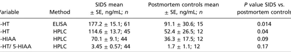
Table 2. Serum data for 5-HT and 5-HIAA (ng/mL) in SIDS cases compared with controls, adjusted for postconceptional age
Variable Method
SIDS mean
±SE, ng/mL;n
Postmortem controls mean
±SE, ng/mL;n
Pvalue SIDS vs.
postmortem controls
5-HT ELISA 177.2±15.1; 61 91.1±30.6; 15 0.014
5-HT HPLC 114.6±13.7; 45 52.4±26.5; 12 0.04
5-HIAA HPLC 70.1±9.1; 44 36.3±17.5; 12 0.09
5-HT/ 5-HIAA HPLC 3.45±0.57; 44 1.7±1.1; 12 0.17
All postmortem serum data are adjusted for postconceptional age at death.
MEDICALSCIENCES
5-HT measurements. In our analysis examining time of death and serum 5-HT, the Loess curves suggested that serum 5-HT was slightly higher in the morning (data not shown). The confidence intervals, however, reflected too much variation to permit conclusions of an effect. In the HPLC analysis of serum, levels of the 5-HT breakdown product 5-HIAA were also obtained. Although there was a numerical increase in 5-HIAA levels in SIDS cases compared with control cases, this increase did not reach statistical significance (P=0.09) (Table 2).
There was also no statistically significant difference in the ratio of 5-HT/5-HIAA between the two groups (Table 2).
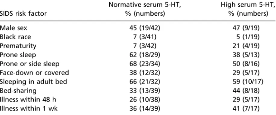
Definition of the SIDS Subset with Elevated Serum 5-HT.After finding the significant elevation in serum 5-HT in this SIDS cohort, we defined post hoc“high”serum 5-HT as greater than 2 SDs above the mean of the controls as determined by ELISA (≥211.8 ng/mL) and“normative”
serum 5-HT as below this cutoff (<211.8 ng/mL). The percent of SIDS cases with high serum 5-HT was 31% (19/61) (Fig. 1A). There were no controls above this cutoff level of serum 5-HT (Fig. 1A). The percent of SIDS cases with known SIDS risk factors was compared between SIDS cases with high serum 5-HT and those SIDS cases with nor- mative levels. There were no significant differences in risk factors for SIDS between the SIDS subset with high serum 5-HT levels (n=19) and the SIDS subset with normative 5-HT levels (n=42) (Table 3). The frequencies of male sex, prematurity, and discovery in the prone position were not significantly different between the two groups (Table 3). There was no significant difference in postmortem interval (P = 0.58) or length of storage interval (P=0.91) between SIDS cases with high and normative levels of 5-HT.
The 5-HTTLPR Genotype Association with Serum 5-HT Levels. In a second post hoc analysis, we tested the hypothesis that the high serum 5-HT subset was associated with the LL genotype of 5-HTTLPR. Of the SIDS cases, 58 out of 61 cases were geno- typed; all control cases were genotyped. Among SIDS cases and controls, there was no significant difference in the serum 5-HT values across the different genotypes (Table 4). In the SIDS cases, there was no significant difference in the frequency of
cases with high versus normative serum 5-HT levels within the different 5HTTLPR genotypes (Table 5).
Effect of Breastfeeding on Serum 5-HT Levels.There were no sig- nificant differences in serum 5-HT concentrations between (i) control cases who were breastfed vs. control cases who were not breastfed or (ii) SIDS cases who were breastfed vs. SIDS cases who were not breastfed (Table S1andFig. S1).
Serum 5-HT and 5-HIAA Levels in Living Control Infants.As measured by HPLC, living infant controls had higher 5-HT concentrations (300.8±25.0 ng/mL, mean±SE,n=40) than postmortem SIDS (114.6±13.7 ng/mL,n=45) and postmortem control infants (52.4± 26.5,n=12). As measured by HPLC, living infant controls had lower 5-HIAA concentrations (9.9±1.1 ng/mL,n=40) than postmortem SIDS (70.1±9.1 ng/mL,n =44) and postmortem control infants (36.3±17.5,n=12). There were no significant differences in serum values of 5-HT, 5-HIAA, and 5HT/5HIAA between living male in- fants (n=20) and living female infants (n=20).
Effect of Delay in Serum Separation on Serum 5-HT Concentrations.To assess the potential impact of a postmortem delay in serum sep- aration on serum 5-HT values, normal adult blood was collected using serum separator tubes identical to those used for the post- mortem cohort. Of note, it was logistically impossible to obtain IRB-approved, fresh serum samples from normal, living infants.
There was no significant difference in serum 5-HT concentration as determined by ELISA between serum separated immediately (346.3±56.9 ng/mL), at 12 h (319.7±98.7 ng/mL), 24 h (312.1± 102.1 ng/mL), or 36 h (297.6±54.8 ng/mL).
Discussion
This study provides evidence of a blood biomarker for peripheral 5-HT abnormalities in SIDS. We identified an elevation in serum 5-HT levels in SIDS cases compared with postmortem controls, with a subset of 31% of the SIDS cases with levels greater than 2 SDs above the mean of the controls. This study expands upon the brainstem evidence suggesting that SIDS is a 5-HT disorder related to an underlying vulnerability posited in the triple-risk Table 3. Frequency of cases with known SIDS risk factors within SIDS groups
defined by normative and high levels of 5-HT SIDS risk factor
Normative serum 5-HT,
% (numbers)
High serum 5-HT,
% (numbers)
Male sex 45 (19/42) 47 (9/19)
Black race 7 (3/41) 5 (1/19)
Prematurity 7 (3/42) 21 (4/19)
Prone sleep 62 (18/29) 38 (5/13)
Prone or side sleep 68 (23/34) 50 (8/16)
Face-down or covered 38 (12/32) 29 (5/17)
Sleeping in adult bed 66 (21/32) 59 (10/17)
Bed-sharing 33 (13/39) 44 (8/18)
Illness within 48 h 26 (10/38) 29 (5/17)
Illness within 1 wk 36 (14/39) 41 (7/17)
High serum 5-HT levels were defined as greater than 2 SDs above the mean of the control group. Risk factor information was not available on every case.
Table 4. Serum 5-HT values across different 5-HTTLPR genotypes in SIDS and controls
SIDS Controls
5HTTLPR genotype SS SL LL SS SL LL
n 11 27 20 2 4 9
Serum 5-HT±SD, ng/mL 169.5±134.4 157.4±105.9 189.6±146.1 96.6±59.8 78.23±60.9 100.2±63.86 ANCOVA controlling for diagnosis and postconceptional age;P=0.62 (not significant).
model (6). High serum 5-HT levels at autopsy may help to distin- guish the 5-HT-related vulnerability in infants with sudden, unex- pected death from those infants with similar presentation mimicking SIDS, but due to unsuspected infection, inborn errors of metabolism, trauma, and accidental asphyxia. The evidence of brainstem (13–16), and now peripheral, abnormalities in 5-HT pathway markers sug- gests a key role for 5-HT metabolism in SIDS.
Platelets avidly take up 5-HT from the bloodstream via the 5-HT transporter and store it in dense granules, where it is protected from the action of mitochondrial monoamine oxidases. Indeed,
∼95% of the 5-HT in blood is carried in platelet dense granules (18), and serum contains the secretion products of activated platelets, including the contents of their dense granules. An ex- tensive body of investigation has used platelets as a peripheral model of neuronal function in the central nervous system (20).
Multiple neurological disorders demonstrate functional abnor- malities in platelets, including Alzheimer disease, Parkinson dis- ease, various psychiatric disorders, and autism (21–27). The major forces driving these studies are the inherent difficulty of directly accessing brain function in vivo and the critical molecular simi- larities between platelets and central neurons. The presently reported data suggest that measurement of the platelet stores of 5-HT, via measurement of serum 5-HT, may be an accessible means of premortem identification of infants at risk for SIDS.
We did not find any associations of the high serum 5-HT levels in the SIDS cases with any previously known risk factors definable by autopsy and investigative reports in the cohort, including pre- maturity, sleep position when found at discovery, history of minor illness around the time of death, or with LL genotype of the 5-HT transporter (5-HTT) polymorphism. Known polymorphisms of the 5-HTT gene (SLC6A4) have been extensively studied in various disorders, including SIDS (28–30). The 5-HTTLPR is an insertion−
deletion polymorphism that consists of a repeated sequence of 22 to 23 base pairs that is present in 14 copies producing a short or“S”
allele or in 16 copies producing a long or“L”allele. The L allele and the LL genotype are associated with increased gene transcription and protein expression compared with the SS and SL genotypes (31, 32).
Although the association of the LL genotype on 5-HT uptake rate in platelets has been documented (33), the effect of this genotype on serum content of 5-HT is less well understood (33, 34). In the present study, the lack of association between the LL genotype and high values of serum 5-HT suggests that specific 5-HTTLPR genotypes, shown to influence 5-HTT expression levels (31, 32), do not con- tribute to the increased serum 5-HT in SIDS.
The cause(s) of the elevated serum 5-HT levels in the SIDS subset in this study remains unknown. One potential cause is over- production of peripheral 5-HT in SIDS without a proportional in- crease in breakdown. In the HPLC analysis, we found a nonsignificant trend toward an increase in the levels of 5-HIAA in SIDS cases compared with control cases, consistent with an increase in 5-HT peripheral breakdown; the lack of difference in the ratio of 5-HT/5-HIAA further suggests that the breakdown of 5-HT into 5-HIAA is proportionate with the increased 5-HT, and that there is no lack of breakdown of 5-HT to account for the increased serum 5-HT. Other potential sources of elevated serum 5-HT in- clude enterochromaffin (EC) cells of the gut and pulmonary
neuroendocrine cells (PNEC) and neuroepithelial bodies (NEBs) of the lung. Greater than 90% of the peripheral 5-HT is produced in the gut EC cells, and these cells are known to increase their se- cretion of 5-HT under various conditions, including hypoxia (35) and infection (36). PNEC and NEBs cells of the lung function as airway oxygen/carbon dioxide sensors and release 5-HT in a dose- dependent manner in response to hypoxia (37, 38). Although there have been no reports in SIDS of increased 5-HT production from EC cells of the gut, hyperplasia and hypertrophy of PNEC/
NEB within the lungs of infants dying of SIDS have been reported, and secretory products of these PNEC/NEB cells have been pro- posed previously as a potential biological marker of SIDS (38). The relationship in SIDS between increased serum 5-HT and gut/lung synthesis and release of 5-HT requires further research.
There are incomplete data on the maternal use of selective serotonin reuptake inhibitors (SSRIs) in our cohort. In any event, although gestational exposure to SSRIs appears to substantially reduce serum 5-HT in cord blood (39), physiologically meaningful SSRI exposure in infants through breastfeeding by mothers re- ceiving SSRIs is probably infrequent (40–42). Thus, previous studies of maternal SSRI treatment in breastfeeding mother−infant pairs have demonstrated (i) elevated levels of SSRI and major metabo- lites in the maternal plasma, but SSRI and major metabolites at or below the lower limit of quantitation in paired breastfed infants; and (ii) maternal SSRI use significantly decreases serum 5-HT levels in the mother, but does not affect serum 5-HT levels in paired breastfed infants (41, 42). In this study, there were no significant differences in serum 5-HT concentrations between (i) control cases who were breastfed vs. control cases who were not breastfed or (ii) SIDS cases who were breastfed vs. SIDS cases who were not breastfed. Thus, 5- HT in breast milk does not account for the differences in serum 5-HT observed between SIDS cases and controls.
A potential limitation of this study concerns the effects of postmortem interval, postmortem delay in serum separation, and lengthy storage times on the stability of the serum 5-HT. We did not, however, observe an effect of either postmortem interval or the time of serum storage on the levels of serum 5-HT as measured by two independent assays (ELISA and HPLC) (Fig. 1CandD), supporting the relative stability of the monoamine in postmortem samples stored under appropriate con- ditions. Likewise, we did not see an effect of delayed serum separation, albeit necessarily modeled using adult donor serum. A second limitation to this study is the relatively small number of control samples, a problem reflected in any postmortem infant study due to the difficulty in obtaining control tissue. Although all of our control infants died of definable acute disorders, the actual causes of death varied. Despite this variation among the controls, we were able to define a significant subset of SIDS cases with high serum 5-HT levels.
The presently reported 5-HT concentrations in healthy, living infants in the first year of life are data that are not readily available in the literature. Compared with postmortem infant SIDS and postmortem infant control samples, living infant controls had higher concentrations of serum 5-HT and lower concentrations of 5-HIAA. Likely explanations for the higher 5-HT and lower 5-HIAA in sera from living subjects compared with postmortem in- fants include that (i) the lower platelet counts in postmortem samples compared with living controls results in less 5-HT Table 5. Frequency of high or normal serum 5-HT values within each 5-HTTLPR
genotype in the SIDS cases
5-HTTLPR genotype High 5-HT, % (numbers) Normal 5-HT, % (numbers)
SS (n=11) 27 (3/11) 73 (8/11)
SL (n=27) 22 (6/27) 78 (21/27)
LL (n=20) 40 (8/20) 60 (12/20)
High serum 5-HT levels were defined as greater than 2 SDs above the mean of the control group.
MEDICALSCIENCES
available to be released from platelet dense granules into the se- rum, (ii) there is less catabolism of 5-HT to 5-HIAA in living subjects than occurs after death, and (iii) there are differences in plasma clearance rates of 5-HT and 5-HIAA. Because SIDS is, by definition, the sudden, unexplained death of an infant, we were unable to obtain serum samples from living infants who would subsequently die of SIDS, as there is currently no biomarker, biochemical or otherwise, to identify them. The present data do not therefore allow us to confirm that 5-HT levels are higher in living infants who will subsequently develop SIDS than in living infants who will not subsequently develop SIDS. To address this question, a large, long-term, prospective population study needs to be performed. Importantly, however, the present study does allow us to conclude that serum 5-HT is elevated in SIDS infants at postmortem, because we have the appropriate controls: serum from infants at postmortem who died of non-SIDS causes.
In summary, the available evidence of central and peripheral abnormalities in the serotonergic system in SIDS infants pro- vides essential information on a biological vulnerability in a subset of infants who die of SIDS. Further research will be necessary to validate, in an independent cohort, the present finding of increased serum 5-HT in SIDS and to determine the relationship or correlation between central and peripheral 5-HT abnormalities. A high serum 5-HT value may ultimately prove to be a peripheral biomarker of central 5-HT abnormalities in SIDS
cases, and its determination will be useful in identifying such SIDS infants, i.e., as an autopsy biomarker. Postmortem serum analysis of 5-HT by HPLC or ELISA is easier, less expensive, and more efficient than analyses of brain 5-HT neurochemistry, e.g., Western blotting and HPLC on micropunches of discrete and tiny brainstem nuclei. These techniques require frozen brain samples and oftentimes lengthy experiments (months for expo- sure) such as receptor ligand autoradiography. HPLC on Guthrie cards may be an option for future population screening, given that 5-HT has been reliably measured from dried blood spots (43). Further research aimed at understanding the association between the increased serum 5-HT reported here and the decreased brainstem 5-HT reported previously (14) will be essential for a deeper understanding of 5-HT abnormalities as a pathological mechanism underlying SIDS death.
ACKNOWLEDGMENTS.We thank Mark D. Kellogg, PhD, for his assistance, and the following people for critical reading of the manuscript in prepara- tion: Drs. Joseph J. Volpe, MD (Boston Children’s Hospital), Eugene E. Nattie, MD (Geisel School of Medicine), and Holcombe E. Grier, MD (Dana Farber Cancer Institute). This study was supported by the Eunice Kennedy Shriver National Institute of Child Health and Development Grants P01-HD036379 (to H.C.K.), R01-HD20991 (to H.C.K. and R.L.H.), and P30-HD18655 (Develop- mental Disabilities Research Center, Children’s Hospital Boston); Barrett Tallman Memorial Fund; CJ Foundation; Florida SIDS Alliance; Jacob Neil Boger Foundation for SIDS; Jason Lutz SIDS Foundation; and Robert’s Program on Sudden Unexpected Death in Pediatrics.
1. Willinger M, James LS, Catz C (1991) Defining the sudden infant death syndrome (SIDS): Deliberations of an expert panel convened by the National Institute of Child Health and Human Development.Pediatr Pathol11:677–684.
2. Willinger M, Hoffman HJ, Hartford RB (1994) Infant sleep position and risk for sudden infant death syndrome: Report of meeting held January 13 and 14, 1994, National Institutes of Health, Bethesda, MD.Pediatrics93:814–819.
3. American Academy of Pediatrics AAP Task Force on Infant Positioning and SIDS (1992) Positioning and SIDS.Pediatrics89:1120–1126.
4. Trachtenberg FL, Haas EA, Kinney HC, Stanley C, Krous HF (2012) Risk factor changes for sudden infant death syndrome after initiation of Back-to-Sleep campaign.Pediatrics129:
630–638.
5. Matthews TJ, MacDorman MF, Thoma ME (2015) Infant mortality statistics from the 2013 period linked birth/infant death data set.Natl Vital Stat Rep64:1–30.
6. Filiano JJ, Kinney HC (1994) A perspective on neuropathologic findings in victims of the sudden infant death syndrome: The triple-risk model.Biol Neonate65:194–197.
7. Kinney HC, Richerson GB, Dymecki SM, Darnall RA, Nattie EE (2009) The brainstem and serotonin in the sudden infant death syndrome.Annu Rev Pathol4:517–550.
8. Sridhar R, Thach BT, Kelly DH, Henslee JA (2003) Characterization of successful and failed autoresuscitation in human infants, including those dying of SIDS.Pediatr Pulmonol36:113–122.
9. Meny RG, Carroll JL, Carbone MT, Kelly DH (1994) Cardiorespiratory recordings from infants dying suddenly and unexpectedly at home.Pediatrics93:44–49.
10. Franco P, Szliwowski H, Dramaix M, Kahn A (1999) Decreased autonomic responses to ob- structive sleep events in future victims of sudden infant death syndrome.Pediatr Res46:33–39.
11. Schechtman VL, Lee MY, Wilson AJ, Harper RM (1996) Dynamics of respiratory pat- terning in normal infants and infants who subsequently died of the sudden infant death syndrome.Pediatr Res40:571–577.
12. Kahn A, et al. (1992) Sleep and cardiorespiratory characteristics of infant victims of sudden death: A prospective case-control study.Sleep15:287–292.
13. Paterson DS, et al. (2006) Multiple serotonergic brainstem abnormalities in sudden infant death syndrome.JAMA296:2124–2132.
14. Duncan JR, et al. (2010) Brainstem serotonergic deficiency in sudden infant death syndrome.JAMA303:430–437.
15. Kinney HC, et al. (2003) Serotonergic brainstem abnormalities in Northern Plains Indians with the sudden infant death syndrome.J Neuropathol Exp Neurol62:1178–1191.
16. Panigrahy A, et al. (2000) Decreased serotonergic receptor binding in rhombic lip- derived regions of the medulla oblongata in the sudden infant death syndrome.
J Neuropathol Exp Neurol59:377–384.
17. Amireault P, Sibon D, Côté F (2013) Life without peripheral serotonin: Insights from tryptophan hydroxylase 1 knockout mice reveal the existence of paracrine/autocrine serotonergic networks.ACS Chem Neurosci4:64–71.
18. Anderson GM, Feibel FC, Cohen DJ (1987) Determination of serotonin in whole blood, platelet-rich plasma, platelet-poor plasma and plasma ultrafiltrate.Life Sci40:1063–1070.
19. Paterson DS, et al. (2010) Lack of association of the serotonin transporter polymorphism with the sudden infant death syndrome in the San Diego Dataset.Pediatr Res68:409–413.
20. Gurguis NM (2007) Psychiatric disorders.Platelets, ed Michelson AD (Academic, Burlington, Ma.), pp 791–821.
21. Li QX, Master CL (2007) Alzheimer’s disease.Platelets, ed Michelson AD (Academic, Burlington, Ma.), pp 779–790.
22. Ferrarese C, et al. (1999) Reduced platelet glutamate uptake in Parkinson’s disease.
J Neural Transm (Vienna)106:685–692.
23. Reilmann R, Rolf LH, Lange HW (1997) Huntington’s disease:N-methyl-D-aspartate receptor coagonist glycine is increased in platelets.Exp Neurol144:416–419.
24. Hranilovic D, et al. (2009) Hyperserotonemia in autism: Activity of 5HT-associated platelet proteins.J Neural Transm (Vienna)116:493–501.
25. Chugani DC, et al. (1999) Developmental changes in brain serotonin synthesis capacity in autistic and nonautistic children.Ann Neurol45:287–295.
26. Cook EH, Leventhal BL (1996) The serotonin system in autism.Curr Opin Pediatr 8:348–354.
27. Mulder EJ, et al. (2004) Platelet serotonin levels in pervasive developmental disorders and mental retardation: Diagnostic group differences, within-group distribution, and behavioral correlates.J Am Acad Child Adolesc Psychiatry43:491–499.
28. Narita N, et al. (2001) Serotonin transporter gene variation is a risk factor for sudden infant death syndrome in the Japanese population.Pediatrics107:690–692.
29. Weese-Mayer DE, et al. (2003) Sudden infant death syndrome: Association with a promoter polymorphism of the serotonin transporter gene.Am J Med Genet A117A:268–274.
30. Nonnis Marzano F, et al. (2008) Genes regulating the serotonin metabolic pathway in the brain stem and their role in the etiopathogenesis of the sudden infant death syndrome.Genomics91:485–491.
31. Heils A, et al. (1996) Allelic variation of human serotonin transporter gene expression.
J Neurochem66:2621–2624.
32. Greenberg BD, et al. (1999) Genetic variation in the serotonin transporter promoter region affects serotonin uptake in human blood platelets.Am J Med Genet88:83–87.
33. Anderson GM, et al. (2002) Serotonin transporter promoter variants in autism: Func- tional effects and relationship to platelet hyperserotonemia.Mol Psychiatry7:831–836.
34. Pivac N, et al. (2009) The lack of genotype-phenotype relationship between platelet serotonin concentration and serotonin transporter gene promoter polymorphism in healthy subjects.Neurosci Lett462:45–48.
35. Haugen M, et al. (2012) Differential signal pathway activation and 5-HT function: The role of gut enterochromaffin cells as oxygen sensors.Am J Physiol Gastrointest Liver Physiol303:G1164–G1173.
36. Worthington JJ (2015) The intestinal immunoendocrine axis: Novel cross-talk between enteroendocrine cells and the immune system during infection and inflammatory disease.Biochem Soc Trans43:727–733.
37. Livermore S, et al. (2015) Pulmonary neuroepithelial bodies are polymodal airway sen- sors: Evidence for CO2/H+sensing.Am J Physiol Lung Cell Mol Physiol308:L807–L815.
38. Cutz E, Yeger H, Pan J (2007) Pulmonary neuroendocrine cell system in pediatric lung disease-recent advances.Pediatr Dev Pathol10:419–435.
39. Anderson GM, Czarkowski K, Ravski N, Epperson CN (2004) Platelet serotonin in newborns and infants: Ontogeny, heritability, and effect of in utero exposure to selective serotonin reuptake inhibitors.Pediatr Res56:418–422.
40. Epperson CN, Anderson GM, McDougle CJ (1997) Sertraline and breast-feeding.N Engl J Med336:1189–1190.
41. Epperson N, et al. (2001) Maternal sertraline treatment and serotonin transport in breast-feeding mother-infant pairs.Am J Psychiatry158:1631–1637.
42. Epperson CN, Jatlow PI, Czarkowski K, Anderson GM (2003) Maternal fluoxetine treatment in the postpartum period: Effects on platelet serotonin and plasma drug levels in breastfeeding mother-infant pairs.Pediatrics112:e425.
43. Saracino MA, Gerra G, Somaini L, Colombati M, Raggi MA (2010) Chromatographic analysis of serotonin, 5-hydroxyindolacetic acid and homovanillic acid in dried blood spots and platelet poor and rich plasma samples.J Chromatogr A1217:4808–4814.