Characterization of the genetic interactome of prohibitins in S. cerevisiae
I n a u g u r a l - D i s s e r t a t i o n
zur
Erlangung des Doktorgrades
der Mathematisch-Naturwissenschaftlichen Fakultät
der Universität zu Köln
vorgelegt von
Christof Osman aus Gräfelfing
Köln, 2009
Berichterstatter:
Professor Dr. Thomas Langer Professor Dr. R. Jürgen Dohmen Professor Dr. Dieter Wolf
Tag der mündlichen Prüfung: 26. November 2008
Für Anne, Elias und Linus
4
Abstract
Prohibitins comprise an evolutionary conserved and ubiquitously expressed family of membrane proteins that is essential for development in higher eukaryotes. Large ring complexes formed in the inner mitochondrial membrane by prohibitins regulate mitochondrial dynamics and function. Roles of prohibitins in cell signaling events across the plasma membrane and transcriptional regulation in the nucleus have been proposed as well. The molecular mechanism of prohibitin function, however, remains elusive. In contrast to higher eukaryotes, prohibitin-deficient yeast cells are viable and exhibit a reduced replicative life-span.
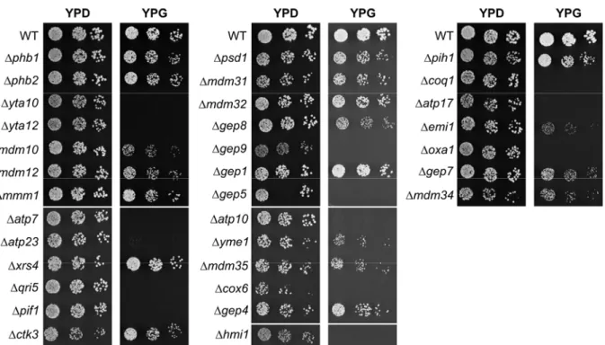
To investigate the functional role of prohibitins in yeast and to identify redundant processes that fulfill the functions of prohibitins in their absence, an unbiased genetic approach was chosen. Synthetic genetic arrays were applied to identify genes showing synthetic lethal interactions with prohibitins. This approach revealed 35 genes required for cell survival in the absence of prohibitins. The assembly of the FO-particle of the F1FO-ATP synthase was identified as one process essential in prohibitin-deficient cells.
Atp23 was characterized as a novel processing peptidase with a dual function in maturation of the mitochondrially encoded subunit Atp6 and its assembly into the functional F1FO-ATP synthase. ~50% of the genes required in prohibitin-deficient cells, including the strongest genetic interactions, are demonstrated for the first time to be required for mitochondrial phospholipid homeostasis. Evidence is provided that members of a conserved protein family, Ups1 and Gep1, coordinately regulate the levels of the non-bilayer forming phospholipids cardiolipin and phosphatidyl- ethanolamine in mitochondria. Additionally, an uncharacterized putative phosphatase was identified that is required for cardiolipin biosynthesis and might represent the last missing enzyme in the biosynthesis pathway of cardiolipin in yeast.
The genetic interactome of prohibitins defined in this thesis suggests that prohibitins serve scaffolding functions in the inner mitochondrial membrane and define functional
5
microdomains composed of proteins and non-bilayer forming lipids. This function becomes essential, when levels of cardiolipin and phosphatidylethanolamine are limiting in mitochondria. In the absence of prohibitins and decreased levels of non-bilayer forming lipids, essential processes dependent on these microdomains are compromised.
6
Table of contents
1 INTRODUCTION ... 9
1.1 Cellular compartmentalization ... 9
1.2 Import of mitochondrial proteins ... 10
1.3 F1FO-ATP synthase assembly ... 11
1.4 Mitochondrial quality control system ... 13
1.5 Phospholipids, the major components of mitochondrial membranes ... 15
1.6 Functional membrane domains ... 19
1.7 The SPFH-family of proteins ... 20
1.8 Prohibitins, proteins with a variety of functions ... 22
2 AIM OF THESIS ... 27
3 MATERIAL AND METHODS ... 28
3.1 Molecular Biology ... 28
3.2 Cloning procedures ... 28
3.3 Yeast genetic procedures ... 32
3.4 Yeast strains and growth conditions ... 33
3.5 Preparation of crude mitochondrial fractions ... 37
3.6 In organello labeling of mitochondrial translation products ... 38
3.7 Blue native PAGE analysis ... 38
3.8 Lipid analysis ... 39
7
3.9 Microscopy ... 41
3.10 Miscellaneous ... 41
4 RESULTS ... 42
4.1 A synthetic lethal screen to integrate prohibitins into a genetic network ... 42
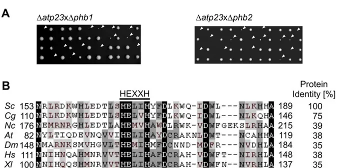
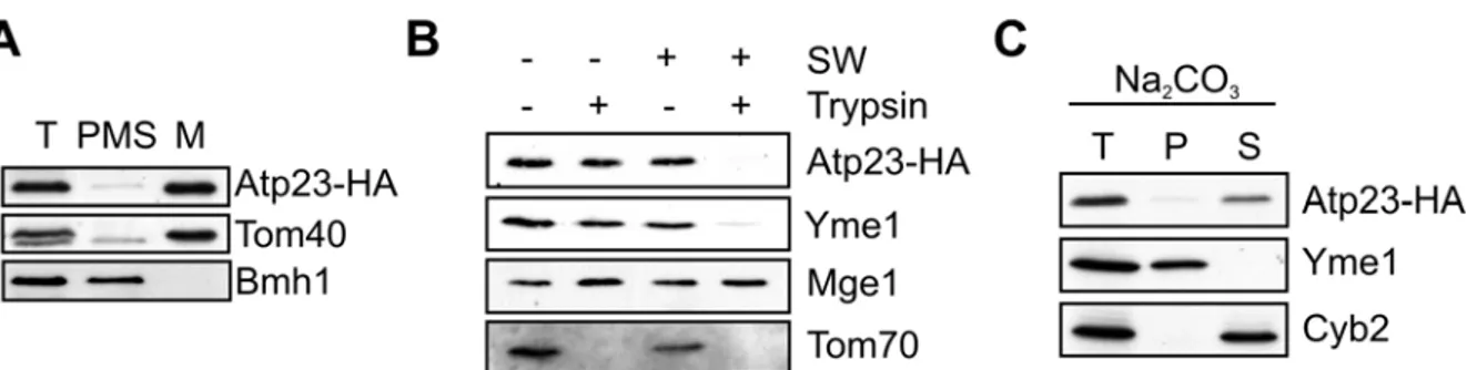
4.2 Atp23, a metallopeptidase in the mitochondrial intermembrane space ... 44
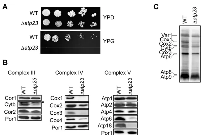
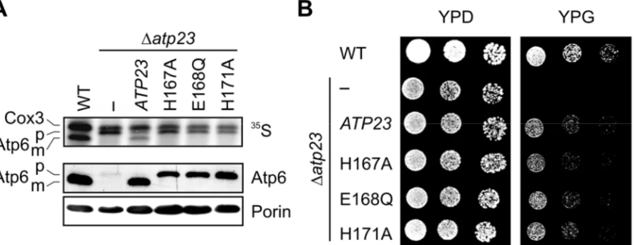
4.3 Atp23 mediates maturation of newly synthesized Atp6 ... 49
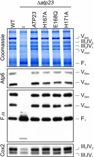
4.4 The assembly of the FO-particle of the ATP Synthase depends on Atp23 but not its proteolytic activity ... 51
4.5 Atp23 and Atp10 affect FO-assembly at a similar step ... 53
4.6 Is efficient respiratory chain assembly crucial in prohibitin-deficient cells? ... 55
4.7 Gep4, a putative phosphatase is essential for normal mitochondrial cardiolipin levels ... 57
4.8 Gep1, an uncharacterized open reading frame synthetically lethal with prohibitins ... 61
4.9 Mitochondrial inner membrane integrity depends on Gep1 and prohibitins ... 63
4.10 Gep1-like proteins are localized to the intermembrane space of mitochondria ... 68
4.11 Gep1 is required for normal mitochondrial phosphatidylethanolamine levels ... 70
4.12 Gep1 function is evolutionary conserved ... 73
4.13 Gep1 is required for the stability of mitochondrially synthesized phosphatidylethanolamine ... 75
4.14 Lipid-specific functions of Gep1-like proteins for phosphatidylethanolamine and cardiolipin ... 78
4.15 Survival of prohibitin-deficient cells depends on the lipid composition of mitochondrial membranes ... 81
5 DISCUSSION ... 83
8
5.1 Maintenance of mitochondrial PE and CL levels is essential in prohibitin-deficient cells .. 83
5.2 Prohibitins as membrane scaffolds in the inner mitochondrial membrane ... 85
5.3 Gep4, a putative phosphatase regulating CL biosynthesis ... 87
5.4 Gep1-like proteins are novel regulators of mitochondrial phospholipid composition ... 88
5.5 Prohibitins and the assembly of FO-particle of the F1FO-ATP synthase ... 93
5.6 Atp23, a novel processing peptidase and chaperone for the F1FO-ATP synthase ... 95
6 ZUSAMMENFASSUNG ... 99
7 REFERENCES ... 101
8 LIST OF ABBREVIATIONS ... 118
9 APPENDIX ... 120
10 ACKNOWLEDGEMENT ... 121
11 EIDESSTATTLICHE ERKLÄRUNG ... 122
12 LEBENSLAUF ... 123
9
1 Introduction
1.1 Cellular compartmentalization
One of the key steps for life to become possible was the first sequestration of organic molecules that allowed the formation of a certain organization protected against the exterior environment. This very day the formation of aqueous compartments is still one of the most fundamental principles for the development of living cells. Besides a plasma membrane that demarcates a cell from the surrounding environment, eukaryotic cells have evolved an intricate system of intracellular compartments that separate and enable processes with complete distinct biochemical requirements. One of these specialized compartments is represented by the mitochondrium that distinguishes itself from other organelles (excluding chloroplasts) as it is surrounded by two different cellular membranes that originate from an endosymbiotic event between a eukaryotic cell and an α-proteobacterium (Wallace, 2005). Four different compartments can thus be distinguished in mitochondria: the outer mitochondrial membrane, the intermembrane space, the cristae forming inner mitochondrial membrane and the matrix space. The best known role of mitochondria is their function in the generation of cellular ATP by oxidative phosphorylation that is accomplished by high molecular weight multisubunit complexes in the inner membrane. Besides this, mitochondria also harbour the Krebs cycle, enzymes required for heme and Fe/S cluster biosynthesis as well as the metabolism of certain amino acids. Furthermore, mitochondria also exert crucial functions during programmed cell death and calcium signaling (Chan, 2006; McBride et al., 2006). Consistent with roles of mitochondria in all these processes, a variety of human disorders have been linked to mitochondrial dysfunction (Chan, 2006; Wallace, 2005). It has to be noted that mitochondria are essential organelles in all species (Lill and Kispal, 2000). Therefore, any severe perturbation of mitochondrial biogenesis impacts dramatically on the viability of the whole cell.
The traditional textbook view of mitochondria as bean-shaped and static organelles was renewed by studies in the last years, which show that mitochondria can adopt a variety
10
of different shapes in different cell types. Additionally, mitochondria constantly move and undergo fusion and fission events that are required for maintenance of mitochondrial morphology and inheritance. Mitochondria need to ensure the coordinated fusion or fission of the outer and the inner membrane. Consistently, elaborated molecular machines have evolved and several conserved components have been identified in the past years that mediate these steps (Hoppins et al., 2007). The importance of the regulation of mitochondrial dynamics is highlighted by the observation that defects in this process have been linked to a variety of diseases (Detmer and Chan, 2007).
1.2 Import of mitochondrial proteins
While compartmentalization is crucial for living cells, it also imposes the problem on cells to ensure import of proteins that are synthesized in the cytosol to their sites of action. The import of mitochondrial proteins has been extensively studied in yeast in the recent years and signal sequences and protein complexes required for the import of proteins into the four different mitochondrial compartments have been identified (Becker et al., 2008; Bolender et al., 2008). Proteins destined for mitochondria are synthesized on cytosolic ribosomes and are recognized by receptors at the outer membrane (Abe et al., 2000; Young et al., 2003). Subsequently, all mitochondrial proteins are transported through the outer membrane import pore, termed the TOM complex (Becker et al., 2005). At that stage, import routes diverge depending on the final localization of the protein to be imported. Many proteins resident in the outer mitochondrial membrane are released from the TOM complex into the lipid bilayer (Gabriel et al., 2003). For a different set of proteins that form β-barrel structures in the outer membrane the action of another complex is required, the SAM complex (Wiedemann et al., 2003). Inner membrane and matrix proteins are in contrast further transferred to the presequence translocase complex (TIM23 complex) in the inner membrane (Chacinska et al., 2005).
Proteins taking this route typically have a positively charged N-terminal leader sequence that is responsible for recognition of the protein at the outer membrane and also for further import by the TIM23 complex. The energy that is required for import is derived from two sources. The membrane potential, formed by the respiratory chain, exerts an
11
electrophoretic effect on the positively charged presequence. Additionally, ATP is used by another complex, the presequence translocase-associated motor (PAM) complex, to either trap the imported protein in the matrix or to actively exert a pulling force (Krayl et al., 2007). Multispanning membrane proteins are integrated into the inner membrane by the help of yet another complex, termed TIM22 complex (Rehling et al., 2003). This import pathway is also dependent on the membrane potential. A fourth import route was identified recently that ensures efficient import of proteins into the intermembrane space. This pathway involves the reversible formation of disulfide bonds between components of the import machinery and the imported protein (Chacinska et al., 2004;
Mesecke et al., 2005).
1.3 F1FO-ATP synthase assembly
In the course of evolution, the majority of the mitochondrial genome was lost or transferred to the nuclear DNA and only a small percentage of the genome, encoding for eight or thirteen proteins in S. cerevisiae or humans, respectively, is still present in mitochondria. Consequently, mitochondria still have transcription and translation machineries for the expression of mitochondrial genes (Chen and Butow, 2005). The respiratory chain complexes and the F1FO-ATP synthase in the inner membrane are composed of proteins that are encoded in the nuclear and the mitochondrial genome.
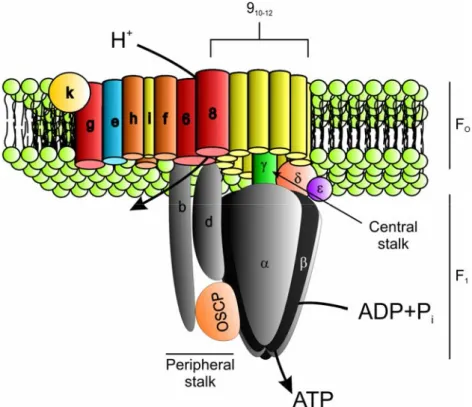
Therefore, mitochondria not only have to ensure correct import of nuclearly encoded proteins but they also have to coordinate this import with the expression of mitochondrially encoded genes. This is exemplified by the F1FO-ATP synthase, a multisubunit complex in the inner membrane that utilizes the electrochemical gradient, formed by the respiratory chain complexes, for the generation of ATP. The functional F1FO-ATP synthase in yeast is composed of at least 21 different structural subunits of which three are mitochondrially encoded (Velours and Arselin, 2000). The complex can be structurally separated into the membrane embedded FO-sector and the hydrophilic F1-particle (Figure 1). The FO-part contains the proton transducing channel and a peripheral stator stalk. The F1-part contains a central stalk and the catalytic headpiece responsible for the catalysis of the reversible formation of ATP from ADP+Pi. For ATP synthesis, protons are translocated through the FO-sector and this results in the rotation
12
of the central stalk, which sequentially interacts with the six subunits constituting the headpiece, which causes conformational changes that result in the synthesis of ATP (Nakamoto et al., 1999). During this process the headpiece is fixed to the peripheral stator stalk to avoid futile rotation (Nakamoto et al., 1999).
The FO- and the F1-part appear to have the ability to assemble in the absence of the respective other one and both have their own set of assembly factors that act at different steps, ranging from gene transcription to the final assembly steps (Ackerman and Tzagoloff, 2005). While henceforth nine genes were identified to facilitate steps at mtDNA or RNA levels, only four genes function on the protein level. Atp11 and Atp12 are two proteins that are required for proper assembly of the F1-part. Current models suggest that Atp11 and Atp12 maintain the unassembled subunits of the F1-headpiece F1β and F1α, respectively, in an assembly-competent state (Ackerman, 2002).
Figure 1 Schematic model of the F1FO-ATP synthase of S. cerevisiae (Adapted from Ackerman and Tzagoloff, 2005)
13
The knowledge about the biogenesis of the membrane embedded FO-moiety is likewise limited. Atp6, Atp8 and Atp9 are mitochondrially encoded subunits that form the core structure of the proton transducing channel and are believed to be inserted into the inner membrane co-translationally, but evidence for the responsible proteins mediating this insertion is lacking. Interestingly, yeast Atp6 is synthesized with a 10 amino acid long extension at the N-terminus, which is cleaved off following its integration into the inner membrane (Michon et al., 1988). The peptidase responsible for this maturation, however, has not been identified to date. Subsequent, to its integration into the membrane Atp9 has the ability to self-assemble into a ring structure (Arechaga et al., 2002). The next step of assembly is chaperoned by Atp10 that binds Atp6 and assists its assembly with the Atp9 ring (Tzagoloff et al., 2004). This step needs to be tightly controlled, as imprecise assembly could lead to proton leakage through the proton channel formed by the Atp9 oligomer and Atp6. How subsequent assembly processes are coordinated, specifically at what step the proton transducing channel assembles with the peripheral stalk and the F1-moiety and whether this is under the control of specific proteins is currently unknown.
1.4 Mitochondrial quality control system
The above described considerations point out that biogenesis of mitochondrial protein complexes is a highly intricate process that requires assistance by a multitude of proteins. Removal of misfolded or damaged proteins by mitochondrial proteases is of high importance to maintain integrity of mitochondrial membranes. Many proteases have been identified in the proteome of mitochondria and it became evident that the function of these proteins is not restricted to control of protein quality but that they also play essential roles in regulatory processes. Most of the proteases identified in the mitochondrial proteome have been studied and can be grouped into three different classes: Processing peptidases, ATP-dependent proteases and oligopeptidases (Koppen and Langer, 2007). Several other proteases, though, still await their assignment into one of these functional classes.
14
Many mitochondrial proteins that are synthesized in the nucleus possess N-terminal signal sequences that ensure correct targeting, but are not required for the function of the proteins. These sequences are removed by the hetero-dimeric mitochondrial processing peptidase (MPP), a metallopeptidase in the mitochondrial matrix (Gakh et al., 2002). Subsequently, many proteins are further processed by the mitochondrial intermediate peptidase (MIP) that removes an octapeptide from the pre-proteins. The inner membrane protease IMP exposes its catalytic domains to the intermembrane space and is required for maturation of a variety of pre-proteins that are localized in this mitochondrial compartment (Gakh et al., 2002). A processing peptidase with a regulatory role is represented by the rhomboid protease Pcp1 in the inner membrane that cleaves polypeptide bonds within the membrane bilayer (McQuibban et al., 2003).
Two substrates of Pcp1 have been identified in yeast: Mgm1, a GTPase required for mitochondrial fusion, is processed by Pcp1 which results in the generation of a short isoform. Processing of the reactive oxygen scavenger protein Ccp1, the other known substrate of Pcp1, is mediated by the help of the m-AAA protease that dislocates Ccp1 through the inner membrane to expose the target sequence to the catalytic center of Pcp1 (Esser et al., 2002; Tatsuta et al., 2007).
Members of the second class of proteases, the ATP-dependent proteases, have been identified in all mitochondrial compartments with the exception of the outer membrane.
The homo-oligomeric PIM1 protease is present in the mitochondrial matrix (Van Dyck et al., 1994). The m-AAA protease and the i-AAA protease are anchored in the inner membrane and expose their catalytic centers to the matrix or the intermembrane space, respectively (Langer, 2000). Hydrolysis of ATP is used by these proteases for the unfolding of substrates or the extraction of proteins from membranes that has to precede degradation. Yeast cells devoid of either of these proteases display severe phenotypes (Van Dyck and Langer, 1999). The nature of all these phenotypes is, however, not fully understood to date, but they appear to reflect two roles of these proteases. On one hand, ATP-dependent proteases remove damaged or otherwise harmful proteins and on the other hand, they exert important regulatory activities that are indispensable for mitochondrial biogenesis (Koppen and Langer, 2007; Nolden et
15
al., 2005). Oligopeptidases represent a third class of mitochondrial proteases that encompasses members localized to the matrix and the intermembrane space of mitochondria. These enzymes function in the degradation of oligopeptides that are generated by processing peptidases and ATP-dependent proteases (Kambacheld et al., 2005).
1.5 Phospholipids, the major components of mitochondrial membranes Research on the import of proteins into the various mitochondrial compartments, their assembly into functional complexes and their degradation has identified a plethora of components that facilitate all possible steps in these processes. Consequently, a fairly well defined picture has emerged. This, however, only represents half of the story of mitochondrial biogenesis. Phospholipids that are the major components of mitochondrial membranes have to be synthesized and assembled into functional membranes as well.
Elucidation of these processes, however, has attracted less interest in the past years and lags behind (Voelker, 2004). Mitochondria, like other cellular membranes harbour many more lipid species than would be required for simply establishing water impermeable barriers. All these species have distinct properties that serve distinct functions, of which only the minority has been elucidated (van Meer et al., 2008).
The spectrum of mitochondrial lipids in S. cerevisiae comprises glycerophospholipids and to a minor extent the sterol ergosterol (Zinser and Daum, 1995). In contrast, sphingolipids that are present in other cellular membranes do not occur in mitochondria.
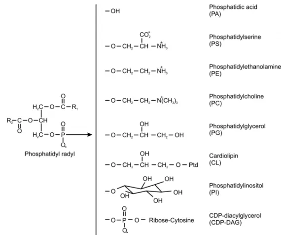
Phosphatidic acid is the basic building block of glycerophospholipids and consists of a glycerol backbone whose three hydroxyl groups are esterified with two fatty acids and a phosphate. Different species of these phospholipids are defined by the attachment of different head groups to the phosphate, which comprise choline, ethanolamine, serine, inositol and glycerol (van Meer et al., 2008) (Figure 2). Additionally, mitochondria possess cardiolipin, a phospholipid that is not found elsewhere in the cell and is somewhat special in its architecture as it contains two phosphatidic acid moieties that are held together by a glycerol bridge (Schlame, 2008).
16
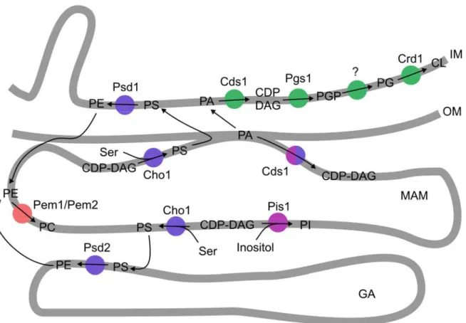
Phosphatidylcholine (PC) and phosphatidylethanolamine (PE) account for ~70% of total mitochondrial phospholipids with roughly equal distributions between outer and inner mitochondrial membranes. The main synthesis pathway starts for both phospholipids in a specialized fraction of the endoplasmic reticulum, known as the MAM (mitochondria associated membrane) compartment, where the phosphatidylserine synthase Cho1 synthesizes phosphatidylserine (PS) from serine and CDP-diacylglycerol (CDP-DAG) (Achleitner et al., 1999; Gaigg et al., 1995) (Figure 3). Two PS-decarboxylases have been described in S. cerevisiae that are capable of converting PS into PE. None of these enzymes is localized in the endoplasmic reticulum and therefore PS needs to be transported to either the inner mitochondrial membrane or the Golgi apparatus to be decarboxylated by Psd1 or Psd2, respectively (Clancey et al., 1993; Trotter et al., 1993;
Figure 2 Representation of mitochondrial phospholipids. A glycerol backbone is esterified to two fatty acids (R1 and R2) that can vary in length and saturation. Different head groups are attached to the glycerol backbone via a phosphodiester, which gives rise to different phospholipids. (Adapted from Voelker, 2004)
17
Trotter and Voelker, 1995). After this step, PE is either transported to its final target membrane or subjected to further modifications that result in PC formation. In this case, PE again requires retranslocation to the endoplasmic reticulum, where the final methylation reactions for the production of PC can take place (Kuchler et al., 1986;
Zinser et al., 1991). Once synthesized, PC has to be shuttled to membranes of all cellular compartments. Besides this described biosynthesis pathway, PE and PC can also be formed via the so-called Kennedy pathway, where exogenously added ethanolamine or choline is linked to DAG by a different set of enzymes (Kent, 1995).
This pathway, however, contributes very little to the mitochondrial phospholipid supply (Birner et al., 2001).
The third most abundant lipid within mitochondria is cardiolipin, which is preferentially localized to the inner membrane, but small amounts were also identified in the outer membrane (Schlame, 2008). Cardiolipin has been recognized to be required for a variety of different functions in yeast that include aging and apoptosis, mitochondrial protein import, mitochondrial bioenergetics, translational regulation and cell wall biogenesis in yeast (Joshi et al., 2008). Consequently, cardiolipin and its impact on several cellular processes are being studied extensively. For its synthesis, phosphatidic acid synthesized at the outer mitochondrial membrane, needs to cross the intermembrane space to be activated by Cds1 in the inner mitochondrial membrane to CDP-DAG (Kuchler et al., 1986), which together with glycerol-3-phosphate (G3P) in turn is converted to phosphatidylglycerolphosphate (PGP) by the enzyme Pgs1 (Chang et al., 1998a) (Figure 3). Dephosphorylation by a yet unidentified enzyme yields phosphatidylglycerol (PG). Finally, the cardiolipin synthase Crd1 catalyzes the synthesis of CL from PG and CDP-DAG (Chang et al., 1998b; Tuller et al., 1998). CL is afterwards subjected to postsynthetic modifications that lead to substantial changes in the acylchain composition. The best characterized enzyme involved in this remodeling process is a transacylase localized in the outer mitochondrial membrane and the outer leaflet of the inner membrane, which is termed Taz1 (Claypool et al., 2006).
Considerable interest in Taz1 has recently arisen, when it was demonstrated that the human homolog is frequently mutated in patients suffering from Barth Syndrome, a
18
disease characterized by cardiomyopathy, neutropenia and muscle weakness (Bione et al., 1996). The biosynthesis of phosphatidylinositol (PI) requires fewer steps than the CL synthesis. In a reaction that is similar to the production of PS by Cho1, inositol is attached to CDP-DAG in the MAM compartment (Gaigg et al., 1995). Like other lipids, PI requires transport to various target membranes within the cell.
As it is evident from the cellular localization of the enzymes involved in the biosynthesis of different phospholipid species, several translocation steps are required for efficient supply of different cellular organelles with lipids. While most organelles are connected to the vesicular transport system, which represents a way to also distribute lipids besides
Figure 3 Phospholipid synthesis in S. cerevisiae. Enzymes required for CL, PE, PC and PI synthesis are shown in green, blue, red and violet, respectively. The synthesis of PA by a two-step acylation of glycerol- 3-phosphate is not shown. Transport routes of PI and PC or PE, synthesized in the Golgi apparatus, to mitochondria are not indicated. IM – inner mitochondrial membrane, OM – outer mitochondrial membrane, MAM – mitochondrial associated membranes (subfraction of the endoplasmic reticulum), GA – Golgi apparatus (for phospholipid abbreviations see figure 2) (Adapted from Voelker, 2004)
19
proteins into various cellular compartments, mitochondria have to ensure their lipid supply by other means. In yeast this process is poorly understood and only one protein, namely Met30, has been demonstrated to be important for import of phospholipids into mitochondria. Met30 is an F-box protein and a subunit of an ubiquitin ligase complex (Schumacher et al., 2002). At what stage ubiquitination plays a role during import of lipids into mitochondria or if Met30 has a role independent of its function in the ubiquitin ligase complex, however, remains unresolved (Choi et al., 2006a). Similarly, it is not known how lipids cross the intermembrane space and if this is protein assisted or regulated to any extent. Another empty spot in our understanding of mitochondrial phospholipid homeostasis regards the regulation of the specific lipid composition of mitochondrial membranes that varies significantly under different growth conditions. It is unclear, whether this is determined exclusively on the level of transcription of genes encoding for enzymes required for lipid biosynthesis or also at later steps. Furthermore, the contribution of lipid turnover to mitochondrial lipid composition has not been studied extensively to date and awaits identification of components that mediate or regulate this step.
1.6 Functional membrane domains
Cellular membranes have long been looked at as homogenous fluids, where different lipids are equally distributed. This view, however, has been challenged with the proposal of functional domains within a membrane that are capable of separating processes occurring in the same membranes. Initially, the idea of a lipid raft was put forward to explain the differential sorting of proteins and lipids to the plasma membrane in morphologically polarized cells, where apical and basal membrane differ in their protein and lipid composition. This model proposes crucial roles for cholesterol and sphingolipids in forming membrane clusters destined for the apical membrane in the Golgi apparatus, which are then recognized as a unit and sorted to the target membrane by the cellular trafficking system (Simons and van Meer, 1988; van Meer and Simons, 1988). This idea was supported, when it was demonstrated that such domains survive extraction with the detergent Triton-X100 at 4°C, which raised the term
“detergent resistant membranes” (DRM) (Brown and Rose, 1992). Additional evidence
20
was derived from experiments where cells were deprived of cholesterol, which prevented lipid raft isolation (Cerneus et al., 1993). Although, insolubility with Triton- X100 and cholesterol depletion have been used in a number of experiments to investigate lipid rafts, it is still a matter of debate, whether insights obtained with these approaches truly reflect the physiological state of a cell or whether these methods are prone to experimental artifacts (Heerklotz, 2002; Munro, 2003). Nevertheless, successful reconstitution of lipid rafts in model membranes and sophisticated optical microscopy based approaches that allow the direct detection of lipid rafts in living cells, greatly support the existence of lipid rafts (Brown and London, 1998; Dietrich et al., 2001; Jacobson et al., 2007; Samsonov et al., 2001). Questions still remain regarding the physiological relevance of lipid rafts as the latest models envision lipid rafts as almost invisible entities that are extremely small and short-lived (Edidin, 2003; Shaw, 2006; Simons and Vaz, 2004).
The current available methods have been extensively used for the characterization of lipid rafts and this has led to the identification of several proteins that are selectively enriched in these microdomains. The most studied protein is represented by caveolin that is responsible for the formation of characteristic structures known as caveolae (Thomas and Smart, 2008). Other examples are GPI-anchored proteins, acylated proteins, cholesterol or phospholipid binding proteins and heterotrimeric G proteins (Rajendran and Simons, 2005). Lipid rafts harbouring these proteins were demonstrated to play important roles in caveolae mediated endocytosis, non-caveolar internalization, sorting in polarized epithelial cells, virus budding or immune receptor signaling (Rajendran and Simons, 2005). Common to the function of lipid rafts in all these processes is the idea that they compartmentalize cellular processes in a membrane that leads to a concentration or segregation of specific proteins.
1.7 The SPFH-family of proteins
A group of proteins that share the same domain, termed SPFH-domain (named after the founding members Stomatin, Prohibitin, Flotillin and HflK/HflC) were repeatedly found in DRM’s (Langhorst et al., 2005; Morrow and Parton, 2005). ~1800 proteins
21
harbouring this domain were identified in species ranging from prokaryotes to higher eukaryotes by computational analysis (Browman et al., 2007; Huber et al., 2006).
Interestingly, it was recently reported that the SPFH-domain emerged independently in different proteins, which suggests that it plays a role in a membrane process of fundamental importance (Rivera-Milla et al., 2006). Proteins belonging to the SPFH- family share a few characteristics (Browman et al., 2007): First, all proteins studied to date are membrane attached, although some variations in the exact membrane association are evident. Second, SPFH-proteins form large homooligomeric or heterooligomeric complexes. Third, members of the SPFH-family appear to be lipid raft associated. Despite these similarities, cellular localization and proposed functions clearly vary among these proteins. Flotillins, stomatins and erlins, all members of the SPFH-family, have been studied in some detail and the current knowledge is summarized below.
The term flotillins is used for two highly homologous proteins, named flotillin-1 and flotillin-2 that are localized to the plasma membrane and endosomes (Lang et al., 1998;
Morrow et al., 2002). Both proteins have been identified in DRM’s and since then are commonly used as marker proteins for lipid microdomains (Lang et al., 1998). Many different roles have been attributed to both proteins that include signal transduction, vesicle trafficking and cytoskeletal rearrangement (Morrow and Parton, 2005). Recently, strong evidence has been provided that supports a function of flotillin-1 in a novel endocytosis pathway that is independent of the two known endocytosis mediating proteins clathrin and caveolin (Glebov et al., 2006). A different idea is emerging for the function of members of the stomatin-family within the SPFH-superfamily that encompasses podocin, stomatin and stomatin-like proteins (e. g. SLP-3, MEC2). All proteins have been localized to the plasma membrane (Huber et al., 2006; Morrow and Parton, 2005; Wetzel et al., 2007). The common model at this state of investigations ascribes a function to stomatins in the regulation of ion-channels that appears to be required for mechanosensation in several cases (Gillespie and Walker, 2001; Huber et al., 2006; Wetzel et al., 2007). Strikingly, it was demonstrated for MEC-2 and podocin that this function depends on the ability of these proteins to bind cholesterol that in turn
22
depends on a conserved proline residue in the N-terminal part of these proteins (Huber et al., 2006). The latest contribution to the SPFH-family of proteins was the identification of the two human proteins erlin-1 and erlin-2 that were recovered from DRM’s (Browman et al., 2006). In contrast to the flotillins and stomatins, erlin-1 and erlin-2 are localized in the endoplasmic reticulum, which is at odds with the lipid raft definition that assumes a requirement for cholesterol and sphingolipids in lipid rafts. These lipids are, however, low abundant in the endoplasmic reticulum. These observations prompted the authors to conclude that lipid rafts are also present in the endoplasmic reticulum in the absence of large quantities of cholesterol and sphingolipids.
1.8 Prohibitins, proteins with a variety of functions
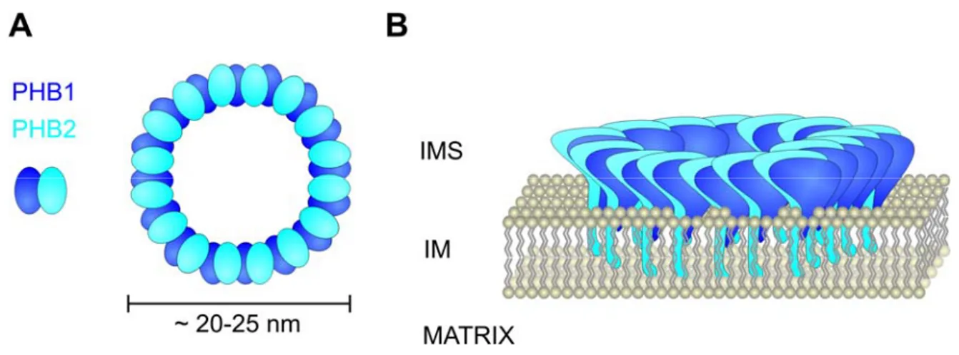
Further members of the SPFH-family are the prohibitins that encompass two proteins known as Phb1 and Phb2. These proteins are introduced in more detail below, because of their central role in the present thesis. Phb1 and Phb2 are homologous proteins that are evolutionary conserved and were identified in most eukaryotic species including plants (Merkwirth and Langer, 2008). Prohibitins are membrane attached proteins that form higher order oligomers. The N-termini of both proteins display hydrophobic regions that are required for membrane anchorage while C-terminal coiled-coil regions, present in both proteins, mediate hetero-oligomerization. A ring-shaped high molecular weight complex that is formed by Phb1 and Phb2 has recently been successfully purified and analyzed by single particle electron microscopy (Tatsuta et al., 2005). In this complex 16-20 subunits of each Phb1 and Phb2 are arranged in an alternative manner, which is supported by an interaction of both proteins that was demonstrated in yeast and mammalian cells (Coates et al., 1997; Steglich et al., 1999) and by crosslinks exclusively detected between different prohibitin subunits (Back et al., 2002) (Figure 4).
Moreover, both proteins are interdependent and depletion of one protein results in the degradation of the respective other one in various organisms, which is in accordance with a quantitative assembly of both proteins observed in immune depletion experiments (Artal-Sanz et al., 2003; Berger and Yaffe, 1998; Coates et al., 2001;
Kasashima et al., 2006). Hence, the prohibitin complex represents the physiologically active structure.
23
Two characteristics that are shared by SPFH-family members, namely membrane attachment and the formation of heterooligomeric complexes, also hold true for prohibitins. Lipid raft association that was demonstrated for various members of the SPFH-family, however, is currently less obvious for prohibitins. The bulk of information present supports a mitochondrial localization of prohibitins that is incompatible with the classical view of lipid rafts. According to this, the formation of lipid rafts depends on the presence of cholesterol and sphingolipids, two lipids that are either very low abundant or completely absent in mitochondria, respectively (Zinser and Daum, 1995).
Studies in yeast exclusively support a mitochondrial localization of prohibitins. The targeting signals of Phb1 and Phb2 have been identified and reside in the N-terminal part of both proteins (Tatsuta et al., 2005). Neither of the proteins is processed upon import (Nijtmans et al., 2000). Phb2 harbours a bipartite targeting signal that consists of an amphipathic helix and a hydrophobic domain, reminiscent of other intermembrane space proteins. Phb1, in comparison, does not display any characteristics of known mitochondrial targeting signals but, nevertheless, the N-terminal region is sufficient to target a hybrid protein to mitochondria (Tatsuta et al., 2005). Studies on the submitochondrial localization show that the prohibitin complex is anchored to the inner
Figure 4 Schematic representation of the prohibitin complex A Dimers of Phb1 and Phb2 are the building blocks of the prohibitin complex. Heterodimers assemble into ring-like complexes. B The prohibitin complex is anchored to the inner mitochondrial membrane via N-terminal hydrophobic stretches. The C- terminal domains are exposed to the intermembrane space. (Courtesy of Carsten Merkwirth)
24
membrane and exposes large domains into the intermembrane space (Berger and Yaffe, 1998; Steglich et al., 1999).
Despite extensive effort, the function of the prohibitin complex in mitochondria is not well understood, which is in part explained by the very mild phenotype of yeast cells lacking prohibitins that affects replicative life-span. Insights were provided by experiments that identified prohibitins as binding partners of the m-AAA protease in the inner membrane (Steglich et al., 1999). Interestingly, absence of prohibitins results in an accelerated degradation of substrates by the m-AAA protease suggesting an inhibitory function of prohibitins on m-AAA protease function (Steglich et al., 1999). This finding is reminiscent of the prokaryotic AAA-protease homolog FtsH that is regulated by the SPFH-proteins HflK and HflC (Kihara et al., 1996; Kihara et al., 1997). In addition to the biochemical interaction, synthetic lethal interactions between PHB1 or PHB2 and the genes encoding for the subunits of the m-AAA protease exist (Steglich et al., 1999).
This suggests that prohibitins have functions beyond regulation of the m-AAA protease, because it appears counterintuitive that absence of a regulatory factor has an effect in the absence of the protein it regulates.
Genetic approaches have linked prohibitins also to other mitochondrial processes. It was demonstrated that prohibitins are essential in the absence of proteins encoded by MMM1, MDM10 and MDM12 (Berger and Yaffe, 1998). These proteins were initially identified to be essential for mitochondrial morphology maintenance (Berger et al., 1997; Burgess et al., 1994; Sogo and Yaffe, 1994). The function in this process, however, remained elusive. More recent work, suggests that disturbance of mitochondrial morphology in mutants lacking these proteins is a secondary consequence from defects in the primary function, which is the assembly of β-barrel proteins in the outer membrane together with the SAM complex (Meisinger et al., 2007).
Yet another function has been linked to prohibitins, which is mitochondrial phospholipid metabolism. Psd1, required for PE biosynthesis in the inner membrane, is another protein essential in the absence of prohibitins (Birner et al., 2003). Although these genetic analyses provide first starting points for the investigation of the functional
25
process where prohibitins are involved, it is currently not possible to pinpoint the function of these proteins.
The current status of investigations in mammalian cells adds another degree of complexity to the field. Starting with the localization, prohibitins have been shown to be residents in the plasma membrane, the nucleus or mitochondria in mammalian cells (Fusaro et al., 2003; Kolonin et al., 2004; Merkwirth et al., 2008). A model uniting all these localizations is currently not available. Very recent work has closely examined the function of prohibitins within mouse embryonic fibroblasts (Merkwirth et al., 2008).
Depletion of PHB2 in these cells results in a proliferation defect that can be rescued by the reexpression of PHB2. This complementation, however, strongly depends on the integrity of the mitochondrial targeting sequence. Mutation of a nuclear localization signal, present in the sequence, did not interfere with restoration of cell proliferation, suggesting a major role of prohibitins in mitochondria. Another phenotype in prohibitin depleted cells, identified in the same study, is a defect in mitochondrial cristae morphogenesis (Merkwirth et al., 2008). One of the key players in this process is Opa1, the functional homolog of the intermembrane space GTPase Mgm1 in yeast (Herzig and Martinou, 2008). Prohibitin depletion led to the selective loss of the longest isoform of Opa1. Expression of a non-cleavable long isoform of Opa1 was capable of rescuing the morphology defect, leading the authors to conclude that Opa1 is a major target of prohibitin function in mouse embryonic fibroblasts (Merkwirth et al., 2008).
Despite growing evidence for a mitochondrial function of prohibitins in a variety of cells, prohibitin functions in the nucleus or the plasma membrane are supported by a series of experiments from independent groups. For PHB1 as well as PHB2 a nuclear function in transcriptional regulation has been proposed. The available data suggest a scenario in which PHB1 associates with p53 or retinoblastoma and inhibits expression of genes that are under the control of the E2F transcription factor (Wang et al., 1999a; Wang et al., 1999b). Similar findings have been obtained for PHB2 that demonstrate an inhibitory effect of PHB2 on the transcriptional activity of estrogen receptors that involves physical association of both proteins (Montano et al., 1999). The repressive effect of PHB1 and
26
PHB2 on transcription was shown to be mediated by the recruitment of diverse co- repressors in both cases (Kurtev et al., 2004; Wang et al., 2002a; Wang et al., 2002b;
Wang et al., 2004). It has to be stated, that evidence for a nuclear function of prohibitins was derived in many studies from experiments that involved overexpression of either PHB1 or PHB2 without concurrent overexpression of the respective other subunit.
Accumulation of a single subunit might lead to an artificial situation because it has been shown that the prohibitin complex is the functional active structure (see above).
The proposed functions of prohibitins at the plasma membrane are all connected to signal transduction processes, where prohibitins either directly act as receptors or are associated with a receptor complex. Back in the early years of prohibitin research, PHB1 and PHB2 were co-purified together with the IgM antigen receptor (Terashima et al., 1994). Further reports demonstrated that a synthetic pro-apototic peptide is recognized directly by PHB1 in white fat vasculature of mice (Kolonin et al., 2004) and that prohibitin associates with a recognition complex responsible for binding to a virulence antigen of Salmonella typhi in a human intestinal epithelial cell line (Sharma and Qadri, 2004). The latest observation implicated prohibitin in Ras/Raf signaling and the authors suggested that prohibitin might localize to the inner leaflet of the plasma membrane for this function (Rajalingam and Rudel, 2005; Rajalingam et al., 2005).
Interestingly, two of these studies isolated prohibitins from DRM’s, which is reminiscent of other SPFH-proteins and raises the possibility that prohibitins might engage in the formation of lipid microdomains (Sharma and Qadri, 2004; Terashima et al., 1994).
Clearly, further experimental evidence is required to establish such an activity of prohibitins.
27
2 Aim of thesis
Prohibitins comprise an evolutionary conserved and ubiquitously expressed family of membrane proteins that is essential for development in higher eukaryotes. This suggests an involvement of these proteins in a cellular process of fundamental importance. Despite extensive research during the last years, the molecular function of prohibitins remains poorly understood. The research on prohibitins in yeast is complicated by the very mild phenotype associated with the loss of prohibitins that is restricted to a shortened replicative life-span. A possibility to investigate the function of genes, whose deletion does not result in an apparent phenotype, is the discovery of synthetic lethal interactions that unmask redundant processes required in the absence of the protein under investigation.
The aim of this thesis was the identification and characterization of the genetic interactome of prohibitins. To achieve this, a comprehensive genetic approach was chosen that allowed a systematic analysis of the viability of double mutants with a deletion of prohibitin combined with the deletion of every non-essential gene. In the ideal case, such an approach should identify functionally related genetic interactors, which would link prohibitin function to a certain cellular process. Furthermore, the integration of so far uncharacterized genes into this genetic network would offer ideas to unravel their function as well. Every elucidation of an uncharacterized gene in this genetic interactome would further contribute to the functional characterization of prohibitins.
28
3 Material and methods
3.1 Molecular Biology
Standard methods of molecular biology were performed according to established protocols (Sambrook and Russell, 2001). Chemicals were purchased either from Sigma or Merck unless stated otherwise. Enzymes used in this study were purchased from NEB or Invitrogen.
3.2 Cloning procedures
For expression of myc-tagged Gep4 variants in yeast, the coding region of GEP4 with 541 bp of the promoter was amplified from genomic DNA. The C-terminal myc-tag and restriction sites were introduced by polymerase chain reaction (PCR) using the oligonucleotides TL4072 and TL4073 (for oligonucleotide sequences see table 1).
Subsequently, a BamHI/PstI DNA fragment containing the promoter and the myc- tagged ORF were cloned into the corresponding restriction sites of the digested 2µ plasmid Yep352 (for vectors used in this study see table 2). Point mutations were introduced with degenerated oligonucleotides listed in table 1 and the site-directed mutagenesis kit (Stratagene). GEP2, UPS1 and CHO1 were cloned into the vector Yeplac181 to confirm their suppressive effect on the lethality of Δgep1Δphb1 cells.
GEP2, including ~500 bp of the promoter, was amplified from genomic DNA. Restriction sites and a C-terminal myc-tag were introduced by PCR using the oligonucleotides TL3987 and TL3989. The fragment resulting from BamHI/PstI digestion was cloned into the respective restriction sites of Yeplac181. CHO1, including ~500 bp up- and ~150 bp downstream of the ORF, was amplified from genomic DNA. Restriction sites were introduced by PCR with the primer pair TL3990/TL3991 and the digested fragment was cloned into the BamHI/PstI restriction sites of Yeplac181. UPS1, including ~500 bp up- and
~250 bp downstream of the ORF was excised from the overexpression clone of the Yep13 library by BamHI and NheI and cloned into the compatible BamHI and XbaI sites of Yeplac181.
29
For overexpression, GEP1, GEP2 and UPS1 were cloned into the 2µ plasmid pESC-URA under the control of the strong galactose inducible promoters Gal1 and Gal10. The ORF’s of GEP1, GEP2 and UPS1 excluding the stop codons were amplified from genomic DNA with the primer pairs TL3374/TL3375, TL3376/TL3377 and TL4604/TL4605, respectively. GEP1, GEP2 and UPS1 containing DNA fragments were cloned into the SalI, the NotI or the BamHI/SalI restriction sites of pESC-URA, respectively. These cloning procedures resulted in an in-frame fusion of GEP1 and UPS1 to the sequence encoding a myc-tag in pESC- URA. GEP2 cloning resulted in an in frame fusion to an FLAG-tag encoding sequence in pESC-URA.
Table 1 Oligonucleotides used in this study
Primer Description Sequence
TL224 Disruption PHB1, S1 GGTACGAAACTTACATTCAAAATCAATAATTTACTTTAGAAAAG ACGTACGCTGCAGGTCGAC
TL225 Disruption PHB1, S2 AAAAATTTTCTCCCCTAGTTTATTGTGTTCATAGCTTTTCCAGA ATCGATGAATTCGAGCTCG
TL226 Disruption PHB2, S1 AAATAAAAGCAAGCGGCTGCTAGAAAAGAATATAATTTAGTGC TGCGTACGCTGCAGGTCGAC
TL227 Disruption PHB2, S2 AGAGCTAGCCAAGATCTTTGCAATGTCCCTGGCTGTATCTAAT CTATCGATGAATTCGAGCTCG
TL2085 Disruption ATP23, S1 TTTGAGTGGTGGAGACGGACCATGCAGTACAAGACTGGTATG ACGTACGCTGCAGGTCGAC
TL2086 Disruption ATP23, S2 TTAGTTCTCGAGCTTTAGAGTCGTCTTCGTATCGAGTTTTATCG ATGAATTCGAGCTCG
TL2387 C-terminal tagging ATP23, S3
TTGCTTCGCCGATACGAGACCGTTTGATGAGATTTACAGACGG ATCCCCGGGTTAATTAA
TL2388 C-terminal tagging ATP23, S2
ATATTTTCTATTATAGAATATTGTCATTTATTACATTGGTGAATT CGAGCTCGTTTAAAC
TL2024 Disruption GEP1, S1 AGACTAAGATAAAATAATCGAGAATAATTAAAAGACGATACGTA CGCTGCAGGTCGAC
TL2023 Disruption GEP1, S2 GTAGTATGCAGTGCCATGCGGGATCAAGGAATTTGTATCTATC GATGAATTCGAGCTCG
30
Primer Description Sequence
TL2391 C-terminal tagging GEP1, S2
AAATATTGACTTGTTTAGAGACGCATACAACCACGAAAATCGG ATCCCCGGGTTAATTAA
TL2392 C-terminal tagging GEP1, S3
GTAGTATGCAGTGCCATGCGGGATCAAGGAATTTGTATCTGAA TTCGAGCTCGTTTAAAC
TL3367
Disruption or N- terminal tagging GEP2, S1
AAATATTTCTATTAGTCATATATCTCTCGAGCTTATATATTAATA ATG
TL3368
Disruption or C- terminal tagging GEP2, S2
AAATCGTTGAGAAAAAATCGTTGAGAAAAGATGTATTTTTTTTT AATCGATGAATTCGAGCTCG
TL3369 C-terminal tagging GEP2, S3
AGTATTCTAGCAATGTTCAACGATATCTGGAAAAATGCTAACGA ACGTACGCTGCAGGTCGAC
TL4610 N-terminal tagging GEP1, S1
TCAGACTAAGATAAAATAATCGAGAATAATTAAAAGACGATAAT GCGTACGCTGCAGGTCGAC
TL4611 N-terminal tagging GEP1, S4
CTGGTCCCATGGATAGTTGAAATCGTAACTGTTTTGAAACAATT TCATCGATGAATTCTCTGTCG
TL4612 N-terminal tagging GEP2, S4
TTTCTCCCATGGGTAATCGAATTCATAAGATTTTTGAAATGATT TCATCGATGAATTCTCTGTCG
TL4613 N-terminal tagging UPS1, S1
TCTGGCTTCTGAGACGGCGGTAAGATATCCTTAAGAGTTGCAA TGCGTACGCTGCAGGTCGAC
TL4614 N-terminal tagging UPS1, S4
GGCAAAATCGGTAGGAAATATATGTGTGCTTTTGTGTAAAAGG ACCATCGATGAATTCTCTGTCG
TL4072 GEP4-myc cloning CGCGGATCCATATCTTTGACATAAAATCC
TL4073 GEP4-myc cloning AAAACTGCAGTCACAAATCTTCTTCAGAAATCAACTTTTGTTCA AATCCCAAAAAGTTGTATAAT
TL4102 GEP4D45N forward
mutagenesis primer CGTGGTCTTGGATAAGAACAACTGCATCGCCTTCC
TL4103 GEP4D45N reverse
mutagenesis primer GGAAGGCGATGCAGTTGTTCTTATCCAAGACCACG
TL4104 GEP4D47N forward
mutagenesis primer CCGTGGTCTTGAATAAGGACAACTGC
TL4105 GEP4D47N reverse
mutagenesis primer GCAGTTGTCCTTATTCAAGACCACGG
31
Primer Description Sequence
TL3987 Cloning of GEP2,
Yeplac181 CGCGGATCCTATGATGACAATGATAGTGC
TL3988 Cloning of GEP2, Yeplac181
AAGCTTTTACTCGAGGTCTTCTTCGGAAATCAACTTCTGTTCTT CGTTAGCATTTTTCCAGATATCG
TL3990 Cloning of CHO1 CGCGGATCCATGATTATAGAGCTTATAGC TL3991 Cloning of CHO1 AAACTGCAGGTAACCATAGTCAGATGTGG
TL3374 Cloning of GEP1 GTCGACCGATAATGAAATTGTTTCAAAACAGTTACG TL3375 Cloning of GEP1 GTCGACATTTTCGTGGTTGTATGC
TL3376 Cloning of GEP2,
pESC-URA GCGGCCGCTAATGAAATCATTTCAAA
TL3377 Cloning of GEP2,
pESC-URA GCGGCCGCTTCGTTAGCATTTTTCCAGATATCGTTG
TL4604 Cloning of UPS1 GGATCCATGGTCCTTTTACACAAA TL4605 Cloning of UPS1 GTCGACAAACTGAGGATTTCTCGC
Table 2 Plasmids used in this study
Description Reference
pFL38-ATP23 (Wilmes, 2006)
pFL38-ATP23H167A (Wilmes, 2006)
pFL38-ATP23 E168Q (Wilmes, 2006)
pFL38-ATP23 H171A (Wilmes, 2006)
Yep352 (Hill et al., 1986)
Yep352-GEP4 This study
Yep352-GEP4D45N This study
Yep352-GEP4D47N This study
pCM189-PHB1 (Osman, 2005)
pYX142-PHB1 (Tatsuta, T., unpublished)
Yeplac181 (Gietz and Sugino, 1988)
Yeplac181-GEP2-myc This study
Yeplac181-CHO1 This study
Yeplac181-UPS1 This study
32
Description Reference
Ycplac181-ADH (Dip, 2008)
Ycplac181-ADH-SLMO1-myc (Dip, 2008)
Ycplac181-ADH-SLMO2-myc (Dip, 2008)
Ycplac181-ADH-PRELI-myc (Dip, 2008)
Ycplac181-ADH-GEP1-myc (Dip, 2008)
pESC-URA (Stratagene)
pESC-URA-GEP1-myc This study
pESC-URA-GEP2-myc This study
pESC-URA-UPS1-myc This study
pFA6a-kanMX6 (Longtine et al., 1998)
pFA6a-3HA-kanMX6 (Longtine et al., 1998)
pFA6a-hphNT1 (Janke et al., 2004)
pAG25 (Goldstein and McCusker, 1999)
pYM46 (Janke et al., 2004)
pYM-N23 (Janke et al., 2004)
3.3 Yeast genetic procedures
For transformation of yeast cells, a lithium acetate/single-stranded carrier DNA/polyethylene glycol method was used (Gietz and Woods, 2002). The SGA was performed as described previously (Osman, 2005; Tong et al., 2001). Synthetic genetic interactions were confirmed by sporulation and tetrad dissection. In order to identify high-copy suppressors of synthetic lethal interactions, Δphb1Δgep1[PHB1] cells were transformed with a genomic Yep13 high-copy library. After growth for one day at 30°C on SC-Leu, plates were replicated on plates containing 5’FOA (1 mg/ml) to counterselect against cells containing the pCM189-PHB1 expression plasmid. Clones were analyzed after two days and the suppressor gene identified by subcloning.