J. Clin. Chem. Clin. Biochem.
Vol. 18,1980, pp. 585-590
Adsorption of Parathyrin:
Pitfall for Solid Phase Assays Using Radiolabelied Antibodies?
By H. Jüppner, H. Mohr and R.-D. Hesch1)
Abteilung für Klinische Endokrinologie, Dept. Innere Medizin, Medizinische Hochschule Hannover (Received December 20,1979/March 26,1980)
Summary: The physicochemical behaviour of parathyrin on surfaces was investigated. Parathyrin is bound to the wall of polyethylene, polypropylene and flint glass tubes. This adsorptive binding of the hormone, detected by labelled antibodies, is a time-dependent process which is complete within one hour.
The addition of plasma decreases but does not prevent adsorption. Coating of the tubes with an anti-N-regional or anti-C-regional antiserum decreases the sensitivity of the detection of 1-34, 53-84 and 1-84 intact parathyrin, when performed in buffer. In the presence of plasma the increase of radiolabelled antibody detecting the parathyrin was more pronounced when tubes were precoated with an anti-parathyrin antibody. The sensitivity of the 1-84 para- thyrin assay was also reduced when incubation was carried out in the presence of high concentrations of parathyrin- fragments, calcitonin, somatostatin or bovine serum albumin. Assay specificity seems to be related to the specificity of the labelled antibody for the respective hormonal fragment, not to that of the antibody used for coating the tubes.
This investigation illustrates the necessity for the careful control of parathyrin assay conditions, e. g. non-specific binding of native parathyrin and radiolabelled tracers to laboratory reaction vessels.
Adsorption von Parathyrin:
Werden dadurch Solid-Phase-Assays in Frage gestellt?
Zusammenfassung: Untersucht wurde das physikochemische Verhalten von Parathyrin an verschiedenen Oberflächen- strukturen. Parathyrin bindet sich an Polyethylen, Polypropylen und Flintglas. Diese Adsorption des Hormons wurde gemessen unter Verwendung von 125I markierten Antikörpern. Der zeitabhängige Bindungsprozeß ist innerhalb einer Stunde abgeschlossen. Zusatz von Plasma vermindert die Adsorption, kann diesen Effekt jedoch nicht völlig verhin- dern. Die Assaysensitivität für 1—34-, 53—84- und intaktes Parathyrin wird reduziert, wenn die verwendeten Inkuba- tionsgefäße zuvor mit anti-N- oder anti-C-regionalen Antiseren beschichtet werden und der Assay in Puffer durchge- führt wird. Bei Zusatz von Plasma bewirkt die vorherige Antikörperbeschichtung der Reaktionsgefäße schon bei nied- rigen Hormonkonzentrationen einen steilen Anstieg des paraihyringebundenen 125I markierten Antikörpers. Redu- ziert wird die Sensitivität des l—84-Parathyrin-Assays auch durch hohe Konzentrationen von Parathyrinfragmenten, Calcitonin, Somatostatin und Rinderserumalbumin. Die Spezifität der Nachweismethode wird anscheinend determi- niert von der Spezifität des verwendeten radioaktivmarkierten Antikörpers für das zu messende Hormonfragment;
nicht von dem Antikörper, der zur Beschichtung der Inkubationsgefäße verwendet wird. Die Untersuchungen ver- anschaulichen die Notwendigkeit der besonderen Überwachung von Parathyrinnachweismethoden; insbesondere sollte die unspezifische Bindung von markiertem und nativem Hormon an Reaktionsgefäße kontrolliert werden.
Introduction In 1971 another principle for the determination of hormone concentrations was introduced by Addison et The adsorption of antibodies to polymeric surfaces was al ^
first utilized for solid-phase radioimmunoassay by Catt, -.*·*· r · r- ^ *-u A Niall & Tfegear (1) and Catt & Tregear (2) in 1967 to *thls modrfication of immuno-assays, a first antibody
determine human growth hormone and human placental was adsorbed to the surface of plastic tubes as described lactogen. In this system labelled and native antigen "
reacted directly with the solid phase antibody adsorbed i) Supported by the Deutsche Forschungsgemeinschaft (DFG):
to the plastic tubes. He 593/10·
0340-076X/80/0018-0585$02.00
© by Walter de Gruyter & Co. - Berlin - New York
(l, 2). In a second reaction the respective hormone was extracted onto the solid phase antibody. Bound hormone was then detected in a third incubation by a labelled second antibody. Thus, the amount of radioactivity meas- ured depended on the concentration of cold hormone which was extracted during the second reaction. Binding of the hormone was a function of the amount of the first antibody coated to the tube. The kinetics of the complex reactions have been examined in more detail by Rod- bard & Feldman (4). This "two-site sandwich" method proved to be suitable for detecting insulin^ human growth hormone, follicle stimulating hormone and parathyrin (5,6,7). Our results presented here suggest that, in contrast to other observations, parathyrin-binding is not regulary related to the amount of antibody coated onto the tubes. This was not surprising since Hamilton et al.
(8) as well as Barrett et al. (9) were able to demonstrate that parathyrin is strongly adsorbed to solid phases, e. g.
glass ware, polyethylene, polypropylene and polystyrene.
When utilizing this effect we were able to set up a solid phase assay for intact parathyrin and some of its frag- ments without prior coupling of a fkst anti-N-regional antibody to the surface. The sensitivity of such a system is not diminished compared to the "two-site sandwich"
assay; at high hormone concentrations (1000 pmol/1), the sensitivity of the assay is distinctly increased. Al- though our data are unusual at a first glance, carefully evaluation of the system made it suitable for measuring 1-84 and 1-34 parathyrin in the physiological range, when performed in buffer, and 53-84 and 44-68 only in higher molar concentrations. Certainly this phenom- enon invalidates some traditional radioimmunochemical determinations of parathyrin.
Materials and Methods
1-84 bovine parathyrin was from Hormon-Chemie, M nchen, GFR and revealed a purity of 10% compared to MRC standard 71/324 (NBSB, London). Synthetic 1-34 human parathyrin (Brewer sequence) was from Dr. Rittel, Ciba-Geigy, Basel. All standards were stored in stock solutions containing 0.1 Mg/50 μΐ of parathyrin at - 60 °C and diluted in assay-buffer just before assay. All results refer to dose response curves performed using standard dissolved in buffer but not in plasma if not stated other- wise. 28-48 bovine parathyrin, 44-68 human parathyrin, 53-84 bovine parathyrin and 53-84 human parathyrin were kindly provided by /. Zanelli, NBSB, London, and Dr. H. Keutmann, Boston, Mass., USA. Antibody A VIII-3, primarily reacting with the C-regional sequence of intact bovine parathyrin and human parathyrin as well, was from Dr. Bouillon, Rega Institut, Leuven, Belgium (10). Antiserum goat 1711 (bleed 5/15/74) against 1-34 human parathyrin was a generous gift from Dr. C. Arnaud, Mayo Clinic, Rochester, USA. AS 262 containing anti-N-regional and anti-C-regional bovine parathyrin populations was from Wellcome, Beckenham, England. Antiserum goat 108; also being anti-N- and Anti-C-regional, was kindly provided by Dr. Wood- head, Welsh National School of Medicine, Cardiff, Wales. This antiserum was raised against trichloroacetic acid extracts of human parathyroid adenomata. Antisera were labelled with 125I as described (11,12). For the antibody goat 1711 an immunoad- sorbent containing 1-34 human parathyrin of the Brewer sequence was used to extract antiserum for radioiodination. Incubation was carried out in polyethylene (Sarstedt 46/6), polypropylene (Sarstedt 46/6 P) or flint glass tubes (Minnerst dter Glaswaren-
fabrik). Somatostatin (cyclic form) was from Serono, Freiburg, GFR. Salmon calcitonin from Armour Pharmaceutical Company Ltd., Eastbourne, England. In lin was from Merck, Darmstadt, GFR. Bovine and human serum albumin was from Behring- Werke, Marburg/Lahn, GFR. All other reagents were of the highest purity available.
Detection of1-84 bovine parathyrin
Tubes were incubated with 200 μΐ of preextracted anti-N-regional populations of the anti-bovine parathyrin-antiserum AS 262.
For this purpose whole antiserum was extracted onto 1-34 bovine parathyrin and eluted as described to yield anti-N-regional populations (13).
Control tubes remained untreated. After 18-24 h at room temperature the coated tubes were washed three times with a washing solution (0.15 mol/1 NaCl, 0.1 g/1 Merthiolate, 5 g/1 bovine serum albumin). Incubation with 1-84 bovine parathyrin was carried out simultaneously with control tubes in a final volume of 200 /xl. Hormone concentrations ranged from 8.5 to 2630 pmol/1 diluted in NIRG-buffer (NIRG-buffer: 50 mmol/1 sodium barbitone, 5.0 g/1 NaCl, 0.5 g/1 NaN3, 20 mg/1 non- immune-bovine-globulin, 5.0 g/1 human serum albumin, pH 8.0).
Substances tested for their influence on the 1-84 binding were dissolved in NIRG-buffer and added in 100 μΐ. Control tubes received 100 μΐ of the same buffer. After another 18-24 h at 4 °C ail tubes were washed twice with 300 μΐ NIRG-buffer and reacted with 200 μΐ labelled antibody Bouillon A VIII-3 (5000 counts/mm per tube) at 4 °C for 24 h. Tubes were then washed twice with 300 μΐ NIRG-buffer and counted for radio- activity. For time course experiments incubation periods and temperature were as indicated.
Detection of 1-34 human parathyrin
Tubes were coated with anti-N-regional populations of the anti- serum goat 108 extracted onto 1-34 human parathyrin-immuno- adsprbent (Brewer sequence) and eluted as described (13). 1^34 human parathyrin was added in concentrations ranging from 95-11900 pmol/1. After incubation and washing, labelled anti- body goat 1711 was added. All other conditions were as de- scribed for 1-84 bovine parathyrin.
Detection of 53-84 bovine parathyrin
Tubes were coated with S 478 VI (14), a primarily anti-C- regional antiserum, diluted 1:1000. Concentrations of 53-84 bovine parathyrin were in the range 0.21-135 nmol/1. After incubation and washing, labelled antibody Bouillon A VIII-3 was added. All other assay conditions were as described for
1-84 bovine parathyrin.
Experiment for extraction specificity
Untreated tubes were incubated either with intact bovine para- thyrin (0.039-5.0 μg/l) or hormone fragments (50.0-1000 μg/l) dissolved in NIRG-buffer. All other conditions were as de- scribed for 1-84 bovine parathyrin.
Results
Parathyrin binding to different materials was investigated using reaction vials of polyethylene, polypropylene and flint glass. Bound parathyrin was detected by radio- labelled antisera. In figure 1 the binding of 15.5—
500 pmol/1 parathyrin is shown. Only minimal differ- ences of bound radioactivity were observed between both plastic materials, while the flint glass assay exhibited a reduced extraction of parathyrin. In subsequent expert merits only polyethylene tubes were used.
The time course of 1-84 bovine parathyrin binding to polyethylene tubes at 4 °C demonstrates a rapid satura- tion of the tubes, which is practicly complete for varying
1200]
1000
11000
600
400
200
15 25 50 100 250 500 Bovine parathyrin [pmol/lj
Fig. 1. Increasing concentrations of bovine parathyrin (16.5—
525.0 pmol/1) were incubated in polyethylene ·—o, polypropylene ο—ο and flint glass +— + tubes for 18-24 h at 4 °C. Parathyrin bound to the different tubes was detected by the labelled antibody.
600
400 200
I
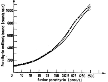
0 10 19 39 78 156 312.5 625 1250 2500 Bovine parathyrin [pmol/l]
Fig. 3. To illustrate that bovine parathyrin is bound very rapidly to polyethylene, the tubes were incubated with increasing concentrations of parathyrin (5.0—2500.0 pmol/1) for 1-6 h at 4 °C. Bound hormone was determined by labelled antibody.
I h 2 h 4 h 6 h
800
600
1 400
,200
2 4 6 8 10 12 16 20 24 36
Incubation time [h] 48
J- ,
Incubation time [h]
Fig. 2. Three concentrations of bovine parathyrin (50, 250 and 500 pmol/1) were incubated in polyethylene tubes at 4 °C for increasing periods (30 min - 6 h). Parathyrin bound after the respective time was detected by labelled antibody, o^-o 500 pmol/1 ·—· 250 pmol/1 +,—+ 50 pmol/1.
concentrations of hormone within 2—3 h (fig. 2). The same result was observed when incubation was carried out at room temperature (data not shown). To further elucidate these findings, increasing amounts of 1=84 bovine parathyrin were incubated for 1-6 h at 4 °C (fig. 3). This showed identical dose^response-curves over the whole range of hormone concentration. To describe the kinetics of the fixation of the radiolabelled
Fig. 4. Bovine parathyrin (5000 pmol/1) was incubated in poly- ethylene tubes for 18-24 h at 4 °C. Parathyrin bound to tubes was then detected by labelled antibody. This second reaction was carried out for 2-48 h at room temperature « · and 4 °C ο ο.
antibody we followed its binding onto bovine parathyrin- coated tubes over a time period ranging from 2—48 h (fig. 4). It was found that this reaction finishes at room temperature after about 24 h, whereas at 4 °C about 36 h are required. Half maximal binding is reached at 3 h and 4 h respectively.
We next investigated the behaviour of bovine parathyrin extraction onto uncoated and antibody precoated tubes detected by radiolabelled antiserum as described (13).
1-84,1-34 and 53-84 extraction onto the tubes was followed. A remarkable difference was found in the dose-response-curves between untreated and antibody- coated tubes. 1-84 bovine parathyrin extraction (fig. 5)
J. Clin. Chem. din. Biochem. / Vol. 18,1980 / No. 9
3000-
-84
10 25 50 100 1000 10000 Bovine porathyrin (-fragment) [pmol/l]
100000
Fig. 5. In a first reaction l-84-(8.5-2630 pmol/l), l-34-(95.0- 11900 pmol/l) or 53-84-(0.21-135.0 nmol/1) parathyrin was incubated in polyethylene tubes, untreated ο ο or precoated with a first antibody · ·, for 18-24 h at 4 °C. In a second reaction the respective hormone was detected by a labelled second antibody.
was less effective at higher hormone concentrations when antibody coated tubed were used. For the 1-34 and the 53-84 parathyrin fragments, the loss of extrac- tion efficiency by using antibody-coated tubes was even more pronounced. In the 1-34 study the increase of radioactivity was reduced to 96.5% at 0.95 nmol/1 and to 56.7% at 11.9 nmol/1. In the 53-84 study these figures are 57.3% at 2.1 nmol/1 and 36.3% at
135.0 nmol/1, respectively. For the 1-84 human para- thyrin the reduction is only 10% at 2.65 nmol/1, whereas at lower concentrations no significant difference be- tween both methods of tube preparation was observed.
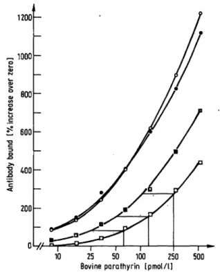
Most interesting, however, is that coupling of a first antibody to the tube does not increase the sensitivity or specificity of the detection method for the different parathyrin-peptides. In a final series of experiments, the adsorption of 1-84 bovine parathyrin to antibody coated and noncoated tubes was followed, either in the presence or absence of plasma, to elucidate potential protective effects of plasma proteins on parathyrin adsorption. If the incubation was carried out in the presence of plasma (1+1-NIRG-buffer + plasma), the adsorption efficiency was reduced compared to the buffer incubation (fig. 6).
In this case prior coating with an anti-N-regional anti- body (extracted and eluted as described) increased the adsorption of parathyrin onto the wall, compared to untreated tubes. The adsorption rate difference was 96.5% higher at 31.25 pmol/l and 93.2% at 250 pmol/l, i.e. a parathyrin value of 25 pmol/l determined in a precoated tube would react as 62.5 pmol/l in a tube where parathyrin was adsorbed onto the tube without antibody precoating. However, the addition of plasma certainly did not prevent the adsorptive binding of 1-84 bovine parathyrin; yet it is added for this purpose in the determination of parathyrin in many assay systems.
Because of the inhibitory effect of plasma, the influence of different peptides on the binding of 1-84 bovine p ra-
1200
~ 1000 20)
I ·: BOO ι I
200
25 50 100 250 Bovine parathyrin (pmol/l] 500 Fig. 6. 1-84-bovine parathyrin was incubated for 18-24 h at
4 °C either in antibody-coated or untreated polyethylene tubes in the presence or absence of plasma. Bound para- thyrin was then detected by the labelled second antibody.
ο ο without coating buffers stem
• · with coating α ο without coating
• · with coating plasma system
thyrin was tested. As shown in table 1 high concentra- tions of parathyrin-fragments, calcitonin and somato- statin, reduced the adsorption of native bovine parathyrin onto polyethylene tubes in a dose dependent manner.
Bovine serum albumin at concentrations of 50 g/1 resulted in a reduction of parathyrin adsorption, which was comparable to that of plasma. Simultaneous incubation with inulin (1.0 g/1) or gelatin (up to 2 g/1) had no effect on the binding of parathyrin onto un- coated surfaces. Adsorption without prior antibody coating of the tubes was observed for bovine and human parathyrin, and was not avoided even in the presence of 8 mol/1 urea. The fixation was not decreased when parathyrin was oxidized with H202 (data not shown).
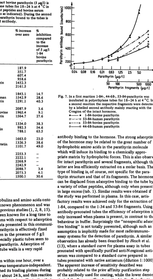
Figure 7 illustrates the specificity of the labelled anti- body used for the experiments. Only intact hormone is determined in physiological concentrations, whereas fragments are detected only in high, supraphysiological ranges. The 2848 and the 1-34 (up to 1 mg/1) peptides did not react at all with the C-regional radiolabelled anti- body (data not included in fig. 7)..Finally it should be mentioned that coating of the tubes with appropriate non-irmnune-gamma-globulin did not influence the parathyrin extraction, or the fixation of the radio- labelled antibody gamma globulin. Repeated washing of the tubes onto which parathyrin had been adsorbed at 4 °C did not influence the amount of parathyrin detected by the radiolabelled antibody in the second incubation, thus indicating firm physiological bindingv
Tab. 1. In the first reaction, intact bovine parathyrin (5 Mg/1) is
incubated in polyethylene tubes for 18-24 h at 4 °C in — inhn the presence of different peptides and bovine serum '|
albumin (concentrations as indicated). During the second Ϊ»
reaction intact bovine parathyrin bound to the tubes is "g detected by the labelled antibody. g 800
standard curve
5.0 Mg/1 bovine1-84 parathyrin
0.156 0.3125 Mg/1 0.625 1-84 1.25 bovine 2.55.0
2.55.0 25.0 250.0 500.0 2500.0 25.05.0 50.0 25.05.0 50.0 25.05.0 500.050.0 2500.0 5000.0 50000.0
paratnynn mg/11-34 human parathyrin Mg/153-84 bovine parathyrin mg/11«
calcitonin
mg/1l|
so m at ο statin
mg/1bovine serum albumin
% increase over zero
187.9 351.7 607.4 958.6 1452.3 2161.5 1843.1 1542.9 1291.1 2087.9 1965.4 1564.7 1334.0 982.3 788.1 1663.0 1326.3 1101.7 2253.3 2222.1 2268.9 2242.9 2088.7 2073.3 1500.1
% I
inhibition ^ 600 compared ·§"to the g increase "g of 5 Mg/1 ° 40° 1-84 'fe bovine ~ <
parathyrin fe 200
28.614.7 40.3 3.69.1 27.6 38.354.6 63.5 23.038.6 49.0
—- 4.13.4 30.6
*
; /
Ί ι ι ι ι ι ι ι ι !_^
0.04 αθθ 0.16 031 0.63 125 2.5 5D ~~
PoTQthyrin I/ig/l)
1 1 1 \
50 100 500 1000 ^ Porothyrin fragments I/ig/IJ Fig. 7. In a first reaction 1-84-, .44-6 8-, 53-84-parathyrin was
incubated in polyethylene tubes for 18-24 h at 4 °C. In a second reaction the respective fragments were detected by a labelled second antibody mainly reacting with the C-region of the intact hormone.
• • l-Of-DOVlIiv ρϋΓαΙΠγΠΠ
ο ο 5 3-84-human parathyrin χ χ 53-84-bovine parathyrin ο α 44-68-human parathyrin
antibody binding to the hormone. The strong adsorption of the hormone may be related to the great number of hydrophobic amino acids in the parathyrin molecule which will induce its binding to a chemically appro- priate matrix by hydrophobic forces. This is also observed for intact parathyrin and several fragments, although the latter are less efficiently extracted on a molar basis. The type of binding is, of course, not specific for the para- thyrin structure and that of its fragments. The hormone
Discussion
The binding of peptides, globulins and amino acids onto different surfaces is a well known phenomenon and was the subject of a number of previous studies (1,2, 8,15).
Especially parathyrin has been known for a long time to impose considerable problems with respect to adsorption onto surfaces (8,9). The data presented in this communi- cation demonstrate that parathyrin is effectively fixed to a variety of materials even in the presence of 5 g/1 human serum albumin. Especially plastic tubes seem to have a high ability to bind parathyrin. Adsorption of parathyrin onto untreated tube walls is a very rapid process.
Maximal parathyrin fixation within one hour, over a wide concentration range, was temperature-independent.
The labelled antibody reached its binding plateau during the second incubation after about 24 h, and this reaction was found to be temperature-dependent, illustrating that parathyrin binding to the tube is distinctly different from
can be displaced from adsorptive binding to the tube by a variety of other peptides, although only when present in large excess (tab. 1). Similar results were obtained if the study was performed in plasma. In this case, satis- factory results were achieved only for the extraction of
1-84, compared to the 1-34 and 53-84 fragments. Using aritibody-precoated tubes the efficiency of adsorption is only increased when plasma is present, in contrast to the behaviour in buffer. Surprisingly the "nonspecific adsorp- tive binding" is not totally prevented, although such an assumption is implicitly made for most radioimmuno- assay systems for the detection of parathyrin. A similar observation has already been described by Hesch et al.
(13), where a standard curve for plasma assay in tubes precoated with specifically purified anti-N-regional anti- serum was compared to a standard curve prepared in tubes precoated with native antiserum (dilution 1:1000).
The apparent increase of sensitivity described was probably related to the prior affinity purification step of the antibody used for coating, while the lower demon- strated the "nonspecific adsorptive binding" similar to that shown in figure 6. These results indicate that yet
J. Clin. Chem. din. Biochem. / Vol. 18,1980 / No. 9
590 JUppner, Mohr and Hesch: Adsorption of parathyrin unknown plasma fractions interfere with the adsorption
of parathyrin to the plastic surface. Surprisingly this inhibitory plasma effect was more pronounced for the 1-34 arid 53-84 fragments and may indicate either the inhibition of adsorption, or an increased binding of fragments onto plasma protein fractions. Furthermore, plasma proteins may mask binding sites within the hormone which are specific for the labelled antibody.
From our observations it seems possible that adsorption occurs preferentially at the NH2-region (residues 1-34).
This part of the sequence may penetrate more into the matrix and undergo noncovalent binding to the plastic material. The remaining part of the molecule, which is more compact (16), will then be detected by the labelled antibody. This offers a further explanation for the more intense binding of intact parathyrin, in contrast to the parathyrin fragments. The difference in adsorption between fragment 1-34 and 53-84 is evident. It can best be explained by different chemical groups undergoing binding to plastic surfaces. The 1-34 parathyrin frag- ment is a more hydrophobic, whereas the 53-84 frag- ment is a more hydrophilic peptide. Even if the binding sites of the antisera are not fully exposed to the peptides this would not account for the differences observed. Our results have several implications:
i. Intact parathyrin is adsorbed onto surfaces to a con- siderable extent. In contrast to another study published very recently on the same phenomenon, we have analyzed unlabelled parathyrin peptides, which certainly behave differently from 125I labelled intact hormone in which the conformation is altered (16). Degradation of the parathyrin at the temperature used here has never been observed and therefore cannot invalidate our results.
ii. In assays for intact parathyrin, appropriate controls must be run to show that effective protection against ad- sorption is achieved. From our results one may expect
that different plasma samples will have different effects on the loss of parathyrin from the reaction milieu. In a direct radioimmunoassay, the adsorption of fragments present, for example, in uremic sera in large excess, may considerably interfere with the reaction of intact hor- mone. It would be an advantage of the "two-site- coated-tube assay" that its specificity is determined by the detection of the CÖOH end of the molecule in the reaction.
iii. Some criticism is attached to the assumptions of
"two-site-coated-tube assays". In contrast to what is assumed from results of such assay systems for parathyrin (7,13), coating of the tubes is riot relevant for specificity, or for sensitivity when intact parathyrin is determined in buffer solution. Specificity of such assay seems to be related very much to the specificity of radiolabelled antiserum as shown in figure 7. Sensitivity is reduced in an assay where intact parathyrin is determined in plasma samples, but this only may partly be improved by precoating the reaction tube with an anti-N-terminal antiserum. This effect is, however, poorly understood.
Whatever the final explanation may be, the design of the "two-site-coated-tube assay", although theoretically universal in specificity and sensitivity, presents several technical problems. This must be taken into account when interpreting results from such assays.
A more reliable approach would be to couple specific first antisera to inert solid phases. However, the assay is very suitable and specific, when carried out in buffer, and includes the advantage that even high concentrations (8 mol/1) of urea do not interfere. For this reason and the facility in handling this system, it may be very useful for several biochemical purposes (for example: during the purification of the hormone). Our findings demon- strate the necessity to further elucidate the adsorptive binding of native parathyrin or its fragments and radio- labelled parathyrin tracers used for radioimmunochemistry.
References
1. Catt, K. J., Niall, H. D. & Tregear, G. W. (1967), Nature 213, 825-827.
2. Catt, K. & Tregear, G. M. (1967), Science 756, 1570-1571.
3. Addison, G. M. & Hales, C. N. (1971), Hoim. Metab. Res. 3, 59-60.
4. Rodbard, D. & Feldmann, . (1978), Immunochemistry 15 (2), 71-76.
5.. Addison, G. M., Hales, C. N., Woodhead, J. S. & O'Riordari, J. L. H. (1971), J. Endocrinol. 49, 521-530.
6. Woodhead, J. S., Davies, G. M., Hales, C. N. & Lehmann, H.
(1977), J. Endocrinol. 75, 279-288.
7. Readhead, C, Addison, G. M., Hales, C. N. & Lehmann, H.
(1978), J. Endocrinol. 59, 313-323.
8. Hamilton, J. W., Spierto, F. W., MacGregor, R. R. & Cohn, D. V. (1971), J. Biol. Chem. 246, 3224-3233.
9. Barrett, P. Q. & Neumann, W. F. (1978), Biochim. Biophys.
Acta 541, 223-233.
10. Bouillon, R., Koninckx, Ph. & DeMoor, P. (1974), title in:
"Radiqimmunoassay and Related Procedures in Medicine", IAEA, Vienna, Vol. 1, 353-365.
11. Woodhead, J. S., Addison,.G. M. & Hales, C. N. (1974), Br.
Med. Bull. 59, 44-49.
12. Miles, L. E. M. & Hales, C. N. (1968), Biochem. J. 108, 611-618.
13. Hesch, R. D., Mclntosh, C. H. S. & Woodhead, J. S. (1975), Horm. Metab. Res. 7, 347-351.
14. Hehrmann, R., Wilke, R., Nordmeyer, J. P. & Hesch, R. D.
(1976), Dtsch. Med. Wochenschr. 101,1726-1729.
15. Mizutanii T., Mizutani, A. (1977), Anal. Biochem. 83, 216-221.
16. Fiskin, A. M., Cohn, D. V. & Peterson, G. S. (1977), J.
Biol. Chem. 254, 8261-8268.
Prof. Dr. R.JX Hesch
Abt. für Klinische Endokrinologie Dept. Innere Medizin
Medizinische Hochschule Hannover Karl-Wiechert-Allee 9
D-3000 Hannover, 61