J. Clin. Chem. Clin. Biochem.
Vol. 27, 1989, pp. 781-786
© 1989 Walter de Gruyter & Co.
Berlin · New York
Biochemical Diagnosis of an Hereditary Aminolaevulinate Dehydratase Deficiency in a 63-Year-Old Man
By A. Hassoun, L. Verstraeten
Department of Clinical Biochemistry, Universite Catholique de Louvain, Cliniques Universitaires St Luc, Bruxelles, Beigigue
R. Mercelis and /.-/. Martin
Department of Neurology, Universitaire Instelling Antwerpen, Universitair Ziekenhuis Antwerpen, Antwerpen, Belgie
(Received April 5/June 30, 1989)
Summary: Porphyrin metabolism was investigated in a 63-year-old male patient who developed a subacute onset polyneuropathy with predominance of motor signs in the upper limb.
The screening for lead, cadmium, mercury, aluminium and thallium was negative. The study of porphyrin metabolism showed remarkable abnormalities, particularly a very high level of plasmatic 5-aminolaevulinic acid contrasting with a normal level of porphobilinogen and a nearly complete loss of activity of aminolae- vulinic acid dehydratase with no regenerative response to dithiothreitol or zinc ions. The other causes of aminolaevulinic acid dehydratase deficiency (tyrosinaemia, alcoholism, smoking, cirrhosis, renal insufficiency, diabetes mellitus) were ruled out.
The diagnpsis of primary aminolaevulinic acid dehydratase deficiency was proposed and confirmed by the familial study, which revealed the existence of several heterozygous members in this family.
Introduction
Hereditary aminolaevulinic acid dehydratase (por- phobilinogen synthase)
1) deficiency is a very rare cause of acute hepatic porphyria. Üntil now, to our knowledgej only three homozygous knöwn cases have been reported (l, 2). Lead ititoxication, which is a common phenomenon, also causes »a loss of activity of atninoläevulinic acid dehydratase, but it is revers- ible by dithiothreitol or zinc ions (3).
Enzymes
Aminolaevulinate dehydratase = porphobilinogen synthase (EC 4.2.1.24)
Porphobilinogen deaminase (EC 4.3.1.8)
To our knowledge, our patient had not suffered from any serious illness, even neurological or haematolog- icäl. Then, at the age of 63, he developed a subacute polyneuropathy for which he was hospitalized. The shoulder girdle muscles and the extensors of the wrist were severely affected, and the lower limb muscles were affected to a lesser degree. Sensory impairment was minimal. At the same time, early signs of mye- loproliferative disease were found.
This study describes the contribution of biochemical investigations to the diagnosis of this rare type of porphyria. We also report the results of the investi- gation of the family for this enzyme defect.
J. Clin. Chem, Clin. Biochem. /Vol. 27,1989 / No. 10
Materials and Methods
Plasma and urinary 5-aminolaevulinic acid and porphobilino- gen were determined spectrophotometrically, after elution from ion-exchange resins (Βίο-Rad, West Germany), according to the Mauzerall & Granick method (4), adapted s follows for plasmatic determinations: 4 ml of plasma are adjusted to pH 6.5 with acetic acid l mol/1 or NaOH l mol/1, then centrifuged.
The usual procedure for urine is then followed. After ion- exchange Separation, the eluate must be filtered on a Millex- SR 0.5 μηι filter (Millipore, France) before adding Ehrlich*$
reagent.
Urinary porphyrins: uroporphyrin fraction, coproporphyrin fraction "and faecal porphyrins: coproporphyrin and protopor- phyrin were determined spectrophotometrically after liquid ex- traction (5). Urinary and faecal porphyrins were separated by high performance liquid chromatography (HPLC) using a method derived from those of Ford et al. (6) and de Verneuil &
Nordmann (7). The Chromatographie System consisted of a SP 8700 solvent delivery System (Spectra-Physics, U. S. A.), a KS Nucleosil 120-5 C 18 (100 mm) column (Macherey-Nagel, West Germany), a fluorimeter Fluoromat FS 950 (Kratos, U. S. A.) s detector (excitation: 400 nm; emission: 600 nm) and a single r nge recorder BD 7 (Intersmat, U. S. A.). The internal Standard was mesoporphyrin IX dimethylester (Porphyrin products, U. S. A.).
Erythrocyte zinc protoporphyrin was measured with a haema- tofluorometef> (8).
For aminolaevulinate dehydratase (EC 4.2.1.24), the Berlin &
Schaller method was used (9). The enzyme was reactivated by addition of both 20 mmol/1 dithiothreitol (10) and 0.1 mmol/1 zinc Chloride (11).
Erythrocyte porphobilinogen deaminase (EC 4.3.1.8) was de- termined fluorimetrically with a fluorimeter Fluoromat FS 950 (Kratos, U. S. A.) according to Deybach, C., Grandchamp, B. &
Nordmann, Y. (unpublished).
Blood and urine analyses for lead were determined according to Blanke & Decker (12) with a graphite furnace atomic ab- sorption spectrometer 2380 (Perkin Eimer, U. S. A.) and a HGA 400 oven (Perkin Eimer, U. S. A.).
Urine mercury was determined by atomic absorption spectro- metry according to Coyle & Hartley (13).
Urine cadmium and thallium were determined according to Kubasik & Volosin (14) with an atomic absorption 4000 spec- trometer (Perkin Eimer, U. S. A.) and a HGA 500 oven (Perkin Eimer, U. S. A.). Plasma aluminium was determined by atomic absorption spectrometry according to Valentin et al. (15).
Urine and serum amino acid analyses were performed by an ion-exchange Chromatographie method, according to Bremer et al. (16), using a TMS l amino acid analyser (Technicon, U. S. A.). The ethylenediaminetetraacetate (EDTA) mobiliza- tion test for lead excretion was performed by i. v. administration of 1600 mg of calcium EDTA in 30 minutes.
The red blood cell, white blood celi, platelet count, haematocrit, blood haemoglobin and white blood cell differentiation were performed with a H-l Technicon analyzer (Technicon, USA).
Plasma glucose and creatinine were performed with a Hitachi 717 analyzer (Hitachi, Japan).
Results
The investigation was started with toxicological screening for heavy metals involved in toxic polyneu- ropathy. The urinary levels of lead, cadmium, mercury
and thallium were within the normal ranges, s were plasma lead and aluminium and the results of the EDTA lead mobiHzati n test (tab. 1).
However, the urinary screening for porphyrins indi- cated an increased excretion of these compounds (tab. 2).
• l
Tab. 1. Heavy metal determination.
Blood Pb (μπιοΙ/1) Urine Pb (μηιοΐ/day) EDTA Pb test (μιηοΐ/day) Plasma AI (μπιοΐ/ΐ) Uririe Cd (μηιοΐ/day) Urine Hg (μπιοΐ/g creatinine) Urine Tl (nmol/1)
Results 0.340.05 0.070.33 0.010.02 N. D.
Reference values
< 1.21
< 0.39
< 0.39
< 0.74
< 0.13
< 0.10
<10.0 EDTA: ethylenediaminetetraacetate
N. D.: not detected
Tab. 2. Porphyrin met bolisnL
Results Reference values Plasmatic precursors
5-Aminolaevulinate (μπιοΐ/ΐ) Porphobilinogen (umol/1) Urine pprphyrins and precursors
5-Aminolaevulinate (μηιρί/ΐ) Porphobilinogen (μήιοΐ/ΐ)
Uroporphyrin fraction (nmpl/day) Coproporphyrin fraction (nmol/day) HPLC determination
Uroporphyrin (%)
Heptacarboxyporphyrin (%) Hexacarboxyporphyrin (%) Pentacarboxyporphyrin (%) Coproporphyrin (%) Faecal porphyrins
Coproporphyrin (nmol/g dry weight) Protoporphyrin (nmol/g dry weight) HPLC determination
Coproporphyrin (%) Protoporphyrin (%)
0.06.8
8005 2333244
43 156 72
174187 4555
0- 0.6 0- 0.2 0- 30 0- 90- 6 0-230 0— 10 0^- 29 0- 90- 8 54-100 0- 60 0- 60 35^- 80 19- 60 Blood zinc protoporphyrin (nmol/g Hb) 19 4 Erythrocyte porphobilinogen deaminase *
(uroporphyrin, pmol/h · mg Hb) 665 . 103 — 243 Erythrocyte aminolaevulinate dehydratase
(5-aminolaevulinate, umol/min · l ery- throcytes)
without additions 0.3 20— 60 after addition of 20 mmol/1 0.6 20- 60 dithiothreitol
after addition of 0.1 mmol/1 0.4 2 0 — 6 0 zinc Chloride
Red biood cells (1012/1) 4.9 4.0-6.1
r* Result obtained with two diifferent samples.
J. Clin. Chem. Clin. Biochem. /Vol. 27, 1989 / No. 10
A more complete study of porphyrins in urine, blood and faeces, and their precursors was performed. The results obtained (tab. 2), especially the significant increase of 5-aminolaevulinate in urine and plasma with porphobilinogen concentrations within normal limits, suggested a deficiency of aminolaevulinic acid dehydratase. The activity of aminolaevulinic acid de- hydratase was therefore determined in erythrocytes, in the presence and in the absence of both dithio- threitol and zinc ions s reactivators. We also deter- mined the activity of porphobilinogen deaminase
1) in erythrocytes. The results obtained are presented in table 2. Tyrosinaemia was ruled out s a possible cause of aminolaevulinic acid dehydratase deficiency, because the levels of plasma tyrosine (55 μηιοΐ/ΐ) were within normal· limits (50—76 μιηοΐ/l), and urine ty- rosine (81 μιηοΐ/g creatinine; reference values: 30 — 220 μπιοΐ/g creatinine) was also normal. Four relatives were also studied: one sister, one brother, the daughter and the grand-daughter of the patient. The results are presented in table 3.
On admission, the erythrocyte count was 4.9 χ 10
12/1 (reference values: 4.0—6.1 χ 10
12/1), the haematocrit was 0.44 (reference values: 0.37—0.52) and haemo- globin was 152 g/l (reference values: 120 — 180 g/l).
The white blood cell count was 11.9 χ 10
9/1 (reference values: 4.0—10.8 χ 10
9/1) with a 0.85 granulocyte fraction. The platelet count was 768 χ 10
9/1 (reference values: 150—450 χ 10
9/1). Bone marrow biopsy re- vealed a local increase in the number of megacary- ocytes. This case was first described s an incipient myeloproliferative disorder and it was not treated. A year later, the following haematological values were found: erythrocyte count: 7.27 χ 10
12/1, haemoglobin:
171 g/l, haematocrit: 0.55, leukocyte count: 9.8 χ 10
9/1 and platelet count: 661 χ 10
9/1. On cytolog- ical and histological ex minati n, the p tient's bone
marrow was hypercellular and had an increased num- ber of megacaryocytes with occasional hypersegmen- tation of the nuclei. There was a slight loss of gran- ulation of the granulocytes and a relative eosinophilia.
The red cell line was hyperplastic. According to these results, the patient was considered to have a myelo- proliferative disease of primary polycythaemia type.
Plasma glucose was 5.5 mmol/1 (reference values:
3.9 — 5.8 mmol/1) and plasma creatinine was 88 μηιοΐ/ΐ (reference values: 35 — 132 μπιοί/1). After oral loading with 100 g of glucose, plasma glucose was 10.8 mmol/1 after 2 hours, and 8.8 mmol/1 after 3 hours.
Discussion
The activity of aminolaevulinic acid dehydratase is most frequently reduced by a heavy metal intoxica- tion, commonly lead, but it is also decreased in var- ious conditions: chronic ethylism (17, 18), cirrhosis (19), smoking (20, 21), renal insufficiency (22) and diabetes mellitus (23). All these causes were ruled out in our patient by anamnestic, clinical and biochemical investigations. However, the oral glucose tolerance test was abnormal, but the plasma glucose levels re- mained always between normal limits during the hos- pitalization of the patient.
Heavy metals other than lead, i. e. aluminium (24), inercury (25) and cadmium (26), do not decrease the activity of aminolaevulinic acid dehydratase in con- centrations that are toxic in vivo. In vitro, very much higher concentrations of these metals can cause an Inhibition (24, 27).
A rare cause of aminolaevulinic acid dehydratase in- hibition is succinylacetone, a metabolite found in ty- rosinaemia (28, 29, 30).
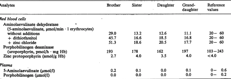
Tab. 3. Laboratory results of the familial study.
Analytes Brother Sister Daughter Grand- Reference
daughter values Red blood cells
Aminolaevulinate dehydratase
(5-aminolaevulinate, μητοΐ/πύη · l erythrocytes)
without additions 29.0 13.2 12.6 11.1 20- 60 + dithiothreitoi 45.7 16.6 18.5 16.8 20- 60 -h zinc Chloride 51.3 18.6 20.5 17.7 20-60 Porphobilinogen deaminase
(uropoiphyrin, pmol/h - mg Hb) 193 178 162 197 103-243 Zinc protoporphyrin (nmol/g Hb) 2.7 4.0 3.5 4.0 <4.0 Plasma
5-Aminolaevulinate (μπιοί/1)
Porphobilinogen (umol/1) 0.20.0 0.1
0.0 0.0
0.0 0.1
0.0 0-
0- 0.6 0.2
J. Clin. Chem. Glin. Biochem. / Vol. 27,1989 / No. 10
Nomial serum and urine amino acid patterns excluded tyrosinaemia, and therefore the presence of succinyl- acetone. The failure of dithiothreitol and zinc to re- store aminolaevulinic acid dehydratase suggests a pri- mary loss of activity of the enzyme, rather than an Inhibition by lead or succinylacetone.
We therefore presumed that our patient suffers from a primary aminolaevulinic acid dehydratase defi- ciency.
During the investigation of certain members of the family, we found an intermediate aminolaevulinic acid dehydratase activity in a sister, in the daughter and in the grand-daughter of the patient, which indicates that this loss of activity is hereditary (tab. 3). Our results are in accordance with an autosomal recessive inheritance, s shown in the three other cases (l, 2), although our data do not exclude an X-linked inher- itance.
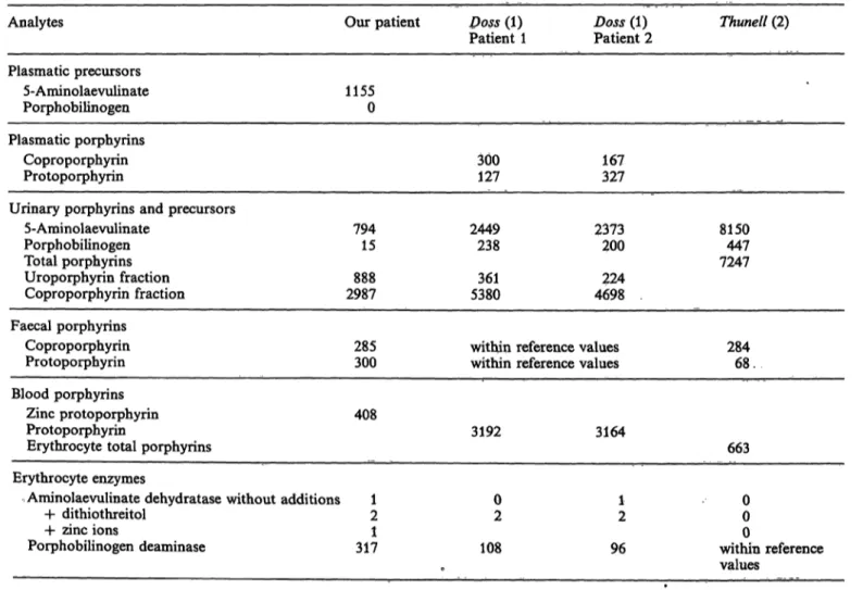
Table 4 compares the results of biochemical investi- gations of pprphyrin metabolism in the homozygous patients described in the literature (l, 2) with those obtained by us. All these patients showed a strong
increase of 5-aminolaevulinate in the urine, together with a mild increase or a normal level of porphoWU linogen in the urine. The plasma of our patient showed a dramatic increase of 5*aminolaevulinate and no detectable porphobilinogen. The remarkable discord- ance between 5-aminolaevulin te and porphobilino- gen points to the existence of ap enzymatic blockade at the aminolaevulinic acid dehydratase level. This enzymatic activity was not increased by reactivators.
In all patients, the urinary porphyrin profile sfaowed a predominant coproporphyrin fraction, with a smaller incre se of the uroporphyrin fraction. This type of profile can also be observed m heavy metal intoxic tio (1). The faecal porphyrin profile was less typical than that of urine; a ^ρΓοροφΙινπη inerease was found in two patients and a protopoiphyrin in- crease in one patient. The erythrocyte porphyrins (protoporphyrin, zinc protophorphyrin, total por- phyrins) were increased in all patients.
The porphobilinogen deaiiiinase activity in erythrp- cytes was strongly increased in our patient, although no haemolytic anaemia was noticed. The three other
Tab. 4. Comparison of porphyrin metabolism in the homozygous aminolaevulinate dehydratase deficient patients.
All results are expressed in % of the upper reference values of each author.
Anaiytes
Plasmatic precursors 5-Aminolaevulinate Porphobilinogen Plasmatic porphyrins
Coproporphyrin Protoporphyrin
Urinary porphyrins and precursors 5-Aminolaevulinate
Porphobilinogen Total porphyrins Uroporphyrin fraction Coproporphyrin fraction Faecal porphyrins
Coproporphyrin Protoporphyrin
Our patient
11550
794 15 2987888
285300
Doss (1) Patient 1
300127
2449238
5380361
within reference within reference
Doss (1) Thunell (2) Patient 2
167327
2373 8150 200 447 224 7247
4698
values 284 values 68 . Blood porphyrins
Zinc protoporphyrin Protoporphyrin
Erythrocyte total porphyrins
408
3192 3164
663 Erythrocyte enzymes
Aminolaevulinate dehydratase without additions + dithiothreitol
+ zinc ions
Porphobilinogen deaminase
12 3171
02 108
•
12 96
00
within reference0 values
J. Clin, Chem. Clin. Biochem. / Vol. 27,1989 / No. 10
patients, showed no increase of porphobilinogen de- aminase activity. Does this represent an attempt of the cell to diminish the blockade of the enzymatic pathway by speeding up the transformation of por- phobilinogen to uroporphyrinogen? On the other band, could a myeloproliferative disorder provoke some modification of haem biosynthesis? An associ- ation of porphyria cutanea tarda with a chronic gran- ulocytic leukaemia has been described before (31, 32).
The immunological characterization of the two pa- tients of Doss showed a cross-reactive immunological material which corresponded to 20% and 33% of the control level (33). Therefore the molecular basis for the deficiency of aminolaevulinic acid dehydratase in these patients is a structurally modified enzyme (33).
The methods used to determine the relative molecular mass (by Western blotting) and the isoelectric point (by chromatofocusing) of the mutants revealed no differences between the modified and normal enzymes (33). In the patient of Thunell, the immunoreactive enzyme protein in the child's erythrocytes was de- creased to 28% of the normal control, suggesting the presence of positive cross reactive material (34). These results suggest that in these cases, the aminolaevulinic acid dehydratase deficiency is associated with the pro- duction of a catalytically abnormal enzyme protein (33, 34).
Finally, the occurrence of the Symptoms of porphyria for the first time at the age of 63 remains an enigma.
Presumably the low activity of aminolaevulinic acid dehydratase had been present since birth, and to our knowledge this patient had never presented clinical Symptoms of porphyria until the age of 63.
Conclusion
case of aminolaevulinic acid dehydratase hereditary deficiency is reported in a 63-year-old man affected by a motor polyneuropathy. The diagnosis was made:
1) from the increase of urinary and plasmatic ami- nolaevulinate, with non-detectable plasmatic por- phobilinogen levels;
2) from the absence of heavy metal intoxication, ty- rosinaemia, alcoholism, smoking, cirrhosis, renal in- suficiency and diabetes mellitus;
3) from the failure to regenerate the activity of eryth- rocyte aminolaevulinic acid dehydratase. The familial study confirmed the hereditary autosomal recessive nature of this enzymatic deficiency.
Acknowledgement
We thank Ms. Ledoux for her efficient technical assistance in the investigation of porphyrin metabolism.
References
1. Doss, M., von Tiepermarm, R., Schneider, J. & Schmid, H.
(1979) New type of hepatic porphyria with porphobilinogen synthase defect and intermittent acute manifestation. Klin.
Wochenschr. 57, 1123-1127.
2. Thunell, S., Holmberg, L. & Lundgren, J. (1987) Amino- levlilinate dehydratase porphyria in infancy. A clinical and biöchemical study, J. Clin, Chem. Clin. Biochem. 25, 5—14.
3. Doss, M., Schneider, J., Tiepermann, R. & Brandt, A.
(1982) New type of acute porphyria with porphobilinogen synthase (deltaTamiiiolevulinate dehydratase) defect in the homozygous stäte. Clin. Biochem. 15, 52^-55.
4. Mauzerall, D, & Granick, S. (1956) The occurrence and determination of delta-aminolevulinic acid and porphobi- linogen in ilrine. J, BioJ. Chem. 219, 435-446.
5. Gajdos, A. & Gajdos-OTörök^ M. (1969) Porphyries et por- phyrines, pp. 207--234, Masson, Paris.
6. Ford, R, Ef, Chin-Nan, Ou & Ellefson, R. D. (1981) Liquid Chromatographie analysis pf urinary porphyrins. Clin.
Chem. 27, 397-40L
7. de Verneuil, H. & Nordmann, Y. (1979) Chromatographie liquide a haute performance (CLHP) Porphyrines et por- phyries. Feuillets de biologie 20, 87-88.
8. Blumberg, W. E., Eisinger, J., Lamola, A. A. & Zuckerman, D. M. (1977) the hematofluorometer. Clin. Chem. 25, 270-274.
9. Berlin, A. & Schaller, K. H. (1974) European standardized method for the determination of delta-aminolevulinic acid dehydratase in blood. Z. Klin. Chem. Klin. Biochem. 72, 389-390.
10. Granick, J. L., Sassa, S., Granick, S., Levere, R. D. &
Kappas, A. (1973) Studies in lead poisoning. II. Correlation between the ratio of activated to inactivated delta-amino- levulinic acid dehydratase of whole blood and blood lead levels. Biochem. Med. 8, 149-159.
11. Doss, M., Benkmann, H. G. & Goedde, H. W. (1986) Delta- aminolevulinic acid dehydratase (porphobilinogen syn- thase) in two families with inherited enzyme deficiency.
Clinical Genetics 30, 191 -198.
12. Blanke, R. V. & Decker, W. J. (1986) Analysis of toxic substances. In: Textbook of Clinical Chemistry (Tietz, N.
W., ed.) pp. 1714-1715, W. B. Saunders Company, Phil- adelphia
13. Coyle, P. & Hartley, T. (1981) Automated determination of mercury in urine and blood by the Magos reagent and cold- vapor atomic absorption spectrometry. Anal. Chem. 53, 354-356.
14. Kubasik, N. P. & Volosin, M. T. (1973) A simplified deter- mination of urinary cadmium, lead, and thallium, with use of carbon red atomization. Clin. Chem. 19, 954—958.
15. Valentin, H., Preusser, P. & Schaller, K. H. (1976) Die Analyse von Aluminium im Serum und Urin zur Überwa- chung exponierter Personen. Int. Arch. Occup. Env. Health.
38, 1-17.
16. Bremer, H. J., Duren, M., Kamerling, J. P, Przymbel, H.
& Wadman, S. K. (1981) Disturbance ofamino acid metab- olism: clinical chemistry and diagnosis» pp. 458—465, Urban
& Schwarzenberg, Baltimore-Munich.
J. Clin. Chem. Clin. Biochem. / Vol. 27,1989 / No. 10
17. Abdulla, M., Haeger-Aronsen, B. & Svenson, S. (1976) EfTect of ethanol and zinc on ALA-dehydratase activity in red blood cells. Enzyme 27, 248-252.
18. Kondo, M., Urata, G. & Shimizu, Y. (1983) Decreased liver delta-aminolaevulinate dehydratase activity in porphyria cutanea tarda and in alcoholism. Clinical Science 65, 423 ·*- 19. Allain, R, Froussard, E. & Boyer, J. (1977) Relation entre428.
le taux de zinc dans le sang et l'activite ALAD erythrocy- taire chez rhomme. Clin. Chim. Acta 78, 183-189.
20. Salle, H. J. A. & Zielhuis, R. L. (1977) Influence of smoking on ALA-D activity, hematocrit and lead in blood in adult urban woman. Int. Arch. Occup. Environm. Health 40, 111-115.
21. Vivoli, G., Vecchi, G., Ferrari, L. R. & Borella, P. (1974) Comportamento dell'attivita 5 aminolevulinico dehydra- tasica in rapporto delle abitudini fumatori. Riv. Ital. Ig.
34, 26-36.
22. Buchet, J. R, Lauwerijs, R., Hassoun, A., Dratwa, M., Wens, R., Collart, F. & Tielemans, C. (1987) Effect of aluminium on porphyrin metabolism in hemodialyzed pa- tients. Nephron 46, 360—363.
23. Djordjevic, V. (1985) Delta-aminolevulinic acid dehydratase activity in erythrocytes of diabetic patients. Arch. Intern.
Physiol. Biochim. 93, 285-290.
24. Abdulla, M., Svenson, S. & Haeger-Aronsen, B. (1979) Antagonistic effects of zinc and aluminium on lead inhi- bition of delta-aminolevulinic acid dehydratase. Arch. En- viron. Health 34, 464-468.
25. Lauwerijs, R. & Buchet, J. R (1973) Occupational exposure to mercury vapors and biological action. Arch. Environm.
Health 27, 65-68.
26. Lauwerijs, R., Buchet, J. P. & Roels, H. A. (1973) Com- parative study of effect of inorganic lead and cadmium on blood delta-aminolevulinic acid dehydratase in man. Brit.
J. Ind. Med. 30, 359-364.
27. Calissano, R, Cärtasegna, C. & Bonsignore, D. (1965) Azione di alcuni metalli neu deidratasi erytrodtaria puri- flcata da sangue umano. Lav. Umano 17, 493—497.
28. Sassa, S. & Kappas, A. (1983) Hereditary tyrosraemia and the heme biosynthetic pathway. Profound Inhibition of delta-aminolevulinic acid dehydratase activity by sueeinyl- acetone. J. Clin. Invest. 71, 625—634.
29. Lamon, J.-M., Frykholm, B. C. & Tschudy, R R (1978) Tyrosinemia with aminolevulinic dehydratase deficiency. J.
Pediatr. 91, 346. ' f
30. Strife, F. C, Zuroweste, E. L. & Emett, E. A. (1977) Tyrosinemia with acute intennittent porphyria aminolevu- linic acid dehydratase deficiency related to elevated urinary aminolevulinic acid levels. J. Pediatr. 90, 400—404.
31. Kyle, R. A., Bowie, R. J. W. & Bnmsting, L. A. (1964) Porphyria cutanea tarda associated with refractory anemia (ineffective erythropoiesis) and thfömbocythemia, Mayo Clinic Proceedings 39, 750-760.
32. Kyle, R. A. & Dameshek, W. (1964) Porphyria cutanea tarda associated with chronic granulocytic leukemia treated with busulphan. Blood 23, 766—785.
33. de Verneuil, H., Doss, M,, Brusco, N., Beaumont, C &
Nordmann, Y. (1985) Hereditary hepatic prophyriä with delta-aminolevulinic dehydrase deficiency: immunological characterization of the noüK;atalytic enzyme. Hum. Genet.
69, J74-177.
34. Fuyita, H-, Sassa, S., Lundgren, J., Holmberg, L., Thuneü, S. & Kappas, A. (1987) Enzymätic defect in a child with hereditary hepatic po hyria due to homozygous delta- äminölevulinic acid dehydratase deficiency: immunochem- ical studies. Pediatrics 80, 880-885.
A. Hassoun
Service de Biochimie Medicale Cliniqües Universitaires St Luc 10, avenue Hippocrate
B-1200 Bruxelles
J. Clin. Chem. Clin. Biochem. / Vol. 27,1989 / No. 10» t