T HE OCCURRENCE OF WATERBORNE PATHOGENIC PROTOZOA IN ENVIRONMENTAL WATER SAMPLES , THEIR
REDUCTION BY WASTEWATER TREATMENT AND DISSEMINATION IN THE HYDROLOGICAL CIRCUIT
Inaugural-Dissertation zur
Erlangung des Doktorgrades
der Mathematisch-Naturwissenschaftlichen Fakultät der Universität zu Köln
vorgelegt von
Carmen Gallas-Lindemann aus Krefeld
Köln
2012
Berichterstatter: Prof. Dr. Hartmut Arndt Prof. Dr. Sigrun Korsching
Tag der mündlichen Prüfung: 15. Juni 2012
Die vorliegende Arbeit wurde unter Anleitung von Herrn Prof. Dr. P. Karanis an der Universität zu Köln durchgeführt. Ihm gilt mein ganz besonderer Dank für das in mich gesetzte Vertrauen bei der Überlassung des Themas, für die Möglichkeit zur Promotion und für anregende Gespräche und Diskussionen.
Herr Prof. Dr. H. Arndt und Frau Prof. Dr. S. Korsching danke ich herzlich für die Begutachtung der Promotion.
Ein großer Dank geht an Frau Sania Sotiriadou und Frau Dr. Judit Plutzer für die Hilfe und Unterstützung bei theoretischen und praktischen Fragestellungen.
Ein herzliches Dankeschön möchte ich auch an Frau Dr. Ute Wingen als Leiterin des LINEG-Zentrallabors und Herrn Dr. Udo Kosmac als ihren Stellvertreter richten, die mich immer wieder ermutigten und diese Arbeit ermöglichten.
Den Kollegen des Fachbereichs Biologie der Linksniederrheinischen Entwässerungs- genossenschaft möchte ich dafür danken, dass sie mir den Rücken freigehalten haben. Mein besonderer Dank gilt Herrn Rainer Köster für die Unterstützung bei den Probenahmen.
Bei der Linksniederrheinischen Entwässerungsgenossenschaft in Kamp-Lintfort
bedanke ich mich für die Bereitstellung der im Rahmen dieser Arbeit benötigten
Sachmittel.
1 Introduction ... 8
1.1 General Introduction ... 8
1.2 Introduction of the target parasites... 12
1.2.1 Cryptosporidium... 12
1.2.2 Toxoplasma gondii ... 16
1.2.3 Giardia duodenalis... 18
1.3 Loop-mediated isothermal amplification (LAMP) ... 20
1.4 References ... 22
2 Circulation of Cryptosporidium and Giardia in wastewater and the surface, drinking and ground waters in the Lower Rhine, Germany ... 28
2.1 Introduction ... 29
2.2 Materials and methods ... 29
2.2.1 Geography and description of the sampling sites... 29
2.2.2 Sample collection ... 34
2.2.3 Sample preparation for microscopic examinations ... 35
2.2.4 Statistics ... 36
2.3 Results ... 36
2.3.1 Results of the examination of waste water treatment plants ... 36
2.3.2 Results from the recreational swimming area ... 39
2.3.3 Results of the examination of the drinking water supply... 40
2.4 Discussion ... 41
2.5 References ... 50
3 Giardia and Cryptosporidium species dissemination during wastewater treatment and comparative detection by IFT, nPCR and LAMP ... 57
3.1 Introduction ... 58
3.2 Materials and Methods ... 59
3.2.1 DNA Extraction... 60
3.2.2 Detection methods... 60
3.2.3 Gel electrophoresis ... 64
3.2.4 Statistics ... 64
3.3 Results ... 64
3.4 Discussion and outlook ... 68
3.5 References ... 74
4 Detection of Toxoplasma gondii oocysts in sewage by Loop Mediated Isothermal Amplification (LAMP)... 79
4.1 Introduction ... 80
4.2 Materials and methods ... 81
4.2.1 Study area ... 81
4.2.2 Sample collection and preparation ... 81
4.2.3 DNA Extraction... 82
4.2.4 LAMP... 82
4.3 Results and discussion... 82
5 Kurzzusammenfassung... 95
6 Abstract ... 99
7 Appendix ... 103
a Year
BMJ Bundesministerium der Justiz -Federal Ministry of Justice CFU Colony-forming units
d Day
DAPI 4′,6-Diamidino-2-phenylindole DDW Double distilled water
df Degrees of freedom
DICM Difference interference contrast microscopy DMSO Dimethyl sulphoxide
EC European Commission
EDTA Ethylenediaminetetraacetate
EU European Union
FITC Fluorescein isothiocyanate
h Hour
HO Hoerstgen
HPC Heterotrophic plate count IFT Immonofluorecence test IMS Immunomagnetic separation
KL Kamp-Lintfort
LA Labbeck
LAMP Loop-mediated isothermal amplification
LANUV NRW Landesamt für Natur, Umwelt und Verbraucherschutz Nordrhein-Westfalen - State Agency for Nature, Environment and Consumer Protection North Rhine Westphalia
LINEG Linksniederrheinische Entwässerungs-Genossenschaft MBR Membrane bioreactor
MG Moers-Gerdt
n Number
NABU Naturschutzbund Deutschland e.V. – Nature and Biodiversity Conservation Union Germany
nPCR Nested polymerase chain reaction
p Level of confidence
PBS Phosphate buffered saline
PCR Polymerase chain reaction
RB Rheinberg
RH Rheinhausen
RKI Robert Koch Institut
RT-PCR Real-time polymerase chain reaction
t0 Dimension of distances between arithmetic means in relation to standard deviation
UN United Nations
USEPA U.S. Environmental Protection Agency
UV Ultra-violet
WHG Wasserhaushaltsgesetz - Water Resources Act WHO World Health Organisation
WVN Wasserverbund Niederrhein WWTP Wastewater treatment plant
XL Xanten-Lüttingen
XV Xanten-Vynen
1 Introduction
1.1 General Introduction
Water plays an important role in the transmission of pathogens. Waterborne pathogens are of high relevance in medicine research since human beings investigate in diseases. Pathogens are roughly categorised in five groups: viruses, bacteria, protozoa, fungi and helminths. Bacterial diseases were researched well in the last two centuries by Robert Koch, when cholera outbreaks were on a high level (Grüntzig et al. 2010, Erer et al. 2010). Extensive investigations in drinking water treatment, sewage treatment, monitoring of drinking water sources regulated by law etc. led to higher hygienic standards. Thus, the problem was widely brought under control in the developed countries. Drinking water is the best investigated foodstuff in industrialized countries.
Nevertheless, 884 million people worldwide live without access to safe drinking water and sanitation (UNICEF and WHO, 2008). Therefore, the human right to water and sanitation has been accepted by the UN resolution in July 2010 (UN, 2010). This was an important step, because waterborne pathogens are relevantly causative agents that induce diarrhoeal illness, which is a main cause of mortality in children under the age of five years and otherwise immunocompomized people in the developing world (Sheth et al., 2010; Parashar et al., 2003).
Cryptosporidium and Giardia duodenalis are major pathogens in waterborne transmission of infections. The worldwide annual new infection rate of G. duodenalis is 2.8 x 10
8and of Cryptosporidium it is 300 000, respectively (Lane and Lloyd, 2002;
Mead et al., 1999). Studies about the prevalence of G. duodenalis and Cryptosporidium spp. are rare in the developing countries. The infrastructure for health care is not well established. Laboratories are often under-resourced and there is a lack of skilled employees. Monitoring and safety plans for health care of the population are generally not available.
From deficiency of toilets and WWTPs contamination of the surface water, which
serves as reservoir for drinking water, transmission of waterborne pathogens is
possible (Snelling et al., 2007; Lim et al., 2007).
Therfore, high prevalence of giardiasis and cryptosporidiosis is significant in developing countries. Especially children, AIDS patients and malnourished people are affected. Diarrhea is the leading cause of deaths; 30 to 50% of childhood mortality is caused by diarrhea (Snelling et al., 2007).
In a Malaysian study wastewater from two WWTPs were investigated. G. duodenalis was present in 100% of influent and effluent samples and Cryptosporidium in 50 % of the influent and 25% of the effluent samples (Lim et al., 2007).
Compared to the developed countries WWTPs do not have state-of-the-art technology in the developing world. E.g. in Malaysia 50% of the WWTPs have only primary treatment process. In rural areas WWTPs are not available (Lim et al., 2007).
In developing countries necessary funds are rare to invest in WWTPs, sanitation, drinking water treatment and expensive test kits for monitoring programs (Snelling et al., 2007).
Cryptosporidium and Giardia are distributed worldwide and cause diseases of the intestinal tract in vertebrates (Mircean et al., 2011). Affected hosts include humans (Thompson and Smith, 2011; Mircean et al., 2011) as well as wild (Ravaszova et al., 2011; Siembieda et al., 2011; Bitto and Aldras, 2009; Levecke et al., 2011) and domestic animals (Budu-Amoako et al., 2012; Ferreira et al., 2011; Coklin et al., 2010; Mark-Carew et al., 2010). Infection causes diarrhea and is self limiting within a few days (Petry et al., 2010). Due to this fact patients usually do not seek medical advice. The actual epidemiological situation can only be estimated. Giardiasis and cryptosporidiosis are life threatening infectious complications that occasionally occur in immunosuppressed patients like children under the age of five years, HIV-infected patients, patients undergoing chemotherapy or organ transplantation and elderly people (Furio and Wordell, 1985). Autoinfection, which has negative effects on health for these patients, is possible (Stürchler 1987). Also extra intestinal infections have been described (Nagazaki et al. 2011). (Oo)cysts, the parasitic stages which are excreted with faeces, are resistant against environmental and chemical influences (e.g. chlorination and UV radiation). The treatment with drugs is possible for giardiasis (Gardner and Hill, 2001; Solaymani-Mohammadi et al., 2010) but not for cryptosporidiosis.
In livestock, economic losses in productivity and animal mortality are often observed,
especially in juvenile cattle (Tiranti et al., 2011). The costs of health care and the
non-productive time of employees in case of illness are presumptively enormous (Karanis et al., 2007).
(Oo)cysts are able to persist in the environment for months, different transmission cycles are possible; one of the most important is waterborne distribution. The occurrence of Cryptosporidium oocysts and G. duodenalis cysts in different types of water has been confirmed, and a considerable number of waterborne outbreaks has been reported worldwide (Karanis et al., 2007; Baldursson and Karanis 2011).
Toxoplasma gondii (Phylum Apicomplexa) is a protozoan pathogen that is phylogenetically closely related to Cryptosporidium. T. gondii also occurs worldwide and infects humans as well as other vertebrates. During disease progression, tissue cysts are formed, followed by the multiplication of the organism within the host cell cytoplasm (Hutchison et al, 1970). Swelling of the lymph nodes, muscle pain and fever are the most common symptoms of toxoplasmosis, while cysts seldom occur in heart, liver and spleen. Intrauterine infection may exert negative effects on a foetus if the mother is infected for the first time during the third trimester of pregnancy (Kaye, 2011; Olariu et al., 2011). Toxoplasmosis is a self-limiting disease in immunocompetent individuals. Among immunocompromised patients, it often results in morbidity and mortality (Bruck et al., 2010; Nissapatorn, 2009; Utsuki et al., 2011).
T. gondii oocysts are excreted with the faeces of Felidae (Dubey, 1998). Oocysts are able to enter and circulate in terrestrial and aquatic environments. Moreover, these robust parasitic stages are capable of persisting for an extended time in the environment and are highly resistant to various chemicals and disinfection methods that are commonly used by the water supplying industry (e.g. filtration, chlorination, ozonation and radiation) (Dubey, 1998). Water plays an important role in the dissemination of human toxoplasmosis (Dubey, 1998). Therefore, the analysis of T.
gondii contamination in water samples provides insight into the potential risk of waterborne infections that affect humans and animals.
Several waterborne toxoplasmosis outbreaks have been documented since 1979, including cases in Panama (Benenson et al., 1982), British Columbia (Bowie et al., 1997), Brazil (Keenihan et al., 2002) and four additional outbreaks described in a recent review by Baldursson and Karanis (Baldursson and Karanis, 2011).
There is a rising interest in waterborne diseases and many international scientific
investigations are published worldwide. In the past investigations were concentrated
on the United States and United Kingdom, but only a few publications are recognized
from Germany. One reason might be the intricate preparation of samples to find parasitic stages in water matrices. The distribution via waterways could be enormous.
The study area of this work is located in Germany and belongs to the administrative district of Düsseldorf in the federal state of North Rhine-Westphalia, which is, in the geographical classification of natural landscapes, a part of the lowland plain of the Lower Rhine (Paffen et al., 1963).
On Lower Rhine area densely populated urban structures exist as well as intensive and extensive used agricultural structures. In the geological history glacial and interglacial changes left a typical lower Rhine terrasse landscape depending on changes of the River Rhine (Schirmer 1994, Schirmer 1990). Many slow running water bodies are distinctive for the Lower Rhine area. Hundreds of kilometres of running water pick up different kinds of material by erosion, losses by surface runoff and other influences. Treated wastewater of eight municipal sewage water plants in the study area drain into them and distribution of parasites could be implicated.
Reasoned by the small difference between the surface of the landscape and the groundwater level, an influence of protozoan parasites on the groundwater has been supposed. This could also be due to riverbank filtration of the River Rhine situated nearby. In addition the catchment area for the drinking water supply is located in close vicinity to the River Rhine.
The purpose of this work was to investigate the occurrence and distribution of parasites on the Lower Rhine in Germany.
Wastewater treatment plants are considered to be a vast source of parasitical contaminations. Immission of protozoan parasites into surface water, groundwater, raw and drinking water is considered probable. The scales of parasites in the different water matrices and their retention should be investigated. Based on the results the risk to human health should be discussed in the relation to drinking water consumption or bathing in surface waters.
Depending on the expected contamination different sampling techniques should be used. The equivalence of the molecular assay PCR and an emerging assay (LAMP) to the conventional Immunofluorescence Test (IFT) and Difference Interference contrast Microscopy (DICM) should be compared.
The LAMP should be further tested for the detection of T. gondii. The investigations
aimed to raise the attention on the risk of toxoplasmosis outbreaks.
1.2 Introduction of the target parasites 1.2.1 Cryptosporidium
Cryptosporidium is an apicomplexan protozoon (Phylum: Apicomplexa, Class:
Sporozoea, Subclass: Coccidia, Order: Eucoccidia, Suborder: Eumeriina, Family Cryptosporidiidae, Genus: Cryptosporidium) (Plutzer and Karanis, 2009; Mehlhorn and Piekarski, 1998). To date 21 valid species of the genus Cryptosporidium are known (Table 1; Plutzer and Karanis, 2009; Smith et al., 2010). The parasite is capable of infecting humans and other vertebrates. The species and their designated hosts are listed in Table 1. Eight Cryptosporidium species are known as human pathogens (C. hominis, C. parvum, C. meleagridis, C. felis, C. canis, C. suis, C.
muris, and C. andersoni) (Smith et al., 2010). Additionally, five out of the 61 genotypes with uncertain species status (Plutzer and Karanis, 2009) infect immunocompetent and immunocompromized humans (Smith et al., 2010).
Cryptosporidium was firstly described in 1907, but at that time it was not noticed as a pathogen (Tyzzer, 1907). In the first reported outbreak diarrhoeal disease of calves infected by Cryptosporidium was mentioned (Panciera et al., 1971). In 1976 a three year old child (Nime et al., 1976) and an immunosuppressed patient (Meisel et al., 1976) were infected by Cryptosporidium causing diarrhoeal disease. Later it was recognized that water plays an important role in the dissemination of Cryptosporidium. In 1993 Cryptosporidium challenged attention, when the largest documented outbreak of waterborne disease in the United States caused an epidemic with 403 000 infected people and with potentially 112 deaths (MacKenzie et al., 1994). Since then Cryptosporidium was of highest interest in scientific research.
A worldwide overview of the waterborne outbreaks is given in Karanis et al (2007)
and Baldursson and Karanis (2011). Cryptosporidium was the etiological agent in
60.3% of the outbreaks (Baldursson and Karanis, 2011).
Species Major host(s) Minor host(s) Site of infection
1 C. andersoni Cattle, Bactrican camel Sheep Abomasum
2 C. baileyi Poultry Quails, ostriches, ducks Cloaca, bursa, trachea
3 C. bovis Cattle Sheep Small intestine
4 C. canis Dogs Humans Small intestine
5 C. fayeri Red kangaroo Small intestine
6 C. felis Cats Humans, cattle Small intestine
7 C. fragile Amphibia Stomach
8 C. galli Finches, chicken Proventriculus
9 C. hominis Humans Dugong, sheep Small intestine
10 C. macropodum Eastern grey kangaroo Small intestine
11 C. meleagridis Turkey, humans Parrots Small intestine
12 C. molnari Fish Stomach (and intestine)
13 C. muris Rodents
Humans, rock hyrax,
mountain goat Stomach
14 C. parvum Cattle, livestock, humans
152 mammalian species,
deer, mice, pigs Small intestine
15 C. ryanae Cattle, Bos taurus Small intestine
16 C. scophthalmi Fish Intestine (and stomach)
17 C. serpentis Lizards, snakes Stomach
18 C. suis Pigs Humans Small and large intestine
19 C. varanii syn. saurophilus Lizards Snakes Stomach and small intestine
20 C. wrairi Guinea pig Small intestine
21 C. xiaoi Sheep Yak, goat Not known
Table 1: Cryptosporidium species with information on organ locations, major and minor hosts.
(Modified - Plutzer et al, 2009, Smith and Nichols, 2009).
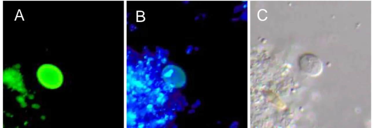
The robust oocysts (round to ovoid, 5 – 6 µm; Figure 2) are the infective stages of the
parasite, which shed into the environment by hosts and are able to persist there for
month. Oocysts are extremely virulent; only few oocysts (1 to 10) are capable to
infect the host. Mainly, faecal-oral transmission and foodborne and waterborne
infections are possible.
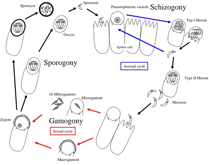
Figure 1: Life cycle of Cryptosporidium parvum (Mehlhorn and Piekarski, 1998, modified).
The monoxenous lifecycle is coccidian-like with division into the three stages schizogony (or merogony), gamogony and sporogony Figure1). The cycle begins with the ingestion of the sporulated oocysts (transmitted into the sporocyst-form).
Each sporocyst contains four sporozoites. In the small intestine of the host the sporozoites leave the sporocyst. After that each sporozoite penetrates an epithelial cell, forming an intracellular but extracytoplasmatic parasitophorous vacuole. In the vacuole cell division generating the schizont or meront type I (each including 8 merozoites) is visible. The liberated merozoites are able to penetrate non-infected epithelial cells for further asexual multiplication (schizogony or merogony). In addition the developement of meront types II is possible. Mature type II meronts with four merozoites, which liberate the vacuole and infect new host cells, undergo either further merogony or initiate gamogony (sexual reproduction). For this purpose they invade host cells again and differentiate into the female macrogamont or into the male microgamont. The microgamont develops into a microgametocyte with up to 16
Epithel cells
Sporocyst Sporozoit
Merozoit
Gamogony Sporogony
Schizogony
16 Mikrogametes
Macrogamont
Parasitophorous vacuole
Zygote
Typ I Meront
Oocyst
Type II Meront Asexual cycle
Sexual cycle
Microgamont
microgametes. Those sperm cell equivalents later fertilize the macrogamonts. A zygote is formed after fertilization and the parasite undergoes asexual multiplication in the sporogony. The sporogony results in the development of sporulated oocysts each containing four sporozoits. The zygote is able to form thin- or thick-walled oocysts. The thin-walled oocysts represent about 20% of all newly sporulated oocysts and are responsible for autoinfection. The thick-walled oocysts are excreted by the faeces in order to infect other hosts. Excretion of sporulated and unsporulated oocysts is possible. The sporulation of this unsporulated form into infective sporocysts then emerges in the environment (Mehlhorn and Piekarski, 1998).
A B
C
Figure 2: Cryptosporidium oocysts fluorescently labelled A: with monoclonal antibodies (fluorescein isothiocyanate, FITC and B: with 2-(4-amidinophenyl)-6-indolecarbamidine di-hydrochloride, DAPI and C. DICM (magnification 1000x).
Symptoms of the disease
The incubation period varies between two and ten days. Clinically asymptomatic run of the cryptosporidiosis in immunocompetent persons is possible, although excretion of oocysts can not be excluded (Arrowood, 1997).
The main symptoms of the disease are watery diarrhea, dehydration, fever, anorexia, weight loss, weakness, abdominal cramps, vomiting, lethargy, general malaise and progressive loss of overall condition (Hunter et al., 2004).
The disease is self-limiting and the symptoms usually last from several days to two
weeks. Depending on the immune status symptoms may be more acute in
immunocompromised persons often leading to mortality (Clifford et al., 1990). Anti-
cryptosporidial drugs are not available and preventive vaccination is not possible.
1.2.2 Toxoplasma gondii
Toxoplasma gondii is an apicomplexan protozoon (Phylum: Apicomplexa, Class:
Sporozoea, Subclass: Coccidia, Order: Eucoccidia, Suborder: Eumeriina, Genus:
Toxoplasma) (Mehlhorn and Piekarski, 1998). T. gondii is the only species of this genus.
T. gondii was firstly described after it has been discovered in an African rodent in 1908 (Nicolle and Manceaux, 1909).
The first case of toxoplasmosis of a human being was reported in 1923, when neonatal infection occurred in an 11-month old child (Wolf et al., 1941). Since the 1980s, toxoplasmosis gains higher interest due to the increase of immunosuppressed patients. An outbreak associated with water appeared in 1979 in Panama. The infection has been assumed to be related to the consumption of uncooked creek water contaminated with oocysts by jungle cats; 32 soldiers were affected (AWWA, 1999). The greatest outbreak so far affecting thousands of people was associated with municipal drinking water in British Columbia during September 1994 and March 1995. The contamination was possible due to treatment deficiencies in a water reservoir serving the population with potable water (Bowie et al., 1997).
The robust oocysts (round to ovoid, 9 to 15 µm) are the infective stages of the parasite, which are shed by hosts into the environment and are able to persist for month. Oocysts are extremely virulent; only few oocysts (1 to 10) are enough to infect the host.
The heteroxenous lifecycle is coccidian-like with the division into the three phases schizogony (or merogony), gamogony and sporogony, including facultative change of host (Figure 3; Mehlhorn and Piekarski, 1998).
The asexual replication is possible in almost any warm-blooded animal. Sexual
development only occurs in felids (Fritz et al., 2012). The main hosts of T. gondii are
felidae in which all phases of the coccidian lifecycle appear until the shedding of the
oocyts. Infection of the cats is possible by the three stages oocyst, pseudocyst or
tissue cyst. After sporulation in the environment infection of a further felid host or
another intermediate host is possible. Subsequent to the excretion of the oocysts
sporulation of two sporocysts each containing four sporozoites arises.
Figure 3: Life cycle Toxoplasma gondii (1-11 Cycles of the intermediate hosts, 5.1 and 9.1 Diaplacental transmission, EN = Endodyogeny, HC = Host cell, NH = Nucleus of the host, OC = oocysts, PC = Primary cyst wall, PV = Parasitophorous vacuole, RB = Residual body, SP = Sporozoit, SPC = Sporocyst; Mehlhorn and Piekarski, 1998).
Subsequently the ingestion the sporozoites release the oocysts in the intestine of the
intermediate host. Invasion of different tissue cells is possible where the sporozoites
penetrate the epithelial cells. Multiple cell divisions (endodyogeny) in the
parasitophorous vacuole form a so-called pseudocyst. Additionally tissue cysts
mainly in brain and muscle cells of the intermediate host emerge. After multiple
endodyogeny the tissue cyst is filled with infective cystmerozoites (dormozoites,
bradyzoites). Merozoites of the prey develop to schizonts if they are ingested by the
cat. Altenatively reproduction via pseudocyts is possible, if they are ingested by another carnivore intermediate host.
A characteristical feature in the life cycle of T. gondii is the diaplacental transmission and intrauterine infection of foetus. The sexual cycle in the cat’s intestine is similar to the gamogony of Cryptosporidium in the differentiation of macro- and micro-gametes that form a zygote after fertilization (see above). With the development of the zygote the sporogony is initiated (Mehlhorn and Piekarski, 1998).
Symptoms of the disease
In immunocompetent persons toxoplasmosis generally runs clinically asymptomatic.
Swelling of the lymph nodes, muscle pain and fever are the most common symptoms of acute toxoplasmosis, while cysts seldom occur in heart, liver and spleen.
Intrauterine infection may exert negative effects on the fetus like hydrocephalus, chorioretinitis and calcifications in the brain if the mother is infected for the first time during the third trimester of pregnancy (Kaye, 2011; Olariu et al., 2011; Mehlhorn and Piekarski, 1998). Toxoplasmosis is a self-limiting disease in immunocompetent individuals. Among immunocompromised patients, it often results in morbidity and mortality (Bruck et al., 2010; Nissapatorn, 2009; Utsuki et al., 2011).
Treatment with drugs is possible in considerable course of the disease or in pregnant women, but preventive vaccination is not available.
1.2.3 Giardia duodenalis
Giardia duodenalis (Syn. G. lamblia, G. intestinalis) is a diplomonadid flagellated protozoan parasite. New systematical classification is based on genetic, structural and biochemical data: Phylum: Metamonada, Subphylum: Trichozoa, Superclass:
Eopharyngia, Class: Trepomonadea, Subclass: Diplozoa, Order: Giardiida, Family:
Giardiidae (Cavalier-Smith, 2003).
Six species of the genus exist (G. duodenalis, G. agilis, G. muris, G. psittaci, G.
ardae and G. microti). G. duodenalis is further divided in different genotypes,
assemblages and subassemblages (A – G), resulting from sequence differences in
the genes. Only the assemblages A (subassemblages AI and AII) and B
(subassemblages BIII and BIV) are recognized as human pathogens (Plutzer et al., 2010).
The organism is able to infect humans and other vertebrates. The genus was firstly detected by Anthony van Leuwenhoek (1681), when he microscopically examined his own stool after he had a diarrhoeal sickness. The first detailed description of the trophozoite in 1859 was made by Lambl, and in 1879 Grassi detected robust parasitic stages (cysts) as a part of the lifecycle (Ansari, 1954; Mehlhorn und Piekarski, 1998).
The former name of the genus Lambl has been changed to Giardia. The whole name (genus and specific epithet) was determined by Stiles 1915 to appreciate the French zoologist Alfred Giard (Ansari, 1954).
The excreted resistant cysts (Figure 4; ovoid, about 10 to 12 µm) are the infectious stages of the parasite and are able to persist in the environment for months (Mehlhorn and Piekarski, 1998). Faecal-oral infection is possible including foodborne and waterborne transmission. The parasite is highly virulent; only 1 to 10 cysts are capable to cause giardiasis (Rendtorff, 1954).
A B C
Figure 4: Giardia duodenalis cysts fluorescently labelled A: with monoclonal antibodies (fluorescein isothiocyanate, FITC and B: with 2-(4-amidinophenyl)-6-indolecarbamidine di-hydrochloride, DAPI and C. DICM (magnification 1000x).
After oral ingestion rupture of the cysts and duplication of the cell into two
trophozoites develops. The trophozoites attach on the epithelium cells of the
duodenum with their ventral side. Nutrition by phagocytosis occurs on the dorsal side
of the trophozoite. Multiple binary fission of the trophozoites resulting in manifold
reproduction leads on to invasive growth of the trophozoites in the intestine.
Trophozoites reaching the rectum form a robust cyst wall, retract the flagella and division of the nucleus follows before the cysts are excreted with the faeces.
While nucleus division already appears in the cysts, the ultimate cell division takes place only after the infection of the new host. In the gut of the host, cysts release trophozoites beginning a new life cycle (Mehlhorn und Piekarski, 1998).
Symptoms of the disease
The incubation period varies between one and twelve days, rarely weeks. Giardiasis is a self-limiting disease in immunocompetent individuals. Clinically asymptomatic run of the giardiasis in immunocompetent persons is possible, although excretion of cysts can not be excluded.
The main symptoms are diarrhoea, bloating, weight-loss, malabsorption, flatulence, abdominal cramps, nausea and vomiting, fatigue, anorexia and chills (Thompson, 2000).
Treatment with drugs is possible in considerable course of the disease or in chronically giardiasis, but preventive vaccination is not available.
1.3 Loop-mediated isothermal amplification (LAMP)
The newly emerging amplification method based on the specific detection of genomic DNA called loop-mediated isothermal amplification (LAMP) has not become common in Germany, yet.
Worldwide LAMP had been utilized for a broad spectrum of applications in the biomedical field including the detection of viruses, bacteria, fungi and parasites, as well as genetically modified organisms, the identification of embryo sex and tumor detection (Karanis and Ongerth, 2009; Fu et al., 2010). LAMP is highly specific, efficient, simple and rapid and the amplification runs under isothermal conditions. No specialized heating equipment is required and the amplification of the target is complete within 60 min (Notomi et al., 2000; Karanis and Ongerth, 2009; Fu et al., 2010).
LAMP is a molecular nucleic acid amplification technique that uses a polymerase
with strand displacement activity and there is no need to use heat denaturation of
double-stranded DNA products to initiate the next amplification step as stringently
required in the Polymerase Chain Reaction (PCR). LAMP runs under isothermal conditions and the reaction may take place in a waterbath instead of an expensive thermal cycler. LAMP benefits the PCR by the use of four or six primers that identify six or eight distinct regions of the target DNA segments resulting in a higher specifity (Notomi et al., 2000).
In the first step of the LAMP all primers were needed to receive the loop formed
structure on both ends of the target. This dumb-bell formed DNA strand converts
immediately into a stem-loop DNA which serves as the template of the ultimate
reaction (LAMP cycling). Further cycling processes are resulting in a great amount of
copies of the target DNA with multiple loops (cauliflower-like structure) (Notomi et al.,
2000). The positive LAMP reaction can either be visualized by naked eyes, because
of the white magnesium pyrophosphate precipitation in the test tube, or by
performing the gel electrophoresis (Goto et al., 2009).
1.4 References
Ansari, M.A.R., 1954. An epitome on the present state of our knowledge of the parasitic duodenal flagellate of man--Giardia intestinalis (Lambl, 1859). In: Pak. J.
Health 4: 131–158.
Arrowood, M., 1997. Diagnosis. In: Cryptosporidium and cryptosporidiosis; Fayer, R.
(ed.). CRC Press: New York, NY. Chapter 2.
AWWA, 1999. Committee report: Emerging pathogens – viruses, protozoa and algal toxine. J. Am. Wat. Wks. Assoc. 91: 110-121.
Baldursson, S., Karanis, P., 2011. Waterborne transmission of protozoan parasites:
Review of worldwide outbreaks - An update 2004-2010. Wat. Res. 45: 6603-6604.
Benenson, M.W., Takafuji, E.T., Lemon, S.M., Greenup, R.L., Sulzer, A.J., 1982.
Oocyst-transmitted toxoplasmosis associated with ingestion of contaminated water. N. Engl. J. Med 307: 666–669.
Bitto, A., Aldras, A., 2009. Prevalence of Giardia and Cryptosporidium in muskrats in northeastern Pennsylvania and New Jersey. J Environ. Health 71: 20–26.
Bowie, W.R., King, A.S., Werker, D.H., Isaac-Renton, J.L., Bell, A., Eng, S.B., Marion, S.A., 1997. Outbreak of toxoplasmosis associated with municipal drinking water. Lancet. 350: 173-177.
Bruck, P.E., Das, S., Allan, P.S., 2010. Evidence-based practice: Management of combined toxoplasma meningo-encephalitis and Pneumocystis pneumonia in HIV. Virulence 1: 350–352.
Budu-Amoako, E., Greenwood, S.J., Dixon, B.R., Barkema, H.W., Hurnik, D., Estey, C., McClure, J.T., 2012. Occurrence of Giardia and Cryptosporidium in pigs on Prince Edward Island, Canada. Vet. Parasit. 184: 18-24.
Cavalier-Smith, T., 2003. Protist Phylogeny and the high-level classification of Protozoa. European. J. Protist. 39: 338-348.
Clifford, C. P.; Crook, D. W.; Conlon, C. P.; Fraise, A. P.; Day, D. G.; Peto, T. E., 1990. Impact of waterborne outbreak of cryptosporidiosis on AIDS and renal transplant patients. In: Lancet 335: 1455–1456.
Coklin, T., Farber, J.M., Parrington, L.J., Coklin, Z., Ross, W.H., Dixon, B.R., 2010.
Temporal changes in the prevalence and shedding patterns of Giardia duodenalis cysts and Cryptosporidium spp. oocysts in a herd of dairy calves in Ontario. Can.
Vet. J. 51: 841–846.
Dubey, J.P., 1998. Toxoplasma gondii oocyst survival under defined temperatures. J.
Parasitol. 84: 862–865.
Erer, S., Erdemir, A.D., 2010. Preventive measures and treatments for cholera in the 19th century in Ottoman archive documents. Vesalius. 16: 41-48.
EU – European Union, 1975. Council Directive of 8 December 1975 concerning the quality of bathing water (76/160/EEC).
Ferreira, F.S., Pereira-Baltasar, P., Parreira, R., Padre, L., Vilhena, M., Távora Tavira, L., Atouguia, J., Centeno-Lima, S., 2011. Intestinal parasites in dogs and cats from the district of Évora, Portugal. Vet. Parasitol. 179: 242–245.
Fritz, H.M., Buchholz, K.R., Chen, X., Durbin-Johnson, B., Rocke, D.M., Conrad, P.A., Boothroyd, J.C., 2012. Transcriptomic analysis of toxoplasma development reveals many novel functions and structures specific to sporozoites and oocysts.
In: PLoS ONE. 7: 29998.
Fu, S., Qu, G., Gou, S., Ma, L., Zhang, N., Zhang, S., Gao, S., Shen, Z., 2010.
Applications of loop-mediated isothermal DNA amplification. Appl. Biochem.
Biotechnol. 163: 845-850.
Furio, M.M.; Wordell, C.J., 1985. Treatment of infectious complications of acquired immunodeficiency syndrome. In: Clin. Pharm. 4: 539–554.
Gardner, T.B.; Hill, D.R., 2001. Treatment of Giardiasis. In: Clinical Microbiology Reviews. 14: 114–128.
Goto, M., Honda, E., Ogura, A., Nomoto, A., Hanaki, K., 2009. Colorimetric detection of loop-mediated isothermal amplification reaction by using hydroxy naphthol blue.
In: BioTechniques 46: 167–172.
Grüntzig, J.W., Mehlhorn, M., 2010. Robert Koch. Seuchenjäger und Nobelpreisträger. Spektrum Akademischer Verlag, Heidelberg, 1087 S.
Hunter, P.R., Hughes, S., Woodhouse, S., Raj, N., Syed, Q., Chalmers, R.M. et al., 2004. Health sequelae of human cryptosporidiosis in immunocompetent patients.
Clin. Infect. Dis. 39: 504–510.
Hutchison, W.M., Dunachie, J.F., Siim, J.C., Work, K., 1970. Coccidian-like nature of Toxoplasma gondii. Br. Med. J. 1: 142–144.
Karanis, P., Ongerth, J., 2009. LAMP - a powerful and flexible tool for monitoring
microbial pathogens.Trends Parasitol. 25: 498-499.
Karanis, P., Kourenti, C., Smith, H., 2007. Waterborne transmission of protozoan parasites: a worldwide review of outbreaks and lessons learnt. J. Wat. Health 5: 1- 38.
Kaye, A., 2011. Toxoplasmosis: diagnosis, treatment, and prevention in congenitally exposed infants. J. Pediatr. Health Care. 25: 355–364.
Keenihan S.H., Schetters, T., Taverne, J., 2002. Toxoplasma in Brasil. Trends Parasitol. 18: 203-204.
Lane, S., Lloyd, D., 2002. Current trends in research into the waterborne parasite Giardia. Crit. Rev. Microbiol. 28: 123–147.
Levecke, B., Meulemans, L., Dalemans, T., Casaert, S., Claerebout, E., Geurden, T., 2011. Mixed Giardia duodenalis assemblage A, B, C and E infections in pet chinchillas (Chinchilla lanigera) in Flanders (Belgium). Vet. Parasitol. 177: 166–
170.
Lim, Y.A.L., Wan, H.W.I., Nissapatorn, V., 2007. Reduction of Cryptosporidium and Giardia by sewage treatment processes. Trop. Biomed. 24: 95–104.
Mac Kenzie, W. R., Hoxie, N. J., Proctor, M. E., Gradus, M. S., Blair, K. A., Peterson, D. E. et al., 1994, A massive outbreak in Milwaukee of cryptosporidium infection transmitted through the public water supply. N. Engl. J. Med. 331: 161–167.
Mark-Carew, M.P., Khan, Y., Wade, S.E., Schaaf, S., Mohammed, H.O., 2010.
Incidence of and risks associated with Giardia infections in herds on dairy farms in the New York City Watershed. Acta Vet. Scand. 52: 44.
Mead, P.S., Slutsker, L., Dietz, V., McCaig, L.F., Bresee, J.,S., Shapiro, C. et al.
(1999). Food-related illness and death in the United States. Emerging Infect. Dis.
5: 607–625.
Mehlhorn, H., Piekarski, G., 1998. Grundriß der Parasitenkunde: Parasiten des Menschen und der Nutztiere. Spektrum Akad. Vlg., Hdg.; Auflage: 5., überarb. A.
Meisel, J.L., Perera, D.R., Meligro, C., Rubin, C.E., 1976. Overwhelming watery diarrhea associated with a cryptosporidium in an immunosuppressed patient.
Gastroenterology 70: 1156–1160.
Mircean, V., Györke, A., Cozma, V., 2011. Prevalence and risk factors of Giardia duodenalis in dogs from Romania. Vet. Parasitol.
Nagasaki, T., Komatsu, H., Shibata, Y., Yamaguchi, H., Nakashima, M., 2011. A rare case of gallbladder cancer with giardiasis. Nihon Shokakibyo Gakkai Zasshi 108:
275–279.
Nicolle, M.M.C., Manceaux, L., 1909. Sur un protozoaire nouveau du gondi (Toxoplasma n. sp.). Arch. Inst. Pasteur Tunis. 2: 97
Nime, F.A., Burek, J.D., Page, D.L., Holscher, M.A., Yardley, J.H., 1976. Acute enterocolitis in a human being infected with the protozoan Cryptosporidium.
Gastroenterology 70: 592–598.
Nissapatorn, V., 2009. Toxoplasmosis in HIV/AIDS: a living legacy. Southeast Asian J. Trop. Med. Public Health 40: 1158–1178.
Notomi, T., Okayama, H., Masubuchi, H., Yonekawa, T., Watanabe, K., Amino, N., Hase, T., 2000. Loop-mediated isothermal amplification of DNA. Nucleic Acids Res. 28: E63.
Olariu, T.R., Remington, J.S., McLeod, R., Alam, A., Montoya, J.G., 2011. Severe congenital toxoplasmosis in the United States: clinical and serologic findings in untreated infants. Ped. Inf. Dis. J. 30: 1056-1061.
Paffen, K., Schüttler, A., Müller-Miny, H., 1963. Die Naturräumlichen Einheiten auf Blatt 108/109 Düsseldorf/Erkelenz. In: Naturräumlliche Gliederung Deutschlands.
[Hrsg.] Bundesanstalt für Landeskunde und Raumforschung. Bad Godesberg.
Panciera, R.J., Thomassen, R., Garner, F.M., 1971. Cryptosporidial infection in a calf. Vet. Pathol. 8: 479-484.
Parashar, U.D., Bresee, J.S., Glass, R.I., 2003. The global burden of diarrhoeal disease in children. Bull. World Health Organ. 81: 236.
Petry, F., Jakobi, V., Tessema, T.S., 2010. Host immune response to Cryptosporidium parvum infection. Exp. Parasitol. 126: 304–309.
Plutzer, J., Karanis, P., 2009. Genetic polymorphism in Cryptosporidium species: an update. Vet. Parasitol. 165: 187–199.
Plutzer, J., Ongerth, J., Karanis, P., 2010. Giardia taxonomy, phylogeny and epidemiology: Facts and open questions. Int. J. Hyg. Environ. Health. 213: 321–
333.
Ravaszova, P., Halanova, M., Goldova, M., Valencakova, A., Malcekova, B., Hurníková, Z., Halan, M., 2011. Occurrence of Cryptosporidium spp. in red foxes and brown bear in the Slovak Republic. Parasitol. Res. 110: 469-471.
Rendtorff, R. C., 1954. The experimental transmission of human intestinal protozoan parasites. II. Giardia lamblia cysts given in capsules. Am. J. Hyg. 59: 209–220.
Schirmer, W., 1990. Rheingeschichte zwischen Mosel und Maas. deuqua-Führer, 1:
295 S.; Hannover (Deutsche Quartärvereinigung).
Schirmer, W., 1994. Der Mittelrhein im Blickpunkt der Rheingeschichte – In:
Koenigswald, Wv & Meyer, W (Hrsg.): Erdgeschichte im Rheinland Fossilien und Gesteine aus 400 Millionen Jahren. Verlag Dr. Friedrich Pfeil, München.
Sheth, A.N., Russo, E.T., Menon, M., Wannemuehler, K., Weinger, M., Kudzala, A.
C. et al., 2010: Impact of the Integration of Water Treatment and Handwashing Incentives with Antenatal Services on Hygiene Practices of Pregnant Women in Malawi. Am. J. Trop, Med. Hyg. 83: 1315–1321.
Siembieda, J.L., Miller, W.A., Byrne, B.A., Ziccardi, M.H., Anderson, N., Chouicha, N., Sandrock, C.E., Johnson, C.K., 2011. Zoonotic pathogens isolated from wild animals and environmental samples at two California wildlife hospitals. J. Am. Vet.
Med. Assoc. 238: 773–783.
Smith, Huw V., Nichols, R.A. B., 2010. Cryptosporidium: detection in water and food.
Exp. Parasitol. 124: 61–79.
Snelling, W.J., Xiao, L., Ortega-Pierres, G., Lowery, C.J., Moore, J.E., Rao, J.R. et al., 2007. Cryptosporidiosis in developing countries. J. Infect. Dev. Ctries. 1: 242–
256.
Solaymani-Mohammadi, S., Genkinger, J.M., Loffredo, C.A., Singer, S.M., 2010. A meta-analysis of the effectiveness of albendazole compared with metronidazole as treatments for infections with Giardia duodenalis. PLoS Negl. Trop. Dis. 4:
e682.
Stürchler, D., 1987. Parasitic diseases of the small intestinal tract. Baillieres Clin.
Gastroenterol. 1: 397–424.
Thompson, R.C., 2000. Giardiasis as a re-emerging infectious disease and its zoonotic potential. Int. J. Parasit. 30: 1259-1267.
Thompson, R.C.A., Smith, A., 2011. Zoonotic enteric protozoa. Vet. Parasitol. 182:
70-82.
Tiranti, K., Larriestra, A., Vissio, C., Picco, N., Alustiza, F., Degioanni, A., Vivas, A., 2011. Prevalence of Cryptosporidium spp. and Giardia spp., spatial clustering and patterns of shedding in dairy calves from Córdoba, Argentina. Rev. Bras.
Parasitol. Vet. 20: 140– 147.
Tyzzer, E.E., 1907. A sporozoon found in the peptic glands of the common mouse.
Proc. Soc. Exp. Biol. Med. 5: 12-13.
UN – United Nations, 2010. The Human Right to Water and Sanitation, G.A. Res.
64/292, U.N. Doc. A/RES/64/292 (July 28, 2010), available at http://daccess- ods.un.org./access.nsf/Get?Opern&DS=A/RES/64/292&Lang=E
UNICEF and WHO, 2008, Progress on drinking water and sanitation. Special focus on sanitation. New York, Geneva: UNICEF; World Health Organization. Available online at http://www.worldcat.org/oclc/243769346.
Utsuki, S., Oka, H., Abe, K., Osawa, S., Yamazaki, T., Yasui, Y., Fujii, K., 2011.
Primary central nervous system lymphoma in acquired immune deficiency syndrome mimicking toxoplasmosis. Brain Tumor Pathol. 28: 83–87.
Wolf, A., Cowen, D., Paige, B.,H., 1941. Fetal encephalomyelitis: Prenatal inception
of infantile toxoplasmosis. Science 93: 548–549.
2 Circulation of Cryptosporidium and Giardia in wastewater and the surface, drinking and ground waters in the Lower Rhine, Germany
Abstract
A total of 396 samples from different water sources were collected in the period from July 2009 to January 2011. In- and outflow samples were collected over 24 h from sewage plants. Wastewater samples (2 l of influent and 5 l of effluent) were collected and purified by aluminium sulphate flocculation. Cryptosporidium oocysts and Giardia cysts were further concentrated by sucrose centrifugation. Up to 400 l of surface water and 6400 l of ground water and drinking water were collected by micro fibre filtration over a 24-h period for each sample.
(Oo)cysts were identified by IFT in combination with DAPI and DICM at magnifications of 400x and 1000x. Out of 95 raw wastewater samples, 78 (82%) were found to be positive for Giardia cysts and 46 (44%) were positive for Cryptosporidium oocysts. Out of the 111 samples of treated wastewater, 56 (50%) were positive for cysts and 18 (16%) were positive for oocysts. Parasite numbers ranged from 0-2436 cysts and 0-1745 oocysts /l in influent samples and 0-56 cysts and 0-36 oocysts /l in effluent samples. The elimination rate of (oo)cysts during wastewater treatment was approximately 92%.
Ten out of the 113 drinking water samples were found to be positive for Cryptosporidium oocysts (0–6.64 /100 l), and only one sample was found to be positive for Giardia cysts (0.54 /100 l). Nine out of the 77 surface water samples were found to be positive for Cryptosporidium oocysts (0-2000 /100 l) and eight for Giardia cysts (0-4000 /100 l).
This study provides substantial evidence that Giardia (lamblia) duodenalis cysts and
Cryptosporidium spp. oocysts are able to enter and circulate in the aquatic
environment with negative implications on public health.
2.1 Introduction
Cryptosporidium and Giardia parasites are distributed worldwide and cause diseases of the intestinal tract in vertebrates (Mircean et al., 2011). Affected hosts include humans (Thompson and Smith, 2011; Mircean et al., 2011) and wild (Ravaszova et al., 2011; Siembieda et al., 2011; Bitto and Aldras, 2009; Levecke et al., 2011) and domestic animals (Budu-Amoako et al., 2011; Ferreira et al., 2011; Coklin et al., 2010; Mark-Carew et al., 2010). Infection causes diarrhea and is self limiting within a few days (Petry et al., 2010). In livestock, economic losses in productivity and animal lethality are often observed, especially in juvenile cattle (Tiranti et al., 2011).
Cryptosporidium and Giardia duodenalis are major pathogens in the waterborne transmission of infections. Because the robust (oo)cyst form of the pathogens is able to persist in the environment, different transmission cycles are possible, and one of the most important is waterborne distribution. The occurrence of Cryptosporidium oocysts and G. duodenalis cysts in different types of water has been confirmed, and a considerable number of waterborne outbreaks has been reported worldwide (Karanis et al., 2007; Baldursson and Karanis 2011; Mons et al., 2009).
Circulation of (oo)cysts from waste water to surface and ground water and ultimately to drinking water is possible. The presence of the target pathogens in all investigated water sources demonstrates the risk of waterborne transmission for human health.
This study reveals the process of pathogen removal in the hydrological cycle from the originating source to the drinking water. The purpose of this work was to investigate the occurrence and distribution of parasites on the Lower Rhine in Germany.
2.2 Materials and methods
2.2.1 Geography and description of the sampling sites
2.2.1.1 The River Rhine
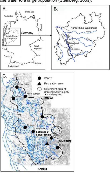
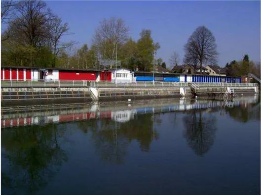
The River Rhine is one of the longest rivers in Europe (Lendering, 2011). The Lower
Rhine section (Figure 5A-C) comprises densely populated urban structures and rural
regions (NABU -Naturschutzstation e. V, 2011). The River Rhine and its tributaries
collect different types of materials due to erosion and from the faeces of wild and domestic animals. The deposition of enteric pathogens from the running water discharges from municipal sewage water plants is possible (Figure 5C). The retention of pathogens by riverbank filtration or by geological layers is most likely reduced because of the short distance between the groundwater level and landscape surface (depth to water table 0.5 to 1.0 m, partly, LINEG, unpublished observations). The Lower Rhine catchment area contains a drinking water supply (Figure 5C), which provides potable water to a large population (Steinberg, 2009).
Germany
North Rhine- Westphalia
North Rhine-Westphalia
Cologne Düsseldorf Low
R er hine
Rur
Ruhr
Rh ine
Lippe Netherlands
Belgium
France
Austria Czech Republic North Sea
Baltic Sea
Poland
Switzerland
A. B.
C.
Figure 5: Study area: North Rhine-Westphalia in the western part of Germany (A) and Lower Rhine
area as a part of it (B). Position of the eight municipal wastewater treatment plants, recreational area,
and catchment area of drinking water supply (C); (ArcGIS 9, ArcMap Version 9.3.1, Esri, Germany).
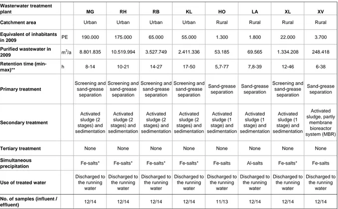
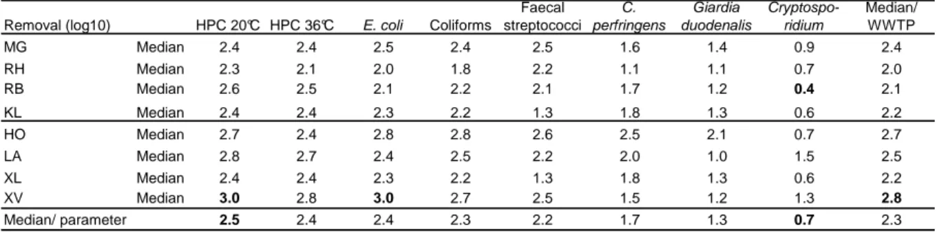
2.2.1.2 Investigated wastewater treatment plants
Eight municipal wastewater treatment plants (WWTPs) were investigated over a one- year period from 2009-2010 [see description in Table 2; WWTPs: Moers-Gerdt (MG), Rheinhausen (RH), Rheinberg (RB), Kamp-Lintfort (KL), Hoerstgen (HO), Labbeck (LA), Xanten-Lüttingen (XL) and Xanten-Vynen (XV)]. All sewage treatment plants included secondary sewage treatment: four had a two-stage activated sludge treatment process, three had a single-stage activated sludge purification and one had a membrane bioreactor (MBR). Based on German legislation, the WWTPs had state- of-the-art technology, with fixed effluent values only for chemical parameters (LANUV NRW, 1995; 2006; BMJ, 2009; EC, 2000), (Figure 5C) but not for microbiological pollutants.
Wasterwater treatment
plant MG RH RB KL HO LA XL XV
Catchment area Urban Urban Urban Urban Rural Rural Rural Rural
Equivalent of inhabitants
in 2009 PE 190.000 175.000 65.000 55.000 1.300 1.800 22.000 3.700
Purified wastewater in
2009 m3/a 8.801.835 10.519.994 3.527.749 2.411.336 53.185 69.565 1.334.208 248.418
Retention time (min-
max)** h 8-14 10-21 14-27 17-50 5,7-77 7,8-39 12-46 6-38
Primary treatment
Screening and sand-grease
separation
Screening and sand-grease
separation
Screening and sand-grease
separation
Screening and sand-grease
separation
Sand-grease separation
Sand-grease separation
Screening and sand-grease
separation
Sand-grease separation
Secondary treatment
Activated sludge (2 stages) and sedimentation
Activated sludge (2 stages) and sedimentation
Activated sludge (2 stages) and sedimentation
Activated sludge (2 stages) and sedimentation
Activated sludge (1 stage) and sedimentation
Activated sludge (1 stage) and sedimentation
Activated sludge (1 stage) and sedimentation
Activated sludge, partly
membrane bioreactor system (MBR)
Tertiary treatment None None None None None None None None
Simultaneous
precipitation Fe-salts* Fe-salts* Fe-salts* Fe-salts* Fe-salts Al-salts Fe-salts* Fe-salts
Use of treated water
Discharged to the running
water
Discharged to the running
water
Discharged to the running
water
Discharged to the running
water
Discharged to the running
water
Discharged to the running
water
Discharged to the running
water
Discharged to the running
water No. of samples (influent /
effluent) 12/14 12/14 12/14 12/14 11/13 12/14 12/14 12/14
* In case of dominance of the filamentous Bacteria Microthrix parvicella FeCl3 is substituted with Polyaluminate; PE people equivalents; Data from LINEG, Germany, unpublished.
** Depending from precipitation.