INAUGURAL-DISSERTATION
zur
Erlangung des Doktorgrades
der Mathematisch-Naturwissenschaftlichen Fakultät der Universität zu Köln
vorgelegt von
Deenadayalan Bakthavatsalam aus
Chennai, Indien
Köln, 2004
Referees/Berichterstatter: Prof. Dr. Angelika A. Noegel Prof. Dr. Martin Huelskamp Date of oral examination: 10.02.2005
Tag der mündlichen Prüfung
The present research work was carried out under the supervision of Prof. Dr. Angelika A.
Noegel, in the Institute of Biochemistry I, Medical Faculty, University of Cologne, Cologne, Germany from Nov 2001 to Feb 2005.
Diese Arbeit wurde von Nov 2001 bis Feb 2005 am Biochemischen Institut I der
Medizinischen Fakultät der Universität zu Köln unter der Leitung von Prof. Dr. Angelika
A.Noegel durchgeführt.
In the preparation of this doctoral thesis I gratefully acknowledge the contribution made by one and all, supervisors, colleagues, well-wishers and friends.
First of all I would like to express my heartfelt gratitude to my supervisor Prof. Angelika Noegel for her diligent guidance and constant encouragement throughout the course of my study. She strove to seek the very best in me with her expertise and knowledge, which provided the backbone of this research. Thank you very much for giving me this privilege.
I am also greatly indebted to Dr. Ludwig Eichinger for his unfailing patience and valuable suggestions especially with all the microarray studies. Thanks a lot Ludwig for sparing your
valuable time whenever needed.
I also owe so very much to Dr. Francisco for his continual support, enthusiasm and encouragement throughout the course of my work. Thank you Francisco for your invaluable
support and timely help. Special thanks are due to Dr. Derrick and Dr. Schlatterer for helping with the characterization of the mutant.
I thank Dr. Gomer for useful discussions and my thanks also to Dr. Devreotes, Dr. Nellen and Dr.
Gundersen for providing antibodies.
I would like to mention my special thanks to Patrick for his never-ending support in microarray preparations and analysis. My thanks are also due to Dr. Budi Tunggal for all the help with the
computers. I thank the secretaries Bettina and Dörte for their help in administration issues.
I am also thankful to Maria, Berthold, Rosie, Kathrin, Rolf and Martina for helping in more than one way and contributing towards this work.
I also highly acknowledge the financial support received from the DFG.
I owe a special thanks to my friend Sunil for being beside me throughout my stay.
Last but not at all the least I would like to thank my parents for never losing faith in me and being by my side always. Thank you mom and dad for everything.
My thanks will not end without mentioning Sharada for her constant support and helping hand in preparing the thesis. Thank you Sharada for your patience and smiling face during tough times
and always being an inspiration.
I also thank all my friends and colleagues in the department for making my stay memorable. I will never forget the Christmas and Carneval parties.
Chapter Description Page(s) Abbreviations 1-2
I. Introduction 3-10
1.1 G Protein Coupled Receptors 3
1.2 Frizzled proteins 4 1.2.1 Functions of Frizzled proteins 4
1.2.2 Frizzled role in development and diseases 6
1.3 Do Frizzled receptors exist in Dictyostelium? 6 1.3.1 Dictyostelium as a model system 8
1.3.1.1 Early development 8
1.3.1.2 Calcium role in early development 9
1.3.1.3 cAMP signaling 9 1.4 Aim of work 10
II. Materials and Methods 11-51 1. Materials 11
1.1. Laboratory materials 11
1.2. Instruments and equipments 12
1.3. Kits 13
1.4. Enzymes, antibodies, substrates, inhibitors and antibiotics 14
1.5. Chemicals and reagents 15
1.6. Media and buffers 16
1.6.1 Media and buffers for Dictyostelium culture 16
1.6.2 Media for E. coli culture 17
1.6.3 Buffers and other solutions 18
1.7 Biological materials 19
1.7 Plasmids 19
1.8 Oligonucleotide primers 19
2 Cell biological methods 20
2.1 Growth of Dictyostelium 20
2.1.1 Growth in liquid nutrient medium 20
2.1.2 Growth on SM agar plates 20
2.2 Development of Dictyostelium 21
2.2.1 Development in suspension culture 21
2.2.2 Development on phosphate-buffered agar plates 21
2.3 Transformation of Dictyostelium 21
2.4 Preservation and revival of preserved Dictyostelium cells 22
3 Molecular biological methods 23
3.6 Purification of plasmid DNA 23
3.7 Digestion with restriction enzymes 23
3.8 Dephosphorylation of DNA fragments 23
3.9 Setting up of ligation reaction 23
3.10 Isolation of Dictyostelium genomic DNA 24
3.11 DNA agarose gel electrophoresis 25
3.12 Recovery of DNA fragments from agarose gel 25
3.13 Southern blotting 25
3.14 Total RNA and cDNA preparation 26
3.9.1 Isolation of total RNA from Dictyostelium cells 26
3.9.2 Generation of cDNA 26
3.15 RNA formaldehyde agarose gel electrophoresis 27
3.16 Northern blotting 28
3.17 Radiolabelling of DNA 28
3.13 Chromatography through Sephadex G-50 spin column 28
3.14 Hybridization of Southern or northern-blot with radiolabelled DNA probe 29
3.15 Transformation of E. coli 29
3.15.1 Transformation of E. coli cells by the CaCl
2method 29
3.15.2 Glycerol stock of bacterial culture 30
3.15.3 DNA sequencing 30
3.16 Quantitative PCR 30
4. Microarray Analysis 31
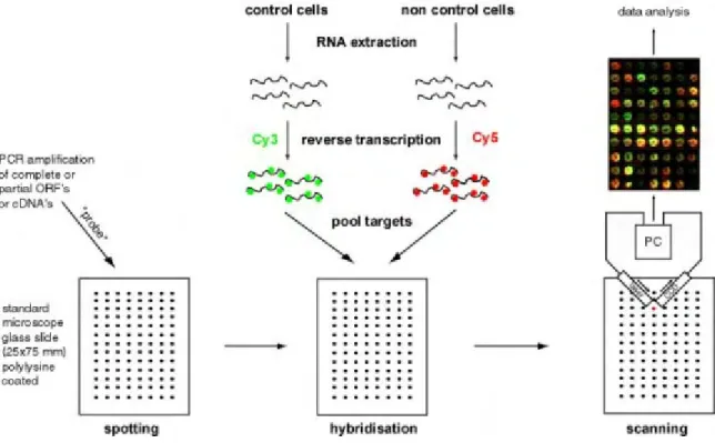
4.1 Principle 31
4.2 The Dictyostelium discoideum DNA microarray 31
4.2.1 Prehybridization 32
4.2.2 Sample preparation and cDNA preparation 32
4.2.3 Spiking of internal mRNA controls 33
4.3 cDNA generation and Fluorescent labeling of cDNA 34
4.4 Hybridization of microarray slide 34
4.4.1 Buffers and solutions 34
4.5 Scanning of microarray slides 36
4.6 Data Analysis 36
5.0 Construction of vectors 37
5.1 Amplification and cloning of the partial and
full-length cDNA 37
5.2 Vector for expression as a GFP-fusion protein 37
5.3 Vector for expression of PIP5K domain as a GST-fusion protein 38 5.4 Gene replacement vectors 38 5.4.1 Screening of FrzA
-mutant 39 6. Biochemical methods 40
6.1 Cell culture of Dictyostelium 40
6.2 cAMP pulsing experiment 40
6.3 Subcellular fractionation 40
6.4 Preparation of a membrane-enriched cell fraction for cAR1 analysis 40
6.5 PLC assay 41
6.6 cGMP assay 41
6.7 SDS-polyacrylamide gel electrophoresis 41
6.8 Coomassie blue staining of SDS-polyacrylamide gels 42
6.9 Drying of SDS-polyacrylamide gels 43
6.10 Western blotting using the semi-dry method 43 6.11 Ponceau S staining of western blots 43 6.12 Immunodetection of nitrocellulose membrane-bound proteins 44 6.13 Enzymatic chemiluminescence (ECL) detection system 44 6.14 Expression and purification of GST fusion protein 45 6.14.1 Small-scale protein expression 45 6.14.2 Large-scale protein expression 45 6.14.3 Preparation of cell homogenates 46 6.14.4 Analysis of GST fusion protein in fractionated cell lysate of E. coli 46 6.14.5 Affinity purification of polyclonal antibodies by the blot method 47 7 Cell biological methods 47 7.1 Measuring cell size 47 7.2 Growth rate measurement 47 7.3 Fluid uptake analysis 48 7.4 Conditioned medium preparation and starvation under submerged condition 48 7.5 Light scattering measurements and [Ca
2+]
idetermination 48
7.6 Prespore differentiation assay 48
7.7 Video imaging and chemotaxis assay 49
7.8 Indirect immunofluorescence of Dictyostelium cells 49
7.8.1 Preparation of Dictyostelium cells 49
7.8.2 Methanol and PFA fixation 49
7.8.3
Immunolabeling 50
7.8.4
DAPI and phalloidin staining of fixed cells 50
7.8.5
Mounting 51
8 Computer analyses 51
III. Results 52-98
1.0 Results 52
1.1 Search for polarity genes in Dictyostelium 52 1.2 Sequence analysis and generation of the complete sequence for FrzA 52 1.2.1 Dictyostelium Frizzled like proteins 53 1.2.2 Comparison of FrzA protein structure to a typical Frizzled receptor 54
1.2.3 Domain analysis of the FrzA protein 55
1.3 Northern blot analysis for studying the gene expression pattern
of the FrzA 57
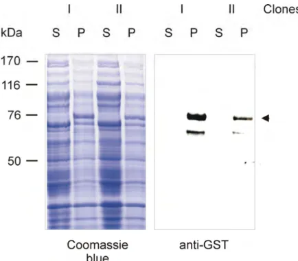
1.4 Expression of GST fusion proteins 57
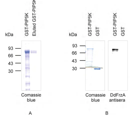
1.5 Purification of GST-PIP5K fusion protein 59 1.5.1 Characterisation of FrzA polyclonal antibodies 59
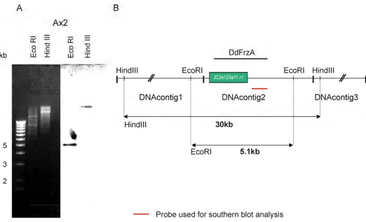
2.0 Generation of FrzA
−mutant cells 60
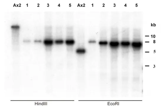
2.0.1 Southern blot analysis 61
2.1 Characterisation of the FrzA
−mutant 62
2.1.1 Measurement of cell size of Ax2 and FrzA
−62
2.1.2 Growth in axenic medium 63
2.1.3 Cytokinesis in FrzA
−64
2.1.4 Fluid uptake in wild type and FrzA
−64
2.1.5 Growth on a lawn of Klebsiella 65
2.2 Aggregation and development of FrzA
−mutant 66
2.2.1 Aggregation on a plastic surface 66
2.2.2 Development on phosphate agar plates 66
2.3 Characterisation of
FrzA−development 67
2.4 Role of FrzA in cell adhesion and aggregation 68 2.4.1 Can exogenous cAMP pulses also induce ACA expression? 70 2.5 Synergy between FrzA
−and wild type cells 71 2.6 Cell motility and chemotaxis of FrzA
−mutant 72 2.7 Does FrzA play an important role even before aggregation? 74 2.7.1 Cell density factor in FrzA
−mutant cells 74 2.7.2 Do FrzA
−cells produce and/or sense cell density factors? 75
2.7.3 Expression of CMF and CMFR1 in FrzA
-76
2.7.3 Is CMF mediated signaling in FrzA
−mutant cells affected? 77 3.0 cAMP signal transduction in FrzA
−cells 78 3.1 Function of FrzA in cell differentiation at low cell density 80 3.2 Production of Inositol Phosphate-3 (IP3) and cGMP in wild type
and FrzA
−cells 81
3.3 Calcium oscillation in FrzA
−cells 82
3.4 Complementation analysis for FrzA
−83
3.4.1 Expression of PIP5K-GFP fusion proteins 83 3.4.2 Subcellular localization of GFP-PIP5K in vegetative cells 84 3.4.3 Development of Ax2 and FrzA
−mutant expressing the
PIP5K domain 86
3.5 Microarray analysis 87
3.5.1 Experimental design 87
3.5.2 Comparison of the expression profile of vegetatively
growing cells 88
3.5.3 Comparison of the expression profile during
early development 89
3.5.3.1 Differential gene regulation after 4 hrs of starvation 90 3.5.3.2 Differential gene regulation after 8 hrs of starvation 92 3.5.3.3 Comparison of differentially regulated genes under
HCD and LCD conditions 94
3.5.3.4 Cluster analysis 95
3.5.3.5 Developmental regulation of genes in cluster 2
in comparison to Ax2 and the ACA
−mutant 97 IV. Discussion 99-106
1.0 Discussion 99
1.1 FrzA role in aggregation and development of Dictyostelium 100
1.2 csA mediated cell adhesion is controlled by FrzA 102
1.3 FrzA plays a major role in chemotaxis 102
1.4 Function of FrzA in cAMP signaling 103
1.5 FrzA affects calcium oscillation 104
1.6 The FrzA acts as a GPCR like CMF Receptor 104
1.7 Microarray analysis of FrzA
−mutant 106
V Summary 108-110
VI. References 111-121
APS ammonium persulphate ATP adenosine 5’-triphosphate
bp base pair(s)
BCIP 5-bromo-4-chloro-3-indolylphosphate BSA bovine serum albumin
Bsr blasticidin resistance cassette cAMP cyclic adenosine monophosphate
cDNA complementary DNA
CIAP calf intestinal alkaline phosphatase dNTP deoxyribonucleotide triphosphate DABCO diazobicyclooctane
DEPC diethylpyrocarbonate
DMSO dimethylsulphoxide
DNA deoxyribonucleic acid
DNase deoxyribonuclease
DTT 1,4-dithiothreitol
ECL enzymatic chemiluminescence EDTA ethylenediaminetetraacetic acid
EGTA ethyleneglycol-bis (2-amino-ethylene) N,N,N,N-tetraacetic acid ELISA enzyme linked immunosorbent assay
G418 geneticin
GFP green fluorescent protein GST glutathione S-transferase HRP horse radish peroxidase
IgG immunoglobulin G
IPTG isopropyl-β-D-thiogalactopyranoside
kb kilo base pairs
MES morpholinoethansulphonic acid
β-MEbeta-mercaptoethanol
MOPS Morpholinopropanesulphonic acid
Mw molecular weight
NP-40 nonylphenylpolyethyleneglycol pNPP para-nitrophenyl phosphate
OD optical density
ORF open reading frame
PAGE polyacrylamide gel electrophoresis PCR polymerase chain reaction
PMSF phenylmethylsulphonylfluoride
RACE PCR random amplification of cDNA ends polymerase chain reaction RT-PCR reverse transcript polymerase chain reaction
RNA ribonucleic acid
RNase ribonuclease
rpm rotations per minute SDS sodium dodecyl sulphate
TEMED N,N,N’,N’-tetramethyl-ethylendiamine
U unit
UV ultra violet
vol. volume
v/v volume by volume
Units of Measure and Prefixes
Unit Name Symbol Prefix (Factor)
°C degree Celsius k kilo (10
3)
kDa Dalton c centi (10
-2)
g gram m milli (10
-3)
hrs hour µ micro (10
-6)
ml millilitre n nano (10
-9)
m meter p pico (10
-12)
min minute
sec second
V volt
mol mole
1. Introduction
1.1 G Protein Coupled Receptors
Multicellular organisms have highly evolved based on the capacity of their cells to communicate with each other and with their environment. Over years of discovery the membrane-bound receptors have been categorized into four or five families belonging to one large G protein-coupled receptor (GPCR) superfamily. GPCRs are among the oldest signal transduction machinery present in plants (Plakidou-Dymock et al., 1998), yeast (Dohlman et al., 1991) and slime mold (Dictyostelium discoideum) (Devreotes, 1994), which control the activity of enzymes, ion channels and transport of vesicles via the catalysis of the GDP–GTP exchange on heterotrimeric G proteins (Gα–βγ). A large number of genes code for GPCRs in vertebrates and Caenorhabditis elegans that are mainly involved in the recognition and transduction of messages as diverse as light, Ca
2+, odorants, small molecules including amino- acids, nucleotides and peptides, as well as proteins (Figure I). Although sequence comparison between the different GPCRs revealed the existence of different receptor families sharing low sequence similarity they have a central core domain constituted of seven transmembrane helices (TM I-VII).
Figure I. Illustration of the GPCR mediated signal transductions (Bockaert and Pin, 1999).
Two cysteine residues in the extracellular loops (e1 and e2) are conserved in most GPCRs that
form a disulfide link which is probably important for the packing and for the stabilization of a
restricted number of conformations of these seven TMs (Figure I). GPCRs differ in the length
and function of their N-terminal extracellular domain, their C-terminal intracellular domain
and their intracellular loops. Each of these domains provides specific properties to the various
receptor proteins and any conformational change in the core domain will affect the
intracellular pocket formed between the TM III-VI that plays a major role in binding and activating the G proteins (Bockaert and Pin, 1999).
1.2 Frizzled proteins
Frizzled proteins belong to the cell surface proteins functioning as the receptor for the Wnt ligand and range in length from about ~500 to 700 amino acids. The amino terminus of Frizzled protein is predicted to be extracellular and contains a cysteine-rich domain (CRD) that is required for binding of Wnt molecules (Bhanot et al., 1996), followed by a hydrophilic linker region of 40-100 amino acids. The proteins also contain seven hydrophobic domains that are predicted to form transmembrane α-helices. The intracellular carboxy-terminal domain has a variable length and is not well conserved among different family members (Wang et al., 1996). Moreover, the presence of seven hydrophobic domains has categorized the protein to be related to the GPCR superfamily. The sequence similarity to GPCRs is low, because similarity is restricted to the seven transmembrane regions that have a high frequency of hydrophobic residues.
1.2.1 Functions of Frizzled proteins
Frizzled proteins are exclusively found at the cell surface to bind Wnt and get internalized to regulate the extracellular level of Wnt protein (Chen et al., 2003). The binding of Wnt ligands to the Frizzled receptors induces a canonical and two non-canonical β-catenin dependent Wnt/Fz pathways, the planar cell polarity (PCP) and the Wnt/Ca
2+pathway.
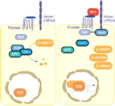
I Canonical Wnt/β-catenin pathway
The canonical Wnt/β-catenin pathway is characterized by stabilization of β-catenin in response to ligand binding. Wnt proteins act on the target cells by binding to the Frizzled (Fz)/low-density lipoprotein (LDL) receptor-related protein (LRP) complex at the cell surface.
These receptors transduce a signal to several intracellular proteins which mainly includes
Dishevelled (Dsh) and the transcriptional regulator, β-catenin. Cytoplasmic β-catenin levels
are normally kept low through continuous proteasome-mediated degradation but the
degradation pathway is inhibited when cells receive Wnt signals and consequently β-catenin
accumulates in the cytoplasm and nucleus. This accumulated β-catenin in the nucleus
interacts with the transcription factors such as the lymphoid enhancer-binding factor 1/Tcell-
specific transcription factor (LEF/TCF) to affect transcription (Figure III). A large number of
Wnt targets have been identified that include members of the Wnt signal transduction
pathway itself, which provide feedback control during Wnt signaling (Logan and Nusse, 2004).
Figure III. Canonical Wnt signaling (Logan and Nusse, 2004)
II Non-canonical planar cell polarity
The PCP pathway regulated genes when mutated result in cell polarity defects in a planar tissue. A non-canonical Frizzled (fz) activity controls the coordinated cell polarity decisions in the Drosophila cuticle where each cell produces a single hair on its apical surface at the distal vertex of the cell, which then grows out distalwards (Figure IIA). In the absence of fz, hairs form in the centre of the apical surface of the cell and no longer invariably grow out distalwards (Figure IIB) (Wong and Adler, 1993). This constitutes the cell-autonomous activity of fz in the wing.
Figure II. Planar cell polarity in Drosophila wing (Strutt, 2003).
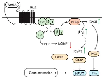
III Non-canonical Wnt/Ca
2+pathway
In the Wnt/calcium pathway Frizzled appears to act through heterotrimeric guanine nucleotide–binding proteins (G proteins) activating phospholipase C (PLC) (Slusarski et al., 1997a) and phosphodiesterase (PDE) (Ahumada et al., 2002), which leads to increased concentrations of free intracellular calcium [Ca
2+]
i(Slusarski et al., 1997b) and to decreased intracellular concentrations of cyclic guanosine monophosphate (cGMP) (Ahumada et al., 2002), respectively (Figure IV). However the newly discovered role of cGMP in Wnt signaling is the least well understood. Therefore Frizzleds appear to function in multiple pathways, including the PCP, Wnt/calcium, and canonical Wnt/ß-catenin pathways and induce different signal transduction pathways that regulate some of the major cellular processes in a cell.
Figure IV. Frizzled mediated Wnt-Ca2+ signaling (Wang and Malbon, 2003).
1.2.2 Frizzled role in development and diseases
Frizzled receptors regulate specification of cell fate, cell adhesion, migration, polarity, and proliferation by binding to Wnt molecules. Their roles in development have been analysed by the phenotypes exhibited in different model systems as a result of gene knockout or mutations which is summarized in Table I (Huang and Klein, 2004). Noticable is a human hereditary disorder, called familial exudative vitreopathy (FEVR), which is caused by mutation in the Frizzled (Fz4) receptor that results in defective vasculogenesis in the peripheral retina (Robitaille et al., 2002).
1.3 Do Frizzled receptors exist in Dictyostelium?
Frizzled’s role is remarkable with extended functions from cell polarity in Drosophila to the
human diseases. The highly conserved signal transduction pathways identified in many
primitive organism like Hydra vulgaris to highly evolved species such as humans prompted us to search for Frizzleds in Dictyostelium. Although Dictyostelium has a β-catenin, a canonical β-catenin dependent pathway is not described.
Table I. Frizzleds function in different model systems. All the phenotypes exhibited by the organisms due to the lack of Frizzled or mutation in Frizzled presented here is taken from (Huang and Klein, 2004). The number represents the respective references, 1-(Adler, 2002), 2 and 3-(Chen and Struhl, 1999), 4-(Muller et al., 1999), 5-(Kennerdell and Carthew, 1998), 6-(Sato et al., 1999), 7- (Rocheleau et al., 1997), 8-(Harris et al., 1996), 9-(Sawa et al., 1996), 10-(Wang et al., 2002), 11- (Wang et al., 2001), 12-(Ishikawa et al., 2001), 13-(Robitaille et al., 2002), 14-(Deardorff et al., 2001), 15-(Sumanas et al., 2000) , 16-(Winklbauer et al., 2001).
However, cAR1 mediated signaling has been compared to the cell fate determination pathway in Xenopus, Drosophila and C. elegans (Kim et al., 2002). The PCP pathway may be present in Dictyostelium as the cells during development exhibit cell polarity (Williams and Harwood, 2003), but cell polarity is not directly associated to the components of PCP signaling.
Calcium is an important part of Dictyostelium development (Bumann et al., 1984) and all the
components of the Wnt/Ca
2+pathway are present except the Wnt protein and the Frizzled
protein. The Wnt proteins are extracellular signaling molecules that play a central role in cell
fate determination in C. elegans (Nusse and Varmus, 1992). Although Wnt proteins were not
identified in Dictyostelium, CMF, an extracellular glycoprotein was identified that determines
the cell density for aggregation and the cell type jointly with cAMP during development
(Mehdy and Firtel, 1985; Yuen et al., 1995). But when searched for Frizzled in the Dictyostelium we identified 25 Frizzled like receptors.
1.3.1 Dictyostelium as a model system
D. discoideum is a eukaryote, which can exist as a unicellular and multicellular organism.
Various signal-transduction pathways regulates processes such as chemotaxis, cell adhesion and cell fate determination and are closely related to metazoan pathway (Firtel and Chung, 2000; Gerisch, 1968; Kim et al., 2002). Therefore Dictyostelium has been identified as a good model system for studying many biological processes. Dictyostelium cells feed on bacteria and grow as amoebae but with depletion of food they initiate development and achieve multicellularity by the aggregation of cells (Figure V) (Brown and Firtel, 1999).
Figure V. Life cycle of Dictyostelium (Williams and Harwood, 2003)
1.3.1.1 Early development
The amoebae feed on bacteria and grow exponentially until the depletion of the food source.
The status of the food supply is monitored by secretion of a prestarvation factor (PSF). The PSF expression is high till the depletion of the food source and the level goes down with the onset of starvation. The genes that are expressed at low level in response to PSF during the late exponential growth are expressed at higher level in starving cells (Rathi and Clarke, 1992). Discoidin-I is one of them which is induced in response to PSF (Rathi et al., 1991) and continues to be stimulated during early development by conditioned medium factor (CMF), a factor secreted by starving cells (Gomer et al., 1991). The expression level of discoidin-I is downregulated by cAMP during aggregation which is important for the formation of head-to-tail streams by aggregating cells (Crowley et al., 1985; Vauti et al., 1990). Although these factors PSF and CMF are produced at different times in the Dictyostelium life cycle, cells need both glycoproteins to measure their own density and to aggregate.
The developing Dictyostelium cells exhibit chemotactic response by extending pseudopods
towards cAMP during aggregation (Soll et al., 2002). During this process the cells form
radial branching streams, which results from the cAMP wave propagation depending on the
cell density (van Oss et al., 1996). These streams then move coordinately towards the aggregation centre to form a mass of 10
5cells. When the cells move in streams they adhere to each other and express first the cell adhesion protein gp24. In later stage of aggregation a second adhesion molecule, csA, is expressed which is induced by cAMP pulses secreted by the starving cells. Cell-cell adhesion was found to be important for induction of developmentally regulated genes during the aggregation (Faix et al., 1992). The cAMP synthesized by aggregating cells is sensed by a cell surface receptor (cAR1) which through G protein signaling stimulates cAMP production through an adenylyl cyclase (Fontana et al., 1991; Klein et al., 1988). The signaling molecule cAMP acts as a chemoattractant, secreted at the aggregation centres towards which cells make directed migration (Alcantara and Monk, 1974). Similar chemotactic responses are critical for a wide variety of biological processes including immune system responses, neuronal path finding, angiogenesis and metastasis.
Signalling molecules downstream of the receptors that contribute to chemotactic responses include guanylyl cyclase, phosphatidylinositol 3-kinases (PI 3-kinases), phospholipases and mitogen-activated protein kinases (Segall, 1999). Moreover, Dictyostelium and mammalian cells have a common mechanism for migration.
1.3.1.2 Calcium role in early development
The binding of cAMP to its receptor cAR1 also induces a transient entry of external Ca
2+, however G-proteins are not required for this process (Milne et al., 1995). Ca
2+influx increases the intracellular Ca
2+and activates phospholipase C (PLC) to elevate inositol 1, 4, 5- triphosphate (IP3), cAMP and cGMP. Although Ca
2+levels control the motile response it is not required for chemotaxis (Malchow et al., 1996a; Traynor et al., 2000). A slow periodic Ca
2+oscillation was observed during early development in suspension (Bumann et al., 1984) and a wave of Ca
2+is propagated from the tip of the slug towards the rear end in the multicellular slug stage that may help to regulate gene expression and coordinate cell movement in Dictyostelium (Pinter and Gross, 1995).
1.3.1.3 cAMP signaling
When cells are in the preaggregative stage a low level of cAMP is produced in a pulsatile
fashion that augments the expression of aggregative genes coding for cAMP receptor (cAR1)
and contact sites A (csA) (Gerisch and Malchow, 1976). cAR1 null cells show a defect in
aggregation, however it is not clear whether cells produce a signal or whether the signal
produced is not transduced to regulate gene expression. At the aggregation stage the cAMP
produced binds to cAR1 and induces the G protein Gα2 which binds to cAR1 and frees the Gβ causing PI-3 kinase activation that stimulates activation of the adenylyl cyclase (ACA) gene. ACA is expressed at high levels exclusively at the aggregation stage and is necessary for synthesis of the chemoattractant cAMP. The cAMP produced mediates chemotaxis and induces gene expression through cAR1 (van Haastert and Kien, 1983; Wu et al., 1995).
The hydrolysis of cAMP is very important for dynamic signaling. Dictyostelium employs 5 different classes of phosphodiesterase to breakdown cAMP, of which the cAMP phosphodiesterase gene (PDE) shows a pronounced expression during aggregation and is induced by cAMP pulses contributing to the negative feedback loop of oscillatory cAMP signaling (Meima et al., 2003). The level of active PDE is regulated at transcription level and by the phosphodiesterase inhibitor (PDI), which is induced at low levels of cAMP and inhibits the PDE by binding. PDI expression is also controlled by the presence of high levels of PDE (Franke and Kessin, 1992). The expression level of ACA decreases with the progression of aggregation to the mound stage. At this stage the postaggregative and cell-type specific genes stimulate a developmental change to form cell types that are the precusor cells found within the mature fruiting body.
1.4 Aim of work
We identified for the first time Frizzled in Dictyostelium, and found FrzA, one of the 25 Frizzled like receptors. It has a unique C-terminus with a Phosphatidylinositol-4-phosphate 5- kinase (PIP5K) domain. The N-terminus preceeding the Frizzled transmembrane domain is rather short and lacks the extracellular CRD region. Our studies included RACE PCR, RT- PCR and characterisation of the genomic and cDNA database, to assemble the full-length FrzA cDNA. The sequence homology of FrzA showed its close relation to the smoothened receptor, which is a homologue of Frizzled having a structural and functional similarity to the GPCR. To characterize the cell biology of the FrzA protein further we generated a polyclonal antibody against the C- terminus of FrzA. To get further insight in to the function of this protein, a knockout strategy was initiated. Although the knock out cells survive as amoebae, they do not aggregate during the developmental cycle to develop into fruiting bodies. To understand the role of FrzA in Dictyostelium during early development, we analysed the FrzA
−mutant in detail with regard to,
1. Aggregation 4. cAMP and calcium signaling
2. Cell-cell adhesion 5. Cell fate determination
3. Cell migration
1. Materials
1.1 Laboratory materials
Cellophane sheet, Dry ease Novex
Centrifuge tubes, 15 ml, 50 ml Greiner
Coverslips (glass), ∅ 12 mm, ∅ 18 mm Assistant
Corex tube, 15 ml, 50 ml Corex
Cryo tube, 1 ml Nunc
Electroporation cuvette, 2 mm electrode gap Bio-Rad
Gel drying frames Novex
Hybridisation bag Life technologies
Microcentrifuge tube, 1.5 ml, 2.2 ml Sarstedt Micropipette, 1-20 µl, 10-200 µl, 100-1,000 µl Gilson
Micropipette tips Greiner
Multi-channel pipette Finnigan
Needles (sterile), 18G–27G Terumo, Microlance Nitrocellulose membrane, BA85 Schleicher and Schuell Nitrocellulose-round filter, BA85, ∅ 82 mm Schleicher and Schuell
Nylon membrane, Biodyne B Pall
Parafilm American National Can
Pasteur pipette, 145 mm, 230 mm Brand, Volac
PCR softtubes, 0.2 ml Biozym
Petri dish (35 mm, 60 mm, 100 mm) Falcon
Petri dish (90 mm) Greiner
Plastic cuvette, semi-micro Greiner
Plastic pipettes (sterile), 1 ml, 2 ml, 5 ml, 10 ml, 25 ml Greiner
Quartz cuvette, Infrasil Hellma
Quartz cuvette, semi-micro Perkin Elmer
Saran wrap Dow
Scalpels (disposable), Nr. 10, 11, 15, 21 Feather
Slides, 76 x 26 mm Menzel
Syringes (sterile), 1 ml, 5 ml, 10 ml, 20 ml Amefa, Omnifix
Syringe filters (Acrodisc), 0.2 µm, 0.45 µm Gelman Sciences Tissue culture flasks, 25 cm
2, 75 cm
2, 175 cm
2Nunc
Tissue culture dishes, 6 wells, 24 wells, 96 wells Nunc
Whatman 3MM filter paper Whatman X-ray film, X-omat AR-5, 18 x 24 mm, 535 x 43 mm Kodak
1.2 Instruments and equipments Centrifuges (microcentrifuges):
Centrifuge 5417 C Eppendorf
Centrifuge Sigma B. Braun Biotech Instruments Cold centrifuge Biofuge fresco Heraeus Instruments
Centrifuges (table-top, cooling, low speed):
Centrifuge CS-6R Beckman
Centrifuge RT7 Sorvall
Centrifuge Allegra 21R Beckman
Centrifuges (cooling, high speed):
Beckman Avanti J25 Beckman
Sorvall RC 5C plus Sorvall
Centrifuge-rotors:
JA-10 Beckman
JA-25.50 Beckman
SLA-1500 Sorvall
SLA-3000 Sorvall
SS-34 Sorvall
Dounce homogeniser, 10 ml and 60 ml B. Braun Electrophoresis power supply, Power-pac-200, -300 Bio-Rad Electroporation unit, Gene-Pulser Bio-Rad
Freezer (-80°C) Nunc
Freezer (-20°C) Siemens, Liebherr
Gel-documentation unit MWG-Biotech
Heating block, DIGI-Block JR neoLab
Heating block, Dry-Block DB x 20 Techne
Hybridising oven Hybaid
Ice machine Ziegra
Incubators:
CO
2-incubator, BBD 6220, BB 6220 Heraeus
CO
2-incubator, WTC Binder Biotran
Incubator, microbiological Heraeus Incubator with shaker, Lab-Therm Kuehner Laminar flow, Hera Safe (HS 12) Heraeus
Magnetic stirrer, MR 3001 K Heidolph Microscopes:
Light microscope, CH30 Olympus Light microscope, DMIL Leica
Light microscope, CK2 Olympus
Fluorescence microscope, DMR Leica Fluorescence microscope, 1X70 Olympus Fluorescence microscope, CTR/MIC Leica Confocal laser scan microscope, DM/IRBE Leica Stereomicroscope, SZ4045TR Olympus
Oven, conventional Heraeus
PCR machine, PCR-DNA Engine PTC-2000 MJ Research
pH-Meter 766 Calimatic Knick
Refrigerator Liebherr
Semi-dry blot apparatus, Trans-Blot SD Bio-Rad
Shakers GFL, Kuehner
Sonicator (water bath), Sonorex RK 52 Bandelin Speed-vac concentrator, DNA 110 Savant
Spectrophotometer: Ultraspec 2000, UV/visible Pharmacia Biotech
BioPhotometer Eppendorf
Scanarray Express Scanner Perkin Elmer UV- transilluminator, TFS-35 M Faust
Vortex, REAX top Heidolph
Waterbath GFL
X-ray-film developing machine, FPM-100A Fujifilm 1.3 Kits
Nucleobond AX 100 and 500 Macherey-Nagel
NucleoSpin Extract 2 in 1 Macherey-Nagel
Nucleotrap Macherey-Nagel
1 kb DNA-marker Gibco-BRL
pGEM-T Easy Promega
High molecular weight protein marker Amersham Biosciences Low molecular weight protein marker Amersham Biosciences
SMART RACE PCR Kit Clontech
Qiagen RNeasy Mini or Midi Kit Qiagen 1.4 Enzymes, antibodies, substrates, inhibitors and antibiotics Enzymes used in the molecular biology experiments:
Calf Intestinal Alkaline Phosphatase (CIP) Boehringer
Klenow fragment Boehringer
Lysozyme Sigma
Proteinase K Sigma
Restriction endonucleases Amersham, Life technologies, New England Biolabs
Reverse transcriptase, M-MLV Promega
Ribonuclease H (RNase H) Boehringer
T4 DNA ligase Boehringer
Taq-polymerase Life technologies/Boehringer
Primary antibodies:
Goat anti-GST antibody Pharmacia
Mouse anti-GFP monoclonal antibody, K3-184-2 (Noegel et al., 2004)
Polyclonal anti-cAR1 Gift from Dr. Peter Devreotes Polyclonal anti- Gα2 Gift from Dr. Gundersen
Secondary antibodies:
Goat anti-mouse IgG, peroxidase conjugated Sigma Goat anti-rabbit IgG, peroxidase conjugated Sigma Mouse anti-goat IgG, peroxidase conjugated Sigma Sheep anti-mouse IgG, Cy3 conjugated Sigma Inhibitors:
Diethylpyrocarbonate (DEPC) Sigma
Leupeptin Sigma
Pepstatin Sigma Phenylmethylsulphonylfluoride (PMSF) Sigma
Benzamidine hydrochloride Sigma
Proteinase Inhibitor cocktail Sigma Antibiotics:
Ampicillin Grünenthal
Blasticidin S ICN Biomedicals
Chloramphenicol Sigma
Dihydrostreptomycinsulphate Sigma
Geneticin (G418) Life technologies
Kanamycin Sigma, Biochrom
Tetracyclin Sigma
1.5 Chemicals and reagents
Most of the chemicals and reagents were obtained either from Sigma, Fluka, Difco, Merck, Roche, Roth or Serva. Those chemicals or reagents that were obtained from companies other than those mentioned here are listed below:
Acetic acid (98-100%) Riedel-de-Haen
Acrylamide (Protogel: 30:0,8 AA/Bis-AA) National Diagnostics
Agar-Agar (BRC-RG) Biomatic
Agarose (Electrophoresis Grade) Life technologies
Chloroform Riedel-de-Haen
Dimethylformamide Riedel-de-Haen
Ethanol Riedel-de-Haen
Glycerine Riedel-de-Haen
Glycine Riedel-de-Haen Isopropypl-β-D-thiogalactopyranoside (IPTG) Loewe Biochemica
Methanol Riedel-de-Haen
Morpholino propane sulphonic acid (MOPS) Gerbu Peptone Oxoid
Sodium hydroxide Riedel-de-Haen
Yeast extract Oxoid
Roti-Phenol Roth
TRITC Phallodin Sigma
Radiolabelled nucleotide:
α-32
P-deoxyadenosine triphosphate, (10 mCi/ml) Amersham
1.6 Media and buffers
All media and buffers were prepared with deionised water filtered through an ion-exchange unit (Membra Pure). The media and buffers were sterilized by autoclaving at 120ºC and antibiotics were added to the media after cooling to approx. 50ºC. For making agar plates, a semi-automatic plate-pouring machine (Technomat) was used.
1.6.1 Media and buffers for Dictyostelium culture Ax2-medium, pH 6.7:
(Claviez et al., 1982) 7.15 g yeast extract
14.3 g peptone (proteose)
18.0 g maltose
0.486 g KH
2PO
40.616 g Na
2HPO
4.2H
2O
add H
2O to make 1 litre Phosphate agar plates, pH 6.0:
9 g agar
add Soerensen phosphate buffer, pH 6.0 to make 1 litre Salt solution:
(Bonner, 1947) 10 mM NaCl
10 mM KCl 2.7 mM CaCl
2Starvation buffer, pH 6.5:
(Shaulsky et al., 1998) 10 mM MES, pH 6.5 10 mM NaCl
10 mM KCl 1 mM CaCl
21 mM MgSO
4SM agar plates, pH 6.5:
(Sussman, 1951) 9 g agar
10 g peptone
10 g glucose
1 g yeast extract 1 g MgSO
4.7H
2O
2.2 g KH
2PO
41 g K
2HPO
4add H
2O to make 1 litre Soerensen phosphate buffer, pH 6.0:
(Malchow et al., 1972) 2 mM Na
2HPO
414.6 mM KH
2PO
41.6.2 Media for E. coli culture LB medium, pH 7.4:
(Sambrook, 1989) 10 g bacto-tryptone 5 g yeast extract
10 g NaCl
adjust to pH 7.4 with 1 N NaOH add H
2O to make 1 liter
For LB agar plates, 0.9% (w/v) agar was added to the LB medium and the medium was then autoclaved. For antibiotic selection of E. coli transformants, 50 mg/l ampicillin, kanamycin or chloramphenicol was added to the autoclaved medium after cooling it to approx. 50ºC. For blue/white selection of E. coli transformants, 10 µl 0.1 M IPTG and 30 µl X-gal solution (2%
in dimethylformamide) was plated per 90 mm plate and the plate was incubated at 37ºC for at least 30 min before using.
SOC medium, pH 7.0:
(Sambrook, 1989) 20 g bacto-tryptone 5 g yeast extract
10 mM NaCl
2.5 mM KCl
dissolve in 900 ml deionised H
2O adjust to pH 7.0 with 1 N NaOH
The medium was autoclaved, cooled to approx. 50ºC and then the following solutions, which were separately sterilized by filtration (glucose) or autoclaving, were added:
10 mM MgCl
2.6H
2O 10 mM MgSO
4.7H
2O 20 mM Glucose
add H
2O to make 1 litre 1.6.3 Buffers and other solutions
The buffers and solutions that were commonly used during the course of this study are mentioned below
10x MOPS, pH 7.0/ pH 8.0: 41.9 g MOPS
16.7 ml 3 M sodium acetate
20 ml 0.5 M EDTA
add H
2O to make 1 litre 10x NCP-buffer, pH 8.0: 100ml 1M Tris/HCl, pH 8.0
87.0 g NaCl
5.0 ml Tween 20
add H
2O to make 1 litre PBG, pH 7.4: 0.5 % bovine serum albumin
0.1 % gelatin (cold-water fish skin)
in 1x PBS, pH 7.4
1x PBS, pH 7.4: 8.0 g NaCl
0.2 g KH
2PO
41.15 g Na
2HPO
40.2 g KCl
dissolve in 900 ml deionised H
2O
adjust to pH 7.4
add H
2O to make 1 litre, autoclave
20x SSC, pH 7.0: 3 M NaCl
0.3 M sodium citrate
TE buffer, pH 8.0: 10 mM Tris/HCl, pH 8.0 1 mM EDTA
10x TAE buffer, pH 8.3: 27.22 g Tris
13.6 g sodium acetate
3.72 g EDTA
add H
2O to make 1 litre 1.7 Biological materials
Bacterial strains:
E. coli BL21 (DE) Studier and Moffat, 1986
E. coli DH5α Hanahan, 1983
E. coli XL1 blue Bullock et al., 1987
Klebsiella aeorgenes Williams and Newell, 1976
Dictyostelium discoideum strain:
Ax2-214 An axenically growing derivative of wild strain, NC-4 (Raper, 1935) commonly referred to as Ax2.
1.8 Plasmids
pDNeo II Witke et al, 1987
pDGFP-MCS Weber et al, 1999
p1aBsr8 Gräf et al, 2000b
pGEM-T Easy Kit: Promega pGEX-4T1, 2 and 3 Pharmacia Biotech 1.9 Oligonucleotide primers
Oligonucleotide primers were designed on the basis of sequence information available and
ordered for synthesis from Sigma and Metabion companies. Following is a list of the primers
used for PCR or sequence analysis or both during the course of the present investigation. The position and orientation of the primers are indicated in the text.
FullFrzAfp GGT ACC AAA ATG TCT TTT GCT GGA AGA ATT TCA TTA GAT GC FullFrzArp CTC GAG TTG TTG AGT AGC AGA TTT ATG
FrzTMrp CTC GAG TGC AAT ACC ACG TAA TGA ACA
Myrpipkfp GGT ACC ATG GGT TCA TCA AAA TCA AAA CCA AAA GAT CCA....
...TCA CAA CGT CGT CGT TGT TCA TTA CGT GGT ATT GC PIPKfp GAATTCT T GTT CAT TAC GTG GTA TTG C
PIPKrp TTA GTT GAG TAG CAG ATT TAT G FrizcDNAfp CTA TTG GAT GGA GTG GTA TGA FrizcDNArp ACT CGC TTG GTT GGG TGC ATC
14extn GAAC CCATCTTTAA AATTTACTAC
15extn GATTCAAGGGT TTATGAATGC
10extn CTGAAGAGGG TTACCCTCTA AAG
11extn CATCA GGGTTAAACCATATGGATG
8extn CTAC CCCCAACAAT TCTCACATTT G
9extn CCAAA TTATGCTCCC ATACCTTG
RtCMFfp CCAAACCACTGTTGATGAAACAC
RtCMFrp GGTGGAGAATGAACAATTGTACG
RtCMFRfp TCGTTCAACCGGTGTTTGGAC
RtCMFRrp GATGGTGGGTAGAAAACAACACC
frizkoFPa15p CCA ACC CAA GTT TTT TTA AAC C frizkoFPneo1 GAT TGT CGC ACC TGA TTG C frizkoFPneo2 GTT TCC CGT TGA ATA TGG CTC AT frizkoRPtest 1 CTT GAA GTG AAC GAA TTA GTG GTG frizkoRPtest 2 GCA ATA CCA CGT AAT GAA CAT G