Characterisation of Dof, an essential adaptor molecule in fibroblast growth factor signalling in
Drosophila melanogaster
Inaugural-Dissertation
zur Erlangung des Doktorgrades
der Mathematisch-Naturwissenschaftlichen Fakultät der Universität zu Köln
vorgelegt von
Ágnes Csiszár aus Orosháza, Ungarn
Köln 2005
Berichterstatter: Prof. Dr. Maria Leptin
Priv.-Doz. Frank Sprenger
Tag der Disputation: 03. Februar 2005
Table of contents
1. Introduction ... 1
1.1 Adaptor proteins...1
1.1.1 Adaptors in the context of the specificity of cellular processes...1
1.1.2 Modular assembly of adaptor molecules ...2
Structural domains preferentially found in PTK-activity-linked adaptors...3
Classification of PTK-activity-linked adaptors based on their structural domains ...4
1.1.3 Functional dissection – adaptors as molecular interneurons of the cells ...6
1.1.4 Tools to regulate adaptor protein activity ...6
Posttranslational modifications...6
Tyrosine phosphorylation ...6
Serine/Threonine phosphorylation...7
Caspase cleavage...8
Dimerisation/oligomerisation ...9
Intramolecular interactions, conformational changes ...10
Protein degradation ...10
1.2 FGF signalling-dependent morphogenetic processes in flies ...11
1.2.1 Regulation of tracheal morphogenesis...11
1.2.2 Regulation of mesoderm development ...13
1.3 Molecular mechanism of FGF signalling...14
1.3.1 Downstream signalling cassettes activated by FGFR...15
Ras mediated signalling ...15
The Ras-MAPK cascade...16
Other Ras effectors...16
Rap1 – a parallel pathway to activate known Ras effectors ...16
1.3.2 Negative regulators in FGF signalling ...17
1.3.3 Learning from the fly - specialities of FGF signalling in Drosophila ...18
1.4 The protein Dof...18
1.5 Specific aim of this thesis ...19
2. Results... 20
2.1 Revisiting yeast Dof-interactors in Drosophila S2 cells ...20
2.1.1 Summary of Dof interaction data...20
2.1.2 Signalling molecules...22
Dof self-association...22
Heartless...23
i19 ...23
i51 ...24
i249 ...25
i173 ...25
i234 ...27
2.1.3 Cytoskeleton proteins...27
i6 ...27
i8 ...27
2.1.4 Components of the SUMOylation machinery...28
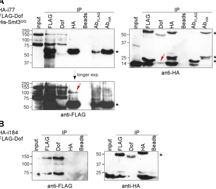
i77 ...28
i184 ...30
2.1.5 Other candidates...30
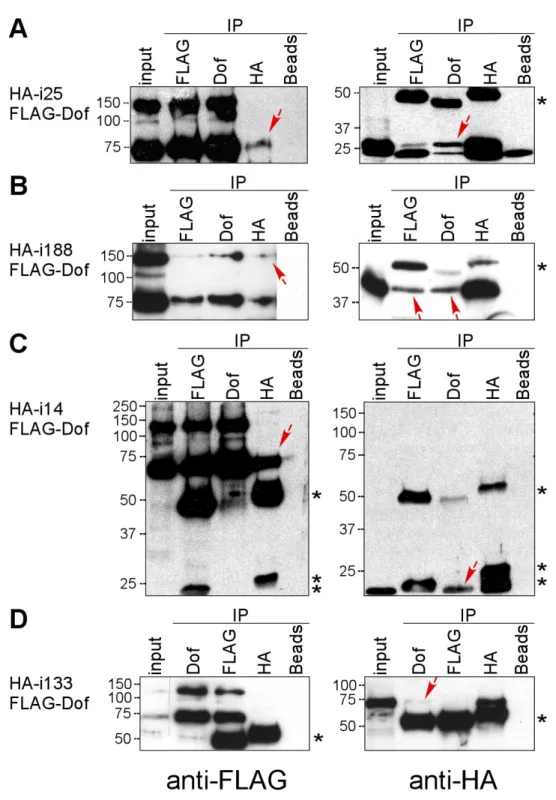
i25 ...30
i188 ...32
i14 ...32
2.1.6 Nuclear proteins ...34
i133 ...34
i163 ...34
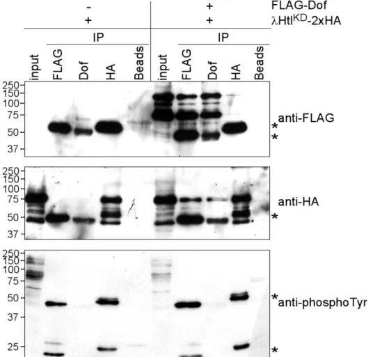
2.2 Interaction study of Dof with the FGF receptor Heartless...35
2.2.1 Interaction of different forms of Heartless with Dof ...35
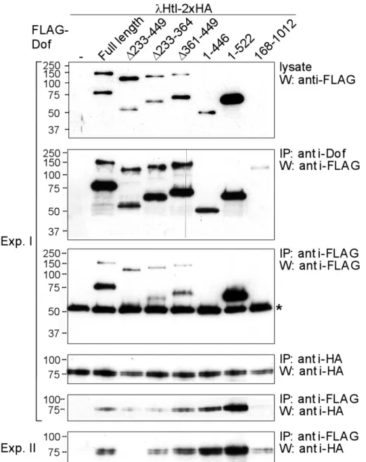
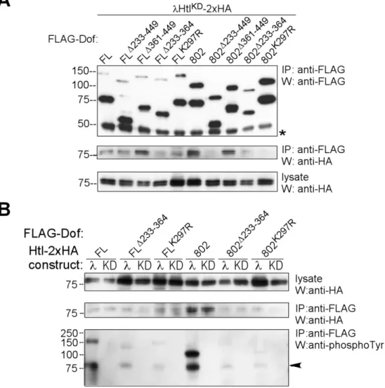
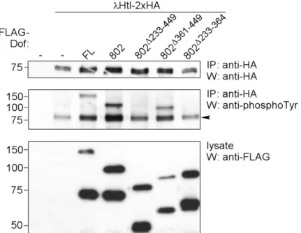
2.2.2 Domain mapping in Dof for receptor interaction...37
2.2.3 The effect of Dof binding on the activation level of Heartless in S2 cells ...39
2.2.4 Large Dof-FGF receptor complexes are present in S2 cells ...41
2.3 Tyrosine phosphorylation of Dof...44
2.3.1 Identification of tyrosine residues phosphorylated in Dof...44
2.3.2 Conservation of Dof phosphorylation sites in Dof homologues...49
2.4 The role of sumoylation in Dof function ...53
2.4.1 Dof sumoylation assay in Drosophila S2 cells ...53
2.4.2 Analysis of the role of the SUMO conjugation pathway in FGF dependent developmental processes ...55
Ubc9...55
Smt3 ...58
2.5 Identification of Dof as caspase substrate and analysis of the role of Dof cleavage ...62
2.5.1 Dof is cleaved in S2 cells...62
2.5.2 Cleavage of Dof depends on caspase activity...63
2.5.3 Cleavage of Dof requires an intact caspase cleavage site in the molecule ...65
2.5.4 Searching for the molecular role of caspase cleavage in Dof function...66
2.5.5 Analysis of the in vivo function of Dof cleavage and caspase activity in FGF signalling...68
2.6 Indications for the translational control of Dof protein levels in S2 cells ...71
2.7 The role of Ras effectors in FGF signalling in Drosophila ...75
2.7.1 The capacity of effector loop mutant Ras transgenes to rescue dof mutant phenotypes ...76
Rescue of mesoderm development ...76
Rescue of tracheal development ...78
2.7.2 Capacity of known downstream effectors of Ras to rescue dof mutant phenotypes...80
2.7.3 Dominant effects of constitutively active Ras effectors...83
2.7.4 Ras and/or Rap? – searching for the in vivo players in FGF signalling...85
3. Discussion ... 89
3.1 Interaction of Dof with yeast two hybrid interactors ...89
3.2 Conclusions from subcellular localisations of Dof...90
3.3 Interaction of Dof with the FGF receptor ...91
3.4 The role of lysine 297 in the interaction of Dof with the FGF receptor ...92
3.5 Sumoylation of Dof...92
3.6 Phosphorylation sites in Dof ...93
3.7 Functional relevance of the identified phosphorylation sites...94
3.8 Phosphorylation as a landmark of full length Dof ...94
3.9 Dof is a substrate for caspases in S2 cells – and in flies? ...95
3.10 Potential translational regulation of Dof and its relationship to caspase cleavage ...95
3.11 The role of different Ras effector pathways in FGF signalling ...97
3.11.1 Mesodermal studies of Ras effectors ...97
3.11.2 Studies of Ras effectors in the tracheae ...98
3.11.3 Implications for a role of Rap1 ...99
4. Materials and Methods ... 100
DNA constructs...100
Expression vectors ...100
Dof constructs for S2 cell expression...100
pUAS-FLAGDof
D587E...101
i-clones for S2 cell expression ...101
Heartless constructs for S2 cell expression ...102
Cell culture, transient transfection, immunoprecipitation and Western blot analysis ...102
Size exclusion chromatography ...102
RNAi in Drosophila S2 cells ...103
Caspase inhibition in transiently transfected Drosophila S2 cells...103
Metabolic [
35S]-methionin radioactive labelling of Drosophila S2 cells ...103
Dof protein stability analysis in Drosophila S2 cells ...104
Real-time RT-PCR...104
Immunocytochemistry ...105
Drosophila strains and genetics ...105
Immunohistochemistry ...106
Eggshell preparation ...106
5. Bibliography... 107
Abstract ... 118
Zusammenfassung ... 119
Abbreviations... 120
Acknowledgements... 121
Erklärung ... 122
Lebenslauf ... 123
1. Introduction
Fibroblast Growth Factor (FGF) signalling is a conserved mechanism from worms to humans.
It regulates several cellular processes including cell growth, survival, differentiation and migration in developing and adult organisms. In Drosophila melanogaster the development of the mesoderm and the tracheal system are processes regulated by FGF signalling. In the search for additional mutations that affect the migration of tracheal and mesodermal cells or the differentiation of mesodermal derivatives, and thus, eventually interfere with the propagation of FGF signalling three groups found mutant alleles of the gene dof/heartless/stumps independently (Imam et al., 1999;
Michelson et al., 1998; Vincent et al., 1998). Homozygous mutant embryos showed similar defects in the mesoderm and the tracheae as mutants of the two known Drosophila FGF receptor genes.
Genetic epistasis analysis showed that dof is an essential component of FGF signalling and placed it downstream of the FGF receptors and upstream or in parallel to Ras in the signalling cascade (therefore the name downstream of FGFR). The gene encodes a large molecule with no structural signs of known enzymatic domains characteristic for protein families with known functions. It shows weak homology to the B-cell receptor adaptors BCAP and BANK. The predicted domains of Dof (two ankyrin repeats and a coiled coil motif) are protein-protein interaction domains. In addition, several tyrosine residues of the sequence are located in consensus sites that could serve as binding sites for signalling molecules in their phosphorylated forms. Biochemical studies showed that Dof is tyrosine phosphorylated in an FGF receptor dependent manner and is able to form a complex with Drosophila FGF receptors (Wilson et al., 2004). Thus, its molecular characteristics, biochemical behaviour as well as genetic interactions indicated Dof to be an adaptor molecule in FGF signalling specific for Drosophila.
1.1 Adaptor proteins
The specific and appropriate response of cells to extracellular stimuli requires the integration of multiple signalling pathways. An emerging class of proteins that are major contributors in the regulation of cellular signalling networks are adaptor proteins. Adaptors are molecules of modular structure without enzymatic activity composed exclusively of multiple protein-protein or protein- lipid interacting domains. They link proteins to other proteins or to the plasma membrane, form large signalling complexes and specify subcellular localisation of other molecules thereby contributing to the specificity and efficiency of cellular responses in a spatio-temporal manner.
Docking and scaffolding molecules are subgroups of the adaptor family with specialised functions.
Docking proteins anchor interacting partners to subcellular structures, often membranes and so frequently contain protein-lipid interaction domains or sites for lipid modifications. Scaffolding proteins serve as interaction platforms for enzymatic cascades of multimeric signalling complexes.
An alternative view also defines molecules as adaptors despite their enzymatic activity if they serve as a multi-modular platform for signalling complex assembly with a “built-in” enzymatic function or if the two functions are clearly distinguishable in different cellular responses.
1.1.1 Adaptors in the context of the specificity of cellular processes
Adaptors are described in studies of signalling and sorting processes. They have pivotal role
in cell surface receptor signalling including receptor tyrosine kinase (RTK) signalling, G protein
coupled receptor (GPCR) signalling, the Toll-like receptor (TLR) mediated inflammatory signalling
pathway as well as T- and B-cell receptor activation during lymphocyte development and immune response. Neurotransmission works with many structural and signalling adaptors on the side of the sensing (post-synaptic density) as well as on the side of the signal release. Adaptors have a general role in membrane and vesicle trafficking, e.g. in protein sorting in the Trans-Golgi network (TGN) and in endocytosis. The regulation of cell adhesion, cytoskeletal dynamics and active cell movements require whole sets of adaptors which act in integrin signalling, focal adhesion assembly and disassembly and actin polymerisation and crosslinking. Nuclear adaptors play role in transcriptional regulation. Even death needs the help of specific adaptors that form the
“apoptosome”, a multiple protein complex essential to promote apoptosis.
Along the study of these different processes a growing body of evidence emerged for the existence of many adaptors which are involved in several signalling events. It is also known that the same canonical signalling cassettes are activated by many different signals. What are actually the tools in the hand of adaptors to increase specificity?
One way is that different combination of adaptors is utilised in different signalling events, called the combinatorial control. This can be emphasized and more specialised in a cell type dependent expression of adaptors. In general, interaction studies pointed out, that large protein complexes are the rule where simultaneous interaction of several components is required for signal transmission and the removal or exchange of a single component might modify the action of the complex. The assembly of signalling complexes might be influenced by mutually exclusive binding properties of the molecules and so factors might be displaced from a complex despite their ability to interact with components of the complex. These interfering interactions can be based on competitory or overlapping binding sites as well as on interaction surfaces which are dependent on conformational changes of the molecule and influenced by other interacting partners or modifications.
No less important is the subcellular compartmentalisation of active and inactive adaptors.
Activation often means the recruitment to specific places of signalling at the cell membrane, the so called lipid rafts (membrane microdomains enriched in glyco-sphingolipids). Another example of specific recruitment is served by scaffolding molecules, binding platforms of enzymatic cascades.
They synergise enzyme activity for one signalling and in parallel can keep away enzymes from other processes.
Even if same signalling cascades are activated by several signals, their amplitude and duration can be characteristic for the appropriate signalling event. This is achieved by the presence/absence of specific scaffolding molecules, by the capability of adaptors to oligomerise and thereby increase the local concentration of signalling molecules or by the fact that activation of cascades can happen in several alternative routes, which might be used with different redundancy in the one or the other signalling process.
Since Dof plays a role in FGF signalling - a subtype of RTK signalling – we focus in this work on adaptors which are involved in RTK signalling or linked directly to other protein tyrosine kinase (PTK) activity.
1.1.2 Modular assembly of adaptor molecules
A characteristic feature of adaptor proteins is the ability to bind at least two different
molecules simultaneously. This capability derives from their multi-modular structure. They have
protein domains with independent folding and binding capacity. Some of these domains are
characteristic for the protein family of PTK-activity-linked adaptors and have defined binding sites
whilst others are widely distributed modules often without preferring a defined binding domain.
Structural domains preferentially found in PTK-activity-linked adaptors
Src homology 2 (SH2) domains are protein modules of about 100 amino acids which bind phospho- tyrosine containing peptide motifs of 5-10 amino acids length, generally C-terminal to a negatively charged region. They do not have affinity for unphosphorylated sequences and so are exclusive modules of tyrosine kinase signalling. The phosphopeptide binding site is bipartite. The pTyr binding pocket is highly conserved, whereas the second binding surface is more variable, and allows specific recognition of amino acids immediately C-terminal to the pTyr. These amino acids are decisive in binding specificity of different SH2 domains (Table 1) (Songyang et al., 1993;
Songyang et al., 1994). Thus, tyrosine phosphorylation functions as an on-off switch for SH2 binding, while enabling the sequence context of the pTyr site to predict which SH2 domains, and so which signalling proteins are bound.
Table 1. Binding specificity of SH2, SH3 and PTB domains of various signalling molecules SH2 domain binding specificities:
Src family: pY-[E/D/T]-[E/N/D]-[I/V/M/L]
Abl: pY-[E/T/M]-[N/E/D]-[P/V/L]
Crk: pY-[D/K/N]-[H/F/R]-[P/L]
Nck: pY-D-E-[P/D/V]
Sem5: pY-[L/V/I/M]-N-[v/p]
Csk: pY-[T/A/S]-[K/R/Q/N]-[M/I/V/R]
3BP2: pY-[E/M/V]-[N/V/I]-x fps/fes: pY-E-x-[V/I]
Drk: pY-[Y/I/V]-N-[F/L/I/V]
Grb2: pY-[Q/Y/V]-N-[Y/Q/F]
SHC: pY-[I/E/Y/L]-x-[I/L/M]
Vav: pY-[M/L/E]-E-P ZAP-70: pY-x-x-I/L
p85 N-term: pY-[M/V/I/E]-x-M C-term: pY-x-x-M
PLCγ N-term: pY-[L/I/V]-[E/D]-[L/I/V]
C-term: pY-[V/I]-[I/L]-[P/I/V]
SHP2 N-term: pY-[I/V]-x-[VILP]
Syk C-term: pY-[Q/T/E]-[E/Q]-[L/I]
HCP N-term: pY-F-x-F
SH3 domain binding sites:
general rule: x-P-p-x-P, x aliphatic Src: R-x-x-P-p-x-P
Abl: T-x-x-P-p-x-P Grb2 C-term: P-x
3-R-x
3-P Grb2 N-term: P-x-x-P
PTB domain binding sites:
Dok: Y/M-x-x-N-x-L-pY minimal consensus: N-P-x-Y
Consensus sequence motifs were generated in a random peptide binding assay. Amino acids in the brackets separated by a slash were found without significant difference in that position. Bold letters remark outranking preference for that amino acid in that position. x stays for any amino acids. pY marks phosphorylated tyrosine residue. Modified and extended from (Songyang et al., 1993; Songyang et al., 1994).
Phospho-Tyrosine Binding (PTB) domains are modules of ~200 amino acid residues. Initially, the
study of the adaptors Shc and IRS-1 showed that they recognise phospho-tyrosine containing
peptide motifs that form a β turn. Binding to target sequences is determined by residues N-terminal
to the pTyr (Table 1). Later findings demonstrated that PTB domains are able to bind
unphosphorylated tyrosine-containing peptides (Margolis et al., 1999) or even do not require
tyrosine for their binding to targets (Ong et al., 2000). Thus, sequence binding preference of PTB
domains is less defined than of SH2 domains, which makes target prediction difficult for this
domain.
Src homology 3 (SH3) domains bind proline rich peptides of approximately 10 amino acids. The core binding sequence contains the consensus x-P-x/p-x-P, where prolines at defined positions are of structural need and ensure high affinity binding. Each SH3 domain has a distinct binding preference and the nature of the non-proline residues in and around the core sequence serves for specificity (Table 1) (Pawson, 1995). SH3-binding does not depend on modification like phosphorylation enabling theoretically a constitutive protein-protein interaction. However, serine and threonine phosphorylation adjacent to the proline-rich motif may influence SH3 domain interactions and result in uncoupling of proteins.
Pleckstrin homology (PH) domains are lipid interacting modules of approximately 100 amino acids length. They bind the charged headgroups of specific poly-phosphoinositides and may thereby regulate the subcellular targeting of proteins to specific regions of the plasma membrane, where their ligand is enriched.
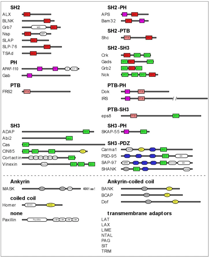
Classification of PTK-activity-linked adaptors based on their structural domains Most of the PTK-activity-linked adaptors contain at least one of the preferentially used domains. Adaptors contain either exclusively the characteristic domains in single or multiple copies or combine these modules with diverse other domains. Some of them lack domains typical for PTK- activity-linked adaptors. Another characteristic feature is that they often harbour proline rich regions and tyrosine motifs – binding sites for SH3 and SH2 domains respectively. Figure 1 gives an overview of the different classes of PTK-activity-linked adaptors based on their structural domains.
Without going into details with each group we can make several general remarks. First, beside the preferential domains several other domains are represented in the different adaptors.
Some of them appear in more then one protein, like PDZ domains, ankyrin repeats or coiled coil motifs whereas others are unique for a particular adaptor molecule, e.g. sorbitol homology (SoHo) domains in the Vinnexin family. Second, if the phospho-tyrosine binding motif SH2 is present in an adaptor then only in a single copy whilst conserved tyrosine motifs, potential binding sites for different SH2 domains are abundant. Thus, an SH2 domain-containing adaptor can bind only to a single phosphorylated partner which might cause a competition of molecules that contain binding sites for the particular SH2 domain. On the other hand tyrosine phosphorylated adaptors are able to recruit many SH2 containing adaptors at the same time. Third, the number of SH3 domains in an adaptor is less well defined and can vary from zero up to three, albeit often with different binding specificities.
The same is true for their binding sites, the proline rich regions. Finally, the presence and order of the different domains is unique for the different adaptor families. In other words, the order of moduls, rather than only the presence of the identical moduls determines homology.
What can one conclude from these structural characteristics for the function of the different
adaptors? The conserved and unique domain structure of the different adaptor families indicates
their important and specific role in signalling events. However, the diversity in the presence and
combination of SH2 and SH3 domains and tyrosine and proline rich motifs alone gives already such
high level of networking potential for these adaptors, that it makes their cellular role in signalling
unpredictable. Indeed recent proteomics data showed the tyrosine phosphorylation of plenty of
cytosolic proteins in the cell upon a single stimulation indicating the wide range of possibilities for
phosphorylation based interactions. The presence of specific domains might be helpful revealing the
function of adaptors, like the actin binding domain in Cortactin and Paxillin or the receptor binding
PTB domain of FRS2.
Figure 1. Classes of PTK-activity-linked adaptors based on their structural domains. Gray bars represent the relative length of the proteins with the exception of IRS and MASK. Colour coded boxes show the domains characteristic for PTK-activity-linked adaptors, colour coded ovals represent domains that are shared by several members of PTK-activity-linked adaptors but do not belong to the characteristic ones and white ovals stand for domains which are unique among these adaptors. Dashed line separates adaptors with characteristic domains (top) from adaptors with non-characteristic domains (bottom). Since transmembrane adaptors contain no recognisable domain in their amino acid sequences beside the transmembrane region they are listed only by name as a separate group. The domain structure of the proteins was constructed
1.1.3 Functional dissection – adaptors as molecular interneurons of the cells
Adaptors are linkers that couple sensing processes of the cell to effector molecules. Their regulation enables to modulate signalling outputs. Thus, the number of adaptors involved in a signalling pathway might reflect the fine-tuning capacity of the appropriate system. Most extreme examples are T- and B-cell receptor signalling events, where up to eight components are utilised to activate the Ras-MAPK cascade (Simeoni, 2004). In other systems e.g. in case of several RTKs a single adaptor is sufficient to activate the same cascade.
It raises an interesting option to distinguish adaptors based on the criterion if they can directly bridge activator and effector, bind activator or effector or function as intermediator by linking adaptors together. It could give a kind of hierarchy of adaptors in the signalling cascades.
However the position of an adaptor might be different in the activation of the one or the other signalling cascade. Taken the example of the adaptor Grb2: it can directly bridge EGF receptors to the Ras activator Sos. In B-cell activation it links the adaptor LAT to the effector Sos, whereas in FGF signalling it links the sensor/receptor binding FRS2 adaptor to Sos. It acts also as an intermediator adaptor in FGF signalling by binding to FRS2 and recruiting the adaptor Gab1, which then activates the effector PI3K. The validity of such a positioning relays on the selective specificity of the interactions; it is not an intrinsic feature of the adaptors. The identification of domains in the adaptors with binding specificity for receptors/activators or effectors might help for positioning but a particular position should not be generalised for all signalling events.
An alternative way to classify adaptors might be their specificity to different signalling pathways. Several adaptors are involved only in one signalling cascade – e.g. scaffolding proteins of enzymatic cascades, many B- and T-cell adaptors or the adaptor FRS2 in mammalian FGF signalling – whilst others are utilised by many signalling events, e.g. members of the Grb2, Crk, Gab or Nck adaptor families. The first group might provide specificity to the different signalling events whereas the second group might enable crosstalk and balancing between signalling events in the cell.
1.1.4 Tools to regulate adaptor protein activity
Adaptor proteins function as molecular switches in signal transduction. Their activity requires a tight regulation to ensure signal propagation or inhibition. Diverse strategies are used for both positive as well as negative regulation including phosphorylation, proteolytic cleavage, proteosomal degradation and changes in the conformation or in the oligomerisation capacity of the molecules. Regulatory changes can have temporary/reversible effects like phosphorylation and conformational changes or definitive/irreversible consequences like cleavage or degradation.
Posttranslational modifications
Tyrosine phosphorylationTyrosine phosphorylation acts as a general switch for activation in signalling cascades. Most PTK-activity-linked adaptors are tyrosine phosphorylated in a stimulation dependent manner and a
using PFAM (http//:www.sanger.uk/Pfam). act.b, actin binding domain; Ank, ankyrin repeat; CARD, caspase- recruitment domain; CC, coiled coil; Cort, cortactin repeat; GK, guanylate kinase-like; GM, Grb7/Mig-10 homology region; KH, Kash homology; LIM, lin-11/isl-1/mec-3 domain; LZ, leucine zipper; MRE,MAGUK recruitment domain; PDZ, PSD-95/Discs-large/ZO-1 domain; PH, Pleckstrin homology; PTB, phospho- tyrosine binding domain; SAM, sterile α motif; SH2, Src-homology 2; SH3,Src-homology 3; SoHo, Sorbitol homology.
cooperative action of kinases and phosphatases balances the actual tyrosine phosphorylation state of these adaptors during signalling. Only very few of them do not show detectable tyrosine phosphorylation, like the adaptor E3B1/Abi2 (Ichigotani et al., 2002).
The primary role of phosphorylated tyrosine residues is to provide signalling-dependent binding surface for the highly specialised SH2 domains thereby recruiting downstream transducers which propagate signalling and activate effectors.
However, we also find inhibitory effects of tyrosine phosphorylation. Cortactin, an actin binding adaptor protein is phosphorylated on tyrosine residues adjacent to its SH3 domain. This blocks the binding capacity of the SH3 domain (Martinez-Quiles et al., 2004). Crk function is regulated by tyrosine phosphorylation-dependent auto-inhibition. Its SH2 domain interacts with a phosphorylated tyrosine residue of the own molecule (Feller, 2001). This has dual consequences:
SH2 domain is occupied for other interactors in a competitive manner and interactors of the SH3 domain are lost due to the conformational changes caused by the intramolecular interaction.
Serine/Threonine phosphorylation
If tyrosine phosphorylation is the general switch then serine/threonine phosphorylation is the modifier in signalling events. It serves as fine tuner in the regulation of adaptor proteins. A broad range of Ser/Thr kinases and phosphatases can act on adaptors. They are either activated by the same signalling the respective adaptor involved in (often as feedback regulators) or by independent signalling cascades thereby providing cross talk between different signalling processes in the cell.
We find examples of constitutive as well as signalling dependent phosphorylation of adaptors on Ser/Thr residues with positive or negative regulatory role. Ser/Thr phosphorylation acts through its allosteric effect on the molecule and can cause diverse functional changes. It can sensitise or desensitise adaptors for specific interactions by inducing changes in the conformation of the molecule (e.g. IRS, FRS2 and Grb7/10/14). A local allosteric effect of Ser/Thr phosphorylation is the disruption of the phospho-tyrosine binding capacity of SH2 domains (Gab1 and IRS). The local effect of Ser/Thr phosphorylation in binding site disruption can cause global conformational changes through the inhibition of intramolecular interaction (Cortactin) or interfering with the auto- inhibitory binding of adaptors (AFAP-110).
IRS proteins are examples for complex roles of S/T phosphorylation. In quiescent cells a basal level of S/T phosphorylation on particular residues is necessary for an effective subsequent tyrosine phosphorylation by the insuline receptor. On the other hand, elevated S/T phosphorylation on different residues inhibits the binding of IRS-1 and -2 to the juxtamembrane region of the insuline receptor impairing their ability to undergo tyrosine phosphorylation. Thus, position of S/T phosphorylation in IRS determines the positive or negative influence on insuline receptor binding affinity. Further negative regulatory mode is the specific phosphorylation of serine and threonine residues adjacent to SH2 binding motifs thereby decreasing the binding affinity of SH2 domain based interactors to IRS. Additional observation is that S/T phosphorylation commits IRS to proteosomal degradation (reviewed in (Johnston et al., 2003)).
The activity of the adaptor FRS2, key mediator of FGF signalling stands also under the dual
control of tyrosine and threonine phosphorylation. FRS2 binds constitutively to FGF receptors in
cells via its PTB domain (Ong et al., 2000). Upon FGF stimulation FRS2 is phosphorylated by the
FGF receptor on several tyrosine residues that serve as docking sites for the MAPK cascade-
activating molecules Grb2 and SHP2 (Ong et al., 1997). Activated MAPK has a negative regulatory
feedback on FRS2 by phosphorylating it on several threonine residues. Threonine phosphorylation
inhibits further tyrosine phosphorylation of FRS2, leading to the downregulation of the signalling
cascade (Lax et al., 2002). Similarly, threonine phosphorylation of FRS2 is induced in response to
other growth factors (e.g. PDGF, EGF). FRS2 is able to bind to the EGF receptor in a ligand-
stimulated manner and undergo threonine and weak tyrosine phosphorylation (Wu et al., 2003).
Thus, MAPK dependent threonine phosphorylation of FRS2 might ensure a controlled dosage of signalling via FGF receptors balanced also by other growth factor signallings. Since FRS2 is constitutively associated with the FGF receptor, threonine phosphorylation might provide an additional tight control to prevent activation of FGF-dependent signalling by random receptor autoactivation.
AFAP-110 and Cortactin are good examples how S/T phosphorylation can lead to conformational changes by altering intramolecular interactions and thereby propagate signalling.
The phosphorylation of AFAP-110 alters its multimerisation behaviour and enables novel downstream interactions (Baisden et al., 2001). Cortactin SH3 C-terminal domain forms an auto- inhibitory loop by binding to proline rich motifs of the same molecule. Upon Erk activation Erk phosphorylates serine residues within the proline rich motif and thereby liberates cortactin SH3 domains to interact with proline rich regions of other molecules (Campbell et al., 1999). As mentioned above phosphorylation of adjacent tyrosine residues block the ability of the SH3 domain for effective binding (Martinez-Quiles et al., 2004). Thus, tyrosine and serine phosphorylations act on the interaction behaviour of the same domain with opposing effects.
Gab1 adaptor function on PI3K activation is negatively regulated by serine/threonine phosphorylation (Lehr et al., 2004). Phosphorylation of serine/threonine residues adjacent to tyrosine motifs which are phosphorylated upon RTK signalling and serve as binding sites for the SH2 domain of PI3K disrupts this interaction.
Much less is known about the role of serine/threonine phosphorylation of paxillin, E3B1/Abi2 and the Grb7/10/14 family adaptors. Phosphorylation of particular serine residues play a key role in cell migration and in the regulation of focal adhesion organisation (Huang et al., 2004).
However, the mechanism of the modulatory effect of these phosphorylations is not uncovered as well as the biological relevance of other phospho-serine/threonine residues in the molecule remains to be elucidated. E3B1/Abi2 becomes hyperphosphorylated on serine residues following mitogenic stimulation (Ichigotani et al., 2002). However little is known about the clear cause and consequence of the phosphorylation. Grb7/10/14 family adaptors are phosphorylated on serine and threonine residues at basal level in quiescent cells which is increased in case of Grb10 and Grb14 upon growth factor stimulation (Han et al., 2001). Grb14 interacts directly with activated insulin receptor and its serine/threonine phosphorylation inhibits insulin signalling probably by maintaining the bound receptor in an inactive conformation (Cariou et al., 2004). Functional significance of serine/threonine phosphorylation of other Grb7 family members is not yet understood.
Caspase cleavage
Signalling can serve as survival factor in cells. Effective shut down of signalling events is therefore a prerequisite of apoptosis. As a consequence many adaptor proteins are targets of caspases which are the major initiator and executor proteases activated in programmed cell death.
Interestingly, caspase activity is observed and required in the regulation of some adaptors not exclusively under apoptotic conditions.
Cas family adaptor proteins are involved in the assembly of multiple protein interactions at
focal adhesion sites (Bouton et al., 2001). Human enhancer of filamentation 1, HEF1, a member of
this family shows a cell cycle dependent regulation. In cells in G1-S-G2 phases full length forms of
the molecule accumulate at focal adhesions, while at G2/M transition HEF1 is cleaved at a caspase
consensus site to generate an N-terminal HEF1 form, p55HEF1, which relocalises to the mitotic
spindle. C-terminal cleavage products are apparently degraded (Law et al., 1998). Additional
finding was that proapoptotic stimuli or HEF1 overexpression lead also to the cleavage of HEF1 by
caspases. The same caspase cleavage site is used as during mitosis to generate the N-terminal 55
kDa and a C-terminal 65 kDa fragment, while a novel caspase cleavage site was identified to be
responsible for the generation of a carboxy-terminal small 28 kDa fragment (Law et al., 2000). This
second caspase site is also present in p130Cas, an other member of the Cas family, which is similarly cleaved at this site to produce a 28 kDa protein species following proapoptotic stimulus (Bannerman et al., 1998). The proapoptotic activity of HEF1 was found to depend ot the expression of the C-terminal 28 kDa region of the protein. These findings together suggest a model in which separate parts of HEF1 have functions distinct from the full length molecule. Regulation of the cleavage and abundance of HEF1 (and p130Cas) might propagate the destruction of focal adhesion sites and regulate onset of mitosis or apoptosis (Law et al., 2000). Thus, modulated caspase activity towards Cas adaptors may be important for cytoskeletal organisation and cell cycle progression, while a dramatic increase or dysregulation of caspase activity is necessary to promote apoptosis.
Another example, how caspase cleavage can regulate signalling events, is served by the adapter molecule Gads. Gads functions specifically in T-cell receptor signalling by linking the adaptor SLP-76 to tyrosine phosphorylated LAT. This linker function of Gads is essential to recruit SLP-76 to the membrane which than upon phosphorylation activates downstream signalling events (Liu et al., 2001). Apoptosis induction results detectable cleavage of Gads at a predicted caspase cleavage site. Cleavage products retain their binding specificity for LAT and SLP-76 respectively.
Expression of Gads cleavage products showed a dominant inhibitory effect on signal propagation.
Hence, the functional relevance of caspase cleavage of Gads is likely to uncouple LAT and SLP-76 interaction (Berry et al., 2001). The surprising finding, that cell death receptor mediated signalling is required for activation induced proliferation of T-cells (Kennedy et al., 1999) rises the hypothesis that cleavage of signalling molecules downstream of the T-cell receptor could modulate the spectrum of activated pathways in a way that propagates proliferation.
Dimerisation/oligomerisation
Oligomerisation of adaptors can have both posive as well as negative effects on their activity. It is mediated either by homophylic interaction of the same domains or by heterophylic interaction between two different regions of the molecules. However, based on the experimental approaches it is not distinguishable in each case, whether these domains are utilised for intra- or for intermolecular interactions.
Preferential domains of homophylic interactions are leuzine zipper domains, coiled coil motifs and helix-loop-helix structures. Less defined are the domains for heterophylic interactions:
the one part is often determined only as a molecular region without known domain structure.
Examples below show that heterophylic interaction of adaptors is usually regulated by tyrosine phosphorylation.
CIN85/CMS family adaptor proteins are involved in the regulation of RTK downregulation via endocytosis (reviewed in (Dikic, 2002)). They are able to homodimerise via their coiled-coil domains. Overexpression of CIN85 or CMS leads to the formation of multiple endosomal-like vesicles in the cells, the existence of which depends on the presence of the coiled-coil domain. One possible explanation is that homo-oligomerisation of CIN85/CMS increases the local concentration of recruited signalling complexes in the proximity of the receptor (cargo) and thereby positively regulates endocytosis.
Members of the Dok adaptor family inhibit the Ras pathway by recruiting RasGAP in a tyrosine phosphorylation-dependent manner. Mutations in Dok that prevent oligomerisation also abrogate inhibitory activity of Dok1 without affecting the overall phosphorylation state of the molecule, indicating that domains involved in oligomerisation play a crucial role in downstream interactions without influencing Dok1 activation itself (Songyang, 2001). It is not undersood at present if the dual requirement of PTB domain and phosphorylated Tyr146 in Dok1 function is primarily due to their capability to induce oligomerisation.
The regulation of the oligomerisation capacity of the adaptor AFAP-110 is also
phosphorylation dependent and involves both inhibitory and activation steps. AFAP-110 has the
intrinsic capability to form multimers via its C-terminal leucine zipper motif. This domain has an auto-inhibitory binding to N-terminal regions of the molecule and this interaction keeps AFAP-110 in a tetrameric complex (Qian et al., 2004). Upon phosphorylation auto-inhibition is released and conformational changes lead to a reduction of the multimeric status of AFAP-110. Dimers are formed probably via new interacting surfaces in the molecule. The new conformation allows the binding and activation of Src kinase and increases the actin crosslinking activity of AFAP-110 (Qian et al., 2002).
Oligomerisation of adaptor molecules can have also inhibitory effect on interactions of signal transduction events. The adaptor Grb10 forms oligomers in a way, that N-terminal amino acid sequences of the one molecule interact with the C-terminal BPS-SH2 domains of the other molecule (Dong et al., 1998). BPS-SH2 domains are important functional modules, which can directly bind activated insulin receptor. Oligomerisation therefore may provide a reservoir of Grb10 that is prevented to bind phospho-tyrosine containing molecules and stimulation may release Grb10 molecules from this pool. Alternatively Grb10 oligomers may have a more active function serving as docking complex to recruit multiple signalling molecules. Another piece to the complexity is that Grb7, member of the same adaptor family was also able to interact with the C-terminal PBS/SH2 domains of Grb10 in yeast two hybrid assay (Liu and Roth, 1998). This potentiates the homo- /hetero-oligomerisation capacity of Grb7 family adaptors as a broader regulatory tool in signal transduction. However, no physiological relevance of these interaction has been shown so far.
The Cas family adaptor protein HEF1 is also able to dimerise via its C-terminal helix-loop- helix (HLH) domain. This motif mediates not only HEF1 homodimerisation, but also heterodimerisation with the HLH domain of the other Cas family member p130cas as well as with the transcriptional regulatory HLH proteins Id2, E12 and E47 (Law et al., 1999). Dimerisation capacity was analysed only for the C-terminal region of HEF1 but not for the full length molecule.
HEF1 is processed in T cells (see above). Thus, it might be possible, that dimerisation is an intrinsic feature of a sole C-terminal fragment of HEF1 but not of the full length molecule. Functional relevance of dimerisation is not understood.
Intramolecular interactions, conformational changes
Conformational changes are stimulation-dependent temporary alterations that affect the activity of adaptors. They are induced upon phosphorylation of the adaptors which causes allosteric distortion or frequently alters intramolecular interactions of the molecule. As discussed earlier with examples phosphorylation can disrupt intramolecular interactions and result an “open” molecular structure where new protein interaction domains become available (e.g. Cortactin). Phosphorylation can also generate new binding sites for own phospho-tyrosine binding domains propagating a
“closed” form of the molecule which will mask interaction surfaces for other binding partners (e.g.
Crk). Thus, conformational change is a potent tool to alter interaction surfaces of adaptors and therefore mutually exclude interacting partners in a signalling dependent manner.
Protein degradation
Protein degradation is an irreversible way to downregulate adaptor activity. The ubiquitine
E3 ligase Cbl plays important role in this negative signalling sending not only adaptors but several
receptors and signalling effectors for degradation (Dikic and Giordano, 2003). It contributes in the
poly-ubiquitination of FRS2 which leads to the proteosomal degradation of this adaptor (Wong et
al., 2002). The adaptor Cin85, involved in the ligand-induced endocytosis of several receptors
undergoes Cbl dependent mono-ubiquitination during this process which provides the sorting signal
for lysosomal degradation of the whole receptor-Cbl-Cin85 complex (Dikic, 2002). SOCS proteins
play a role in proteosomal targeting of associated proteins (Kamura et al., 1998). It has been shown
that they contribute to the proteosomal degradation of IRS adaptors (Johnston et al., 2003). Studies on IRS also showed that S/T phosphorylation promoted its degradation through an unknown mechanism.
The discussed examples show that adaptor proteins are regulated in a complex way.
Regulatory modifications can have opposing effects and can be a prerequisite or inducer of further regulatory changes. Hence, regulatory tools act in concert to achieve an appropriate activity level of adaptors.
1.2 FGF signalling-dependent morphogenetic processes in flies
Initially dof mutants were found in genetic screens affecting mesodermal and tracheal development in the embryo (Imam et al., 1999; Michelson et al., 1998; Vincent et al., 1998). Both processes are controlled by FGF (Ghabrial et al., 2003; Wilson and Leptin, 2000). Later analysis showed that dof is generally involved in developmental processes where FGF signalling plays a role, e.g. also in the migration of midline glia cells (Vincent et al., 1998) and in the formation of air sacs in the pupa (Sato and Kornberg, 2002). Here I will focus on the embryonic development of the mesoderm and the tracheae since the in vivo function of dof and other components of the FGF signalling cascade were studied in these developmental processes to the greatest detail including experimental results presented in this work. Thus, mesoderm and tracheal development are the two FGF-dependent processes in Drosophila about the regulation of which we understand the most at present.
1.2.1 Regulation of tracheal morphogenesis
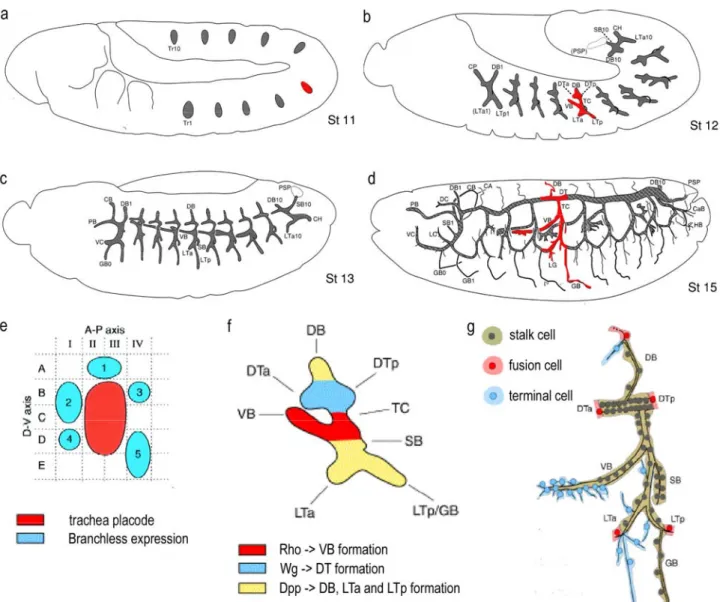
The Drosophila embryonic trachea is a tubular network and serves as the respiratory organ throughout larval life (reviewed in (Affolter et al., 2003; Ghabrial et al., 2003). It derives from ten clusters of dorsal ectodermal cells from both side of the embryo, the so called tracheal placodes (Figure 2a). They invaginate into the mesoderm to form the tracheal pits, each containing approximately 80 cells. Without further cell division the 20 sacs establish independent tracheal metamers with highly similar branching patterns through stereotypic, directed migration and cell shape changes (Figure 2b,c). Six primary branches are formed into different directions and each branch contains a well defined, invariable number of tracheal cells. The metamers become interconnected by subsequent fusion events through specialised fusion cells of particular tracheal branches (Figure 2d). As last step fine subcellular extensions, the terminal branches grow out to deliver oxygen to target tissues guided directly by different oxygen requirement of the cells (Figure 2g). Tracheal branches migrate into different parts of the embryo invading different tissues. The surrounding tissue provides additional specific interactions for the corresponding branches and thereby contributes to the final pattern of the tracheal network (reviewed in (Uv et al., 2003)).
At molecular level the JAK/STAT pathway is responsible to initiate tracheal cell fate. It
induces the expression of the transcription factor Trachealess in the tracheal placodes. Trachealess
is responsible to make tracheal cells competent for migration by inducing the expression of Dof and
the FGF receptor Breathless (Btl). The Btl specific FGF ligand Branchless (Bnl) is expressed in
dynamic pattern in surrounding cells of the tracheal placodes and predefines the directional
outgrowth of the six primary branches (Figure 2e) (Sutherland et al., 1996). Bnl is acting as a
chemoattractant for tracheal cells that is nicely demonstrated by its ability to reroute tracheal cells if
ectopically expressed or to attract fine terminal branches in larvae towards oxygen-starved cells that
Figure 2. Schematic representation of the developmental stages of tracheal morphogenesis during embryogenesis. (a-d) Schematic drawing of Drosophila embryos from lateral view at developmental stages 11, 12, 13 and 15 respectively. Anterior to the left. Gray structures show the morphology of tracheal anlage/tissue at different stages. (a) Invaginated tracheal palcodes prior branching, (b) Initial branching of the six primary branches, (c) trachea metamers with branched out primary branches prior fusion and (d) tracheal network after fusion occurred and secondary branches are formed. (e-g) blow ups of regions in a, b and d highlighted with red. (e) shows the arrangement of Branchless expression clusters (blue) surrounding a trachea placode (red). (f) Regional differentiation of tracheal cells at early primary branching stage. Colour code shows the regionally acting signalling pathways which determine different cellular fates of the developing primary branches according their anterior-posterior, dorsal-ventral position in the placode. (g) tracheal unit of one hemisegment showing the positions of specified tracheal cells for fusion and terminal branching. CA, cerebral anastomosis; CaB, caudal branch; CB, cerebral branch; DB, dorsal branch; DC, dorsal cephalic branch; DTa, dorsal trunk anterior; DTp, dorsal trunk posterior; GB, ganglionic branch; HB, hindgut branch; LC, lateral cephalic branch; LG, lateral group branch G; LTa, lateral trunk anterior; LTp, lateral trunk posterior; PB, pharyngeal branch; SB, spiracular branch; TC, trancverse connective; VB, visceral branch. Figures were taken and modified from (Bate, 1993) (a-d), (Metzger and Krasnow, 1999) (e) and (Uv et al., 2003) (f-g).
express it at high levels. In the absence of Bnl, Btl or Dof cell migration and subsequent development of the tracheae fails to occur (reviewed in (Ribeiro et al., 2003)).
Tracheal cells show a regional differentiation at placode stage as a consequence of the
differentially localised activity of Decapentaplegic (Dpp), Rhomboid (Rho) and Wingless (Wg)
(Figure 2f) (reviewed in (Uv et al., 2003)). Their activity is manifested by a selective expression of
transcription factors that act as sorting markers of tracheal cells for different primary branches. FGF signalling controls motility of tracheal cells by inducing filopodia formation in a spatially specific manner but is not sufficient for successful outgrowth of tracheal branches (Ribeiro et al., 2002).
Regional differentiation might prepare and specialise cells of different branches for oriented migration towards the sources of Bnl along distinct substrates. Signalling systems provided by the regional differentiation integrate cell motility and thereby allowing the correct morphogenesis of individual branches.
1.2.2 Regulation of mesoderm development
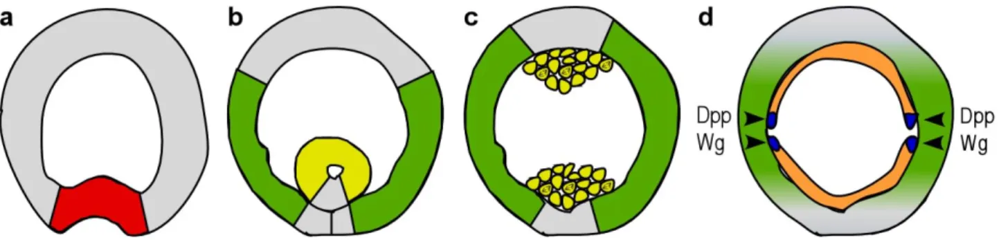
The mesoderm invaginates as an epithelial layer on the ventral side of the embryo driven by cell shape changes (Figure 3a). After the central part of the mesoderm is fully internalised and the ventral furrow is closed, the epithelial tube initiates contact with the ectoderm (Figure 3b), followed by epithelial mesenchymal transition (Figure 3c). Mesodermal cells dissociate, resume mitosis and spread out on the ectoderm to form a single-cell layer (Figure 3d). Based on dorso-ventral inductive signals from the underlying ectoderm mesodermal cells differentiate into specific tissues along the dorso-ventral axis (Figure 3d). Thus, the formation of mesodermal derivatives requires a sequential interplay of cell migration and cell differentiation (reviewed in (Michelson et al., 1998).
Figure 3. Schematic representation of mesoderm morphogenesis. (a-d) Schematic drawings of cross- sections of embryos at four successive stages of mesoderm development. (a) Mesoderm invagination begins with the formation of the ventral furrow by the most ventral cells. Presumptive mesoderm expresses Twist (red). (b) Invaginated mesoderm forms an epithelial tube and begins to make contact with the ectoderm.
Heartless and Dof are expressed in the mesoderm (yellow) and the two FGF ligands in the complementary parts of the ectoderm (green). (c) Mesodermal tube flatten against the ectoderm, cells loose their epithelial integrity and begin to divide. Note that at this time germ band extension is in a progressive stage. (d) Mesodermal cells (orange) migrated out dorsally on the surface of the underlaying ectoderm and start to differentiate. The dorsal-most cells of the mesodermal sheet form Eve-positive cell clusters (blue) upon inductive signals of the ectoderm (Dpp, Wg). FGF signalling is also required for this process; expression of the two FGF ligands becomes higher/stays longer in the dorsal ectoderm (green) and Heartless and Dof expression is upregulated in the dorsal-most mesodermal cells (not shown here). The germ band is in a fully extended stage.
Mesodermal fate is adopted through the action of a series of maternal genes of the so-called
dorsal goup. It results in a nuclear concentration gradient of the Dorsal transcription factor. Specific
expression pattern of zygotic genes is established based on the different binding affinity of their
promoters to Dorsal. On the ventral side of the embryo the highest nuclear levels of Dorsal turn on
twist and snail, the two major sequester transcription factors of mesoderm formation and
development (Figure 3a). Dorsal participates with Twist in activating dof and the FGF receptor
heartless (htl) ventrally (reviewed in (Michelson et al., 1998)). The recently identified Htl specific
FGF ligands FGF8-like1/pyramus and FGF8-like2/thisbe are activated laterally by lower levels of
Dorsal (Gryzik and Muller, 2004; Stathopoulos et al., 2004). It is likely that Snail represses their
expression in the ventral cells thereby generating a strict complementary expression pattern of FGF ligands and their receptor (Figure 3b,c). FGF signalling is immediately needed after invagination to establish initial contact between ectodermal and mesodermal cells and then later for effective and coordinated spreading. It remains to be elucidated how Htl ligands act: in a permissive or in an instructive manner.
In addition to its migratory role Htl has a second function in mesodermal cell fate specification (Michelson et al., 1998). Different regions of the ectoderm send different inductive signals to the contacting mesoderm. The dorsal-most mesodermal cells are induced by Dpp that gives them the competence to differentiate into visceral, cardiac or dorsal somatic muscle derivatives. Superimposed activation of Heartless or Heartless and Drosophila EGF receptor together serves to distinguish the fate of a subgroup of three cells in each hemisegment characterised by Even-skipped (Eve) expression (Figure 3d): two pericardial progenitors and a single dorsal somatic muscle cell respectively. Thus, the presence of Eve-positive mesodermal clusters relays on the dual function of FGF signalling. First, it controls cell migration allowing that mesodermal cells acquire a precise position relative to the ectoderm and second, contributes to cell fate determination.
1.3 Molecular mechanism of FGF signalling
FGF receptors (FGFR) belong to the family of receptor tyrosine kinases (RTK). FGFRs are monomers in the cell membrane. Ligand binding induces dimerisation of the receptors resulting in trans-autophosphorylation of their cytoplasmic domains on several tyrosine residues.
Autophosphorylation is necessary to “wake up” kinase activity, but most auto-phosphorylated tyrosines are located in non-catalytic regions of the receptor molecule. These sites function as binding sites for SH2 or PTB domain containing signalling molecules which then upon recruitment activate diverse signalling pathways commonly used by RTKs (reviewed in (Schlessinger, 2000). A key mediator for FGF receptor functions is the adaptor protein FRS2. It binds constitutively to FGF receptors and is tyrosine phosphorylated upon FGFR activation (Ong et al., 2000). It links the receptor to downstream signalling cassettes by recruiting multiple protein complexes through phospho-tyrosine based interactions (Figure 4). Grb2-dependent interaction of FRS2 leads to the activation of PI3K by the recruitment of the FRS2-Grb2:Gab1-PI3K complex to the FGF receptor (Ong et al., 2001). FRS2-Grb2:Sos-Ras interaction activates the MAPK cassette (Kouhara et al., 1997), but FRS2 is also able to bind to the protein phosphatase SHP2 (Hadari et al., 1998) providing an alternative route for the recruitment of Grb2:Sos-Ras through SHP2. FGF receptors can directly interact with and phosphorylate the adaptor molecule Shb, which contributes to FRS2 phosphorylation in an SHP2 dependent manner (Cross et al., 2002) – a potential additional regulation for FRS2 activation and recruitment.
Beside adaptor molecules FGFR can interact directly with and activate enzymes:
phospholipase Cγ (PLCγ), thereby directly activating the PLCγ signalling cascade (Falasca et al.,
1998) and RasGAP (Cross et al., 2002), a Ras family specific GTPase activating protein. The fact
that FGFR can activate Ras and also recruit RasGAP, an inhibitor of Ras signalling shows tightly
coupled regulatory tools for the timing, amplitude and duration of FGF signalling. Studies on Torso,
a Drosophila RTK showed that SHP2 dephosphorylates RasGAP binding site on Torso thereby
increasing MAPK activity (Cleghon et al., 1998). This might be also utilised in FGF signalling
supported by the fact that phosphatase activity of SHP2 is necessary for the activation of the MAPK
cascade.
Figure 4. Model for the molecular mechanism and regulation of FGF signalling. (A) In non-stimulated cells FGFR is in monomeric form and constitutively binds the adaptor molecule FRS2. The adaptor molecule Grb2 forms constitutive cytosolic complexes with the adaptor Gab1 or the RasGEF protein Sos.
Ras GTPase is in an inactive, GDP bound form. (B) In FGF stimulated cells FGFRs dimerise, trans- autophosphorylate each other and phosphorylate FRS2 and other signalling molecules which are recruited to the plasmamembrane.
These molecules form signalling complexes via phospho-tyrosine- SH2 domain mediated interactions.
Grb2:Sos complexes are membrane recruited by phosphorylated FRS2 or SHP2 and activate Ras that in turn activate the MAPK cascade. As a result phosphorylated Erk enters the nucleus. FRS2 binds also Grb2 in complex with Gab1 which than activates the PI3K cascade. PLCγ binds directly to phosphorylated FGFR and iniciates a cascade that leads to the activation of Protein kinase C (PKC). JAK-STAT pathway is activated by the FGFR and phosphorylated STAT is translocated to the nucleus. Negative regulatory feedback loops depend also on phosphorylation: (i) rasGAP binds to the phosphorylated FGFR and interferes with Ras signalling, (ii) phosphorylated Sprouty inhibits MAPK signalling at several levels and (iii) activated Erk inhibits interaction of FRS2 with the FGFR by phosphorylating FRS2.
1.3.1 Downstream signalling cassettes activated by FGFR
Activated FGFRs transmit the signal to numerous signalling cascades: the JAK/STAT pathway, PLCγ, PI3K and Ras-MAPK cassettes and thus activate effector molecules in the nucleus and in the cytoplasm (Figure 4) (reviewed in (Schlessinger, 2000)). Signalling cassettes work in cross talk creating a network of stimulatory and inhibitory modifying signals. This work focuses on signalling events transmitted by Ras from two reasons: first, the Ras-MAPK pathway is essential for transduction of the FGF signal in Drosophila and second, activated form of Ras can circumvent many effects caused by mutations of Drosophila FGF receptors or Dof (Gisselbrecht et al., 1996;
Imam et al., 1999; Michelson et al., 1998; Reichman-Fried et al., 1994; Vincent et al., 1998) indicating that Ras activity is important for effective FGF signalling.
Ras mediated signalling
Ras is a small molecule with GTP hydrolising activity localised to the plasma membrane
through lipid modification. It has the capacity to recruit effector molecules in a GTP-dependent
manner. FGF signalling activates it by relocating the guanine nucleotide exchange factor Sos into its vicinity thereby enriching its GTP bound, active form. Up today there are seven Ras effectors known: the serine/threonine kinase Raf, the phospholipid kinase PI3K, the guanine nucleotide dissociation stimulator for Ral GTPases RalGDS (White et al., 1995), phospholipase Cε (Wing et al., 2003), the polarity protein AF6/Canoe (Kuriyama et al., 1996), the Ras inhibitor RIN1 (Wang et al., 2002) and IMP, a regulator of MAPK signalling (Matheny et al., 2004).
The Ras-MAPK cascade
The core structure of the MAPK signalling cascade is a module of three kinases: Raf (MAPK kinase kinase), Mek (MAPK kinase) and Erk (MAPK), which become sequentially activated. Phosphorylated Erk enters the nucleus and activates set of transcription factors. The cascade is effectively assembled and localised to the appropriate subcellular site by scaffolding proteins (reviewed in (Morrison and Davis, 2003). In quiescent cells Raf as well as Mek in complex with the scaffolding protein Kinase suppressor of Ras (Ksr) are bound to 14-3-3 proteins through phosphorylated serine residues which keep them in the cytoplasm (Figure 5A). Upon Ras activation they become partially dephosphorylated that allows Raf to relocate to the membrane via interacting with GTP bound Ras. The Ksr complex released from the inhibitory binding of 14-3-3 recruits Erk and also translocates to the membrane where Raf-Mek-Erk activation occurs (Figure 5B). The protein phosphatase PP2A is responsible for the Ras stimulation-dependent dephosphorylation of Raf and Ksr, thus acting as a regulator at the level of Raf activation (Ory et al., 2003). Another regulator of the MAPK cascade at this level is the Ras effector RIN1. It directly competes with Raf for Ras binding (Wang et al., 2002). RKIP (Raf Kinase Inhibitor Protein) and the Ras effector IMP regulate MAPK cascade at the level of Mek activation. RKIP uncouples Raf-Mek signal transmission by directly inhibiting Raf-Mek interaction (Yeung et al., 2000; Yeung et al., 1999) whereas IMP inactivates the scaffolding protein Ksr. Binding of IMP to Ras blocks its inhibitory activity on Ksr (Matheny et al., 2004). Thus, Ras has dual effector inputs on the MAPK cascade:
initiating Raf activation while derepressing Ksr-dependent Raf-Mek complex formation.
Other Ras effectors
Ras is a branching point in signalling since it can activate effectors with diverse functions.
Beside the Raf-MAPK cascade discussed above it activates the lipid modifying enzymes PI3K and PLCε. They produce second messengers by lipid phosphorylation and hydrolysis respectively to propagate signalling.
The Ras effector RalGDS activates the Ras family small GTPase Ral. Ral is involved in membrane trafficking by regulating the formation of the exocyst complex (Lipschutz and Mostov, 2002) and the ligand-induced endocytosis of growth factor receptors (Nakashima, 1999). It influences cytoskeletal organisation and filopodia formation through filamin binding and by controlling the activity of Rac/Cdc42 (Ohta, 1999; Sugihara, 2002). It can also influence gene expression by interacting with the Ras-MAPK cascade (Okazaki, 1997). Thus, Ral GTPase might be a good candidate to interconnect signalling cascades downstream of FGF signalling.
Rap1 – a parallel pathway to activate known Ras effectors
Ras signalling is not the exclusive way to activate known effectors of Ras. In some cases
Ras effectors are reported to be recruited through the activation of Rap1 to the site of action. Rap1
is a Ras family GTPase with a virtually identical effector domain to that of Ras, suggesting that
both proteins theoretically interact with similar effectors. Yeast two hybrid and in vitro binding
assays showed that both can bind Raf, RalGDS and AF6/Canoe (Boettner et al., 2003; Ohtsuka et al., 1996). In overexpression situations they behaved antagonistically, but evidence is accumulating that Rap1 functions independently of Ras protein signalling in vivo, utilising effectors similar or identical to those of Ras (Asha et al., 1999; Boettner et al., 2003; York et al., 1999). The activation is also independent of the two proteins. Ras is activated by the Grb2:Sos complex. Although Rap1 activation follows the same mechanism and thus, requires the binding of GEF proteins, which are recruited upon phosphorylation-dependent interactions to the plasma membrane, it uses different molecules. The Crk adaptor:C3G Rap1GEF compex is recruited to activate Rap1 upon stimulation (Ishimaru et al., 1999). The specific and exclusive affinity of the GEFs to the respective GTPases ensures independent activation of the two pathways. The in vivo role of Rap1 is of emerging importance in the activation of Ral and AF6/Canoe upon EGFR stimulation (Boettner et al., 2003;
Mittar et al., 2004; Mott et al., 2003). However, a direct role of Rap1 in FGF signalling remains to be elucidated.
Figure 5. Model for the regulation of the MAPK pathway by Ras activation. (A) In non- stimulated cells, binding of 14-3-3 to phosphorylated Raf and Ksr retains both complexes in the cytoplasm. In addition, Ksr is docked to Triton-insoluble cellular fraction via IMP interaction which also triggers the inhibitory phosphorylation of Ksr. The PP2A core enzyme (A and catalytic C subunit) is bound to Raf and Ksr. Inactive Mek is also part of the Ksr complex. RKIP inhibits Raf- Mek interaction. (B) Stimulation of the cells results in the assembly of the active PP2A holoenzyme leading to dephosphorylation of particular residues in Raf and Ksr. 14-3-3 is displaced from these sites which facilitates the membrane recruitment of both proteins and enables Raf to interact with Ras.
Simultaneously, IMP releases Ksr upon binding to activated Ras. Interaction of IMP with Ras induces IMP auto-ubiquitination.
Liberated Ksr mediates the complex formation between Raf, Mek and Erk. RKIP is competitory inhibitor of the Raf-Mek interaction. Ras activation recruits an additional negative regulator RIN1. It competes with Raf for the same binding site in GTP-bound Ras.