Identification and characterization of
interactors of RAX1 controlling shoot branching in Arabidopsis thaliana
Inaugural-Dissertation
zur Erlangung des Doktorgrades
der Mathematisch-Naturwissenschaftlichen Fakultät
der Universität zu Köln
Vorgelegt von
Fang Yang
aus Chengdu, China
Köln 2007
Gutachter: Prof. Dr. Klaus Theres Prof. Dr. Wolfgang Werr
Datum der mündlichen Prüfung: 06.02.2008
Index
1. Introduction 1
1.1 SAM is the fountain of plant growth 1
1.1.1 Organization of the SAM 1
1.1.2 Establishment and maintenance of the SAM 2
1.1.3 Shoot meristem maintenance by the CLV-WUS feedback loop 3
1.1.4 The SAM and lateral organs 5
1.1.5 Hormonal regulation of the SAM function 6
1.2 Shoot branches determine the architecture of a plant 7
1.2.1 Formation of axillary meristem 7
1.2.1.1 Ontogeny of axillary meristem 7
1.2.1.2 Regulators of axillary meristem initiation 9
1.2.2 Outgrowth of axillary meristem 12
1.3 Aim of this work 14
2. Material and methods 15
2.1 Materials 15
2.1.1 Chemicals 15
2.1.2 Expendable materials and reagents 15
2.1.3 Enzymes 16
2.1.4 Antibodies 16
2.1.5 Organisms 16
2.1.5.1 Bacteria 16
2.1.5.2 Plants 17
2.1.6 Vectors 18
2.1.6.1 E.coli vectors 18
2.1.6.2 Yeast vectors 18
2.1.6.3 Plant vectors 19
2.1.7 Libraries for yeast two-hybrid screening 20
2.1.8 Oligonucleotides 20
2.1.9 Plasmids 23
2.1.10 Computer programmes and Databases 24
2.2 Methods 25
2.2.1 Isolation of Genomic DNA 25
2.2.2 Isolation and Purification of Plasmid DNA 25
2.2.3 Isolation of RNA from plants 25
2.2.4 cDNA synthesis / RT-PCR 25
2.2.5 Polymerase Chain Reaction 25
2.2.6 RNA in situ hybridization 26
2.2.6.1 Description of probes 26
2.2.6.2 Preparation of tissue sections and hybridization 26
2.2.7 DNA sequencing 27
2.2.8 Incubation conditions for bacteria 27
2.2.9 Incubation conditions for yeasts 27
2.2.10 Bacteria transformation and selection 27
2.2.11 Plant transformation and selection 28
2.2.12 Growth conditions for plants 28
2.2.13 Scanning Electron Microscopy 28
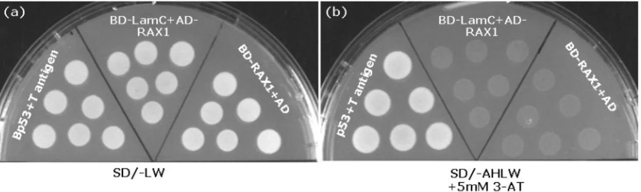
2.2.14 Methods for yeast-two-hybrid system 28
2.2.14.1 Transformation of yeast (LiOAc method) 28
2.2.14.2 cDNA library screening 29
2.2.14.3 Transcription factor (TF) library screening 30
2.2.14.4 Plasmid rescue from yeast 30
2.2.14.5 protein isolation from yeast 30
2.2.15 Recombinant protein expression and purification 31
2.2.15.1 Purification of His-fusion protein 31
2.2.15.2 Purification of GST-fusion proteins 31
2.2.16 GST pull down assay 32
2.2.17 Western blot analysis 32
3. Results 34
3.1 Yeast two-hybrid screening for RAX1 interacting proteins 34 3.1.1 Y2H screen using full-length RAX1 protein as a bait 37 3.1.1.1 A transcription factor library screening 38 3.1.1.2 Analysis of interaction between HAT1 and RAX1 proteins 40 3.1.1.3 Comparison of transcript accumulation of HAT1 and RAX1 in 41
vegetative apices
3.1.1.4 Analysis of the effect of hat1-1 on the branching defect of rax1-3 42
3.1.2 An apex cDNA library screening 44
3.1.2.1 Y2H screening identified YAB1 as a potential partner of RAX1 45
3.1.2.2 Confirmation of the interaction between YAB1 and RAX1 by GST-pull 46
down assay
3.1.2.3 Comparison of transcript accumulation of YAB1 and RAX1 in the shoot 46 apex
3.1.2.4 YAB1 regulates axillary bud formation during the both vegetative and 47 reproductive stages of development
3.1.2.5 Analysis of the pattern of axillary meristem formation in fil-8 rax1-3 50
double mutants
3.2 A role for AtLAX in shoot branching of Arabidopsis thaliana 52
3.2.1 Phylogenetic analysis of LAX-related genes in Arabidopsis 53 3.2.2 Expression profiles of the AtLAX subfamily members 55
3.2.3 Analysis of the expression pattern of AtLAX in the shoot apex 57 3.2.4 Functional characterization of AtlAX in shoot branching 58
3.2.4.1 Analysis of bhlh knockout mutants 59
3.2.4.2 Analysis of atlax knockout mutants 60
3.2.4.3 Analysis of plants expressing the dominant repressor AtLAXSRDX 62 3.2.4.4 Analysis of plants overexpressing AtLAX 65
3.2.5 Interaction between AtLAX and RAX1 66
3.2.5.1 Analysis of atlax-1 rax1-3 double mutants 67 3.2.5.1.1 Analysis of branching pattern of atlax-1 rax1-3 double mutant 68 3.2.5.1.2 Analysis of flowering time of atlax-1 rax1-3 double mutant 70 3.2.5.2 Comparison of transcript accumulation of AtLAX and RAX1 in the 71
shoot apex
3.2.5.3 The interaction of AtLAX and RAX1 proteins 72 3.2.5.4 Analysis of the expression patterns of RAX1 and AtLAX in atlax-1 and 73
rax1-3 mutants
3.2.6 Genetic interaction of AtLAX, RAX1 and LAS 75 3.2.6.1 Analysis of atlax-1 las-4 and atlax-1 rax1-3 las-4 mutants 76 3.2.6.2 Analysis of expression patterns of LAS and AtLAX in atlax-1 and las-4 78
4. Conclusion and discussion 80
4.1 HAT1 and YAB1 are two putative interactors of RAX1 80 4.1.1 Two libraries screens produced different complexities of positive 80 clones
4.1.2 HAT1 may have a function in regulating axillary meristem formation 81 during the vegetative stage of development
4.1.3 YAB1 is a regulator of axillary meristem formation 81
4.2 AtLAX is a regulator of axillary meristem formation 83
4.2.1 AtLAX is the closest homolog of the rice LAX gene in Arabidopsis 83 4.2.2 AtLAX plays a role in axillary meristem formation 84 4.2.3 AtLAX interacts with RAX1 in the process of axillary meristem 86 formation
4.2.4 AtLAX acts downstream of RAX1 and LAS in controlling axillary 88 meristem formation
5. Abstract 91
6. Zusamenfassung 93
7. References 95
8. Erklärung 104
9. Lebenslauf 105
10. Acknowledgements 106
Abbreviation
AA amino acid
AD GAL4 activation domain
AFO ABNORMAL FLORAL ORGANS
AM Axillary meristem A.t. Arabidopsis thaliana
AtLAX LAX-related gene in Arabidopsis AtRHD6 ROOT HAIR DEFECTIVE 6 AtRSL1 RHD SIX-LIKE1 BD GAL4 binding domain bHLH basic helix-loop-helix BA1 BARREN STALK1 from maize bp base pair
Col Columbia CTK Cytokinin
CUC CUPSHAPED COTYLEDON
DNase Deoxyribonuclease E.coli Escherichia coli EGL3 Enhancer of GLABRA3
FIL FILAMENTOUS FLOWER
FM Floral meristem GL1 GLABRA1
GL3 GLABRA3
GST Glutathione S Transferase HAT1 HD-Zip protein 1
HDZip Homeodomain leucine zipper IND INDEHISCENT
IPTG Isopropyl β- D -1-thiogalactopyranoside
KNAT KNOX-Gene from Arabidopsis
LAS LATERAL SUPPRESSOR from Arabidopsis LAX LAX PANICLE from rice
LD Long days LM Lateral meristem
Ls LATERAL SUPPRESSOR from Tomato ORF Open reading frame
Os Oryza sativa (rice)
RAX REGULATOR OF AXILLARY MERISTEMS RT-PCR Reverse transcriptase PCR
SD Short days
TF Transcription factor wt Wild type
WUS WUSCHEL from Arabidopsis Y2H Yeast two hybrid YAB1 YABBY1 from Arabidopsis Zm Zea mays (maize)
1. Introduction
1.1 SAM is the fountain of plant growth
The basic growth axes of flowering plants are established during embryogenesis. On the basis of positional information specified by the apical-basal body axis, the shoot apical meristem (SAM) and root apical meristem (RAM) form at fixed positions early during embryogenesis. After germination, the RAM gives rise to the root system, whereas the SAM is responsible for the development of areal structures of a plant. The SAM may become determinate by forming a terminal flower (sympodial growth), or it may display an indeterminate pattern of growth (monopodial growth) by continuously producing leaves, branches and flowers (Itoh et al., 2006; Kerstetter and Hake, 1997). Shoot branches originate from axillary meristems (AMs) that initiate in the axils of leaf primordia and function like the SAM of the primary shoot. The pattern of shoot branching, as a result of the selective outgrowth of AMs, is a major source of the morphological diversification of flowering plant species.
1.1.1 Organization of the SAM
The SAM consists of a small group of cells from which all the aerial parts above the cotyledons of a plant are derived. SAMs are organized by three functionally different zones with distinct cellular morphologies (Fig. 1a; Steeves and Sussex, 1989; Clark, 1997; Medford et al., 1992;
Kerstetter and Hake, 1997). The central zone (CZ), locating at the summit of the meristem, contains a population of slowly dividing stem cells. Cells in the surrounding peripheral zone (PZ) tend to be larger. They divide more rapidly, and eventually form lateral organs and the outer tissue of the stem. Underlying the CZ is the rib meristem (RZ, rib zone), whose cells also divide and expand rapidly and form the central tissue (pith) of the stem (Golz, 2006).
Another structural feature of the SAM of many angiosperms is the stratified appearance of the cell layers ( Fig. 1a. Kerstetter and Hake, 1997). The cells of outermost layer(s) of the SAM, the tunica, divide in an anticlinal plane (perpendicular to the surface), whereas the cells in the corpus layers also divide periclinally. The tunica layer includes the L1 and L2 layers in most dicots, and the corpus comprises the L3 layer, underneath the tunica. In general, the outermost L1 layer gives rise to the epidermis of shoots, leaves and flowers, the L2 layer produces the ground tissues and germ cells, and the L3 layer contributes to the vascular tissues of the stem and the most internal tissues of leaves and flowers (Clark, 1997;
Laux and Jurgens, 1997; Poethig, 1997; Schiefelbein et al., 1997).
1.1.2 Establishment and maintenance of the SAM
The SAM is formed during embryogenesis and subsequently gives rise to new organs reiteratively during postembronic development (Steeves and Sussex, 1989). Once established, the SAM harbors a population of stem cells over almost the entire life cycle of monopodial plants (Weigel and Jürgens, 2002). Stem cells in the center of the shoot meristem are the ultimate source from which all tissues of the growing shoot are derived (Clark, 1997; Laux and Mayer, 1998; Meyerowitz, 1997). Molecular and genetic studies revealed key regulators controlling establishment and maintenance of the SAM. The first described gene regulating the SAM identity is the maize KNOTTED 1 (KN1), the founding member of the class-I KNOX subfamily of homeobox genes (Kerstetter and Hake, 1997;
Kerstetter et al., 1997; Vollbrecht et al., 2000). A gain-of-function mutation or ectopic expression of KN1 results in meristematic structures in the vicinity of leaf veins (Smith et al., 1992; Sinha et al., 1993). Subsequent loss-of-function studies have demonstrated that KN1 is essential for shoot meristem formation and maintenance. The KN1 mRNA is expressed in the corpus of the SAM and developing stem but not in leaf primordia or mature leaves (Jackson et al., 1994; Kerstetter et al., 1997). In Arabidopsis, the KNOX I subclass of genes
comprise four members, namely SHOOT MERISTEMLESS (STM), KNAT1/BP (KNAT = Knotted in Arabidopsis thaliana; BP=BREVIPEDICELLUS), KNAT2 and KNAT6. Based on null mutant phenotypes and expression patterns of transcripts, STM has a closer functional similarity to KN1 than other KNOXI genes (Long et al., 1996; Vollbrecht et al., 2000). Mutants harbouring the strong loss of function alleles stm-1, stm-5 and stm-11 lack a functional SAM at the base of the cotyledons, and the less severe mutation stm-2 results in disorganized and short-lived SAM. (Barton und Poethig, 1993; Long et al., 1996; Clark et al., 1996; Endrizzi et al., 1996).
Thus, STM is essential for embryonic shoot meristem formation and for the subsequent maintenance of SAM organization. STM is initially expressed in the central apical cells of the globular embryo, later on in the transcripts are detectable throughout the SAM (including the CZ and PZ), in the interprimordial regions and in newly formed lateral meristems (Grbic and Bleecker, 2000; Long and Barton, 2000).
1.1.3 Shoot meristem maintenance by the CLV-WUS feedback loop
Shoot meristems, as the growth pole of a plant, always keep a homeostasis during life which is maintained by a tightly controlled balance between slowly dividing stem cells in the meristem center and cells that are recruited in the periphery and undergo differentiation (Haecker and Laux, 2001). WUSCHEL (WUS) and CLAVATA are identified as components of a negative feedback loop that controls this balance (Schoof et al., 2000). The genes acting in the CLV pathway are CLV1, CLV2 and CLV3 (Clark, 1997; Clark et al., 1993; Kayes and Clark, 1998). Mutations in any of these three genes produce larger SAMs due to an increased number of cells in the CZ suggesting that CLV genes suppress stem cell proliferation and/or promote organ formation. CLV1 is expressed in the L3 layer of the meristem (Clark et al., 1997; Mayer et al., 1998). The transcript of CLV3 is restricted to the so-called stem cells, through the L1 to L3 layers, and hence is often used as a maker for stem cells in the SAM
(Fletcher et al., 1999; Fig. 1). CLV3 is postulated to function together with CLV1 to limit the size of the WUS expression domain underneath (Trotochaud et al., 1999).
Fig. 1. Histology of the shoot apical meristem (SAM) and the CLV-WUS feedback loop. (a) Three cell layers and three zones of an Arabidopsis vegetative SAM. The colored domains depict the different cell layers. In Arabidopsis, the tunica corresponds to the outer two cell layers, L1 and L2, whereas the corpus corresponds to the internal layers L3. The black lines represent the approximate boundaries between the different meristematic zones:
the peripheral zone (PZ) from which lateral organs are formed, the rib zone (RZ), and the central zone (CZ) acting as a ‘source’ of cells for the PZ and RZ, but also for its own replenishment with new cells (Golz, 2006). The PZ and the CZ contain both tunica and corpus cells, whereas the RZ locates in the deeper layers of the corpus. (b) A schematic representation showing the CLV-WUS feedback required for maintenance of a functional SAM. A positive signal from WUS maintains the CLV3 expression domain and stem cell identity in L1, L2, and L3 layers at the summit of the meristem. A signal from CLV3 limits the expression domain of WUS to the organizing center (OC) (Adapted from Taiz & Zeiger 3rd. Fig 16.28. p364).
WUS encodes a homeodomain transcription factor and is expressed in a small region in the centre of the SAM, called the organizing center (OC) (Laux et al. 1996; Mayer et al. 1998).
wus mutants are characterized by a stop-and-go growth, indicating that WUS is necessary for maintenance of the SAM. WUS expression is sufficient to induce the expression of CLV3 and to promote meristem cell identity of overlying neighbor cells. Activation of CLV3 results in a down-regulation of WUS expression. Such a feedback loop allows the SAM to maintain the equilibrium between stem cell proliferation in the CZ and cell loss due to differentiation in the PZ (Brand et al. 2000; Schoof et al. 2000; Fig. 1b). Double mutant combination of wus and clv are almost indistinguishable from the wus single mutant, consistent with the model that WUS is a target for the negative regulation by the CLV genes to maintain a constant number of stem cells in the CZ of the SAM (Laux et al., 1996; Schoof et al., 2000).
1.1.4 The SAM and lateral organs
In seed plants, lateral organs such as leaf and floral primordia are formed from the flanks of the SAM, a defined group of founder cells of apical meristems. KNOX genes are not expressed in developing primordia and have been shown to function antagonistically to the process of leaf development (Brand et al., 2002; Chuck et al., 1996; Dean et al., 2004; Gallois et al., 2002; Lenhard et al., 2002; Lincoln et al., 1994; Pautot et al., 2001). Several genes are responsible for the initial repression of the KNOX genes in lateral organs: Antirrhinum PHANTASTICA (PHAN) (Waites et al., 1998), maize ROUGH SHEATH2 (RS2) (Timmermans et al., 1999; Tsiantis et al., 1999b) and Arabidopsis ASYMMETRIC LEAVES1 (AS1) (Byrne et al., 2002). Loss of PHAN/RS2/AS1 functions is associated with the ectopic expression of KNOX genes in leaves indicating that these genes repress KNOX expression in leaf primordia (Schneeberger et al., 1998). The PHAN/RS2/AS1 genes are initially expressed in the leaf founder cells and later in the growing leaf primordia.
In addition, members of the YABBY (YAB) gene family have also been shown to repress the expression of class-I KNOX genes in leaves (Kumaran et al., 2002). YABBY genes are expressed on the abaxial side of lateral primordia and function in promoting abaxial cell fate of lateral organs (Kumaran et al., 2002; Sawa et al., 1999; Siegfried et al., 1999).
Knockout mutations in both YAB1 and YAB3 result in lobed leaves expressing KNOX genes and ectopic meristems, suggesting an incompatibility between KNOX gene functions and proper specification of abaxial cell fate.
Adaxial-specific transcriptional regulators are also important in establishing a connection between leaf polarity and shoot meristem function (McConnell et al., 2001; Otsuga et al., 2001). The class III HD-ZIP family members PHABULOSA (PHB), PHAVOLUTA (PHV) and REVOLUTA (REV) are adaxial-identity promoting factors in developing leaves (Byrne, 2006).
These genes are expressed in the central zone of the SAM, in the vasculature and on the adaxial side of developing leaf primordia. Loss-of-function mutations in REV result in a reduced meristematic competency (Talbert et al., 1995). Mutants harboring gain-of-function mutations in PHB and PHV produce enlarged SAMs and STM-expressing ectopic meristems.
(McConnell and Barton, 1998). Conversely, simultaneous loss of PHB, PHV and REV function results in leaf abaxialisation and loss of shoot meristem formation/maintenance (Emery et al., 2003).
1.1.5 Hormonal regulation of the SAM function
Recently, several studies lead to a model in which high auxin concentrations in incipient primordia down-regulate the expression of KNOXI genes, inducing lateral organ initiation (Benkova et al., 2003; Reinhardt et al., 2000; Reinhardt et al., 2003). Therefore, auxin is supposed to act as a positive regulator for leaf initiation.
The positive role of cytokinin (CK) on SAM function has been addressed by the analysis of triple mutants of three genes encoding CK receptors: AHK2 (Arabidopsis HISTIDINE KINASE 2), AHK3 and AHK4/CRE1/WOODEN LEG (WOL) (Higuchi et al., 2004; Nishimura et al., 2004;
Riefler et al., 2006). ahk2 ahk3 ahk4 triple mutants display a dramatic reduction in meristem size. New insights into the regulation of CK in meristem function and maintenance come from functional analysis of LONELY LOG (LOG) in rice (Kurakawa et al., 2007). The log mutant has a smaller vegetative meristem and its floral meristems terminate prematurely. LOG is specifically expressed in the shoot meristem tips and encodes an enzyme that controls the final CK biosynthesis step to release active CK.
KNOXI proteins have been shown to negatively regulate gibberellic acid (GA) biosynthesis, causing a suppression of GA activity in the SAM (Chen et al., 2004; Hay et al., 2002;
Sakamoto et al., 2001). The application of exogenous GA suppresses the phenotypes caused by the misexpression of KNOX genes (Hay et al., 2002).
1.2 Shoot branches determine the architecture of a plant
The aerial architecture of flowering plants is determined to a large extent by shoot branches established by the formation and outgrowth of axillary meristems (AMs). These meristems subsequently function as shoot apical meristems (SAMs) to provide plants with an unlimited growth potential. In higher plants, AMs form in the axils of leaves (Sussex and Kerk, 2001;
Ward and Leyser, 2004). Once established, AMs first produce a few leaves to form lateral buds. These buds have a potential to develop into lateral shoots, or remain dormant until outgrowth is triggered. The outgrowth of lateral buds is controlled by the main shoot apex, which is known as apical dominance. Hence, the degree of branching depends not only on the establishment of an axillary meristem but also upon its subsequent activity.
1.2.1 Formation of axillary meristem 1.2.1.1 Ontogeny of axillary meristem
AMs form at the boundary regions between the SAM and lateral primordia. These ‘boundary cells’ display distinctive structural characteristics compared to the surrounding cells (Kwiatkowska and Dumais, 2003). However, the ontogeny of the AM is still in a state of debate. To date, there are two opposite models explaining the originS of AMs. The first suggests that AMs initiate de novo separately from the SAM in leaf axils (‘de novo’ formation concept) (Snow and Snow, 1942). This hypothesis is supported by the observation that in the axils of leaves formed in early development there is no morphologically distinguishable AM at the time of leaf primordium initiation. Further support for the ‘de novo’ formation of AMs
comes from the study on the Arabidopsis phabulosa-1d mutant in which AMs form at the (adaxilaized) abaxial base of leaves (McConnell and Barton 1998). An alternative model suggests that AMs derive from groups of meristem cells that detach from the SAM at the time of leaf primordia initiation and never lose their meristematic identity (‘detached’ meristem concept) (Garrison, 1955; Sussex, 1955). Evidence for this hypothesis comes from the expression of the SAM marker STM in the leaf axils immediately proceeding the appearance of a morphological AM (Long et al., 1996). However, the interprimordial expression of STM makes it difficult to distinguish between the “detached meristem” and “de novo formation”
concepts of AM initiation.
The formation of AM has been suggested to be temporally and spatially specified. In Arabidopsis, not all AMs develop at the same time, some may not develop at all (especially those in the cotyledonary nodes) (Aguilar-Martinez et al., 2007). During prolonged vegetative stage, AMs are initiated in an acropetal order, first in the axils of mature leaves distant from the shoot apex and progressing to younger leaves. Once the SAM is transformed from the vegetative to reproductive stage, AMs are initiated in a basipetal order, first in leaf axils closest to the shoot apex (Hempel and Feldman, 1994; Grbic and Bleecker, 2000; Long and Barton, 2000).
1.2.1.2 Regulators of axillary meristem initiation
The characterization of mutants defective in shoot branching makes it possible to investigate the molecular mechanisms of AM initiation. The well-known regulators for AM initiation are orthologous GRAS transcription factors from tomato LATERAL SUPPRESSOR (Ls), Arabidopsis LATERAL SUPPRESSOR (LAS), and rice MONOCULM1 (MOC1) (Greb et al., 2003;
Schmitz et al., 2002; Schumacher et al., 1999; Li et al., 2003). Mutations in both Ls and LAS resulted in a complete lack of side shoots during the vegetative phase of development. This
phenotype is due to loss of the meristematic competence of the cells in the leaf axils. After the transition to reproductive development, axillary shoots are formed and inflorescence branching is not affected in these mutants. Introduction of a genomic fragment carrying the Arabidopsis LAS gene into the tomato ls mutant led to a full complementation of the mutant phenotype, revealing a conserved working mechanism for AM formation over a large evolutionary distance (Greb et al., 2003). The Arabidopsis LAS gene is expressed in a band-shaped domain at the adaxial base of all primordia derived from the SAM. The Ls/LAS-orthologous gene MOC1 in rice controls vegetative as well as reproductive branching.
Loss of MOC1 function resulted in lack of tillers as well as a reduced number of rachis branches and spikelets (Li et al., 2003). MOC1 is expressed mainly at the leaf axils and the expression extends to the entire tiller buds. Molecular and structural analysis of the barren leaf axils in these mutants suggests that the function of LAS/Ls/MOC1 is probably to retain the meristematic capacity in a group of cells at the adaxial base of leaf primordia. Loss of LAS/Ls/MOC1 function results in an inability to sustain this group of cells, hence the phenotypes of these mutants. This conserved genetic function suggests a single general mechanism of AM formation in different plant species.
In Arabidopsis thaliana, the functionally redundant genes CUP-SHAPED COTYLEDON1 (CUC1), CUC2, and CUC3 regulate embryonic shoot meristem formation and boundary specification of lateral organs including cotyledons (Aida et al., 1997; Hibara et al., 2006;
Takada et al., 2001; Vroemen et al., 2003). Any combination of mutant cuc alleles leads to severe cotyledon fusions and cup-shaped structures, which do not contain a SAM. All three CUC genes encoding NAC transcription factors are expressed in a narrow strip of cells in the boundary domains between the SAM and lateral primordia, very similar to LAS. Such characteristic expression patterns of CUCs in the axils of leaf primordia indicate their roles in AM formation. This hypothesis has been proven by two independent studies on cuc loss-of-function mutants (Hibara et al., 2006; Raman, 2006). The significant roles of CUC2
and CUC3 in AM formation were described by Hibara (Hibara et al., 2006). cuc3 mutants were found to be impaired in tertiary shoot formation (axillary shoots from secondary shoots) at a low frequency, whereas the cuc2 cuc3 double mutant lacked branches in all leaf axils, indicating the redundant functions of CUC2 and CUC3 in AM formation. Raman also showed that CUC3 regulates axillary bud formation (Raman, 2006). Over-expression of MIR164A or MIR164B, targeting the mRNAs of CUC1 and CUC2, in the cuc3-2 mutant caused an almost complete lack in axillary bud formation, revealing a functional redundancy of CUC1, CUC2 and CUC3 in AM formation.
Recently, the roles for three Scarecrow like (SCL) genes (SCL6, 22, 27) in AM development, but not in AM initiation have been characterized by Schulze (2007). In scl27-1 mutants grown in short photoperiods, formation of lateral buds was found to be compromised during the vegetative and reproductive phases of development. Any combination of loss-of-function mutations in three SCL genes led to an enhancement of the branching defect of scl22-7, demonstrating functional redundancy among these three genes. Transcripts of SCL22 and of SCL27 were detected in vegetative and reproductive apical meristems, lateral meristems and the vasculatures of shoots and leaves. SCL6 transcripts were detected in the axils of leaf and floral primordia. The expression of the meristem markers STM and LAS was detected in the interprimordial regions of apices of scl22-1 scl27-1 and scl6-1 scl22-1 scl27-1 mutant plants, demonstrating that the activities of these SCL genes are needed for the development of side shoots after the initiation of AMs (Schulze S., 2007).
Another group of regulators controlling early steps of AM formation comprises members of R2R3-type MYB transcription factors from tomato and Arabidopsis, namely Blind and REGULATORS OF AXILLARY MERISTEMS (RAXs), respectively. In tomato, the allelic blind and torosa mutants display a strong reduction in AM formation during the vegetative and reproductive developmental stages (Mapelli and Kinet, 1992; Schmitz et al., 2002). The
sympodial shoot is often not formed in these mutants and the plants terminate with a single inflorescence. In addition, the number of floral meristems (FMs) is greatly reduced, and flowers are often fused. In Arabidopsis, the Blind-homologous RAX genes (RAX1, 2, 3) have recently been shown to control an early step of AMs formation (Müller et al. 2006; Keller et al., 2006). rax1-3 mutants fail to form lateral buds during the early vegetative developmental stage. Analysis of double and triple rax mutants revealed that RAX genes control shoot branching in overlapping zones along the shoot axis. In addition, the severity of branching defects of rax mutants was shown to be strongly dependent on day-length conditions. The defects observed under short-day conditions were strongly diminished or vanished in long days. RAX1 transcripts accumulate in a ball-shaped domain in the center of the boundary region between the SAM and the leaf primordia. RAX3 displays an expression pattern similar to that of RAX1 during the vegetative stage, whereas RAX2 shows a wide expression in the shoot tip (Müller et al. 2006). Double mutant analysis of ls and blind (bl) in tomato as well as of las-4 and rax in Arabidopsis suggests that at least two independent pathways control the initiation of AMs (Müller et al., 2006; Schmitz et al., 2002).
LAX PANICLE (LAX) and BARREN STALK1 (BA1) were recently characterized as major regulators of shoot and inflorescence branching in rice and maize, respectively (Gallavotti et al., 2004; Komatsu et al., 2003; Komatsu et al., 2001). LAX and BA1 encode homologous basic helix–loop–helix (bHLH) transcription factors. LAX transcripts are excluded from the vegetative SAM and accumulate mainly in the boundary regions between the SAM and the new meristems during the reproductive stage. Plants homozygous for strong lax alleles have severely reduced panicle branches and completely lack lateral spikelets, demonstrating an important role of LAX in regulating reproductive branching. The LAX gene also regulates the formation of vegetative branches (tillers), in a redundant fashion with the SMALL PANICLE (SPA) gene. lax spa double mutants are characterized by a complete lack of tillers (Komatsu et al., 2003). Studies on mutants harbouring a loss-of-function mutation in BA1, the
LAX-orthologous gene from maize, have uncovered a pivotal role of BA1 in AM formation during the vegetative and reproductive stages of development (Gallavotti et al., 2004). ba1 mutants do not form tillers, ears and tassel branches. Accordingly, BA1 transcripts are detectable at the adaxil bases of new meristems.
1.2.2 Outgrowth of axillary meristem
The outgrowth of lateral buds is well known to be inhibited by signals derived from the main shoot tip, a phenomenon named apical dominance (Chatfield et al., 2000; Horvath et al., 2003). The plant hormone auxin (IAA, Indole-3-acetic acid) is an important cause for the inhibition of axillary bud outgrowth. Studies on the AUXIN RESISTANT1 (AXR1) gene of Arabidopsis supply in vivo significance for this fact (Leyser et al., 1993; Lincoln et al., 1990;
Stirnberg et al., 1999). Mutations in AXR1 result in increased shoot branching in both vegetative and floral nodes, and the lateral buds in axr1 are resistant to inhibition by apically or locally applied IAA.
However, the endogenous level of IAA is not always correlated with the degree of shoot branching, suggesting that additional signals could be involved (Beveridge et al., 1994;
Beveridge et al., 2000). Genetic analysis has provided evidence for the presence of a new messenger regulating axillary bud outgrowth. Mutations in the MORE AXILLARY GROWTH (MAX) 1, 3, and 4 genes of Arabidopsis, the RAMOSUS (RMS) 1 and 5 genes of pea, and the DECREASED APICAL DOMINANCE1 (DAD1) gene of petunia result in increased bud outgrowth (Booker et al., 2005; Napoli, 1996; Snowden et al., 2005; Sorefan et al., 2003;
Stirnberg et al., 2002; Turnbull et al., 2002). RMS1, DAD1, and MAX4 are orthologous genes.
The max4 and rms1 mutant buds are resistant to the inhibitory effects of apical auxin, leading to the hypothesis that the MAX/RMS/DAD pathway plays an important role in regulating bud outgrowth (Beveridge et al., 2000; Sorefan et al., 2003). Bennett et al.
demonstrated that the MAX pathway controls shoot branching by regulating auxin transport capacity (Bennett et al., 2006). max mutants have increased levels of PIN proteins, auxin efflux facilitators, and therefore have increased auxin transport capacity. Thus, the MAX/RMS/DAD pathway may act through auxin signaling in the process of lateral bud outgrowth.
Several TCP (TEOSINTE BRANCHED1, CYCLOIDEA, and PCF) transcription factors were also shown to regulate bud outgrowth. These are TEOSINTE BRANCHED1 (TB1) from maize (Zea mays) (Doebley et al., 1997) and its homologous genes OsTB1 from rice (Oryza sativa) (Takeda et al., 2003; Hu et al., 2003), SbTB1 from sorghum (Sorghum bicolor) (Kebrom et al., 2006), and the two Arabidopsis homologs BRANCHED1 and 2 (Aguilar-Martinez et al., 2007). Both TB1 and OsTB1 are expressed in AMs and lateral buds, where they function as negative regulators of lateral branch outgrowth (Hubbard et al., 2002; Takeda et al., 2003).
Loss-of-function mutations in TB1 and OsTB1 (FINE CULM1) caused enhanced outgrowth of tillers. BRANCHED1 (BRC1), one of the closest homologs of TB1 in Arabidopsis, is a recently characterized regulator of bud outgrowth. BRC1 is expressed in different regions of lateral buds throughout axillary bud development (Aguilar-Martinez et al., 2007). Downregulation of BRC1 leads to branch outgrowth as a result of a relief of bud growth repression.
Aguilar-Martinez et al. also showed that BRC1 acts downstream of the MAX pathway and is required for auxin-induced apical dominance. Therefore, BRC1 acts inside the buds as an integrator of branching signals and translates them into a response of cell growth arrest.
1.3 Aim of this work
The aim of this work was to identify and characterize interactors of RAX1, an early regulator of axillary meristem formation in Arabidopsis thaliana. A yeast two-hybrid screening was performed to identify interactors for RAX1 at a large scale. HAT1 (HD-Zip protein1) and YAB1 (YABB1/AFO/FIL) were two candidates and were focused on analysis in the first part of this study. A further aim of this study was to identify novel regulators of axillary meristem initiation. AtLAX (LAX-related gene in Arabidopsis) was isolated based on its high sequence similarity to LAX and BA1, two important regulators of shoot and inflorescence branching in rice and maize, respectively. The role of AtLAX in axillary meristem formation was characterized. In addition, the genetic interaction between AtLAX, RAX1 and LAS, another axillary meristem regulator, was inspected.
2. Material and Methods
2.1 Materials 2.1.1 Chemicals
The following were main sources of supply for chemicals used in this work:
Ambion, Austin, USA
Amersham Pharmacia Biotec, Braunscheig, Germany Biozym, Hess.Oldendorf, Germany
Carl Roth GmbH, Karlsruhe, Germany Invitrogen GmbH, Karlsruhe, Germany
MBI Fermentas GmbH, St. Leon-Rot, Germany
Merck KgaA, Feinchemikalien und Laborbedarf Deutschland, Darmstadt New England BioLabs GmbH, Schwalbach/Taunus, Germany
Operon, Cologne, Germany PIERCE, Rockford, USA QIAGEN, Hilden, Germany Roche, Basel, Switzerland
Sigma Chemical Co., St.Lois, USA
2.1.2 Expendable materials and reagents
The following were the main suppliers of laboratory expendables used during this work:
Incubation tubes and Petri-dishes: Greiner Lobortechnik; Eppendorf-Netheler-Hiny GmbH, Hamburg; Sarstedt AG & Co, Nümbrecht
PVDF membranes: Macherey-Nagel GmbH & Co.KG, Düren
Kits for DNA and RNA extraction and purification: Qiagen, Hildesheim Kits for total RNA extraction from plant: Qiagen, Hildesheim
cDNA synthesis kit: MBI, GmbH, Fermentas RNA probe transcription kit: Austin, USA
pGEM-Teasy for RNA probe transcription: Invitrogen, GmbH, Karlsruhe, Germany Gateway cloning kit: Invitrogen, GmbH, Karlsruhe, Germany
SuperSignal West Dura Extended Duration Substrate, Pierce, Rockford, USA
SuperSignal West Pico Chemiliminescent Substrate, Pierce, Rockford, USA
2.1.3 Enzymes
Enzymes used during the course of this work were from following suppliers:
Invitrogen GmbH, Karlsruhe, Germany
New England BioLabs GmbH, Schwalbach/Taunus, Germany MBI Fermentas GmbH, St. Leon-Rot, Germany
Roche, Basel, Switzerland
Sigma Chemical Co., St.Lois, USA
KOD hot start DNA polymerase, Novagen, Toyobo, Japan.
2.1.4 Antibodies
Anti-Digoxigenin-AP Fab-Fragments (from sheep), Roche, Basel, Switzerland Anti·His HRP conjugates, Hildesheim, QIAGEN
2.1.5 Organisms 2.1.5.1 Bacteria
The following Escherichia coli strains were used during the course of this work. For cloning specific DNA fragements into vectors, DH5α (Hanahan, 1983) was transformed. For large scale fused proteins expression, BL21 (Stratagene) was used. The chemical competent cells were prepared as described by Sambrook und Russell (2001).
DH5 α F-, end A1, hsdR17 (rk-, mk+), gyrA96, relA1, Hanahan, 1983 supE44, L-, recA1, 80dlacZM15, ∆(lacZYAargF) U196
DB3.1 B F– ompT hsdS(rB– mB–) dcm+ Tetr gal l (DE3) Stratagene endA Hte metA::Tn5(KanSr) [argU proL Camr]
BL21- CodonPlus (DE3)-RP-X Stratagene B F– ompT hsdS (rB– mB–) dcm+ Tetr galλ (DE3) endA Hte metA::Tn5
(KanSr) [argU proL Camr]
For plant transformation, Agrobacterium tumefaciens was used.
GV3101 Virulence Plasmid: pMP90RK (Koncz und Schell, 1986) Selection markers: Rifampicin, Gentamycin and Kanamycin.
PMP90RK was included as helper to integrate into plant genome when the binary vectors contain Carb selective marker. Antibiotics used for selection: Rif, Gent, Kan, Carb.
Antibiotics and plant* selection
Antibiotics Working concentration Solvent
Ampicillin (Amp) 100 mg/L H2O
Carbenicillin (Carb) 100 mg/L H2O
Gentamycin (Gent) 50 mg/L H2O
Kanamycin (Kan) 50 mg/L H2O
Rifampicin (Rif) 100 mg/L DMSO
Spectinomycin (Spec) 100 mg/L H2O
Basta * 25 mg/L H2O
2.1.5.2 Plants
The phenotype investigation in this work were carried out on model plant Arabidopsis thaliana, belonging to the ecotypes Columbia, Landsberg and Wassilewskija. Seeds for the different ecotypes were obtained from the Nottingham Arabidopsis Stock Centre (NASC).
The following Arabidopsis thaliana mutants were used during this work:
Mutant Allele Type of mutation Genetic background
Source
atlax atlax-1 Null allele; A T-DNA insertion at the beginning of the bHLH domain (156bp after ATG)
Col N580565
atlax-2 Null allele; A T-DNA insertion at 396bp after ATG (118bp behind of the bHLH domain).
Col N524760
bhlh85 bhlh85-1 A T-DNA insertion at 107bp upstream of ATG.
Col N548849
bhlh86 bhlh86-1 A T-DNA insertion at 164bp upstream of ATG.
Col N465407
bhlh87 bhlh87-1 Null allele; A T-DNA insertion at 486bp after ATG.
Col N566339
bhlh88 bhlh88-1 Null allele; A T-DNA insertion at 543bp after ATG (exactly at the end of the bHLH domain).
Col GABI297B10
hat1 hat1-1 T-DAN insertion in the 2nd intron
Col N506022
lateral suppressor
las-4 Null allele; frameshift arising from deletion of 20bp and addition of a single nucleotide in the begining of the ORF
Col Greb et al., 2003.
rax1 rax1-3 T-DNA insertion in the end of ORF.
Col Müller et al., 2006 yab1 fil-8
(afo-1)
Ds insertion in the 4th exon. Ler Y. Esheld (Kumaran et
al, 1999) fil-1 G to A change in splice site
acceptor sequences.
Ler (Siegfried et al., 1999).
2.1.6 Vectors
2.1.6.1 E.coli vectors
The following vectors were used to clone specific DNA fragements and express fusion proteins in vitro during this work:
GEM4Z Vector for cloning and transcription of DNA Promega fragment under the T7 promotor.
pGEM-Teasy Vector for cloning of PCR products and their Promega transcription under the T7 Promotor .
pDONR201 Vector for cloning of DNA-Fragmenten for Invitrogen use in Gateway System
pDONR221 Vector for cloning of DNA-Fragmenten for use Invitrogen in Gateway System
pET28a Vector for protein expression with N-terminal Novagen His(6)-Fusion
PGEX4T1 Vector for protein expression with N-terminal Pharmacia GST-Fusion
2.1.6.2 Yeast vectors
To investigate protein interactions in yeast, the following vectors were used during this work:
PAS-attR-new Vector for protein expression fused with GAL4-BD (kindly provided by J. Uhrig)
PACT-attR Vector for protein expression fused with GAL4-BD (kindly provided by J. Uhrig)
pGADT7 Vector for protein expression fused with GAL4-AD Clontech pGBKT7 Vector for protein expression fused with GAL4-BD Clontech pCL1 Positive control for yeast two hybrid system, Clontech
coded for GAL4-transcription factor
pGBKT7-53 Positive control for yeast two hybrid system, Clontech coded for fusion protein between p53 from mouse
and the Gal4-BD
pGADT7-T Positive control for yeast two hybrid system, Clontech coded for fusion protein between SV40 T-antigen
and the Gal4-AD
pGBKT7-LamC Negative control for the yeast two-hybrid system, Clontech coded for fusion protein between Lamin C from
humans and the GAL4-BD
2.1.6.3 Plant vectors
For transformation of A. thaliana, the following binary vectors were used:
pGPTV-Bar-35S 35S promoter carrying binary vector (Cardon et al., 1999).
GV3101 was transformed with constructs in this vector backbone.
pGPTV-Bar-AscI GUS carrying binary vector (Überlacker et.al,1996). GV3101 was transformed with constructs in this vector backbone.
pXLSG-strepII C-terminal strepII-tag fusion binary vector harboring attR1, R2
sites for Gateway cloning (Witte et al., 2004). GV3101+pMP90K was used for plant transformation.
2.1.7 Libraries for yeast two-hybrid screening
To identify the interaction partners of RAX1 protein, cDNA libraries screening is powerful and handy way using yeast two hybrid method. The following are two kinds of prey libraries used in this study:
Library Material Density Sourse
Apex cDNA library Shoot tips from both vegetative and reproductive stages
~1x10⁶ Hans Sommer
Transcription factor (TF) library
Annotated transcription factors 1002 REGIA (Paz-Ares et al., 2002)
2.1.8 Oligonucleotides
The oligonucleotides used in this study were mainly synthesized in Operon and Invitrogen.
Oligonucleotides used in the part of yeast two-hybrid screening:
Name 5’-3’ sequences Locus
HAT1-rev GAA GAT CTA TCT AGA GTG ATG At4g17460
HAT1-841R CCA AAG CCA GCT TCT GTT Tc At4g17460
HD-ZIP-EcoRIfo GAT GAA TTC ATG ATG ATG GGT AAA GAG GAT At4g17460 HD-ZIP-NotIre CAT GCG GCC GCT TAA GAC CTA GGA CGC ATC A At4g17460 HD-ZIP-ORFre CTT CTG TTT GGG ATT GAG AGT At4g17460 M37-1036F ATT CAC TAA CGA GAG AAA TGG GAA At5g23000 M37-2549R CAA GAG AGT CTA GAA GAA CTA GGA G At5g23000 M37-1987F CAA TCC CAT CTT CTT CTT ACA ATC C At5g23000
M37-2489R GCT ACC CAT GCT TTT GTT CTC At5g23000
myb37-EcoRIF CAT GAA TTC GGA AGA GCT CCG TGT TGC GAC At5g23000 myb37-XhoIR CAT CTC GAG CTA GGA GTA GAA ATA GGG CAA GC At5g23000 myb37-XhoIre CAT CTC GAG TCA GAG TTT CTT CCT TAG CTT TGT G At5g23000 YAB1-EcoRI-F GTA GAA TTC ATG TCT ATG TCG TCT ATG TCC TC At2g45190 YAB1-XhoI-R GAT CTC GAG TTA ATA AGG AGT CAC ACC AAC G At2g45190
YAB1-ORF-fo TGT GGT TGC TGT ACC AAT CT At2g45190
YAB1-ORF-re GGA CTC TCT GTC TTT TCT CTG At2g45190 YAB1-1309F GTC TCT TTC TGT CTG AGT TTT TG At2g45190
YAB1re AGA AAC CAC AAC TTT TGG ACA T At2g45190
Oligonucleotides used in the part of bHLH140 functional analysis:
Name 5’-3’ sequences Locus
bHLH37-998F GGA GGA TGG ATA ACT CCC AC At3g50330
bHLH37-1699R ATC ATC TAA GAA TCT GTG CAT TTC C At3g50330
bHLH40F CAA CCA CAG CCC CAA AAG A At4g00120
bHLH40R TCT TCT CGC TGA TCC TTT CC At4g00120
bHLH43-for2 TGA ACC CAT CTC TCT TCC AAA At5g09750
bHLH43-rev2 GGC GGT GAA ACC CAT GAC At5g09750
bHLH52-for2 CAA GAG CAA CCG CAA CAT C At1g30670
bHLH52-rev2 AAG ATG CAT TCT TCG GCT TT At1g30670
bHLH53-for2 TTG CTT TAT TCC CGA GAT GG At2g34820
bHLH53-rev2 AAA TAT TCG GGG TTC TGC AA At2g34820
bHLH54-for2 TGG AGT CTC TCT TGG GGA TG At1g27740
bHLH54-rev2 GTA AGC CAA TGG TGC GTA CA At1g27740
bHLH83F ATC CGG TGA AGA TCA TCA CAA At1g66470
bHLH83R AGC GGA TTT AGG CGA AAG AG At1g66470
bHLH84F TCT CCC CTC CAA GGA TTT GT At2g14760
bHLH84R AGA GAA GCA AAA GCC ACC A At2g14760
bHLH85F AGC AAC AAC CTC GGA GGA A At4g33880
bHLH85-For2 TGA AGC CGG TAG CTT CTG TT At4g33880
bHLH85-proF2 ATT ACT CGG ATG CTA ACG AAA C At4g33880 bHLH85R CCA TTG TCT CAT TTT GGT TCT CTT At4g33880 bHLH85-T7R CGA TAA TAC GAC TCA CTA TAG GGC CAT TGT CTC
ATT TTG GTT CTC TT At4g33880
bHLH085-T7R2 TAA TAC GAC TCA CTA TAG GGA TGT CCA TCC CAT
TGA AAG C At4g33880
bHLH86-for2 ATT GCG GAT TAG ACG AAG GA At5g37800
bHLH086F TGC TTC ATC ATT CTT CAC CTT T At5g37800
bHLH086-T7F TAA TAC GAC TCA CTA TAG GGT GCT TCA TCA TTC TTC ACC TTT
At5g37800
bHLH86-proF TGA TCT CAT GCC AGC TTC C At5g37800
bHLH086R GAA AGT TGT GTG TTC TCT CCC At5g37800
bHLH086-T7R TAA TAC GAC TCA CTA TAG GGT GCT TCA TCA TTC TTC ACC TTT
At5g37800
bHLH86-rev2 AAA ACT CAT CGG CTG CAA GT At5g37800
bHLH87-348F ATG GAA GGA TTG GAA TCT GTG T At3g21330 bHLH87-1145R CTC CAC AAT CTC TAA CCC GAA At3g21330
bHLH087-1461R TCA AAA GTT TAT AAT CTG TCA ACA CTC At3g21330
bHLH88F ACT AAC CCT TCT TCT ATC TCT CC At5g67060
bHLH88-T7R TAA TAC GAC TCA CTA TAG GGC TCT CGC TTA TTC TCT CCC TT
At5g67060 bHLH88-116F ACC CAT TTT ATC AGC TTC TCC A At5g67060
bHLH88-519R CTC CTC TTC GGT GGC TTA AC At5g67060
bHLH139F TGC AAT GCT CCA AAT GAG AC At5g43175
bHLH139-T7R CGA TAA TAC GAC TCA CTA TAG GGG CTA TCC CTC
TGT TGG CTT T At5g43175
bHLH137-1040F GGG GAC AAG TTT GTA CAA AAA AGC AGG CTC AAT
GGA TGA TTT CAA TCT TCG TAG At5g01310
bHLH137-1347F GCT TCT CTT CAT AAA CGA CCA C At5g01310
01310-1551R CGA GTC ACG TTC TTG CTC A At5g01310
bHLH140-1931R CAA ATT TAC ATT AAA ACG CCT GTT TAT C At5g01310 bHLH140-Acc65Ire CTA GGT ACC GGA CGA GTC ACG TTC TTG CTC A At5g01310 bHLH140-BamHIre GAT GGA TCC ATG GAT GAT TTC AAT CTT CGT AGC At5g01310 bHLH140-EcoRIfo GAT GAA TTC ATG GAT GAT TTC AAT CTT CGT AGC At5g01310 bHLH140-NotIre CAT GCG GCC GCC TAG GAC GAG TCA CGT TCT TGC At5g01310 bHLH140GW2 GGG GAC CAC TTT GTA CAA GAA AGC TGG GTT GGA
CGA GTC ACG TTC TTG CT
At5g01310
Atls2427F TCC TCT CCC TTA ACT CTT CTC C At1g55580
Atls2717R CCG TTA AAT GAC CGA ACC GA At1g55580
CUC3-1610F TGG AAA GCT ACC GGC AAA At1g76420
CUC3-1983R ATT GAA CAC TCT GCA AAT CA CC At1g76420 LDs10 GGG AAT TCT TTT ACC GAC CGT TAC CGA C Ds-element M37-1036F GGG GAC AAG TTT GTA CAA AAA AGC AGG CTA TTC
ACT AAC GAG AGA AAT GGG AA At5g23000
M37-1688F AAA ATC ACG AGC TAC ATT GAT TCC At5g23000 M37-2624R TTC TCT CTC CTC CCA TAC CCC ATC AAA TC At5g23000 Myb37-GW2 GGG GAC CAC TTT GTA CAA GAA AGC TGG GTA GGA
GTA GAA ATA GGG CAA GCA
At5g23000
T-DNA-LB-Rev GTT ACT AGA TCG ACC GGC A T-DNA
T-DNA-LBfo CCA AAC TGG AAC AAC ACT CAA T-DNA
T-DNA-RBre GCG GTT CTG TCA GTT CCA A T-DNA
T-DNA-RBre2 GAA ATC ACC AGT CTC TCT CTA CA T-DNA
Molecular markers for identification of mutant alleles:
Allele Markers Polymorphism
atlax-1 T-DNA-LB-Rev+bHLH140-1931R; Produces an ~1200bp T-DAN band
bHLH140-EcoRIfo+bHLH140-1931R Produces an 860bp WT band atlax-2 T-DNA-LB-Rev+bHLH140-1931R;
bHLH140-EcoRIfo+bHLH140-1931R
Produces an ~970bp T-DNA band Produces an 860bp WT band bhlh85-1 T-DNA-LB-Rev+bHLH85pro-F2
bHLH85pro-F2+bHLH85R
Produces a ~900bp T-DAN band Produces an 830bp WT band bhlh86-1 GABI-Left+bHLH86R
bHLH86-proF+bHLH86R
Produces an ~870bp T-DAN band Produces a 930bp WT band bhlh87-1 SALK-LB1+bHLH087-1461R
bHLH087-348F+bHLH087-1461R
Produces an ~800bp T-DAN band Produces a 1139bp WT band bhlh88-1 GABI-Left+bHLH088-116F
bHLH088-116F+bHLH88-rev2
Produces an ~600bp T-DAN band Produces a 640bp WT band hat1-1 T-DNA-LB-Rev+HD-Zip-EcoRIfo
HD-Zip-EcoRIfo+HAT1-841R
Produces an ~1000bp T-DAN band Produces a 670bp WT band
las-4 Atls2427F+Atls2717R Produces a 270bp mutant band Produces a 290bp WT band rax1-3 T-DAN-LB-Rev+M37-1688F;
M37-1688F+M37-2624R
Produces an ~930bp T-DAN band Produces a 637bp WT band fil-8 LDs10+YAB1re
YAB1-1309F+YAB1re
Produces an ~970bp Ds band Produces a 1051bp WT band
2.1.9 Plasmids
Listed below are the plasmids used in this study:
Construct Insertion Vector Cloning sites Antibiotics BD-RAX1 full length cDNA of
RAX1
pAS-attR gateway Amp
AD-RAX1 full length cDNA of RAX1
pACT-attR gateway Amp
pFY10 (GST-RAX1)
full length cDNA of RAX1
pGEM-4T-1 BamHI/XhoI Amp
pFY19 (His-RAX1)
full length cDNA of RAX1
pET28a BamHI/XhoI Kan
pFY30 (His-HAT1)
full length cDNA of HAT1
pET28a EcoRI/NotI Kan
pFY63 (anti-HAT1)
1-486 relative to the ATG
pGEM-Teasy T-easy (antisense)
Amp
pFY51 (anti-YAB1)
160-433 relative to the ATG
pGEM-Teasy T-easy (antisense)
AmP
pFY62 (AD-YAB1)
full length cDNA of YAB1
pGADT7 EcoRI/XhoI Kan
pFY35 full length cDNA of pET28a EcoRI/XhoI Kan
(His-YAB1) YAB1
anti-AtLAX 215-510 relative to the ATG
pGEM-Teasy T-easy (antisense)
AmP
anti-bHLH86 216-516 relative to the ATG
pGEM-Teasy T-easy (antisense)
AmP
anti-bHLH87 1-1145 relative to the ATG
pGEM-Teasy T-easy (antisense)
AmP
sense-bHLH87 1-1145 relative to the ATG
pGEM-Teasy T-easy (sense)
AmP
anti-bHLH88 116-519 relative to the ATG
pGEM-Teasy T-easy (antisense)
AmP
pFY98b
(35S:AtLAXSRDX)
AtLAX with 60bp 5’UTR fused with SRDX
pBar-35S SmaI/SbfI Kan
pFY92 (35S:AtLAX)
full length cDNA of AtLAX
pXLSG- StrepII
gateway Amp
pFY48 (His-AtLAX)
full length cDNA of AtLAX
pEF28a EcoRI/NotI Kan
pFY49 (GST-AtLAX)
full length cDNA of AtLAX
pGEX-4T-1 EcoRI/NotI Amp
2.1.10 Computer programmes and Databases
DNA sequence analyses.and restriction enzyme site searching were done by using Wisconsin GCG software (Genetics Computer Group, 1997), Clone Manager6.0 software package. The databases of National Center for Biotechnology Information (NCBI), Bethesda, USA and the Arabidopsis Information Resource (TAIR) (Huala et al., 2001) were used for DNA sequence searches and comparisons. The SIGnAL
"T-DNA Express" Arabidopsis Gene Mapping Tool (http://signal.salk.edu/cgi-bin/tdnaexpress) was used to search for T-DNA knockout
mutant lines.
2.2 Methods
All general molecular biology laboratory methods not mentioned here are as described by Sambrook and Russell (2001).
2.2.1 Isolation of Genomic DNA
Isolation of genomic DNA from plants for genotyping and segregation analyses was done using the quick-prep protocol (Edwards et al., 1991). High quality genomic DNA for mapping, cloning and genotyping was extracted using the DNeasy® 96 Plant Kit (Qiagen, Hilden,) and BioSprint® 96 automated DNA extraction apparatus (Qiagen, Hilden).
2.2.2 Isolation and Purification of Plasmid DNA
Plasmid DNA from bacteria was isolated using either the Plasmid Mini kit or Plasmid Midi kit (Qiagen, Hilden).
Purification of PCR products and vectors were done using Qiaquick PCR Purification kit (Qiagen, Hilden).
2.2.3 Isolation of RNA from plants
RNeasy Plant Mini Kit (Qiagen, Hilden) was used for isolation of total RNA from plants. Subsequently, 5ug RNA was submitted to DNase digestion using DnaseI (Ambion, Cat# 1906) in 50µl reaction to get a concentration of 100ng/µl.
2.2.4 cDNA synthesis / RT-PCR
For first strand cDNA synthesis, RevertAidTM H Minus First Strand cDNA Synthesis Kit (GmbH, Fermentas) was used to transcribe the isolated total RNA according to manufacturer’s protocol. Approximately 500ng of total RNA was used for this reaction. 0.5 to 1µl of the synthesized cDNA was used subsequently for a 50µl PCR.
2.2.5 Polymerase Chain Reaction