Characterization of the LATERAL SUPPRESSOR Promoter and the New Regulator of Axillary Meristem Formation
ENHANCER OF LATERAL SUPPRESSOR 5
Inaugural - Dissertation zur
Erlangung des Doktorgrades
der Mathematisch-Naturwissenschaftlichen Fakultät der Universität zu Köln
vorgelegt von Bodo Raatz aus Paderborn
Köln 2009
(Direktor Prof. Dr. Maarten Koornneef) angefertigt.
Berichterstatter: Prof. Dr. Klaus Theres Prof. Dr. Wolfgang Werr
Tag der mündlichen Prüfung: 17.11.2009
Table of contents
1. Introduction ... 1
1.1. Meristem activities shape the development of plant architecture... 1
1.1.1. Genetic regulation of meristem organization ... 1
1.1.2. Members of the GRAS Gene family control meristem initiation and organization ... 3
1.1.3. Regulators controlling lateral meristem development... 5
1.1.4. Strategies to discover new regulators of AM initiation acting upstream of known genes ... 6
1.1.4.1. Previous work on the LAS promoter... 7
1.2. Transcriptional control of gene expression ... 9
1.2.1. Chromatin modifications and their role in plant development ... 10
1.2.1.1. Role of DNA and histone methylations in plants ... 11
1.2.1.2. Plant SET domain proteins ... 14
1.3. Aim of this work... 15
2. Materials and Methods ... 17
2.1. Materials ... 17
2.1.1. Chemicals ... 17
2.1.2. Enzymes ... 17
2.1.3. Vectors... 18
2.1.4. Antibiotics ... 18
2.1.5. Bacteria... 18
2.1.6. Plant material... 18
2.1.7. Oligonucleotides ... 19
2.1.8. Growth media and buffers ... 21
2.1.9. Software and databases... 22
2.2. Methods ... 23
2.2.1. Incubation conditions for bacteria ... 23
2.2.2. Plant growth conditions ... 23
2.2.3. Crossing Arabidopsis plants ... 24
2.2.4. Isolation of DNA ... 24
2.2.5. Isolation of plasmid DNA... 24
2.2.6. Purification of PCR products... 24
2.2.7. Polymerase chain reaction (PCR)... 25
2.2.8. Cloning of constructs... 25
2.2.9. Sequencing ... 27
2.2.10. Transformation of bacteria ... 28
2.2.11. Transformation of Arabidopsis... 28
2.2.12. Southern blot ... 28
2.2.13. GUS staining ... 28
2.2.14. Positional Cloning ... 29
2.2.14.1. CAPS marker... 29
2.2.15. Isolation of RNA from plants ... 30
2.2.16. cDNA synthesis ... 30
2.2.17. Real-time PCR... 30
Abbreviations ... 31
3. Results... 33
3.1. Part 1: Characterization of the LATERAL SUPPRESSOR promoter... 33
3.1.1. Deletion analysis of the 5’ LAS promoter... 33
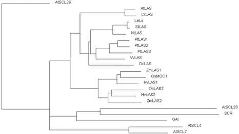
3.1.2. Phylogenetic promoter analysis... 37
3.1.3. Tomato promoter sequences are functional in Arabidopsis ... 40
3.1.4. Determining the significance of selected promoter regions ... 43
3.1.5. Visualization of promoter activities by GUS stainings ... 47
3.2. Part II: Characterization of a new player of axillary meristem formation... 50
3.2.1. The enhancer of lateral suppressor 5 (eol5) mutant ... 51
3.2.2. Positional cloning of eol5 ... 54
3.2.2.1. Rough mapping of eol5: problems and solutions ... 54
3.2.2.2. Fine mapping of eol5 ... 55
3.2.2.3. Annotation of CZS ... 59
3.2.2.4. Confirmation of mapping results... 64
3.2.3. Characterization of eol5... 65
3.2.3.1. Analysis eol5 single mutant alleles ... 65
3.2.3.2. eol5 affects flowering time control... 68
3.2.3.3. Phenotypic variability of eol5 mutants... 71
3.2.4. CZS expression profile ... 72
3.2.4.1. CZS expression in mutant alleles... 73
3.2.4.2. Expression analysis in eol5 mutants... 75
3.2.5. Analysis of CZS homologs ... 80
3.2.6. Analysis of potential downstream factors of CZS ... 83
4. Discussion ... 87
4.1. Part I: Towards understanding the LAS promoter ... 87
4.1.1. Visualization of LAS expression by GUS analyses ... 87
4.1.2. LAS 3`promoter alone is able to confer specific expression... 88
4.1.3. Pinpointing important LAS 3` promoter regions... 91
4.1.4. Relative importance of 5’ and 3’ promoter sequences ... 93
4.2. Part II: Cloning and characterization of the eol5 mutant... 95
4.2.1. Positional cloning of eol5 ... 96
4.2.1.1. Determining the correct CZS gene structure... 96
4.2.1.2. Analysis of eol5 and czs mutant alleles reveals common defects ... 97
4.2.1.3. Complementation of eol5 mutants... 98
4.2.1.4. Phenotypic variability of eol5... 99
4.2.2. Phenotypic analysis of CZS mutants reveals roles in different processes ... 100
4.2.3. Looking into the function of CZS ... 102
4.2.3.1. CZS expression analysis ... 103
4.2.3.2. Investigation of candidate targets of CZS... 103
4.2.3.3. A method of action hypothesis for CZS ... 105
4.2.4. Findings from the analysis of CZS homologs... 108
4.2.5. Putative biological role of interactions between floral induction pathways and AM formation control... 109
5. Contributions of co-workers to this project ... 112
6. Literature ... 113
Abstract ... 120
Zusammenfassung ... 122
Danksagung... Error! Bookmark not defined.
Erklärung ... 124
Lebenslauf ... Error! Bookmark not defined.
Introduction
1. Introduction
1.1. Meristem activities shape the development of
plant architecture
The postembryonic development of flowering plants is based on the activity of meristems, groups of pluripotent cells from which all organs develop. During embryogenesis two groups of meristematic cells are established, the shoot apical meristem (SAM) and the root apical meristem (RAM), giving rise to the major axis of growth.
The RAM will form the main root and later develop lateral roots originating from the pericycle. The SAM will give rise to all aerial structures of the plant, initiating at first leaf and subsequently flower primordia. Lateral meristems develop in the axils of leaves, thereby establishing new growth axis. The controlled outgrowth of these lateral meristems and further SAM activity leads to the vast diversity observable in plant architecture.
1.1.1. Genetic regulation of meristem organization
The SAM is laid out during embryogenesis and consists of a group of self sustaining pluripotent cells. Various genes act in concert to maintain the number and identity of the meristem cell population. Knotted-like homeobox (KNOX) genes keep cells in an undifferentiated state. One of these, SHOOT MERISTEMLESS (STM), is expressed in the Arabidopsis shoot apex and is required for meristem initiation and maintenance (Barton &
Poethig, 1993). Its vital importance can be deduced from stm mutants that fail to produce a SAM or true leaves.
The maintenance of the stem cell population relies on the WUS-CLV loop (Schoof et al.,
2000). The homeodomain transcription factor WUSCHEL (WUS) is expressed in the
organizing centre, specifying the overlaying cells as stem cells. These are marked by
CLAVATA3 (CLV3) expression, a secreted protein expressed in stem cells, acting as a
diffusible extracellular signal. The CLV signaling pathway also comprises the CLV1 CLV2
receptor kinase complex expressed overlapping with WUS. They negatively regulate WUS
expression upon binding of their ligand CLV3. WUS on the other hand activates the CLV
pathway completing the feedback loop controlling the stem cell population. Accordingly
wus mutants loose meristematic activity leading to a stop-and-go growth characterized by terminating and reinitiating of meristems, whereas clv mutants show enlarged meristems (Laux et al., 1996, Clark et al., 1993).
AMs are formed in the axils of leaf primordia. Leaf primordia initiate at the flanks of the SAM, in a spatially and temporally precisely controlled fashion. Early markers of incipient leaf primordia are auxin response maxima, in which auxin flux in the L1 layer is directed towards a convergence point and subsequently inwards, forming a reverse fountain (deduced from intracellular localization of PINFORMED 1, Benkova et al., 2003, Heisler et al., 2005). Other early markers of incipient primordia development are the absence of STM transcript and the expression of leaf identity genes like ASSYMMETRIC LEAVES 1 and AINTEGUMENTA (Byrne et al., 2000, Elliot et al., 1996).
At early stages in primordia development, preceding any morphological changes, genes involved in lateral meristem initiation like REGULATOR OF AXILLARY MERISTEMS 1 (RAX1), REGULATOR OF BRANCHING (ROB/bHLH140), CUP SHAPED COTYLEDON 1 (CUC1) and LATERAL SUPPRESSOR (LAS) are starting to be expressed at, or adjacent to, the position of forming primordia and later on at their adaxial side. Formation of new meristems can be linked to the activity of these proteins, as mutations in these genes show various defects in this process as described below.
At a later stage, around P16 in vegetative Columbia (Col) plants, lateral meristem development has progressed to a stage at which the meristematic marker STM shows a new focused expression (Greb et al., 2003). Establishment of expression domains of other markers of meristem identity like WUS and CLV indicates the formation of a new meristem, which will then commence formation of new leaf and flower primordia.
The exact mechanism promoting axillary meristem fate of a specific cell group is poorly
understood. One of the required signals is presumably to keep cells in an undifferentiated
state. The nature of the signals leading to new cell identities, that may originate from the
primordia, the SAM, or organ boundaries, remain to be uncovered.
Introduction
1.1.2. Members of the GRAS Gene family control meristem initiation and organization
The plant specific GRAS gene family has been shown to play a role in different developmental processes, from meristem maintenance to hormone signaling (Bolle, 2004).
The family is named after the prominent members GAI, RGA and SCR. Specific domains identifying GRAS proteins are a VHIID motif, roughly conserved in all members of the family, the two leucine-rich domains of approximately 100 AA residues length, and homologies near the C-terminus.
The GRAS proteins SCARECROW (SCR) and SHORT ROOT (SHR) are involved in root and shoot radial patterning. Mutants in either gene show, among a range of defects, that cortex and endodermis cell files are not properly established (Sabatini et al., 2003, Helariutta et al., 2000). SHR protein acts non–cell-autonomously and has been shown to upregulate and physically interact with SCR.
A mutation in the petunia gene HAIRY MERISTEM (HAM), which belongs to a different subfamily of GRAS genes, leads to the termination of the SAM and AMs. After cessation of meristem activity a layer of differentiated cells covers the tip of the shoot (Stuurman et al., 2002).
A triple mutant of the homologous Arabidopsis genes SCARECROW-LIKE 22 (SCL22), SCL27, and SCL6 also displays SAM termination and side shoot formation defects. It could be shown in Arabidopsis that the mutations lead to a loss of meristem organization and polarity, as cell groups with meristematic identity are found displaced in lower cell layers (Schulze, 2007). These genes are targeted by miR171, accordingly MIR171 overexpressor plants resemble scl22 scl27 scl6 mutants.
GRAS proteins also act as signal transducers of GA, a plant hormone involved in many developmental processes. GA acts mostly as a differentiation signal, effecting e.g. growth habit, floral development, flowering time and seed germination (Fleet & Sun, 2005).
The DELLA-domain-containing proteins GAI, RGA, and RGA-LIKE 1-3 are negative
regulators of GA response. In the presence of GA these proteins are degraded via the
ubiquitin/proteasome pathway, resulting in the derepression of target genes and thereby
triggering the GA response.
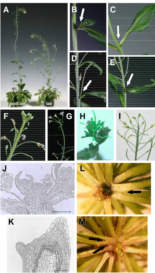
The Arabidopsis GRAS gene LAS is an important regulator of AM development. The las mutant phenotype is characterized by a lack of AM formation during the vegetative phase, while side-shoots develop normally during the reproductive phase (Fig. 1A). During this work only the las-4 allele was used, which carries a 20 bp deletion 365 bp after the ATG, henceforward referred to as las. LAS is expressed in very specific band-shaped domains adaxial of initiating leaf primordia (Greb et al., 2003, Fig. 1C D). The expression domain coincides or lies closely adjacent to those cells, which will later give rise to AMs. As a close homolog of the DELLA domain proteins GAI, RGA, and RGL1-3, LAS may act on the same target genes. As LAS does not contain a DELLA domain, it will not be degraded upon presence of GA. Hence, a possible function of LAS could be to repress the GA response, which primarily means to keep cells in an undifferentiated state in presence of GA.
A B
C D
las las wt wt
Figure 1. Phenotype and expression profile of LAS
A, B, axillary bud formation observed in rosettes of wt (A) and las (B) plants. White arrows point towards buds or barren axils, respectively.
C, longitudinal and D, transverse sections showing LAS mRNA accumulation pattern by in situ
hybridization, in a 28 d old vegetative Col (C) or Ler (D) plant. Pictures from Greb et al., (2003),
bars 200 µm.
Introduction
1.1.3. Regulators controlling lateral meristem development
Next to the GRAS genes mentioned above, several R2R3 MYB genes have been shown to be involved in side shoot development. A mutation in the RAX1 gene results in defects in axillary meristem development during the vegetative phase (Müller et al., 2006). The triple mutant with the close paralogs rax2 and rax3 displays increased lateral meristem formation defects, also affecting cauline leaf axils. The lack of focused STM expression in the axils of later leaf primordia suggests that lateral meristem initiation is compromised early in development. Interestingly the rax mutants are aphenotypic in long day conditions, and mentioned defects only appear when plants have been grown in short days.
ROB/bHLH140 could also be shown to be a regulator of branching, similar to the maize and rice homologs BARREN STALK and LAX PANICLE. rob mutants show minor defects in AM initiation in the rosette but enhance the mutant phenotypes of las and rax1 (Yang, 2007). In concert with the supposed role in aiding AM development, ROB is expressed in specific expression domains adaxial of leaf primordia and ROB overexpressing plants develop accessory side shoots. Experiments indicate that ROB physically interacts with RAX1, which also shares the same expression domains (Yang, 2007).
Another group of genes that show specific expression domains in axils of leaf primordia are the NAC domain factors CUC1, 2, and 3. A loss of function of CUC3 was reported to lead to defects in axillary meristem development (Raman et al., 2008). miR164 is a negative regulator of the close homologs CUC1 and 2 (Rhoades et al., 2002). miR164 overexpression enhances the cuc3 phenotype, revealing redundant functions, while a loss of miR164 function leads to accessory bud formation, interpreted as deregulated, elevated activity of lateral meristems (Raman et al., 2008).
The eol5 mutant was discovered in a screen, designed to find modifiers of the las-4
phenotype (Clarenz, 2004). las-4 mutant seeds were mutagenized with EMS and M2
populations were analyzed for alterations of las-4 phenotype. Two classes of mutants were
isolated during this screen, the so called and enhancers of lateral suppressor (eol), in
which the AM defects were extended into the cauline leaf axils, and the suppressors of
lateral suppressor (sol), whose phenotype was modified to appear more similar to the
wild-type (Clarenz, 2004, Raman, 2006).
This second-site mutagenesis screen was expected to identify mutations in genes that act in some way redundant with LAS, in the same or a different pathway. The limitations of a genetic screen are always lethal mutations and the redundancy of regulatory networks. A second-site screen is designed to partially overcome the problem of redundant factors that might mask low phenotypic changes of single mutants. As las constitutes a sensitized background, mutations could be detected, whose phenotypic changes would be too weak to be spotted in a screen in the wild-type background.
The eol5 mutant was discovered during this second-site mutagenesis screen, as it increases the las loss of function phenotype. When grown in short days, side shoot formation is strongly reduced in cauline leaf axils, while no enhancement of the las phenotype is observable in long day conditions. Additionally, the eol5 las double mutant is reported to accelerate flowering and to develop longer inflorescences (Schulze, 2007).
1.1.4. Strategies to discover new regulators of AM initiation acting upstream of known genes
In order to uncover the genetic network controlling a process like AM initiation, the first step undertaken is usually the analysis of mutants, either derived from screens designed to detect a specific mutant phenotype, or from fortuitous observations. These approaches led to the discovery of various genes involved in AM formation. The currently available information about the process is gathered from their characterizations and interaction studies.
Yet many players cannot be identified this way, due to lethality or to redundancy preventing observable phenotypic alterations in mutants. Applying methods like yeast two- hybrid studies can identify interacting partners. Downstream targets are routinely sought- after utilizing expression arrays, detecting transcript changes caused by mutations.
Identifying transcriptional upstream regulators binding to the promoter of an investigated
gene, is a more challenging task and therefore less common. One way to address this
problem is to devise entirely new screens, e.g. utilizing gene-of-interest reporter gene
constructs, or looking for reversions of gain-of-function mutations. Other techniques like
yeast one-hybrid studies or DNA affinity purification aim to identify proteins binding to a
Introduction
specific promoter fragment. These techniques require knowledge of the promoter regions of the investigated gene. Understanding promoter structure and function can give valuable insight in the process the gene is involved in. Information about timing and position of binding proteins improves understanding of the genetic processes.
Computational methods are not “yet” universally useful in understanding promoters. Most approaches are based on transcription factor binding motifs, which have aided understanding in various cases (e.g. auxin responsive genes, Chapman & Estelle, 2009).
While some binding motifs are well described, others are either not known, or a description of protein-DNA interaction at a sequence level, based on single binding motifs, simply does not reflect the complexity of the underlying process (Florquin et al., 2005).
With reasonable knowledge of the investigated promoter regions, yeast one-hybrid experiments are a suitable choice to find upstream regulators (Li and Herskowitz, 1993).
While broad promoter regions can be used to search for interacting factors, many studies indicate that repeats of short sequences are favorable to produce the desired results (Deplancke et al., 2004, BD Biosciences MATCHMAKER User Manual, 1998). Therefore a detailed promoter study, identifying the essential regions, is a suitable starting point to find upstream interactors and thereby increase understanding of the regulatory network.
In the case of LAS, upstream regulators are of special interest, as LAS is expressed in specific domains, including or neighboring those cells that will later give rise to AMs, and whose cell fate is affected in las mutants. Hence it is plausible that the function of LAS might be largely regulated on transcript expression level. This emphasizes the importance of understanding the establishment of the specific RNA accumulation pattern, i.e.
investigating the LAS promoter and finding upstream regulators. Thus, promoter studies are applied to identify important elements that can later be utilized to find interacting factors by yeast one-hybrid experiments.
1.1.4.1. Previous work on the LAS promoter
Lateral suppressor (Ls) was first studied in tomato, displaying a similar lack of side shoot
formation in the ls mutant as described above for Arabidopsis. Additionally, defects occur
during flower development, like a lack of petals and reduced flower numbers (Schumacher
et al., 1999).
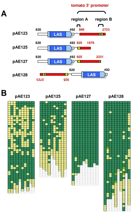
Complementation experiments showed that comparatively large promoter regions are necessary for gene function. 1411 bp of 5’ and 2667 bp of 3’ regulatory sequences driving the Ls open reading frame (ORF) were found to be sufficient to produce a wild-type phenotype when transformed into the ls mutant. In contrast, a shorter construct with only 570 bp of 3’ sequences did not lead to complementation (Schumacher et al., 1999; Schmitt, 1999). The Ls gene was also shown to be functional in tomato, if the 3’ regulatory sequences were in reverse orientation, a property typical for enhancer elements.
In order to individually complement the lack of AMs or the flower phenotype, transgenic constructs were produced, in which the 5’ promoter was exchanged with the PLENA promoter, active in inflorescence meristems (Bradley et al., 1993), or with the CET4 promoter (Amaya et al., 1999), only active in the vegetative meristem. ls mutant plants transformed with these constructs exhibited no complementation. Only constructs carrying also the 3`sequences of Ls were able to confer complementation, leading to restoration of both phenotypes, irrespective of the 5’ promoter (Gregor Schmitz, personal communication). This indicated that the 3’ regulatory sequences are the decisive factor for a functional promoter.
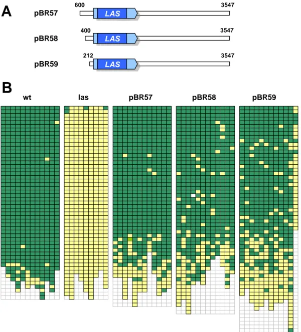
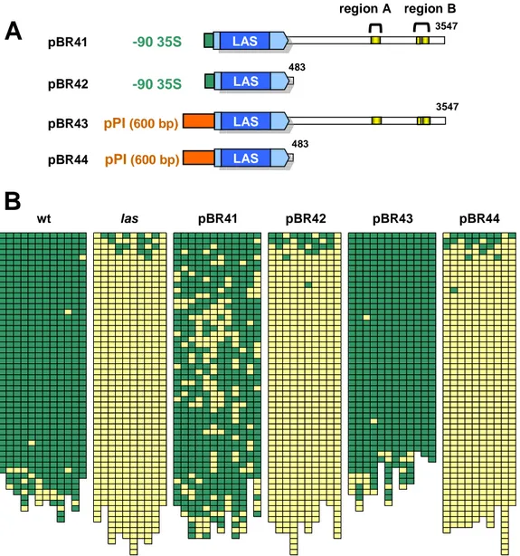
Andrea Eicker (2005) showed that also in Arabidopsis the 3’ promoter of LAS plays an important role. To identify important promoter regions, las Arabidopsis plants were transformed with numerous deletion constructs. In a first experiment constructs with 5’
sequences of varying length were analyzed, all including 4000 bp of 3’ sequences of the LAS gene. Secondly, different sized 3’ promoter fragments were examined for their ability to complement. (In the 5’ promoter distances always refer to the ATG, while 3’ promoter sizes are measured from the stop codon.)
Fig. 2 summarizes the deletion construct analysis results (Eicker, 2005), illustrating that 820 bp upstream and 3547 bp downstream of the LAS gene are necessary for promoter function, whereas shortening of these sequences to 800 bp or 3133 bp respectively, resulted in the loss of complementation ability. These promoter regions shown to contain essential elements are depicted in red in Fig. 2. Additionally, partial complementation could be obtained, also with a short 3’ region (488 bp), when using 2910 bp of 5’
sequences, leading to ~ 60 % of rosette axils sustaining bud formation. These results
indicate the presence of an enhancer element between 1447 and 2910 bp upstream of the
Introduction
-4000 - 3000 - 2000 - 1000 - 0 0 1000 2000 3000 4000
-4000 - 3000 - 2000 - 1000 - 0 0 1000 2000 3000 4000
LAS LAS
2910 - 1447 820 - 800 3133 - 3547
Figure 2. Overview of relevant LAS promoter regions determined by deletion construct analysis.
LAS CDS is depicted in blue, UTRs in light blue, regions shown to contain essential promoter
elements in red. Numbers above parentheses state distance in bp between the indicated regions and the start, respectively stop codon of the LAS ORF. Dashed parenthesis indicates region leading to partial complementation in the absence of long 3’ sequences.
1.2. Transcriptional control of gene expression
Transcriptional activation of genes is dependent on gene promoters, regions of DNA that lead to spatially and temporally specific activation of mRNA formation. That means these sequences result in the assembly of a transcription initiation complex at the transcription start site (TSS). This contains the DNA Polymerase II (PolII), which is responsible for transcribing mRNAs and some small RNAs (Pedersen et al., 1999).
Promoters are commonly divided into 3 parts: the core promoter, the proximal, and the distal promoter (Abeel el al. 2008). The Core promoter usually extends 50 bp around the TSS and provides the platform to assemble the transcription initiation complex. In plants specific core promoter sequence elements seem less conserved than in animals. The most prominent is the TATA box, located ~ 30 bp upstream of the TSS, present at the TSS of ~ 30 % of Arabidopsis genes (Molina & Grotewold, 2005). Initiator elements (Inr) around the TSS have also been reported in several promoters (Shahmuradov et al., 2003). In general no sequence conservation was found that could be used to predict a large number of core promoters (Molina & Grotewold, 2005). The lack of sequence conservation might be replaced by structural information, as Florquin et al., (2005) described different classes of core promoters, based on structural properties. Structural characteristics, like DNA bending properties, may affect positioning of nucleosomes, providing easier access of proteins to certain DNA elements, and histones may aid specific DNA binding proteins by providing binding platforms.
In order to initiate transcription, core promoters need additional elements that provide
binding sites for proteins. The proximal promoter is usually considered to include a region
of a few hundred bp upstream of the TSS, containing various binding motifs for transcription factors. Eukaryotic promoters usually contain binding sites for several TFs that positively or negatively affect formation and activation of the transcription initiation complex (Pedersen et al., 1999).
Distal promoter elements can be localized at distance of several thousand bp and comprise additional regulatory elements named enhancers or silencers. Proteins binding to these elements are assumed to interact with proteins at the core or proximal promoter by looping of DNA. Enhancer or silencer elements can usually act independent of orientation and of their position ahead or behind the transcribed region of the gene. As all regulatory sequences behind the gene belong to the distal promoter, these regions will be referred to as 3’ promoter in this work.
The actual activity of a promoter depends on different aspects. Obviously the number and location of motifs play an important part but only in combination with the composition of TFs present at a certain time. Proximal and distal promoters might also be defined by structural elements like DNA bending properties, influencing the nucleosome positioning and thereby TF binding stability. Another important factor is the chromatin state, limiting the accessibility of DNA to proteins, which is dependent on DNA methylation and histone modifications, as described below. (Pedersen et al., 2005).
1.2.1. Chromatin modifications and their role in plant development
Substantial parts of the genome are in a densely packed state called heterochromatin, in which DNA is inaccessible to TFs. Heterochromatin is inherited through cell divisions.
While large parts of the heterochromatin, like telomeric and centromeric regions, remain in
this state, other regions of densely packed chromatin can convert to euchromatin in
response to developmental cues. Derepression of DNA by unfolding of chromatin is an
important part of gene regulation (Pedersen et al., 1999). Chromatin state depends on
chromatin marks, such as methylations of DNA and histones,
Introduction
Histone and DNA methylation marks require further proteins that translate this information to induce heterochromatin formation. The HETEROCHROMATIN PROTEIN 1 (HP1), originally described in Drosophila, leads to heterochromatin formation and gene repression (Bannister et al., 2001). HP1-like proteins are found in most eukaryotes ranging from S.
pombe (Swi6) to human (HP1h) and plants (LHP1) (Berger & Gaudin, 2003).
The Arabidopsis gene TFL2 e.g. is an HP1 homolog that recognizes H3K9 K27 methylation, leading to the formation of inactive chromatin (Steimer et al., 2004). The tfl2 mutant phenotype shares some similarities with the curly leaf (clf) mutant phenotype (see below), misexpressing homeotic genes and thus, appears to be one of the genes involved in translating histone methylation patterns into repressed chromatin state. Numerous other proteins can be expected to be involved in mediating chromatin condensation in response to heterochromatic methylation marks.
1.2.1.1. Role of DNA and histone methylations in plants
DNA methylation plays a major role in maintaining genome integrity. Accordingly transposons and other repeat elements comprise most of the methylated DNA (Chan et al., 2005). Transcribed regions are usually found to be, if at all, less methylated, e.g. shown for the CpG islands (regions of low CpG methylations) described in vertebrates. CpG islands have also been reported in Arabidopsis but do not seem to play a major role (Shamuradov et al., 2005).
So far DNA methylation has not been shown to play a role in plant development (Schubert et al., 2005). Mutants affected in DNA methylation occasionally show developmental phenotypes, like the AGAMUS (AG) and SUPERMAN mutants, but methylation of these loci has not been shown to play a role in vivo. An exception to this concept are the PHABULOSA and PHAVOLUTA genes, which can be methylated due to the regulation by the miR165 and miR166 (Bao et al., 2004).
Nucleosomes are the fundamental repeating units of chromatin, consisting of 146 bp of
DNA wrapped around histones. Histones are subject to various modifications like
acetylation, phosphorylation, methylation, ubiquitination, or sumoylation, which can be
reversible and associated with regulation of individual genes (Völkel et al., 2007). Histone
methylations belong to these reversible marks, acting as a cellular memory of
transcriptional status, as they are heritable over cell divisions. Proteins that methylate
histones, and thereby affect chromatin state, play a role in many developmental processes like meristem maintenance, phase transition, and embryogenesis (Reyes, 2006).
One of the best studied epigenetic systems in eukaryotes is the Polycomb group (PcG) of proteins and their antagonists the Trithorax group proteins, which are involved in the maintenance of repressed and active transcriptional states, respectively (Bantignies &
Cavalli, 2006). These protein complexes produce epigenetic marks by methylating histones. The effect of histone marks depends on the number of methyl groups and the affected amino acids.
While H3K4 (Lysine 4 of Histone 3), H3K36, and H3K79 methylations are usually associated with expressed genes, H3K9, H3K27, and H4K20 methylations constitute repressive marks (Völkel et al., 2007). Complicating the histone code, lysine residues can carry one, two, or three methyl groups, linked to different enzymes and responses. In wild- type Arabidopsis, monomethyl H3K27 (meH3K27) and dimethyl H3K27 (me
2H3K27) are concentrated preferentially in heterochromatin, whereas trimethyl H3K27 (me
3H3K27) appears to be mostly euchromatic (Schubert et al., 2005).
PcG proteins mediate the cellular memory of transcriptional states over many cell divisions (Steimer et al., 2004). Conserved to their function in animals, PcG proteins elicit tri- methylations of H3K27 on their direct target genes, which is correlated with stable, long- term repression (Farrona et al., 2008). The Arabidopsis genome contains several homologs of members of the conserved Polycomb Repressive Complex 2 (PRC2), well described in animals. The protein group comprises homologs of four genes, first described in Drosophila: Enhancer of Zeste (E[Z]), Suppressor of Zeste 12 (Su[z]12), Multicopy suppressor of Ira (MSI), and Extra sex combs (ESC). In Arabidopsis these proteins are represented in small gene families (Farrona et al., 2008).
E[Z] homologs contain a SET domain (Su(var)3-9, Enhancer-of-zeste, Trithorax), conferring histone methyl transferase (HMT) activity (Berger & Gaudin, 2003). Known Arabidopsis homologs are MEDEA (MEA) involved in seed development (Grossniklaus et al., 1998, Luo et al., 1999), CURLY LEAF (CLF) and SWINGER (SWN), redundantly regulating leaf and floral development, and floral transition (Goodrich et al., 1997).
CLF, SWN, and MEA show a large functional overlap, displaying mainly additive mutant
Introduction
of clf and mea mutants. However, there are also specific regulatory functions, e.g. a MEA containing PcG complex acts on PHERES 1 gene during seed development. Known targets for a CLF containing PcG repressive complex include the KNOX genes, which are found misexpressed in mutants (Schubert et al., 2005).
Su[z]12 homologs are characterized by C2H2 zinc finger motifs, assumed to confer unspecific DNA binding ability (Steimer et al., 2004). FERTILIZATION INDEPENDENT SEED 2 acts in a complex with MEA during seed development (Luo et al., 1999), while EMBRYONIC FLOWER 2 (EMF2) and VERNALIZATION 2 (VRN2) have been shown to affect floral transition (Yoshida et al., 2001, Gendall et al., 2001).
VRN2 has been described to implement stable repression of FLC after cold treatment. FLC is a negative regulator of floral induction, which is itself repressed by the vernalization pathway or the autonomous pathway to enable flowering (Farrona et al., 2008). FLC is strongly activated by FRIGIDA (FRI). As Col or Ler accessions do not possess an active FRI gene, vernalization is not required for flowering. Nevertheless, FLC levels influence floral induction as it is regulated by, and regulates, a large number of genes (Farrona et al., 2008). In wild-type plants, but not in vrn2 mutants, FLC remains repressed after vernalization (Schubert et al., 2005), however, vrn2 mutation does not affect flowering in Ler wild-type background (Gendall et al., 2001).
Mutations in the homologous EMF2 gene flower early under both long days and short days and lead to small, dwarfed plants, indicating participation in a different complex (Chanvivattana et al., 2004). Interestingly, emf2 vrn2 double mutants are not early flowering, showing otherwise additive, pleiotropic phenotypes (Schubert et al., 2005).
FERTILIZATION INDEPENDENT ENDOSPERM (FIE) is an ESC homolog, containing a characteristic WD40 repeat. FIE has been reported to repress floral homeotic genes (Schubert et al., 2005) and to be involved in seed development (Ohad et al., 1999, Chaudhury et al., 1997).
In animals, PRC2 complexes were shown to include the WD40 gene MSI. There are five
homologs (MSI1-5) in Arabidopsis, but so far no experimental data provides evidence that
they are part of PcG complexes (Farrona et al., 2008).
1.2.1.2. Plant SET domain proteins
SET domain proteins form the largest group of lysine HMTs, having different functions in Arabidopsis (Berger & Gaudin, 2003). SET domain proteins can be divided into seven families based on their conserved domains (Ng et al., 2007). The previously mentioned CLF, SWN, and MEA constitute the first family and are the best studied SET proteins, as they are part of the PRC2 homologs. Yet, in recent years also the remaining SET proteins, which have not been associated with these complexes, have attracted attention.
KRYPTONITE (KYP), the first HMT identified in plants, was shown to be involved in DNA methylation control (Berger & Gaudin, 2003). kyp mutations cause a reduction of methylated H3K9, a loss of DNA methylation, and subsequently reduced gene silencing (Jackson et al., 2004). This indicates that KYP mediated methylation of histones results in DNA methylation.
Redundant functions have been reported for KYP/SUVH4 and its homologs SUVH5 and SUVH6, which together control activity of the DNA methyltransferase CMT3 (Ebbs et al., 2006).
CAROTENOID CHLOROPLAST REGULATORY 1 (CCR1/SDG8) is another SET domain protein, reported to be involved in plant development (Dong et al., 2008, Cazzonelli et al., 2009). ccr1 mutants show increased outgrowth of lateral branches, possibly due to altered carotenoid composition.
Another SET domain protein belonging to the same subfamily as KYP is CZS, named after its conserved protein domains C2H2 zinc finger and SET. CZS was identified by its interaction with SWP1, a SWIRM (Swi3p, Rsc8p, Moira) domain Polyamine oxidase (PAO)-like protein (Krichevsky et al., 2007). czs mutants, just like swp1 mutants, show a mild delay in flowering correlated with an upregulation of FLC. Chromatin immunoprecipitation (ChIP) experiments showed that me
2H3K9 and me
2H3K27 marks at the FLC locus are reduced, suggesting that a role of CZS may be to directly repress FLC expression.
PAO containing co-repressor complexes have been shown to be transcriptional regulators
in animals (Jepsen and Rosenfeld, 2002). They specifically silence neuronal genes in non-
neuronal cells. In animals these complexes have been shown to contain LSD1 (lysine-
specific demethylase 1), a protein containing a SWIRM domain and PAO domain that may
Introduction
act as a histone H3 lysine demethylase, the TF REST (Repressor element 1), adapter proteins, histone deacetylases, and a SET domain HMT (Krichevsky et al., 2007b). A homologous complex may be present in plants, as another LSD1 homolog FLOWERING LOCUS D (FLD) represses FLC by histone acetylation, as part of the alternative pathway of flowering regulation. A hypothesis is that CZS and SWP1 act together in a PAO containing co-repressor complex, silencing target genes like FLC (Krichevsky et al., 2007b).
Studies of the close CZS/SUVR5 homologs SUVR4, SUVR1, and SUVR2 revealed that they locate to the nucleus, and that SUVR4 has an in vitro HMT activity, generating me
2H3K9 with a substrate preference for monomethylated H3K9 (Thorstensen et al., 2006).
1.3. Aim of this work
The aim of this project was to obtain a deeper understanding of the process of AM initiation by first, analyzing the LAS promoter and second, the characterization of a new regulator of AM initiation.
The LAS gene was chosen for a detailed promoter analysis because it is a key regulator in AM development. LAS is expressed in very specific domains adaxial of initiating primordia in - or very near to - those cells later giving rise to AMs. This indicates that LAS function might be largely dependant on transcription, emphasizing the importance of understanding the mRNA accumulation pattern, i.e. to understand the composition and localization of the regulatory sequence motifs. The promoter was analyzed by deletion constructs and in silico tools to identify important elements. Additionally, fusion constructs with other promoters were produced to elucidate the relevance of specific promoter regions, and promoter GUS fusions enabled direct visualization of the expression patterns of modified promoter assemblies. Information about position and importance of promoter elements can then be used in yeast one-hybrid studies to identify upstream regulators of LAS, which generate the specific expression pattern.
In a second approach, the gene underlying the eol5 mutant phenotype was to be identified
and characterized. The eol5 mutant was previously obtained in a second-site mutagenesis
screen and reported to enhance the phenotypic defect of las. A map based cloning strategy
was applied for the identification of the underlying gene. The subsequent goal was to characterize the eol5 and eol5 las mutant phenotype, particularly in regard to the effect on lateral meristem initiation, meristem maintenance, and flowering time.
To shed light on the function of the EOL5 gene, RNA expression changes in the mutant were analyzed by real-time PCR. Double mutants with known players in AM initiation were analyzed in order to position the gene function in known regulatory pathways.
Furthermore, homologs of EOL5 were examined for defects in side shoot formation, in
order to reveal a possible general role of HMT containing complexes.
Materials and Methods
2. Materials and Methods
2.1. Materials
2.1.1. Chemicals
The main sources of chemicals used in this work are the following:
Ambion, Austin, USA
Amersham Pharmacia Biotec, Braunscheig, Germany Biozym, Hess. Oldendorf, Germany
Carl Roth GmbH, Karlsruhe, Germany Invitrogen GmbH, Karlsruhe, Germany
MBI Fermentas GmbH, St. Leon-Rot, Germany Merck KgaA, Darmstadt, Germany
New England BioLabs GmbH, Schwalbach/Taunus, Germany Operon, Cologne, Germany
QIAGEN, Hilden, Germany Roche, Basel, Switzerland
Sigma Chemical Co., St.Lois, USA
2.1.2. Enzymes
Enzymes used during this work were obtained from following suppliers:
Invitrogen GmbH, Karlsruhe, Germany
New England BioLabs GmbH, Schwalbach/Taunus, Germany MBI Fermentas GmbH, St. Leon-Rot, Germany
Roche, Basel, Switzerland
Sigma Chemical Co., St.Lois, USA
Novagen, Toyobo, Japan.
2.1.3. Vectors
The following vectors were utilized during the course of this work.
pCR®-Blunt-II-TOPO®: Cloning of PCR products, Invitrogen.
pGEM4Z: Cloning by restriction sites and construct assembly, Promega GmbH, Mannheim, Germany.
pGPTVbar AscI: Binary vector for plant transformation (Überlacker & Werr, 1996).
2.1.4. Antibiotics
Antibiotics during this work were used to select for transformed bacteria in the following final concentrations:
Ampicillin (Amp) 100 µg/L Gentamycin (Gent) 50 µg/L Kanamycin (Kan) 50 µ g/L
2.1.5. Bacteria
The Escherichia Coli strain used for amplification of plasmid DNA was:
DH5 α (Hanahan, 1983): F- end A1 hsdR17 (rk-, mk+) gyrA96 relA1 supE44 L- recA1 80dlacZM15 ∆ ( lacZY AargF) U196
Plants were transformed using the following Agrobacterium tumefaciens strain:
GV3101: Virulence plasmid: pMP90 (Koncz und Schell, 1986)
Selection markers: Rifampicin, Gentamycin and Kanamycin.
2.1.6. Plant material
This work was carried out using the model plant Arabidopsis thaliana.
Table 1: Mutant alleles used in this work
Allele name Allelic variation Background Source
las-4
deletion Col Greb et al., 2003
eol5
SNP Col Clarenz, 2004
czs-1
T-DNA insertion Col SALK N661919
czs-2
T-DNA insertion Col GABI 500A10
Materials and Methods
rob-2
T-DNA insertion Col SALK N52476
mir164a-4
T-DNA insertion Col SM333570
mir164b-1
T-DNA insertion Col SALK N636105
mir164c
transposon insertion Col Baker et al., 2005
suvh1
T-DNA insertion Col SALK N859507
suvr1
T-DNA insertion Col SALK N860017
suvr3
T-DNA insertion Col SALK N662712
swn-7
T-DNA insertion Col SALK obtained from Daniel
Schubert
clf-28
T-DNA insertion Col SALK obtained from Daniel
Schubert
emf2-10
18 bp deletion, weak allele Ws Chanvivattana et al., 2004
vrn2-1
SNP Ler Gendall et al., 2001
swp1-1
T-DNA insertion Col SALK N642477
FRI FLC
active FRI introgressed from San Feliu-2 accession
Col / Sf-2 Searle et al., 2006
2.1.7. Oligonucleotides
Primers were mainly supplied by Invitrogen and Operon
Table 2: Oligonucleotides used in different subprojects
Genotyping and sequencing of plasmidspGPTV-FOR4 caagaccggcaacaggat pGPTV-FOR2 aactgaaggcgggaaacgac pGPTVfor3 aggacgtaacataagggactgac T-DNA-R caatacgcaaaccgcctctc pGPTV-rev2 tccataaaaccgcccagtc pGPTVrev3 gaagcttgcatgcctgcag Plasmid-Forward cacgacgttgtaaaacgacggccag Plasmid-Reverse cacacaggaaacagctatgaccatg
Genotyping and cloning of LAS constructs
35S-F_SacI gatgagctctctccactgacgtaag 35S-R_AvrII ggtcctaggtcctctccaaatgaa AtLs1411F_SacI tgggagctccggcatcagaatctcaac AtLs7116R_SbfI caacctgcaggaaaccagagtcttgtcttc AtLs1831F_XhoI atactcgagaatgtaatgattcacttttctaaaatcat AtLs2019F_XhoI atactcgagcaacttcatctctatccataaaactatgt AtLs2135R_AvrII tatcctaggcctttacctgaaggtatattg AtLs1631F_XhoI atactcgaggtgaatttttatttaattagtatcatttgc AtLs2593muR tggttcgaaacaagaactagt AtLs2599F cagtgtatgcaaagaacagttc
AtLs3070R aacacaattgacggcaatgg AtLs2349F acctccgtcgtcttcttttc AtLs3530F taggagctccaaaatcgtcccctcttctcc AtLs2952R agacctaaagagtcagcgaacc AtLs4051R_SbfI aatcctgcagggacgatttcaatcaatttag AtLs7116R_SacI aacgagctcaaaccagagtcttgtcttctc AtLs4940 F ctaactagtctaaggtttagaggatgatc AtLs5697F_XmaI ttgcccgggataaaacaaaagggtgtgc AtLs5396F_BamHI aatggatccttagggttagtgtcgacaga AtLs1411F_BstBI tggttcgaacggcatcagaatctcaac AtLs5614R_XmaI ccccccgggaatcccttttttacccca AtLs5672R_XmaI gctcccgggtcatccgacaaatcg AtLs5739R_BamHI ataggatcctataacataagtctaaataagcac AtLs6625F_KpnI agaggtaccatttagggttttaggtg
AtLs6798R_SbfI aatcctgcaggtgatcacaaactttggatag AtPI-598-F_SacI tttgagctcaattaattatatacatacacgagtaagc AtLsREV gagacaaagaggacggtcac AtLs4975R tcgcagagatcatcctctaaac
AtPI-1-R_AvrII tctcctaggctttctctctctatctct AtLs3569_AvrII tggcctaggtccaaagagaaggacaa LAS-5UTR -6.2f cgcggatccggcatcagaatctcaac
Mapping primers
cer429966_F ggctcttgagccgaagaaat cer429966_R acgtttcagaccttcgtcgt cer429971_F tcgagagatgttgccatgag cer429971_R cgtgattgttgtcgtcgatt cer44411A_F cggatcagaccgattcaaac cer44411A_R ctccccaaaaagaaacgaca cer44411B_F gttgttgttcggttcggttt cer44411B_R caccgggaaactaccagcta cer445734_F ttgcacctttgccatcatac cer445734_R tgtcaaaacaaaatgacaatgc
cer445742_F ccggagccatcgtagaagta cer445742_R tggttttccacaaaattcca cer44613B_F atcaatatgttgaaaaagctacaccag cer44613B_R cgccaccacaaatctccatc cer44613F aaaataatgggtggggaaatcg cer44613R ttcgaaacacgttggaaaatgac MASC02463F gagtgtcaaaggttacgggttct MASC02463R gcttgaatggtttacacttgacag MASC02627F atgtggttgattcaaagggtg MASC02627R tgaaattgggaggaggattg MASC02866F tagaatttccctgccaacatc MASC02866R gggcttgaagctgttgagac MASC02949F gtttttgaaagtccccggat MASC02949R catggagctggtggtttagc MASC03021F actccgattccaaacacatca MASC03021R ggtatgtgaaatgggttttggt MASC07353F aagcattgctctgtttatcgtc MASC07353R ttcttcttctatagcttttggtctc
Sequencing primers
at2g23347_F cctgataaaagcagcgtcct at2g23347_R agctcctgcacgaagttactg at2g23450_986F tgtttgtttggtggttcaatg at2g23450_2505R aacattgtggtactggttgaaaga at2g23450-499F ccactgggcacgtatcttct at2g23450_1007R cattgaaccaccaaacaaaca at2g23460_2077F tatgagcaggagatgcgaaa at2g23460_3620R ccttggagccaatagaacca at2g23460_648F ccgcagacgaaaggtaagaa at2g23460_2195R accccaagatttccagcag at2g23460-670F aattcgaccccttgacacac at2g23460_789R ccttccttcaggcatagaacc at2g23520_1236F cttgacggattggttggtct at2g23520_2775R ttaccctccttccatttcca at2g23520-178F tcttctcttccgtgaaagtcg at2g23520_1374R cacatccctcacttgagctg at2g23530_717F agacttgtcaccaatgcaggt at2g23530_2232R aacagagagggtcatgtcgaa at2g23530-696R tctctcaagtcaattcaaatcca at2g23530_848R ccatcgtcttctgcctgtaag at2g23640_F tctggtctaagttatcaaattccaa at2g23640_R gacacaggtaaagtcgaccaa at2g23700_1354F cggcatttcaatcaagaaga at2g23700_3258R aacccttcccaagacaatca at2g23700-113F cccaactaagagattcttcttcttc at2g23700_1449R tcgaattgtcgaacaccaga at2g23755-903F gattgatgaaccatttgccata at2g23755_633R ggaactattgatcttccttcaagc at2g23770_287F cccttctggtcaacaagtca at2g23770_1987R tgaccaactccaacacaaca at2g23770-1254F tgtgaaataaatggtgcgtgt at2g23770_405R gtcgttagcaatggcgaaat at2g23780-671F actaatcgatcggcgttcac at2g23780_997R gcatctatcaaccttaaagaatcaaa at2g23790_1140F tggggatggattgattgact at2g23790_2684R aatgatgccaaaagctcgtt at2g23790_-301F tcacctctaccaacccgaac at2g23790_1250R cctccaccaccttcttttga at2g23800-F ccacgaaaagccgttaagtt at2g23800-R cgtccactgctacgtccata at2g23810_406 ggagttgtcttgtggagagca at2g23810_1856R gaacccttcttcattatgtttgatg at2g23810-916F ggctaaggtatgcttttcaaac at2g23810_618R aaaggatcaaaaagctcaatctc at2g23820_896F attgtcaagcttggctgcat at2g23820_2396R ccgtgcaaaatcttgaaaca at2g23820-524F tcaaaacgacatcgtgttaaat at2g23820_1018R gacaaacttcacatcttcaaggatt at2g23830_F ttgcacgggttaaaagttga at2g23830_R gcaagagacatcgctaagagtg at2g23834_F atacatgcctgccgaggac at2g23834_R agaacaccgggatctcagaa at2g23840_829F ttctcctacgggttcgttct at2g23840_2463F cttcgaccgttgcatcttct at2g23840-638F caggttctgcaactttttgg at2g23840_926R gcttcaaactggcaacaaga at2g23860_514F taacaaagaatcggggcatc at2g23860_1954R tcgagacgatatagttgaaataatga at2g23860-903F aaaaattcagcatttcattacattt at2g23860_739R cgagttttgctctggcaatc at2g23910_1134F ctcattttggtcaagattcaatg at2g23910_2651R tcgtggatgcatttgagatt at2g23910-219F ttccaccggtcaatggatta at2g23910_1282R gagcatgccacaactgtgat at2g23920_F tcaggattgtgaagcaggatt at2g23920_R ttcaccacaacatcaaaaatga at2g23930_F acaattggccgcattagaac at2g23930_R tcaaagcagtggatccagagta at2g23950_1457F tggtttatgtaatttgattttgtttg at2g23950 _2985R ttgctttcacaggacctcaa
at2g23950-200F gcgtaggagagacattgcag at2g23950_1506 ccaaaacaaaatactttagacaacaaa at2g23980_1509F gggcttgaaaccagcacata at2g23980_2989R ggaagcaatggcagactctc at2g23980_160F tggactcaaggtactcgcaaa at2g23980_1610R acctgcatgtttccaatgag at2g23980-1217F ttcggcaacgattactctcc at2g23980_260R gatgctttgcttccttgagc at2g23985_F gacgccgtgattgtgtgtaa at2g23985_R caattgggtgatgaatgttttg at2g24030_18F atggcgatacgacgagtttc at2g24030_1543R ggtctctctaatggcattggttat at2g24030-1363F aaaaatcgtttgaaattctcactt at2g24030_137R tgtctgaaaaagttgttgaactg at2g24080_F ttagcggtgtactgcggttt at2g24080_R aacacttagcaatgtcaaatcttca atCLF2299_F gagttgctgagcgagttcct atCLF3857_R taagaaagctccccaaccag atCLF3690_F tgctcctgaaacaacaacaaa atCLF5170_R tagtgcgcgaatcaaatcag atCLF-495_F tcgaaaagctgttgctgaaa atCLF969_R tctccttcgacccactacaga atCLF800_F catgggtttttctggacagg atCLF2400_R atcgctgggtgaacaacttc
atICK1_F aacgggaccactaaaacacg atICK1_R agcgtttagggcggtaagat
atLBD10_-361_F aaaaatgctaaagaatggggtat atLBD10_1856_R tcatttgcttgctttggttg atmiR831a_F gttggggctcagtcatcatc atmiR831a_R tttcgtagtcttggataaaatcagc atSAW2_1615_F aaacgaactaatcacttgaggttt atSAW2_2947_R tgatgaataatgacagaagaaattg atSAW2_2856_F tgtcagtggtacagtttcattgg atSAW2_4182_R cattaaatatggttttgattgtttttg atSAW2_361_F cgcagcaacaacaacacttt atSAW2_1753_R tcgcagtagtggttgtaccg atSAW2_3992_F tctcaaaggaaacacatgtatcataa atSAW2_5355_R tgttaataagtcgatcgggtacg atSAW2_-898_F atggtggtggtttggttcat atSAW2_441_R gtggacggttccgatcata
Materials and Methods
CZS sequencing, genotyping, and cloning of constructs
at2g23740_1113F ctgagtctccaatgcaacca at2g23740_2649R ggctgccacaaggaatacac at2g23740_19F agttggttcttgacgtggatg at2g23740-456F cctgggtttgttgattggtc at2g23740_2507F tggtcgtttagtggatttgc at2g23740_3961R tgggaatctacacctcatgga at2g23740_3847F agctgtcgcagttcagtgtg at2g23740_5303R tcaatccttccaaagagtttcaa at2g23740-1502F tcgctaacataacctgcaca at2g23740-61R tctgcaataagaacaggggaat at2g23740-185F tgacttcgtatgtctgaaaatgc at2g23740_1220R ctgtgctatttcgccaacac
at2g23740-AscIF ataggcgcgcctcgctaacataacctgcaca at2g23740_SbfIR atacctgcaggtcaatccttccaaagagtttcaa atCZS-156F cttccgtgacctagcctttg atCZS_182R ggctcagaaggtgacgactc
atCZS-1668F cttcgtaacaaattttcgctga at2g23740_358R tttcgcacatcttattccagc
atCZS-1668F_AscI ataggcgcgcccttcgtaacaaattttcgctga atCZS_5378R_SbfI atacctgcagggcttccatggttctgcaact atCZS-641R ataaagtgggaacacgaaacaca atCZS_3168R gagcatctgttacgccatca
atCZS-929F tacatgcccaaataccgatg atCZS_5378R gcttccatggttctgcaact
Real-time PCR primers
ANAC83_490F atgcacgaatatcgcctctc ANAC83_679R tcgttcttgttaccggctct atCZScDNA_3938F tcacagctgctcaccaaatc atCZS_4928R tctcgggtgatctcttctcct atCZScDNA_510F gaccagttcccttcagaggtt atCZS_795R cttcacaagcattatcccaaga AtLs3233F atggcgatctttgattcgtt AtLs3297R ccaccgttgctctagggtta AtPP2C_1543F atgggaacagatgagcaacc AtPP2C_1730R tgccatcttcaccagtctcc
DRN_837F gatcgctacgggaattttca DRN_935R tttcttgatacccccactcg
DRNL_732F ccagagagcggttttcagac DRNL_832R cagcccaacctaactctcca FLCcDNA_4156F agccaagaagaccgaactca FLC_4508R cctggttctcttctttcagca LB25_56F accttttcttgttgcgatcc LB25_1296R agtctgacgtgcatttacgc miR164B_9F agggcacgtgcattactagc miR164B_70R ccgcatatatacacgcatttg
PP2A_F taacgtggccaaaatgatgc PP2A_R gttctccacaaccgcttggt
STM_2056F tggagccgtcactacaaatg STM_2802R gccgtttcctctggtttatg
Primers for genotyping mutants
atSWP1_934F gtgatggtgttgaggcaatg atSWP1_1916R ctggaacagagggcttgaac bHLH140-EcoRIfo gatgaattcatggatgatttcaatcttcgtagc bHLH140-1931R caaatttacattaaaacgcctgtttatc
clf-28F ctgccagttcaggaatggtt clf-28R gaagggagctctctgcttgat
emf2-10F gccaggcattcctcttgtta emf2-10R ttgtaagcaaccccacaaca
LB-T-DNA tgaaaagaaaaaccaccccag JL_202 cattttataataacgctgcggacatctac miR164A_171R cacaaacaacgaagagctagtca miR164A-463F cgtgaccggcttcatagg
miR164B-263F tgacataaacaacactcgcactt miR164B_196R acacttgaaccctcgtcgtc miR164C-544F aattacgtcgtgagggttgg miR164C_267R aacacaaaaagtggagtaacaatca SWN_1539F ggataagcagaataccgaggaa SWN_2422R attgggacctcacgctttc
VRN_2323F tgcgttcattaagtaggcaaca VRN_2523R aaggtctttttgtgtgtgttcaag