www.biogeosciences.net/13/4637/2016/
doi:10.5194/bg-13-4637-2016
© Author(s) 2016. CC Attribution 3.0 License.
The role of coccoliths in protecting Emiliania huxleyi against stressful light and UV radiation
Juntian Xu1,2, Lennart T. Bach3, Kai G. Schulz3, Wenyan Zhao1, Kunshan Gao1, and Ulf Riebesell3
1State Key Laboratory of Marine Environmental Science, Xiamen University, Xiamen, Fujian, 361102 China
2Key Laboratory of Marine Biotechnology of Jiangsu Province, Huaihai Institute of Technology, Lianyungang, Jiangsu, 222005 China
3GEOMAR Helmholtz Centre for Ocean Research Kiel, Düsternbrooker Weg 20, Kiel, 24105 Germany Correspondence to:Kunshan Gao (ksgao@xmu.edu.cn)
Received: 14 April 2016 – Published in Biogeosciences Discuss.: 28 April 2016 Revised: 7 July 2016 – Accepted: 20 July 2016 – Published: 18 August 2016
Abstract. Coccolithophores are a group of phytoplankton species which cover themselves with small scales (coccol- iths) made of calcium carbonate (CaCO3). The reason why coccolithophores form these calcite platelets has been a mat- ter of debate for decades but has remained elusive so far.
One hypothesis is that they play a role in light or UV pro- tection, especially in surface dwelling species likeEmiliania huxleyi, which can tolerate exceptionally high levels of solar radiation. In this study, we tested this hypothesis by cultur- ing a calcified and a naked strain under different light con- ditions with and without UV radiation. The coccoliths ofE.
huxleyi reduced the transmission of visible radiation (400–
700 nm) by 7.5 %, that of UV-A (315–400 nm) by 14.1 % and that of UV-B (280–315 nm) by 18.4 %. Growth rates of the calcified strain (PML B92/11) were about 2 times higher than those of the naked strain (CCMP 2090) under indoor constant light levels in the absence of UV radiation.
When exposed to outdoor conditions (fluctuating sunlight with UV radiation), growth rates of calcified cells were al- most 3.5 times higher compared to naked cells. Furthermore, the relative electron transport rate was 114 % higher and non- photochemical quenching (NPQ) was 281 % higher in the calcified compared to the naked strain, implying higher en- ergy transfer associated with higher NPQ in the presence of calcification. When exposed to natural solar radiation includ- ing UV radiation, the maximal quantum yield of photosys- tem II was only slightly reduced in the calcified strain but strongly reduced in the naked strain. Our results reveal an important role of coccoliths in mitigating light and UV stress inE. huxleyi.
1 Introduction
Coccolithophores are a group of marine phytoplankton species which are able to precipitate CaCO3 in the form of small calcitic scales (coccoliths) surrounding the organic part of the cell. They contribute about 1–10 % to marine pri- mary production (Poulton et al., 2007) and approximately 50 % to pelagic deep-ocean CaCO3sediments (Broecker and Clark, 2009). Blooms of coccolithophores can cover up to 8 million km2of the Earth’s surface (Moore et al., 2012) and are considered to be important drivers of biogeochemical cy- cling (Rost and Riebesell, 2004).
Despite intense research on coccolithophore calcification and its biogeochemical relevance during the last decade, it is still an unresolved question why coccolithophores calcify (Young, 1994; Raven and Crawfurd, 2012). One hypothesis is that the layer of coccoliths surrounding the cell (cocco- sphere) protects the organism from excess light and UV ra- diation. This notion is supported by the exceptionally high light tolerance of the surface layer dwelling speciesEmilia- nia huxleyi(Nanninga and Tyrell, 1996; Ragni et al., 2008;
Gao et al., 2009; Loebl et al., 2010).
Physiological studies investigating the light tolerance of E. huxleyishowed that the radiation wavelength matters in this context. The coccosphere does not seem to constitute a protection against very high intensities of photosynthetically active radiation (PAR) since noncalcifying E. huxleyi cells are as resistant to photoinhibition as their calcifying counter- parts (Nanninga and Tyrrell, 1996). This is in clear contrast to the influence of stressful ultraviolet radiation (UVR) on
the cells where results from different physiological experi- ments support a protective role of the coccoliths (Gao et al., 2009, 2012; Guan and Gao, 2010). Protection from UVR or high light exposures by coccoliths may either work by phys- ically shading intracellular organelles or by strongly scat- tering light, which is certainly a feature of coccolithophore blooms (Balch et al., 1996; Voss et al., 1998). The underlying mechanisms, however, are not well understood and warrant further investigations.
UVR strongly contributes to photoinhibition of photosys- tem II (e.g., Hakala-Yatkin et al., 2010) and effectively in- hibits repair processes (Ragni et al., 2008). Therefore, it is likely that the coccoliths protect PSII repair from UV inhi- bition. In this study we explore in more detail how different PAR and UV radiation (280–400 nm) treatments affect calci- fied and naked E. huxleyicells. Specifically we address the question of whether the coccosphere ofE. huxleyihelps the cells to withstand stressful levels of PAR and/or UV radia- tion and whether calcification influences photochemical per- formance.
2 Materials and methods
2.1 Materials and pre-culture conditions
Calcified E. huxleyi (PML B92/11 isolated in the Raune- fjord area, Bergen, Norway) and naked cells (CCMP 2090 isolated in the South Pacific) were used in the experiments.
Both strains were grown in triplicate cultures (300 mL square glass bottles) at 15◦C in 0.2 µm filtered natural seawater (gathered from the Bay of Biscay) at a photon flux density of 500 µmol photons m−2s−1on a 16−8 light–dark cycle. The natural seawater medium was enriched with 64 µmol L−1ni- trate, 4 µmol L−1phosphate, f/8 concentrations of a trace metal and vitamin mixture (Guillard and Ryther, 1962), and 10 nmol kg−1 selenium. Pre-cultures and experimen- tal incubations in semicontinuously diluted batch cultures (> eight generations) ensured exponential growth throughout the experiment.
2.2 Experimental setup
2.2.1 Indoor growth experiments
After pre-culture for at least eight generations, the cells of calcified and naked strains were inoculated in the same glass bottles of 300 mL and cultured under the same conditions as pre-cultures, maintaining the cell concentrations at exponen- tial growth within a range of 3–10×104cells mL−1. 2.2.2 Outdoor growth experiments
Following the indoor growth experiment, the cells were transferred into quartz tubes (100 mL) for the outdoor growth experiment and were exposed to natural solar radiation at
the pier of GEOMAR. The cultures were maintained out- side in a flow-through water tank, where the seawater tem- perature was maintained within a range of 14–16◦C. Af- ter the cells had acclimated for 7 days under the solar ra- diation, aliquots of the cell cultures were transferred to new quartz tubes filled with fresh medium before measure- ments were taken. For the outdoor cultures, the cells re- ceived 60 % full-spectrum solar radiation (the quartz tubes were wrapped with neutral density screens). The daytime av- erage intensities (from 07:00 to 17:00) of PAR, UV-A and UV-B which the cells received during the outdoor experiment were about 260 µmol photons m−2s−1 (about 53 W m−2), 12.4 and 0.34 W m−2, respectively.
2.2.3 Short-term incubation experiments
Short-term incubation experiments were carried out to test UV effects around noontime on a cloudy day and sunny day. Three different radiation treatments were implemented as follows: (1) cells in uncovered quartz tubes, receiving the full spectrum of solar radiation (above 280 nm, PAR+UV- A+UV-B (PAB) treatment); (2) cells in quartz tubes covered with Folex 320 (Montagefolie, Nr. 10155099, Folex, Dreie- ich, Germany), exposed to UV-A and PAR (above 320 nm, PAR+UV-A (PA) treatment); and (3) cells receiving only PAR (P treatment) in quartz tubes covered with Ultraphan film 395 (UV Opak, Digefra, Munich, Germany). The trans- mission spectra of the quartz tubes and the cutoff foils are given by Zheng and Gao (2009). A time-course experiment was also conducted around noon under full solar spectrum conditions.
2.3 Absorptivity of coccoliths
We examined absorption spectra of the cells with or with- out coccoliths to get an indication of how much light and/or UV are blocked by the coccosphere. Therefore, calcified cells, decalcified cells and cells of the naked strain were fil- tered onto Whatman GF/F glass fiber filters (25 mm) and then were subsequently placed at the window near the de- tector of a double-beam UV–VIS–NIR spectrophotometer (PerkinElmer, Lambda950, USA) which can obtain the ab- solute absorbance of coccoliths based on the recaptured scat- tered light. The absorption of the GF/F filter was corrected with a control filter which was soaked with particle-free cul- ture medium (Kishino et al., 1985).
2.4 Growth measurement
Cell densities were measured during a period of 7 days with a particle counter (Coulter Z1, Beckman). The specific growth rate was calculated asµ(d−1)=(lnNt−lnN0)/t, whereN0 andNtrepresent the cell concentrations at the beginning and the end of the incubations andtis the incubation time in days.
2.5 Chlorophyll fluorescence measurement
Parameters of in vivo induced chlorophylla fluorescence of photosystem II were estimated by a phyto-pulse amplitude- modulated fluorometer (Phyto-PAM, Walz). The maximum quantum yield of PSII (Fv/Fm) was calculated asFv/Fm= (Fm−Fo)/ Fm; where Fo is the basal fluorescence under a measuring light of 0.2 µmol photons m−2s−1 and Fm is the maximal fluorescence measured with a saturating light pulse of 5000 µmol photons m−2s−1(0.8 s) in dark-adapted (15 min) cells.
In order to compare the transmission of the same strain with or without coccoliths and to relate this to that of the naked strain, the calcified strain was decalcified with HCl (1 mol L−1, the final concentration is 0.01 mol L−1) for 10 s and subsequent recovery of the pH with equimo- lar amounts of NaOH. Photochemical performance was mea- sured for dark-adapted (15 min) cells in calcified, decal- cified and naked cells. Decalcified cells revealed Fv/Fm
values similar to those obtained prior to decalcifica- tion. The actinic light levels were set to 533, 1077 and 2130 µmol photons m−2s−1, respectively (growth light, sat- urated light and oversaturated light). Non-photochemical quenching (NPQ) was calculated as NPQ=(Fm−Fm0)/Fm0, where Fm was the maximum fluorescence yield after dark adaptation and Fm0 the maximum fluorescence yield under the actinic light levels.
To determine rapid light curves (RLCs; electron transport rate vs. light), the cells were exposed to 10 different PAR lev- els in sequence (87, 140, 263, 382, 449, 611, 778, 993, 1195 and 1391 µmol photons m−2s−1), each of which lasted for 20 s. The relative electron transport rate (rETR) was assessed as rETR=Yield×0.5×PFD, where the yield represents the effective quantum yield of PSII (Fv0/Fm0); the coefficient 0.5 takes into account that roughly 50 % of all absorbed quanta reach PSII; and PFD is the photon flux density of the actinic light (µmol m−2s−1)(Genty et al., 1989).
To examine immediate photochemical responses of the cells to UV radiation, the cells were exposed to the three dif- ferent solar radiations (see above) for 60 min during noon- time under natural solar radiation. The effective quantum yield was calculated asFv0/Fm0 =(Fm0 −Ft)/Fm0, whereFm0 andFt are the maximal fluorescence and steady-state fluo- rescence in the light-adapted cells, respectively.
2.6 Measurement of solar irradiances
Solar PAR was measured using a Quantum Scalar Labo- ratory Irradiance Sensor (QSL-2100/2101, Biospherical In- struments, San Diego, USA). The measured values were recorded every 10 s and saved on a computer. Solar UV- A and UV-B radiation were measured with a radiometer (PMA 2100 Solar Light Co., Glenside, USA); the mean ir- radiances of solar UV-A and UV-B during the experimental
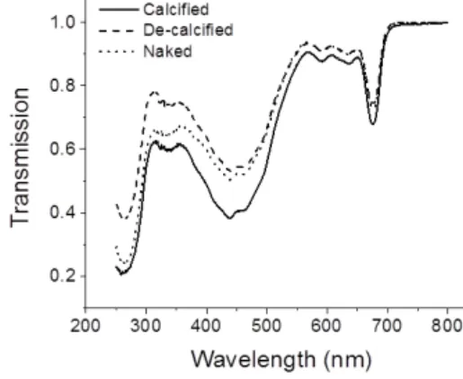
Figure 1.Transmission spectra of cells with (calcified strain) and without (calcified strain with coccoliths removed artificially, decal- cified strain) coccolith cover and naked cells ofEmiliania huxleyi.
periods were confirmed according to the ratios of UV-A/UV- B to PAR at the experimental location.
2.7 Statistics
The data were expressed as the means±standard deviation (SD). The statistical significance of the data was tested with software of Origin 9.0 (one-way ANOVA, Tukey’s post hoc test). A confidence level of 95 % was used in all analyses.
3 Results
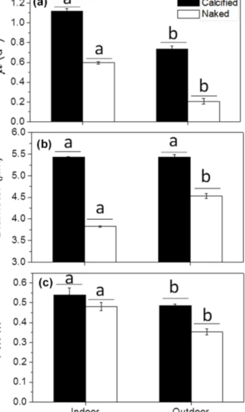
The coccolith layer ofE. huxleyi absorbed both visible and UV radiation. It reduced the transmission of visible radia- tion (400–700 nm) by 7.5 %, that of UV-A (315–400 nm) by 14.1 % and that of UV-B by 18.4 % (280–315 nm) relative to decalcified cells and by 6.5 % for PAR, 6.6 % for UV-A and 5.1 % for UV-B, relative to naked cells (Fig. 1). The specific growth rate of calcifyingE. huxleyistrain (PML B92/11) was about 2 times higher than that of the naked strain (CCMP 2090) (P< 0.05) when grown at 500 µmol photons m−2s−1 of PAR under indoor conditions (Fig. 2a). Growth rates of both strains were significantly (P < 0.05) reduced when the cells were transferred outdoors and exposed to natural solar radiation. However, under outdoor conditions, growth rates of calcified cells were 3.5 times higher than those of the naked cells, indicating that the latter was more harmed by the solar exposure than the former (Fig. 2a). The cell diameter was not significantly different in the calcified cells between the indoor and outdoor conditions (P> 0.05), but an 18 % increase was found in the naked cells after they had grown under outdoor conditions for 7 days (P < 0.05) (Fig. 2b). The maximal quantum yield (Fv/Fm) decreased when the cells were transferred from indoor to the outdoor conditions, re- flecting a harmful effect of solar radiation. The decrease in Fv/Fm, however, was much more pronounced in the naked cells (27 %) compared to calcified cells (11 %) (Fig. 2c).
Figure 2.The specific growth rate (µ)(a), diameter(b)and maxi- mum quantum yield(c)of PSII (Fv/Fm) of the calcified and naked cells ofE. huxleyigrown in indoor and outdoor conditions. Differ- ent letters represent significant differences between the indoor and outdoor experiments. Different horizontal lines represent significant differences between the different strains.
Table 1. Photosynthetic parameters of relative electron transport rate (Fig. 3) as a function of PAR; different letters represent sig- nificant differences (P< 0.05) among the calcified, decalcified and naked cells.
Calcified Decalcified Naked
α 0.23±0.02a 0.20±0.01a 0.17±0.02b rETRmax 90.6±9.0a 73.5±3.5b 42.3±8.5c Ik 1010.8±95.0a 986.3±27.4a 621.8±111.1b
Calcified cells had a significantly higher apparent light use efficiency (α), maximal electron transport rate (rETRmax) and light saturation parameters (Ik) compared to naked cells.
The decalcified cells of the calcified strain showed a remark- able decrease in rETRmax(P< 0.05), and alpha andIk also decreased but not statistically significantly (Fig. 3, Table 1).
Increased actinic light levels (acclimating light during the fluorescence measurement) led to higher NPQ in both the calcified and naked strain (Fig. 4). Furthermore, calcified cells showed higher NPQ values compared to naked cells (p< 0.05).
When exposed to full-spectrum solar radiation, the quan- tum yield of calcified cells showed no significant change dur-
Figure 3.The relative electron rate (rETR) of calcified, decalci- fied and naked cells ofE. huxleyigrown under indoor conditions as function of PAR. The cells were grown for 12–22 generations under 500 µmol photons m−2s−1of PAR.
Figure 4.The non-photochemical quenching (NPQ) of calcified and naked cells ofE. huxleyigrown under indoor conditions. Different letters represent significant differences among the light levels. Dif- ferent horizontal lines represent significant differences among the different type cells.
ing the first 30 min (P> 0.05). After 30 min, the quantum yield quickly dropped from about 0.35 to 0.22 for∼20 min (P< 0.05), followed by a slight recovery in the last 25 min.
A similar trend was observed in the decalcified cells, with the key difference that the sharp decrease already happened during the first 10 min. The quantum yield of the naked cells decreased constantly for the first 50 min and remained at the low level thereafter (Fig. 5).
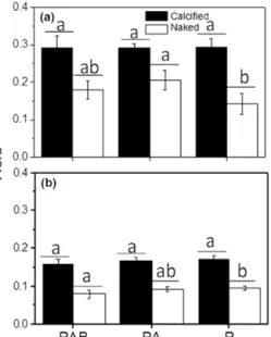
No effect of the radiation treatment (P, PA and PAB radiation) on the quantum yield of calcified cells was observed after the cells grown under indoor conditions were transferred to outdoor solar radiation for 1 h expo- sure (very cloudy day; average PAR, UV-A and UV-B were 481 µmol photons m−2s−1, 22.1 and 0.7 W m−2, re- spectively) (P > 0.05). The quantum yield was significantly higher in the naked cells, however, when they were exposed to UV-A radiation (PA vs.P treatment,P< 0.05; Fig. 6a).
0 10 20 30 40 50 60 70 80 0.0
0.1 0.2 0.3 0.4
Yield
Time (min)
Calcified Decalcified Naked
Figure 5. The time course of quantum yield of calcified, de- calcified and naked cells of E. huxleyi under full-spectrum so- lar radiation (noontime, average PAR, UV-A and UV-B were 1082 µmol photons m−2s−1, 48.1 and 1.6 W m−2, respectively).
Figure 6.The change in quantum yield of the calcified and naked cells ofE. huxleyi when transferred from indoor to outdoor con- ditions, being exposed to PAR alone (P), PAR+UV-A (PA) and PAR+UV-A+UV-B (PAB) for 60 min at around noontime. Panel (a): measured on a cloudy day (average PAR, UV-A and UV-B were 481 µmol photons m−2s−1, 22.1 and 0.7 W m−2, respectively);
panel(b): measured on a sunny day (average PAR, UV-A and UV- B were 1605 µmol photons m−2s−1, 69 and 2.4 W m−2, respec- tively). Different letters represent significant differences among the light treatments. Different horizontal lines represent significant dif- ferences between the different strains.
Similar responses were observed when the same test was done on a sunny day with an average PAR, UV-A and UV- B of 1605 µmol photons m−2s−1and 69 and 2.4 W m−2, re- spectively. Here, the quantum yield of the calcified cells showed no significant difference between the different light treatments, but it decreased significantly under the PAB treat-
ment compared to P treatments in the naked cells (P< 0.05) (Fig. 6b).
4 Discussion
Various hypotheses were proposed for the possible functions of coccoliths, but none of them is supported by sufficient ev- idence (Young, 1994; Raven and Crawfurd, 2012). One im- portant function of coccoliths for surface-dwelling species such asE. huxleyicould be the protection against high photon flux densities, especially UV radiation (Berge, 1962; Young, 1994; Gao et al., 2009).
Some of our results support this hypothesis. The growth rate of the calcified cells of E. huxleyi grown under in- door conditions was about 2 times higher than that of naked cells. This difference came out even more strongly, with growth rates 3.5 times higher in calcified versus naked cells, when the cells were exposed to full-spectrum solar radiation (Fig. 2a). This could potentially be attributed to the screen- ing of PAR, UV-A and UV-B by coccoliths. Although the daytime PAR of solar radiation was reduced to about half of the light level of the indoor test, noontime PAR levels were higher than 500 µmol photons m−2s−1, and the presence of UV could lead to more harm to the naked cells. Light pro- tection by coccoliths is further supported by theFv/Fmmea- surements. The maximum photochemical efficiency of PSII was only slightly reduced in calcified cells but significantly decreased in naked cells when they were exposed to natural solar PAR and UV radiation (Fig. 2c). Furthermore, the pho- tochemical performance of decalcified cells decreased sig- nificantly faster and more strongly with time compared to calcified cells (Fig. 5).
The diameter of calcified cells did not significantly change when they were exposed to the full spectrum of solar radi- ation. The diameter of the naked cells, however, increased significantly (Fig. 2b). Perhaps, the naked cells experienced more DNA damage and so did not enter theS phase regu- larly (Buma et al., 2000). Alternatively, it may reflect a strat- egy to acclimatize to stressful solar UV radiation since it is well known that smaller cells are usually more sensitive to UV than their larger counterparts (Garcia-Pichel, 1994;
Laurion and Vincent, 1998). Some field and laboratory stud- ies showed increased cell size with increased UV exposure (Buma et al., 2000), which can be interpreted as an adaptive or acclimation mechanism for protecting the cells against UV radiation. Furthermore, the naked cells might also employ other strategies, such as synthesizing UV screening com- pounds to ameliorate UV stress, because the naked strain had a lower UV transmittance than the decalcified strain.
Several studies found that coccoliths do not protectE. hux- leyifrom excess PAR (Nanninga and Tyrrell, 1996; Houdan et al., 2005; Trimborn et al., 2007). However, UV radiation was not considered in these experiments. Our results showed that the naked cells were more sensitive to full-spectrum
solar radiation than calcified cells, and even in the same strain, the photochemical performance of decalcified cells decreased significantly when comparing the calcified cells.
This suggests that coccoliths efficiently protect the cells from solar UV radiation.
On the other hand,E. huxleyiappears to be more sensitive to UV-B irradiances than other phytoplankton species, and its growth rate and physiological performances were highly inhibited by UV radiation (Peletier et al., 1996; Buma et al., 2000; Xu et al., 2011). However, competition tests for com- munity changes are rare, and longer-term experiments with less extreme UVR would be more ecologically and evolu- tionarily relevant (Raven and Crawfurd, 2012). In our work, UVR had no significant effect on the quantum yield of cal- cified cells regardless of high- or low-light conditions but it showed inhibition in naked cells when they were exposed to high-solar light (Fig. 6a, b). This provides further evidence for protection by coccoliths against UV radiation.
On the cloudy day, no significant difference was observed among the treatments for the calcified cells; on the sunny day, under the fluctuating light (data not shown) calcified cells manage to refurbish damage to their photosynthetic appara- tus by balancing damage and repair (Gao et al., 2007; Ragni et al., 2008; Loebl et al., 2010). For the naked cells, on the other hand, UV damage was not effectively repaired, lead- ing to the observed negative effect on photosynthetic perfor- mance.
It has to be noted that our experimental data are based on only two strains of a naked and calcifiedE. huxleyi. However, similar trends in photophysiology between naked and decal- cified cells in comparison to calcified cells suggest that the coccoliths ofE. huxleyiplay an important role in protecting this species against harmful solar radiation, especially UV- A and UV-B. Furthermore, the reported absence of photoin- hibition in this alga at high light levels also appears to be connected to the coccosphere of E. huxleyior its calcifica- tion process. In view of ongoing ocean change, the projected shoaling of the upper mixed layer (UML) caused by global warming and progressive ocean acidification that reduces the thickness or the number of coccoliths per cell (Gao et al., 2009; De Bodt et al., 2010) could reduceE. huxleyigrowth rates within the UML due to increased UVR exposure.
The Supplement related to this article is available online at doi:10.5194/bg-13-4637-2016-supplement.
Acknowledgements. This study was supported by the National Natural Science Foundation (41430967; 41476097; 41120164007), the State Oceanic Administration (National Programme on Global Change and Air-Sea Interaction, GASI-03-01-02-04), the Joint project of National Natural Science Foundation of China and Shandong province (no. U1406403) and the Strate- gic Priority Research Program of the Chinese Academy of
Sciences (no. XDA1102030204). The visit of Kunshan Gao to Kiel was supported by DAAD. Kai G. Schulz is the recipient of an Australian Research Council Future Fellowship (FT120100384).
Edited by: T. Treude
Reviewed by: D. Campbell and one anonymous referee
References
Balch W. M., Kilpatrick, K. A., and Trees, E. E.: The 1991 coccol- ithophore bloom in the central North Atlantic. 1. Optical prop- ertiesand factors affecting their distribution, Limnol. Oceanogr., 41, 1669–1683, 1996.
Berge, G.: Discoloration of the sea due to Coccolithus huxleyi
“bloom”, Sarsia, 6, 27–40, 1962.
Broecker, W. and Clark, E.: Ratio of coccolith CaCO3 to foraminifera CaCO3in late Holocene deeper-sea sediments, Pa- leoceanography, 24, PA3205, doi:10.1029/2009PA001731, 2009.
Buma, A. G. J., van Oijen, T., van de Poll, W., Veldhuis, M. J.
W., and Gieskes, W. W. C.: The sensitivity ofEmiliania hux- leyi(Prymnesiophyceae) to ultraviolet-B radiation, J. Phycol., 36, 296–303, 2000.
De Bodt, C., Van Oostende, N., Harlay, J., Sabbe, K., and Chou, L.: Individual and interacting effects ofpCO2and temperature onEmiliania huxleyicalcification: study of the calcite produc- tion, the coccolith morphology and the coccosphere size, Bio- geosciences, 7, 1401–1412, doi:10.5194/bg-7-1401-2010, 2010.
Gao, K., Wu, Y., Li, G., Wu, H., Villafañe, V. E., and Helbling, E. W.: Solar UV radiation drives CO2 fixation in marine phy- toplankton: A double-edged sword, Plant Physiol., 144, 54–59, 2007.
Gao, K., Ruan, Z., Villafane, V. E., Gattuiso, J. P., and Helbling, E. W.: Ocean acidification exacerbates the effect of UV radia- tion on the calcifying phytoplankterEmiliania huxleyi, Limnol.
Oceanogr., 54, 1855–1862, 2009.
Gao, K., Helbling, E. W., Häder, D. P., and Hutchins, D. A.: Re- sponses of marine primary producers to interactions between ocean acidification, solar radiation, and warming, Mar. Ecol.- Prog. Ser., 470, 167–189, 2012.
Garcia-Pichel, F.: A model for internal self-shading in planktonic organisms and its implications for the usefulness of ultraviolet sunscreens, Limnol. Oceanogr., 39, 1704–1717, 1994.
Genty, B., Briantais, J. M., and Baker, N. R.: The relationship be- tween the quantum yield of photosynthetic electron-transport and quenching of chlorophyll fluorescence, Biochim. Biophys. Acta, 990, 87–92, 1989.
Guan, W. and Gao, K.: Enhanced calcification ameliorates the neg- ative effects of UV radiation on photosynthesis in the calcify- ing phytoplankterEmiliania huxleyi, Chinese Sci. Bull., 55, 588–
593, 2010.
Guillard, R. R. and Ryther, J. H.: Studies of marine planktonic diatoms: I.Cyclotella nanahustedt, andDetonula confervacea (cleve) gran, Can. J. Microbiol., 8, 229–239, 1962.
Hakala-Yatkin, M., Mäntysaari, M., Mattila, H., and Tyystjärvi, E.:
Contributions of visible and ultraviolet parts of sunlight to pho- toinhibition, Plant Cell Physiol., 51, 1745–1753, 2010.
Houdan, A., Probert, I., Van Lenning, K., and Lefebvre, S.: Compar- ison of photosynthetic responses in diploid and haploid life-cycle
phases of Emiliania huxleyi(Prymnesiophyceae), Mar. Ecol.- Prog. Ser., 292, 139–146, 2005.
Kishino, M., Takahashi. M., Okami, N., and Ichimur, S.: Estimation of the spectral absorption coefficients of phytoplankton in the sea, Bull. Mar. Biol., 37, 634–642, 1985.
Laurion, I. and Vincent, W. F.: Cell size versus taxonomic compo- sition as determinants of UV-sensitivity in natural phytoplankton communities, Limnol. Oceanogr., 43, 1774–1779, 1998.
Loebl, M., Cockshutt, A. M., Campbell, D. A., and Finkel, Z. V.:
Physiological basis for high resistance to photoinhibition under nitrogen depletion inEmiliania huxleyi, Limnol. Oceanogr., 55, 2150–2160, 2010.
Moore, T. S., Dowell, M. D., and Franz, B. A.: Detection of coccol- ithophore blooms in ocean color satellite imagery: a generalized approach for use with multiple sensors, Remote Sens. Environ., 117, 249–263, 2012.
Nanninga, H. J. and Tyrrell, T.: Importance of light for the formation of algal blooms byEmiliania huxleyi, Mar. Ecol.-Prog. Ser., 136, 195–203, 1996.
Peletier, H., Gieskes, W. W. C., and Buma, A. G. J.: Ultraviolet-B radiation resistance of benthic diatoms isolated from tidal flats in the Dutch Wadden Sea, Mar. Ecol.-Prog. Ser., 135, 163–168, 1996.
Poulton, A. J., Adey, T. R., Balch, W. M., and Holligan, P. M.: Relat- ing coccolithophore calcification rates to phytoplankton commu- nity dynamics: regional differences and implications for carbon export, Deep-Sea Res. Pt. II, 54, 538–557, 2007.
Ragni, M., Airs, R. L., Leonardos, N., and Geider, R. J.: Photoin- hibition of PSII in Emiliania huxleyi(Haptophyta) under high light stress: the roles of photoacclimation, photoprotection, and photorepair, J. Phycol., 44, 670–683, 2008.
Raven, J. A. and Crawfurd, K.: Environmental controls on coc- colithophore calcification, Mar. Ecol.-Prog. Ser., 470, 137–166, 2012.
Rost, B. and Riebesell, U.: Coccolithophores and the bi- ological pump: responses to environmental changes, in:
Coccolithophores- from molecular processes to global impact, edited by: Thierstein, H. R. and Young, J. R., Springer, Berlin, 99–125, 2004.
Trimborn, S., Langer, G., and Rost, B.: Effect of varying calcium concentrations and light intensities on calcification and photo- synthesis inEmiliania huxleyi, Limnol. Oceanogr., 52, 2285–
2293, 2007.
Voss, K., Balch, W. M., and Kilpatrick, K. A.: Scattering and atten- uation properties ofEmiliania huxleyi cells and their detached coccoliths, Limnol. Oceanogr., 43, 870–876, 1998.
Xu, K., Gao, K., Villafañe, V. E., and Helbling, E. W.: Photosyn- thetic responses ofEmiliania huxleyito UV radiation and ele- vated temperature: roles of calcified coccoliths, Biogeosciences, 8, 1441–1452, doi:10.5194/bg-8-1441-2011, 2011.
Young, J. R.: Functions of coccoliths, in: Coccolithophores, edited by: Winter, A. and Siesser, W. G., Cambridge University Press, Cambridge, 63–82, 1994.
Zheng, Y. and Gao, K.: Impacts of solar UV radiation on the photo- synthesis, growth, and UV-absorbing compounds inGracilaria lemaneiformis(Rhodophyta) grown at different nitrate concen- trations, J. Phycol., 45, 314–323, 2009.