DISSERTATION
ANALYSIS OF HUMAN ANTIGEN‐EXPERIENCED CD4 T CELLS ACCORDING TO IL7Rα and CCR7
EXPRESSION
zur Erlangung des akademischen Grades doctor rerum naturalium (Dr. rer. nat.)
im Fach Biologie
eingereicht an der
Mathematisch‐Naturwissenschaftlichen Fakultät I der Humboldt‐Universität zu Berlin
von
Diplom‐Biologin Laura Lozza geb. am 14.02.1976 in Varese (Italien)
Präsident der Humboldt‐Universität zu Berlin Prof. Dr. Dr. h.c. Christoph Markschies
Dekan der Mathematisch‐Naturwissenschaftlichen Fakultät I Prof. Dr. Lutz‐Helmut Schön
Gutachter:
1. ..Prof. Dr. Alf Hamann...
2. ..Prof. Dr. Andreas Radbruch...
3. ..Dr. Michal Or‐Guil...
a nonno Renzo
INDEX
Index
1 INTRODUCTION ... 11
1.1 General introduction to the immune system and T cell biology... 13
1.2 Role of chemokine receptors in regulating T cell traffic ... 15
1.2.1 CCR7 as a key factor to enter the secondary lymphoid organs ... 16
1.2.2 Chemokine receptor expression reflects the functions of T cells ... 17
1.3 Definition of memory T cell subsets: TCM, TEM and TEMRA cells ... 19
1.3.1 Heterogeneity of human TCM cells... 20
1.4 Effector T cells ... 21
1.5 Generation and maintenance of memory T cells... 22
1.5.1 IL‐2, IL‐15, IL‐7 cytokines and their receptors ... 22
1.5.2 Naive T cell maintenance ... 25
1.5.3 T cell activation and clonal expansion... 25
1.5.4 Memory T cell generation and the role of IL7Rα... 25
1.5.5 Maintenance of memory T cells... 27
1.6 Understanding the generation of memory T cells ... 27
1.6.1 Definition of signal strength and fitness ... 27
1.6.2 Lineage relationship of memory T cells... 29
2 AIM OF THE WORK... 33
3 MATERIALS AND METHODS ... 37
3.1 Materials ... 39
3.2 Cell culture handling... 43
3.3 Isolation of PBMC ... 43
3.4 Magnetic cell separation (MACS®) ... 43
3.4.1 Isolation of monocytes... 44
3.4.2 Isolation of CD4 T cells ... 44
3.5 Immunofluorescence, Flow Cytometry and FACS... 45
3.5.1 Analysis of surface molecules ... 47
3.5.2 Intracellular staining... 47
3.5.3 Naive T cell sorting ... 48
3.5.4 Antigen‐experienced T cell sorting... 49
3.6 Labeling of T cells with CFDA‐SE... 49
3.7 Cell culture conditions... 50
Index
3.7.3 Stimulation conditions for analysis of cytokine production... 51
3.7.4 Stimulation conditions for analysis of cell signaling... 52
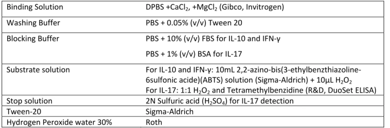
3.8 Enzyme‐linked immunosorbent assay (ELISA)... 52
3.9 Detection of phosphoproteins by immunoblotting ... 53
3.10 Microarray ... 54
3.11 Statistical analysis... 55
4 RESULTS... 57
4.1 Analysis of in vitro generated TSST‐specific CD4 T cells according to IL7R and CCR7 expression ... 59
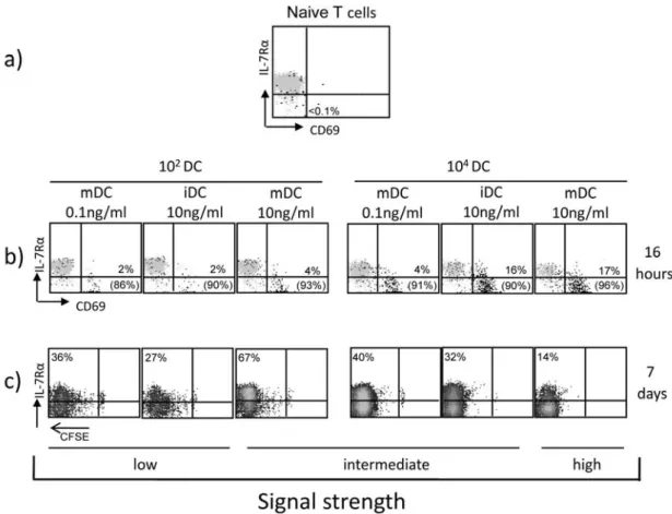
4.1.1 Modulation of IL‐7R(IL7R)expression in human CD4+ T cells primed by different levels of stimulation ... 59
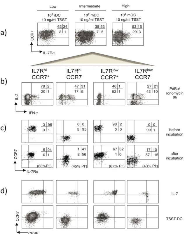
4.1.2 Functional properties of human primed CD4+ T cells defined by IL7R and CCR7 .. 62
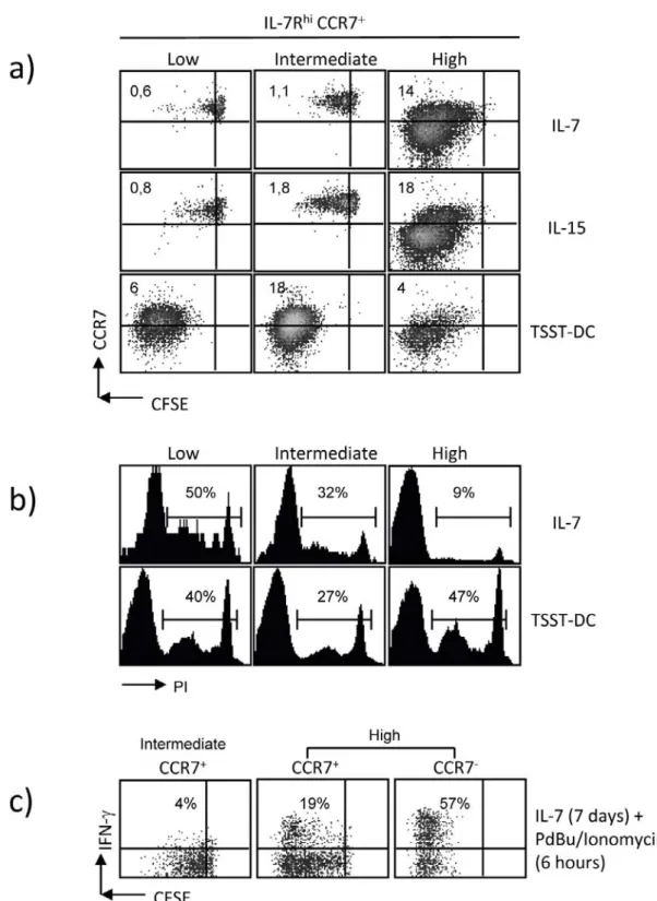
4.1.3 Properties of the IL7RhiCCR7+ subset generated at different strengths of stimulation ... 64
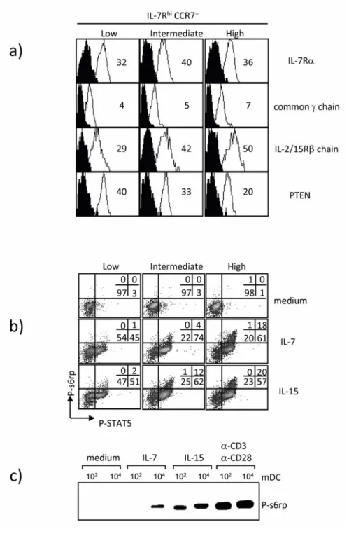
4.1.4 Cytokine receptor signaling capacity of IL7RhiCCR7+ T cells obtained at different levels of stimulation ... 67
4.1.5 Gene expression profile of high stimulated IL7RhiCCR7+ T cells ... 69
4.2 Analysis of ex vivo antigen‐experienced CD4 T cells according to the expression of IL7R and CCR7... 72
4.2.1 Functional properties of ex vivo experienced CD4 T cells sorted according to the expression of IL7R and CCR7... 72
4.2.1.1 Cytokine‐production of IL7R CCR7 subsets upon TCR activation ... 72
4.2.1.2 Proliferation and survival of IL7R CCR7 subsets in the presence of homestatic cytokines or TCR ligands... 73
4.2.2 IL7Rlow CD4+ T cells under steady state conditions contain Foxp3+ and cytotoxic T cells ... 77
4.2.3 Antigen‐specificities of IL7Rhi and IL7Rlow CD4 T cells from healthy donors... 78
4.2.3.1 The addition of IL‐2 enhances antigen‐specific proliferation of IL7Rlow T cells .. 79
4.2.3.2 IL7Rlow T cells respond preferentially to persistent antigens ... 80
4.2.3.3 Cytokine‐production of antigen‐specific IL7Rhi and IL7Rlow T cells ... 82
4.2.3.4 Cytokine‐profile after antigen‐specific proliferation... 82
5 DISCUSSION AND CONCLUSION ... 85 5.1 Functions and phenotype of memory and effector T cells: the contribution of
Index
5.1.1 Summary of the results ... 88
5.2 Lymph nodes homing receptors and IL7Rα expression discriminate between memory‐ and effector‐like CD4 T cell subsets... 89
5.2.1 IL7RhiCCR7+ and IL7RhiCCR7– CD4 T cell subsets ... 89
5.2.2 IL7RlowCCR7+ CD4 T cell subset ... 90
5.2.3 IL7RlowCCR7– CD4 T cell subset ... 91
5.2.4 In vitro primed versus ex vivo isolated human CD4 T cell subsets ... 91
5.2.5 CD4+ Cytotoxic T cells ... 92
5.2.6 TCR triggering and IL‐2 expand effector‐like T cells ... 92
5.2.7 Antigen‐specificity of IL7Rlow CD4 T cells in steady state conditions ... 93
5.2.8 Maintenance of effector T cells... 95
5.3 Role of the signal strength in modulating IL7R surface phenotype and functions of CD4 T cells ... 96
5.4 Conclusion ... 99
6 SUMMARY / ZUSAMMENFASSUNG... 101
6.1 Summary ... 103
6.2 Zusammenfassung... 105
7 REFERENCES... 107
8 SUPPLEMENTARY FIGURES... 129
9 APPENDIX ... 133
9.1 Abbreviations ... 135
9.2 Erklärung ... 137
9.3 Acknowledgments ... 138
9.4 Publications: ... 139
9.5 Congresses attendend:... 141
Index
Index
1 INTRODUCTION
Index
Introduction
1.1 General introduction to the immune system and T cell biology
The immune system defends the host against all kinds of potentially harmful and infectious agents. It is formed by a complex network of cells and organs that interact with each other and their environment. The cells of the immune system are able to discriminate between self-molecules that are components of the organism, from foreign substances, called antigens, that must be eliminated. However, in some pathological situations the immune response can be directed to noninfectious antigens or self-molecules giving rise to allergy or autoimmune disease.
The first defense against foreign molecules is driven by cells of the natural or innate immune system. These cells react fast, do recognize pathogens merely by conserved patterns and do not develop memory of the pathogen encountered. Mast cells, phagocytes, basophils, eosinophils, (natural killer) NK cells and invariant NK T cells belong to this group. In contrast, cells highly specialized to recognize antigens by antigen-specific receptor and to act only after the exposure to them are members of the acquired or adaptive immunity. After the first encounter with a pathogen, these cells develop an immunological memory that enables an efficient and faster response on re-exposure to the same pathogen. T cells, B cells and the antibodies secreted by B cells are part of the adaptive immunity (1, 2).
Whereas B cells and antibodies recognize intact antigens, T cells recognize antigens that are degraded into peptide fragments, processed and presented by antigen presenting cells (APC) in the context of the major histocompatibility complex (MCH) (3, 4). MHC molecules are classified in two types: MHC class I molecules bind peptides deriving from new proteins synthesized and degraded in the cytosol such as fragments of viral proteins.
MHC class II molecules bind peptides deriving from protein degraded inside of endocytic vesicles (5, 6). While most of the nuclear cells have MHC class I molecules only few type of cells are able to internalize and express the antigen by MHC class II molecules. These professional antigen presenting cells comprise dendritic cells (DC), B cells and macrophages. However, only dendritic cells are able to prime naive T cells and thereby initiate a primary immune response (7, 8). DC are generated in the bone marrow and migrate through the blood into tissues where they have the chance to encounter pathogens.
During this phase DC display an immature phenotype and are equipped with a highly
Introduction
Pathogen Associated Molecular Patterns (PAMP), which are recognized by Pathogen Recognition Receptors (PRR) like Nod-like Receptors (NLR), C-type lectin receptors (CLR) and Toll-like Receptor (TLR) (9, 10). Upon stimulation with TLR-ligands DC undergo a process of maturation that results in a migration from the tissue into the T cell areas of secondary lymphoid organs as well as with the up-regulation of MHC and costimulatory molecules on the cell surface (7). T cells present in the T cell areas recognize the MHC-antigen complex by means of the T cell receptor (TCR). The major subset of mature T cells expresses a TCR composed of the two glycoprotein chains α and β which are associated with the glycoprotein CD3 and the co-receptors CD4 or CD8.
CD4+ T cells and CD8+ T cells show different functions and can be subdivided themselves into specialized subsets according to their localization, production of cytokines and functional properties. CD8+ T cells recognize antigen peptide bound to the MHC Class I molecules. These cells are defined as cytotoxic T lymphocytes (CTL) because of their ability to kill infected or tumor cells. CD4+ T cells recognize antigen peptide bound to the MHC Class II molecules and are defined as helper T (Th) cells because of their ability to modulate activation and differentiation of other cells through the production of specific cytokines. The latter are proteins released by cells that mediate and regulate the immune response. However, there are also cytotoxic CD4+ T cells present at low frequency in human peripheral blood (11). Antigens and the cytokine milieu determine effector properties of helper T cells which can be subdivided into the Th1, Th2 (12, 13) and Th17 lineage (14, 15). Th1 cells are generated under direction of the cytokine IL-12 and the T- box transcription factor T-bet. Th1 cell-mediated inflammation is mainly characterized by recruitment of CD4+ T cells that produce IFN-γ and mediate protection against intracellular pathogens such as bacteria and viruses. Conversely, the cytokine IL-4 and the zinc-finger transcription factor GATA-3 drive differentiation towards Th2 cells. Th2 cell- mediated inflammation is characterized by CD4+ T cells producing IL-4, IL-5 and IL-13 that mediate protection against nematodes and contribute to allergic responses. Finally, in the presence of some cytokines such as IL-6, IL-1β, IL-23, IL-21 and TGF-β, CD4+ T cells are induced to up-regulate the expression of the retinoic orphan receptor (ROR)γt and to differentiate into Th17 cells (16-19). These cells produce IL-17, IL-6, IL-22 and TNF-α and are thought to protect against extracellular bacteria and fungi. In addition, involvement of Th17 cells in some autoimmune diseases cells has been observed (20, 21).
Introduction
Another preeminent group of CD4+ T cells are the regulatory T cells (Treg). These cells are characterized by the ability to suppress adaptive T cell responses, preventing autoimmunity (22, 23). According to the conditions of their generation, natural regulatory T cells (nTreg) and adaptive (aTreg) or inducible Treg (inTreg) are distinguished. Natural Treg develop in the thymus and express the forkhead-winged helix transcription factor Foxp3 (22) whereas adaptive or inducible Treg develop from CD4 T cells in the periphery in parallel to effector T cells and do only partly express Foxp3 (24, 25).
1.2 Role of chemokine receptors in regulating T cell traffic
After maturation in the thymus, naive T cells enter the blood and continuously recirculate through secondary lymphoid organs in search of antigen presented on APC. Priming of naive T cells involves recognition of an antigen presented on DC and the subsequent activation of T cells which then start to proliferate and differentiate into effector and memory cells. Effector T cells are specialized in the fast elimination of pathogens and migrate to the tissue where they can interact with other leukocytes. On the other hand memory T cells maintain the memory for already encountered antigens and provide a rapid and efficient response against a secondary challenge. They are able to reach both the secondary lymphoid organs as well as inflamed tissues (26). The migration of DC and T cells during an immune response is finely controlled by chemokines and adhesion molecules that are localized on the appropriate cells and at the right place.
Leukocyte migration can be subdivided into two steps. The first step consists of the extravasion of leukocytes from the blood to the lymph nodes (LN), Peyer’s patches (PP) or inflamed tissues. In this step adhesion molecules such as selectins and integrins are involved (27, 28). The interaction between selectins on T cells and their ligand on endothelial cells allows leukocytes to roll on the endothelium wall until a specific chemokine receptor is recognized by their related ligand on the surface of the endothelial cells. The ligation of chemokine receptors and the activation of integrins lead to a stable adhesion of the cells, followed by the transmigration of leukocytes into the secondary lymphoid organs and tissues. An example of L-selectin is CD62L that recognizes some molecules expressed on high endothelial venules (HEV). These are highly specialized
Introduction
In the second step leukocytes are guided within the secondary lymphoid organs or the inflamed tissue by means of a chemotactic or chemokinetic gradient formed by chemokines (30, 31). Chemokines are recognized by seven-transmembrane domain receptors, which signal via heterotrimetric GTP-binding proteins (32, 33). Based on the site of production and the functional role chemokines can be classified into
“inflammatory” and “homeostatic” (26, 34). Inflammatory chemokines are produced under conditions of inflammation by endothelial, epithelial and stromal cells as well as by leukocytes. They regulate the recruitment of effector leukocytes and thus, determine the composition of inflammatory infiltrates. In contrast, homeostatic chemokines are produced constitutively within lymphoid tissues where they control the homeostatic cell traffic.
Importantly, some of the chemokines can have different roles depending on the context in which they are produced (26).
1.2.1 CCR7 as a key factor to enter the secondary lymphoid organs
Naive T cells enter the lymph nodes (LN) and Peyer’s Patches (PP) via high endothelial venules (HEV). All naive T and B lymphocytes express the chemokine (C-C motif) receptor 7 (CCR7) (35, 36) while its two ligands CCL19/ELC (EBV-induced molecule 1 ligand) and CCL21/SLC (secondary lymphoid tissue chemokine) are presented at the entrance of the luminal side of HEVs and afferent lymphatic vessels and inside the T zone areas of lymph nodes and Peyer’s Patches. In particular CCL21 is constitutively expressed by endothelial cells of HEVs and by stromal cells in the T cell zone in secondary lymphatic organs (37, 38), while CCL19 is expressed on interdigitating DC (39, 40). The presence of CD62L and CCR7 on the surface of naive lymphocytes leads naive T and B cells into the LNs and PPs. Moreover, during the maturation phase DC lose responsiveness to inflammatory chemokines such as CCR5 and instead up-regulate CCR7 in order to leave the periphery and enter the lymph nodes through the afferent lymphatics (37, 39).
The importance of CCR7 and its ligands in regulating the entry of T cells and DC into the LNs was demonstrated in studies of mice genetically deficient in CCR7 (41) and in mice homozygous for the spontaneous mutation plt that results in a defective production of CCL21 (42). These mouse strains show a profound defect of naive T and B cells homing due to their inability to adhere and pass through HEVs. Moreover, it has been shown that
Introduction
the intradermal injection of CCL21/SLC in plt/plt mice, followed by its transport and localization into the HEVs leads naive T cells to enter the LNs, again (43).
It must be noted that in the case of inflammation of the lymph nodes themselves the expression of the chemokines previously described is modulated. CCR7 ligands are donwregulated, whereas ligands for CXCR3 (such as CXCL9, CXCL10) are up-regulated on the luminal side of lymph node HEVs. This mechanism supports the recruitment of NK cells and Th1 cells at the site of inflammation (44, 45).
1.2.2 Chemokine receptor expression reflects the functions of T cells
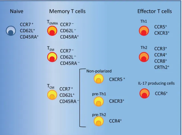
Antigen-stimulation induces the differentiation of specialized T cells with different chemokine receptor patterns reflecting their homing potential. T helper cells that lose CCR7 expression but up-regulate CXCR5 are found in the tonsils (46-48). These cells, called follicular B helper T (TFH), are able to go into the B cell areas by interaction with the ligand CXCL13 (49) and provide help for antibody production by B cells. TFH differ from CXCR5+ T cells found in the blood, which maintain CCR7 and CD62L expression.
This population of non-polarized circulating cells contains antigen-specific cells at low frequency that might have been recently activated (50, 51). Th1 polarized cells express preferentially the chemokine receptors CCR5 and CXCR3 that guide to sites of inflammation (52-55) (Fig. 1.1). CCR5 is also expressed on monocytes and macrophages which explains their colocalization with Th1 cells. Th2 polarized cells frequently express CCR3, CCR4, CCR8 and the prostaglandin D2 chemoattractant receptor CRTh2 (Fig.
1.1). This receptor is known to be required to migrate to sites of allergic reactions (56-58).
CCR3 is also expressed on eosinophilis and basophils whereas its ligand eotaxin is produced by mucosal tissue during an allergic inflammation. The shared expression of CCR3 may allow these three cell types to colocalize at sites of eotaxin production (59).
Finally, the chemokine receptor CCR6 is expressed on IL-17 producing T cells in human peripheral blood (60).
The expression of adhesion molecules and chemokine receptors identifies different types of tissue-specific T cells. For instance the simultaneous expression of cutaneous lymphocyte-associated antigen (CLA) and CCR4 identifies skin homing T cells (61), whereas the expression of integrin α4β7 and CCR9 is characteristic of gut-homing T cells
Introduction
It should be noted that though some chemokine receptors are preferentially expressed on either Th1 or Th2 cells they may also be found at lower levels on the other subset, respectively (34, 52). In addition, the pattern of chemokine receptors here described applies mainly to the resting state of Th1 and Th2 cells due to the fact that some chemokine receptors are modulated upon T cell reactivation (58, 63). Finally, other subsets such as Treg can express the same chemokine receptors as Th1, Th2 and Th17 cells depending on the cytokine milieu in which they are activated (64). The overlap of the chemokine receptor expression profile between Treg and effector T cells subsets is believed to allow Treg to colocalize with the targeted effector T cells.
Fig. 1.1 Surface markers of naive, memory and effector T cells. Each subset preferentially expresses some chemokines receptors and lymph node homing receptors (see text).
Introduction
1.3 Definition of memory T cell subsets: TCM, TEM and TEMRA cells
Memory T cells are characterized by the ability to rapidly proliferate after re-exposure to pathogen and to persist long-term in the absence of antigen by undergoing homeostatic proliferation (65).
Depending on the migratory and functional properties two subsets of memory T cells have been identified both in humans and in mice (66-69). Memory T cells that constitutively express CD62L and CCR7 and are able to recirculate through secondary lymphoid organs are called T central memory (TCM) cells (Fig. 1.1). On the contrary, memory T cells that lack CCR7 expression, are predominantly negative for CD62L expression and are preferentially located in non-lymphoid tissues are called T effector memory (TEM) cells (Fig. 1.1) (70). Phenotypically, TCM cells can be further distinguished from naive T cells by the expression of CD45RO that is one of two isoforms of the leukocyte common antigen (CD45) (71). Naive T cells express the isoform CD45RA that is progressively substituted by the isoform CD45RO following TCR triggering. Primed T cells are mainly CD45RO+ while unprimed T cells are CD45RA+. Nevertheless, a small subset of human memory CD8+CCR7–CD45RA+ T cells (CD8+ TEMRA) (Fig. 1.1) has been described in response to human cytomegalovirus antigens (HCMV) (72-74) and to lytic Epstein-Barr virus antigens (75). On the contrary, human immunodeficiency virus (HIV)-specific memory CD8+ T cells are largely TEM (CD45RA–CCR7–) cells (73) . In vitro data suggest that CD45RA re-expression on antigen-experienced CD8+ T cells is promoted by homeostatic cytokines and require the absence of antigen (76).
TCM cells produce mainly IL-2 and lack immediate effector functions but, after secondary challenge, efficiently proliferate and differentiate into effector T cells (66). In contrast, TEM cells show immediate effector functions and produce inflammatory cytokines such as IFN-γ, TNF-α, IL-4, IL-5 and, in the case of CD8 TEM cells, perforin. Moreover, TEM cells express various inflammatory chemokines in order to move into inflamed tissues.
Although both TCM and TEM cells have high responsiveness to antigenic stimulation, the expansion potential, which means the capacity to proliferate and survive, decreases from TCM towards the TEM phenotype and is very low in TEMRA cells (66, 76). The decreasing expansion potential of these three subsets correlates with a decrease in telomere length and with an increased susceptibility to apoptosis (76, 77). On the contrary, responsiveness to
Introduction
It is important to remark that the phenotypic definition of TCM and TEM cells can be applied only to resting cells. In fact, CCR7 can be transiently up-regulated on TEM cells upon cell activation (63, 73). Several studies have shown that human T cell subsets stimulated in vitro express CCR7, suggesting that activated cells are able to migrate to secondary lymphoid organs upon activation (58, 79). In addition, cytokine-producing cells obtained from murine inflamed tissues are found to be either CCR7+ or CCR7– without a significant difference in their function (80-82). Although an intrinsic difference of CCR7 expression on memory T cell between human and mouse cannot be excluded, the definition of TCM and TEM cells in the human always refers to the steady state condition.
Recently, it has been demonstrated that mouse CD8 T cells showing properties of TCM and TEM cells can be discriminated as IL7RαhiCD62L+ and IL7RαhiCD62L– cells, respectively (83, 84). The importance and the role of IL7Rα and IL-7 in memory T cell development were described further on.
1.3.1 Heterogeneity of human TCM cells
Single cell analysis shows that the human TCM subset is not a homogeneous population but rather is composed of functionally different cells (85). Indeed, TCM cells can behave in different ways according to the stimulus given. After TCR stimulation in the presence of the polarizing cytokines IL-12 or IL-4, TCM cells efficiently proliferate and differentiate into Th1 or Th2 effector cells, respectively (66, 70, 85). On the other hand, some TCM cells that proliferate with the homeostatic cytokines IL-7 and IL-15 spontaneously lose CCR7 expression and are able to produce IFN-γ or IL-4 upon restimulation (76, 78). These cells behave as precursors of Th1 and Th2 cells and can be distinguished according to the expression of the chemokine receptors CXCR3 and CCR4 (Fig. 1.1) (51). In particular, pre-Th1 cells express CXCR3, produce lower amounts of IFN-γ than CXCR3+ TEM cells and spontaneously differentiate into Th1 effector cells in the presence of homeostatic cytokines. Similarly, pre-Th2 cells express CCR4, produce lower levels of IL-4 than CCR4+ TEM cells and acquire Th2 effector functions in the presence of homeostatic cytokines. In contrast to that, TCM cells lacking CXCR3 and CCR4 but expressing CXCR5 remain non-polarized in the presence of homeostatic cytokines. Although both TCM and the TEM CD4 cells can be generated after encounter with a pathogen a different distribution of pre-Th1 and pre-Th2 TCM cells is observed. Tetanus toxoid (TT)-specific
Introduction
with the notion that tetanus vaccination induces a mixed Th1/Th2 response (86). On the other hand, human cytomegalovirus (HCMV) and vaccinia virus (VV) are found to promote a Th1 cell polarization (87, 88); in fact, specific cells for HCMV or VV are found among CXCR3+ T cells in the TCM compartment. Because it was observed that TEM cells survive and proliferate less than TCM, it has been suggested that the different distribution of pathogen-specific TCM cells might be a mechanism to replenish TEM cells in the absence of antigen (89).
1.4 Effector T cells
The function of effector T cells is to rapidly eliminate the pathogen upon infection.
As explained before, effector T cells are generated following antigenic stimulation and extravasate into peripheral tissues at the site of infection according to the chemokine receptor pattern. Effector functions involve the production of inflammatory cytokines and the degranulation of cytolytic enzymes such as granzymes and perforin in order to kill infected cells (90). Although predominantly effector T cells are expected to express activation markers there are no specific phenotypic surface markers. Thus, effector T cells are usually distinguished by low survival rates and the inability to proliferate in the presence of homeostatic cytokines such as IL-7 or after TCR restimulation (91).
Importantly, the presence of antigen is required to perform effector functions and once the pathogen is cleared, effector T cells rapidly become apoptotic and die (92). This mechanism avoids that, in the absence of a target, effector T cells cause immunopathological damage in a bystander manner.
Recently, it has been shown that short-lived effector T cells generated after acute infection in mice down-regulate CD62L and express low amounts of the IL-7 receptor α-chain (IL7Rα) (83). Therefore, these cells differ from long-lived IL7RhiCD62L+ TCM cells and IL7RhiCD62L– TEM cells.
In the human system, two subsets of memory T cells show properties similar to effector T cells. CD8 TEMRA cells are CD27–CD28–CCR7–, produce high levels of perforin and Fas ligand and show low proliferative capacity. Although these cells have properties similar to effector cells they are described as memory T cells (66, 93, 94) since they descend
Introduction
monoculeosis have shown to combine cytotoxic activities with a residual expansion potential (75, 94). A similar subset of ‘terminally differentiated’ human CD45RA+ memory T cells is recently found in the CD4 T cell compartment associated with protracted antigen exposure and low viral load (95, 96). These cells do not express CD27, CD28 and CCR7, proliferate poorly and produce IFN-γ.
CD8+ T cells generally are considered to exert the major cytotoxic activity. Nevertheless CD4+ T cells with granules containing granzyme B and perforin are detected at low frequencies in the blood of healthy individuals (97). Cytotoxic CD4+ T cells do not express the costimulatory receptor CD28 and are mainly CD45RO+ (98). In addition CD4+CD28– T cells are negative for CCR7 and CD62L (66). Finally, most of CD4+CD28– T cells do not express markers of activation and proliferation like CD38, CD69, HLA-DR and Ki67 (97-100). In vitro generated human cytotoxic CD4+ T cells are shown to recognize a variety of pathogens including HCMV, Epstein-Barr virus (EBV), vaccinia virus and human immunodeficiency virus (HIV) (11). Direct ex vivo specific cytotoxic CD4+ T cells have been found in responses to HCMV (97, 101, 102).
In chronic infection resting effector CD8+ T cells are described to survive in an antigen- dependent manner (92). If the chronic antigen stimulation remains high, cells might progressively lose effector cytokine production, cytolytic activity and proliferation potential (103, 104) towards a complete clonal exhaustion (105, 106). Understanding whether T cell dysfunction induced by persistent antigens is reversible, constitutes an important point in vaccine strategies against chronic infections. In this regard, it has been demonstrated that the PD-1-PD-L (receptor Programmed cell Death 1- receptor Programmed cell Ligand) pathway contributes to maintain CD8+ T cell dysfunction during chronic infections (107).
1.5 Generation and maintenance of memory T cells
1.5.1 IL-2, IL-15, IL-7 cytokines and their receptors
Despite the generation of new T cells, the maintenance of circulating T cells and the clonal expansion of antigen specific cells, the number of peripheral T lymphocytes remains relatively constant. This is possible by means of a balance among proliferation, survival
Introduction
γ-chain’ (γc or CD132) family play a major role in promoting and maintaining the T lymphocyte population (109). Among these cytokines, IL-2, IL-15 and IL-7 act as growth and survival factors during the generation and the maintenance of memory T cells. IL-2 binds the heterotrimetric receptor composed of IL-2Rα, IL-2/15Rβ and γc. The expression of the IL-2Rα chain (CD25) confers a high affinity for IL-2, whereas IL-2/15Rβ (CD122) chain is recognized also by IL-15. Similar to the IL-2 receptor, the IL-15R is also heterotrimetric and contains γc, IL-2/15Rβ and exclusively the subunit IL-15Rα. In contrast, IL-7R is composed of two subunits: γc and IL-7Rα (CD127). Those three cytokines share the signaling pathway of the cytokine-receptor common γ-chain: the interaction between cytokines and receptors induces the phosphorylation of the janus kinase 3 (JAK3) that works as a bridge between γc and the signal transducer and activator of transcription 5 (STAT5). Activation of JAK3 induces the phosphorylation of a crucial tyrosine residue leading STAT5 to dissociate from its receptor and to dimerize. STAT5 dimers translocate to the nucleus and promote the transcription of target genes.
The subunits IL-2/15Rβ and IL-7Rα bind JAK1 and are able to activate both STAT5 and other pathways. In fact, every single cytokine use different signaling pathways in order to confer distinct signals to the cell. For instance, IL-2R triggers and supports T cell proliferation and survival through the phosphatidylinositol 3-kinase (PI3K) pathway but also sensitizes cells to FAS-mediated activation-induced cell death (AICD) via STAT5 (110). AICD is a process that leads to the elimination of activated T cells through engagement of cell-surface-expressed death receptors such as CD95 (FAS) or the tumor necrosis-factor receptors (111). On the other hand, the interaction of IL-7 with its receptor induces reciprocal phophorylation of JAK3 and JAK1 and activation of STAT5 resulting in an increased expression of survival factors such as B-cell lymphoma 2 protein (Bcl-2).
Another pathway used by IL-7R is via PI3K/AKT signaling. AKT downstream targets are involved in several functions encompassing cell survival (112, 113) cell-cycle and cell growth regulation (114, 115) and induction of glucose transporters (116). Through the PI3K/AKT pathway, IL-7R induces the phoshorylation and subsequent inactivation of the pro-apoptotic protein Bcl-2-antagonist of cell death (BAD) (113). Moreover, some evidences suggest that IL-7 might have a role in regulating cell cycle progression, proliferation and maintenance of glucose metabolism (117).
Introduction
naive and memory T lymphocytes (117). IL-7 is constitutively produced by stromal tissues and this seems not to be influenced by extrinsic stimuli so the limiting factor for the amount of available IL-7 is only dependent on its consumption (118). In contrast, IL-7Rα expression on the surface of T cells is influenced by extrinsic stimuli. IL-7Rα is expressed on resting T cells and can be transiently down-regulated upon IL-7 binding (119). This mechanism ensures that T cells, that have already received signals from IL-7, will not compete with T cells that have not yet encountered IL-7, increasing the chances of survival for each T cell clone. In this way, the heterogeneity and the number of peripheral CD4+ and CD8+ T cells can be maintained (119, 120). In addition, IL-7R can be down- regulated by other signals such as IL-2 and TCR triggering as will be explained further on.
Due to the common IL-2/15Rβ chain, IL-2 and IL-15 have several similar functions such as the induction of proliferation of CD4+ and CD8+ T cells, or the generation of CTL (121). However, IL-2 and IL-15 play different roles. IL-2, which is produced mainly by activated T cells, has a unique role in AICD and in maintaining peripheral Treg. In contrast, IL-15 that is produced notably by monocytes and DC is important for the maintenance of memory T cells. One fact that mirrors the different functions of IL-2 and IL-15 is the differing distribution of the high-affinity α-chains. IL-2Rα is expressed mostly by activated T cells, whereas IL-15Rα is expressed predominantly on activated monocytes and DC (122). In addition, exposure of T cells to IL-2 induces up-regulation of IL-2Rα, whereas exposure of cells to IL-15 induces down-regulation of IL15Rα. Finally, the distinct mechanisms of action contribute to manifest different functions of IL-2 and IL-15.
Soluble IL-2 binds the pre-formed high-affinity heterotrimetric receptor at the surface of a single activated cell (123, 124). In contrast, IL-15 requires a cell-cell contact to induce signals (124). According to the trans-presentation model, monocytes and DC coordinately express IL-15 and the IL-15Rα chain on the surface. In this way, the IL-15Rα chain presents IL-15 in trans to target cells that in turn, express the IL-2/15Rβ chain and γc (124). That model could explain why wild-type CD8+ T cells transferred into IL-15Rα- deficient mice lose their proliferative capacity, whereas IL15Rα-deficient CD8+ T cells proliferate normally in wild-type host indicating that injected CD8+ T cells require the trans-presentation of membrane-bound IL-15 by monocytes and DC (125, 126).
Introduction
1.5.2 Naive T cell maintenance
Naive T cells express IL-7R, low levels of IL-15Rα and IL-2/15Rβ but not IL-2Rα. The role of IL-7 in sustaining the survival of naive T cells was shown by in vitro and in vivo experiments in which the inhibition of IL-7 signals with antibodies induces a reduction of Bcl-2 levels and a short half-life of peripheral T cells in thymectomized mice (127-130).
In addition, IL-7 signals can also support the homeostatic proliferation of naive T cells after adoptive transfer into lymphopenic mice (130). It is less clear whether also IL-15 is involved in sustaining naive T cell survival, but IL-15Rα- and IL-15-deficient mice contain approximately half of the normal number of naive CD8+ T cells, suggesting a role for IL-15 in naive CD8+ T cell survival (131, 132).
1.5.3 T cell activation and clonal expansion
Activation of naive T cells occurs in a context in which antigen and costimulation signals participate to determine the fate of the cells and thereby the outcome of the immune response. As described further on, the strength of signals received by naive T cells determines whether the cells become anergic or able to differentiate into effector and memory T cells. In the case of optimal T cell activation IL-2 that is produced predominantly by activated CD4+ T cells induces the down-regulation of IL-7Rα and the up-regulation of several cytokine receptors including IL-2Rα, IL-4Rα and IL-15Rα. The expression of these receptors enables responding T cells to survive during the expansion phase independently of IL-7 (127). Also TCR triggering has been shown to induce down- regulation of IL7Rα, even if the function of this down-regulation is not yet clear (127, 133, 134).
Some studies show that autocrine IL-2 controls clonal expansion of CD8+ T cells through combined effects on cell growth and death (135). In addition, it is possible that IL-15, induced by monocytes and DC during the infection, contributes to enhance T cell proliferation upon TCR stimulation. Indeed, the clonal expansion of CD8+ T cells in IL-15 deficient mice infected with vesicular stomatitis virus (VSV) is almost halved compared to wild type mice (136).
1.5.4 Memory T cell generation and the role of IL7Rα
Introduction
showed that during acute infection the majority of antigen-specific CD8+ T cells generated by clonal expansion die after antigen clearance. However, some cells survive during the contraction phase and become long-lived memory cells. During acute infection with lymphocytic choriomeningitis virus (LCMV) or Listeria monocytogenesis, 5 to 15% of virus-specific cells survive after the peak of CD8+ T response and progressively change their gene expression profile gaining memory cell qualities (67, 137). These qualities include the ability to rapidly expand during a secondary response, to produce IL-2, to express increased levels of Bcl-2 and secondary lymphoid homing receptors and to persist on a long term in the presence of homeostatic cytokines. While most of the antigen- specific effector cells that undergo the contraction phase are IL-7Rαlow, the precursors of memory cells express high levels of IL-7Rα (83, 133, 138). In agreement with the expression of IL-7Rα, IL-7 seems to be crucial for the transition from effector to memory T cells since memory CD8+ and CD4+ T cells fail to develop in the absence of IL-7 signals (133, 139). The presence of IL-7 in the intact host can sustain the survival of memory T cells without inducing proliferation (133, 139). In contrast to that, in lymphopenic hosts, where the competition for IL-7 is low, or in models where IL-7 is overexpressed, memory T cells are shown to also proliferate in an IL-7-dependent manner (140-142). Nonetheless, in some conditions, including lymphopenia, the generation of memory T cells can be sustained by IL-15 in the absence of IL-7 (136, 142, 143).
It has been shown that the presence of CD4+ T helper cells supports memory CD8+ T cell development and a successful long-term survival of CD8+ T cells. Indeed, in the absence of adequate CD4 help, CD8+ T cells are still able to proliferate, but display impaired effector and memory functions (83, 144). In the same way, IL-2 sustains to some extent memory CD4+ T cell development (138).
Although in some experimental conditions the expression of IL-7Rα correlates with a subset of memory cell precursors, in other studies, this correlation is not observed.
Notably, it has been shown, both in mice and in humans, that in the presence of a persistent viral infection, antigen-specific T cells fail to re-express IL7Rα and remain IL7Rαlow with characteristics of effector T cells (91, 144-146). The reduction of IL7Rα could be due to continuous TCR stimulation owed to the presence of viral antigen or cytokines such as IL-2. In addition there is evidence that in the presence of a weaker stimulation, such as stimulation with peptide-pulsed DC, memory development proceeds
Introduction
that factors such as the level of stimulation and inflammation are implicated in the generation of memory T cells and raise the question about the role of IL-7R in memory T cell generation.
1.5.5 Maintenance of memory T cells
The homeostasis proliferation of memory T cells is a dynamic process in which a low rate of proliferating T cells is in equilibrium between survival and death. The turnover of memory T cells is independent from antigen binding although contact of TCR ligands and IL-7 seem to be important for the maintenance of murine memory CD4+ T cells (148). IL- 7 is also required for long-lived CD8+ T cells since memory CD8+ T cells survive and proliferate poorly when transferred into IL-7 deficient mice (127). An important role in maintaining memory CD8+ T cells has also IL-15, consistent with the high expression levels of IL-2/IL-15Rβ on these cells compared to naive T cells (149, 150). Murine memory CD4+ T cells, express lower levels of IL-2/IL-15Rβ than do memory CD8+ T cells and appear to be less dependent on IL-15 signals in maintaining homeostasis (149).
However, a role of IL-15 in maintaining low CD4+ memory cell-turnover has been recently demonstrated (151).
Homeostatic proliferation of human memory CD4+ T cells is regulated by IL-7 and IL-15 in a TCR-independent manner (78). Moreover, both in the CD4+ and the CD8+ T cell population, the capacity to proliferate in response to IL-15 increases from naive T cells towards T central memory cells (TCM) and finally T effector memory cells (TEM), consistent with the higher expression of IL-2/IL-15Rβ in these subsets (76, 78).
1.6 Understanding the generation of memory T cells
1.6.1 Definition of signal strength and fitness
Antigenic stimulation leads to different outcomes that range from the deletion of antigen- specific cells and the induction of tolerance, to the generation of effector T cells and the development of immunological memory. One hypothesis argues that T cell differentiation is determined by the ‘signal strength’ which is the overall amount of signals that a cell
Introduction
of TCR triggering, the concentration of co-stimulatory molecules (154-156) which regulates the extent of signal amplification and the duration of interaction between T cell and APC which affects the duration of the signaling process (157, 158). Cumulative data support the concept that a short stimulation can be enough to induce the commitment of naive T cells if they are stimulated by high doses of antigen in the presence of co- stimulatory signals. On the other hand, when the T cells are exposed to low doses of antigen and costimulatory signals they require a longer interaction to be committed (157).
According to the concept of signal strength, the exposure of T cells to immature DC, which have low amounts of peptide-MHC complexes on their surface and provide less costimulatory signals leads to abortive T cell proliferation and T cell tolerance (159, 160).
In contrast, naive T cells are primed efficiently by mature DC that express high amounts of antigen and costimulatory molecules and produce polarizing cytokines engaging T cells in a stable contact. However, exhausted mature DC fail to produce polarizing cytokines and can interact with T cells only transiently, inducing an intermediate activation of T cells (161).
Another factor that limits a full T cell stimulation is the competition for DC contact. For instance, in the presence of high frequencies of naive T cell precursors the number of available DC could be a limiting factor and T cells with a high-avidity TCR would be favored over low-avidity T cells (162, 163). In addition, the presence of inflammatory cytokines or CD4 T helper cells contributes to the total amount of stimulation that a single naive T cell receives and further modulates T cell differentiation (83, 164-167).
The overall amount of signals that a cell receives, converges in coordinated transcriptional programs that control cell cycle, response to cytokines, migratory capacity, effector functions and susceptibility to programmed cell death and determines the state of differentiation of the T cell. In this regard, “fitness” describes to what degree T cells possess survival properties such as resistance to cell death in the absence of antigen and responsiveness to homeostatic cytokines in order to develop an efficient immunological memory (168). It has been shown that the fitness correlates with the expression of cytokine receptors such as IL-2/15Rβ and anti-apoptotic molecules such as Bcl-2 that are characteristic of memory T cells (89, 168).
Introduction
1.6.2 Lineage relationship of memory T cells
The lineage relationship of naive, memory and effector T cells is not well defined yet and different models are proposed according to different evidences observed.
A major divergence between the models is due to different concepts of cell division of the precursors. The traditional models are based on the notion that single naive T cells differentiate into daughter cells that possess the same characteristics (‘one cell-one fate’).
The two prominent traditional models are represented by the ‘linear differentiation model’
and the ‘progressive differentiation model’. The ‘linear differentiation model’ (Fig. 1.2a) is based mostly on studies of murine CD8+ T cells. According to this model memory T cells generate from cells that previously have displayed effector function (67). This model is supported by experimental evidences in which naive T cells become fully cytotoxic effector T cells after activation. The majority of effector cells die during the contraction phase, but some cells gradually acquire the capacity to proliferate, to produce IL-2 and to express Bcl-2 and CD62L consistent with a conversion from TEM cells (CD62L–) to TCM
cells (CD62L+) (67, 133, 169, 170). Thus, this model implies a process of
“dedifferentiation” into long-lived memory cells and considers TEM as an intermediate cell type between the effector and the memory transition. However, all of these studies are performed adoptively transferring high numbers of naive T cell precursors (1x10⁴-1x10⁶ cells) that leads to a high competition for antigen and could eventually result in an ablated differentiation program into TEM and TCM cells. When using physiological numbers of naive T cell precursors (< 500 cells) and therefore presumably in the absence of antigen competition, no conversion of the TEM into TCM phenotype is observed (171). A variation of the linear differentiation model is represented by the ‘decreasing potential model’ (Fig.
1.2b). Hypothesis of this model is that effector cells progressively lose their memory potential moving gradually towards terminal differentiation (172). The loss of memory potential is inversely correlated with the duration of antigen stimulation. Therefore, short antigen stimulation favors the development of TCM over effector or TEM cells, whereas longer stimulation promotes the differentiation of terminal TEM cells or short-lived effector T cells. In the mouse model this model allows to discriminate between effector CD8+ T cells, that quickly die upon antigen-stimulation and memory precursor effector cells, from which long-lived memory T cells are generated (173).
Introduction
amount of signals encompasses the levels of peptide-MHC affinity, the expression of costimulatory molecules, the antigen concentration, the presence of inflammatory cytokines and the time of interaction with APC (89, 168). Therefore, naive T cells reach different activation stages according to the signal strength. The heterogeneity of T cell fate and phenotype are the result of a stochastic process: weakly stimulated cells die by neglect or become tolerogenic whereas increasing signal strength leads to memory or effector T cells. TCM and TEM cells are defined as possessing high fitness because they persist in order to confer long-lived protection. In contrast, effector T cells are terminally differentiated and die once the antigen is cleared. According to this model, high frequency of cells with effector properties in chronic infection such as HCMV or HIV infection (74, 92, 174, 175) can be explained by the strong and repetitive stimulation by persistent antigens that leads to continuous proliferation and terminal differentiation of T cells. In vivo observations support the progressive differentiation model showing that stable contact between T cell and mature DC induces T cell priming , whereas contact with tolerogenic immature DC induces T cell deletion (160, 176). Related to this model it has been suggested that TCM cells develop from naive T cell that are recruited at later stage of the immune response thus receiving weaker priming compared to early engaged naive T cells (177, 178). However, these studies are done using adoptive transfer of high numbers of naive T cells. Therefore an alteration of the differentiation program towards the TCM
phenotype, as mentioned above (171, 179), cannot be excluded.
A new approach to define the lineage commitment is the ‘one cell-multiple subsets model’
(Fig. 1.2d). This model sustains that the heterogeneity of T cells is the result of asymmetric cell division (180). According to this model the first cell division of naive T cells results in redistribution of the cellular components into the first two daughter cells.
Thus, the daughter cell proximal to the APC-T cell interface receives strong signaling and then becomes a precursor of terminally differentiated effector cells, whereas the daughter cell distal to the APC-T cell synapse may receive less signaling and thus gives rise to memory cells. In addition, subsequent interaction with antigen, cytokines and other environmental factors can contribute to further states of differentiation (181, 182).
Introduction
Introduction
Fig. 1.2 Hypothetical models of memory T cell generation (second page).
2 AIM OF THE WORK
Aim of the work
While the functional subsets of memory T cells can be discriminated according to the different expression of the surface markers CD62L and CCR7, these markers do not allow to distinguish memory from effector T cells. Thus, effector T cells are mainly defined by the absence of memory T cell characteristics such as an efficient survival and the ability to self-renew in the absence of antigens. Recent studies in mice, suggest that the expression of IL7Rα (IL7R) identify long-lived memory T cells upon acute infection whereas the lack of IL7R and CD62L expressions is associated with short-lived effector T cells. Moreover, in the presence of persistent viral infection murine T cells remain IL7Rlow and show characteristics of effector cells. Similarly, in human, the loss of IL7R on antigen-specific cells seems to be associated at the presence of viral persistence. It has been proposed that the strength of stimulation during the priming might regulate the development of memory and effector T cells.
The aim of this work was to assess if CCR7 and IL7R can be used as markers to discriminate human memory- and effector-like cells in the CD4+ T cell compartment and to analyze how the generation of memory-like and effector-like cells is affected by the signal strength during the priming.
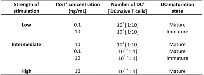
For analyzing if the generation of memory- and effector-like CD4 T cells depends on the strength of stimulation during the priming, an in vitro system was established utilizing the superantigen TSST. Varying the signal strength of the priming the phenotypical and functional properties of the subsets according to IL7R and CCR7 expression and in dependence on the strength of stimulation were determined. Moreover, in order to learn if cells that show the same phenotype after priming with different signal strengths have the same properties, memory characteristics as well as signaling and gene expression were analyzed.
In order to understand the situation under steady state conditons antigen-experienced CD4 T cells were isolated from peripheral blood of healthy donors and phenotype and functions of the different IL7R CCR7 subsets were analyzed. These ex vivo isolated cells reflect the physiological pattern of antigen-experienced CD4 T cells that have encountered the antigen under various conditions in vivo. To understand the priming history of the ex vivo T cell subsets their responses to vaccination and persistent and self-antigens were assessed.
3 MATERIALS AND METHODS
Materials and methods
3.1 Materials Table 3.1: Reagents
Brefeldin A Sigma‐Aldrich, Saint Louis, USA
Candida albicans extract Provided by Prof. Gerna, Servizio di Virologia, Hospital of Pavia
CD14 beads Miltenyi Biotec GmbH, Germany
CD4+ T Cell Isolation Kit II Miltenyi Biotec GmbH, Germany
Horseradish peroxidase (HRP)‐streptavidin conjugate
For IL‐10 and IFN‐γ: from Zymed Laboratory, San Francisco USA.
For IL‐17: from DuoSet ELISA kit R&D System, Germany
Human cytomegalovirus (HCMV) lysate from AD169 strain
Provided by Prof. Gerna, Servizio di Virologia, Hospital of Pavia
Ionomycin calcium salt from Streptomyces conglobatus
Sigma‐Aldrich, Saint Louis, USA
Pepmix MelanA /MART‐1 JPT Peptide Technologies GmbH, Berlin, D Phorbol 12, 13‐dibutyrate (PdBu) Sigma‐Aldrich, Saint Louis, USA
Propidium iodide (PI) Sigma Aldrich, Saint Louis, USA Purified protein derivative of
Mycobacterium tuberculosis
Provided by Dr. Jacobsen, MPI Immunology, Berlin
rhGM‐CSF Strathmenn Biotec, Germany
rhHemagglutinin‐Influenza A Virus H1N1 from Texas 36/91 strain (Flu)
Prospec‐Tany TechnoGene, Rehovot, Israel
rhIFN‐γ R&D System, Wiesbaden, Germany
rhIL‐10 R&D System, Wiesbaden, Germany
rhIL‐15 R&D system, Wiesbaden, Germany
rhIL‐17 DuoSet Elisa Kit , R&D System , Wiesbaden,
Germany
rhIL‐2 R&D System, Wiesbaden, Germany
rhIL‐4 R&D System, Wiesbaden, Germany
rhIL‐7 R&D system, Wiesbaden, Germany
rhMART‐1/MelanA protein Assay designs, (BioTrend), Köln, Germany Streptavidin APC crosslinked or Pacific Blue‐
conjugated
Molecular Probes (MoBiTec), Germany
Tetanus toxoid extract (TT) Provided by Chiron Siena (Italy) Toxic shock syndrome toxin‐1 (TSST) from
Staphylococcus aureus
Sigma‐Aldrich, Saint Louis, USA Ultrapure LPS from E. coli 0111:B4 strain Invivogen, San Diego, USA Vybrant™ CFDA SE Cell Tracer Kit Molecular Probes (Invitrogen)
Materials and methods
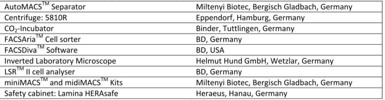
Table 3.2: Instruments used for cell handling and cell separation
AutoMACSTM Separator Miltenyi Biotec, Bergisch Gladbach, Germany
Centrifuge: 5810R Eppendorf, Hamburg, Germany
CO2‐Incubator Binder, Tuttlingen, Germany
FACSAriaTM Cell sorter BD, Germany
FACSDivaTM Software BD, USA
Inverted Laboratory Microscope Helmut Hund GmbH, Wetzlar, Germany
LSRTM II cell analyser BD, Germany
miniMACSTM and midiMACSTM Kits Miltenyi Biotec, Bergisch Gladbach, Germany Safety cabinet: Lamina HERAsafe Heraeus, Hanau, Germany
Table 3.3: Buffers, media and solutions used for cell culture and flow cytometry
Cell fixation solution PBS
2% or 1% (v/v) Formaldehyde (Merck, Germany) Cell fixation solution for Foxp3 staining 4X Fixation/Permeabilization Buffer
Fixation/Permeabilization Buffer Diluent (eBioscience, Natutec, Germany) Cell permeabilization solution PBS
0.5% BSA
0.5% Saponin from Quillaja bark (Sigma‐Aldrich) Cell permeabilization solution for Foxp3 staining 10X Permeabilization Buffer
deionized /distilled water Lymphocyte separation medium Ficoll‐Paque LSM 1077
(PAA Laboratories) PBS/HS (Human Serum) buffer 1% Human Serum
(AB Serum, Bio Whittaker, Lonza, USA)
PBS buffer (pH 7,2‐7,4) 2.7 mM KCl
1.5 mM KH2PO4 137 mM NaCl 8.1 mM Na2HPO4 PBS/BSA (Bovine Serum Albumin) buffer 5 g/L BSA
(PAA Laboratories, Pasching A) PBS/BSA/EDTA (ethylenediaminetetraacetic acid)
buffer or PBS/HS/EDTA buffer
2 mM EDTA in PBS/BSA or in PBS/HS (Merck, Germany)
RPMI medium Rosewell Park Memorial Institute medium 1640 + Glutamax‐ITM (Gibco BLR, USA)
5% (v/v) Human Serum
1% (v/v) 100mM Sodium Pyruvate (Gibco)
1% (v/v) 100X MEM non‐essential aminoacid solution (Gibco)
100 U/mL Penicillin (PAA Laboratories) 0.1 mg/mL Streptavidin (PAA Laboratories) 0.1% (v/v) 50mM 2‐Mercaptoethanol (Gibco)
RPMI medium/ HS 5% Human Serum
(AB Serum, Bio Whittaker, Lonza, USA) RPMI medium/FBS (Fetal Bovine Serum) 10% FBS (HyClone, France)