regulatory and conventional T cells
Dissertation zur Erlangung des Doktorgrades der
Naturwissenschaften (Dr. rer. nat.) der Fakultät für Biologie und vorklinische Medizin der Universität Regensburg
vorgelegt von Christian Schmidl
aus Tauberfeld
im Jahr 2012
Das Promotionsgesuch wurde eingereicht am: 9. Oktober 2012
Die Arbeit wurde angeleitet von: Prof. Dr. Michael Rehli
Unterschrift:
______________________________________
(Christian Schmidl)
1 SUMMARY 1
2 INTRODUCTION 3
2.1 EPIGENETICS 4
2.1.1 DNA METHYLATION 4
2.1.2 CHROMATIN 5
2.1.3 NON-‐CODING RNAS 7
2.1.4 CIS-‐REGULATORY MODULES 8
2.1.5 EPIGENETICS AT CIS-‐REGULATORY MODULES AND THE IMPACT ON GENE REGULATION 11
2.2 T HELPER CELLS 14
2.2.1 EPIGENETICS IN TH DEVELOPMENT 15
2.2.2 REGULATORY T CELLS 16
2.3 OBJECTIVES 24
3 CHAPTERS IDENTICAL TO MANUSCRIPTS 26
3.1 LINEAGE-‐SPECIFIC DNA METHYLATION IN T CELLS CORRELATES WITH HISTONE METHYLATION AND
ENHANCER ACTIVITY 27
3.2 ISOLATION OF INTACT GENOMIC DNA FROM FOXP3-‐SORTED HUMAN REGULATORY T CELLS FOR
EPIGENETIC ANALYSES 71
3.3 EPIGENETIC REPROGRAMMING OF THE RORC LOCUS DURING IN VITRO EXPANSION IS A DISTINCTIVE
FEATURE OF HUMAN MEMORY BUT NOT NAÏVE TREG CELLS 84
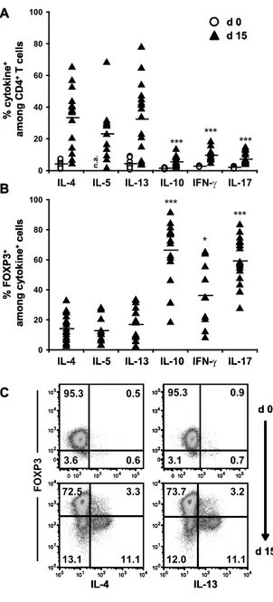
3.4 DOMINANT TH2 DIFFERENTIATION OF HUMAN REGULATORY T CELLS UPON LOSS OF FOXP3
EXPRESSION 106
3.5 THE ENHANCER AND PROMOTER LANDSCAPE OF HUMAN REGULATORY AND CONVENTIONAL T CELL
SUBPOPULATIONS 133
4 DISCUSSION 165
4.1 GENERAL INSIGHTS INTO CELL TYPE-‐SPECIFIC GENE REGULATION IN TREG AND TCONV 165 4.1.1 DISTRIBUTION OF DIFFERENTIAL DNA METHYLATION IN REGULATORY AND
CONVENTIONAL T CELLS 165
4.1.2 DMRS ARE ASSOCIATED WITH HISTONE MARKS, NOVEL PROMOTERS AND ENHANCER FUNCTION 166 4.1.3 ENHANCER PROFILING IDENTIFIES KEY REGULATORS IN T CELL SUBPOPULATIONS 170 4.2 PLASTICITY, STABILITY AND HETEROGENEITY OF HUMAN T CELL POPULATIONS 171
4.2.1 METHODOLOGY ADVANCEMENTS 171
4.2.2 DNA METHYLATION ANALYSIS AND GENE EXPRESSION PROFILING OF T CELL SUBPOPULATIONS 172 4.2.3 CAP ANALYSIS OF GENE EXPRESSION EXTENDS THE INFORMATION CONTENT OF GENE
EXPRESSION ANALYSIS 174
4.3 TREG IN THE CLINIC AND FUTURE PERSPECTIVES 175
5 REFERENCES 179
6 PUBLICATIONS 197
7 DANKSAGUNG 198
1 Summary
Complex multicellular organisms give rise to a wide range of cell types and tissues, even though all the cells share the same DNA sequence. Key to this diversity is differential gene expression in the different types of cells. Gene expression is orchestrated by regulatory DNA sequences, which can be bound by transcription factors mediating the activation or repression of a target gene.
These processes interplay with epigenetic mechanisms including DNA methylation and histone modifications that shape the chromatin structure and control its accessibility for transcription factors and other accessory proteins. Here, regulatory and conventional T cells (Treg and Tconv, respectively) were utilized as a model system to get basic insights in differential gene expression and how it is affected by epigenetic mechanisms. Treg can suppress the activation, proliferation and function of a wide range of immune cells and are thus indispensable for immune
homeostasis and tolerance to self-‐antigens. Tconv develop into different T helper (Th) cells that boost specialized immune reactions. Both Treg as well as Tconv are closely related CD4+ T cells and, due to their variable abilities a suitable model to study differential gene expression.
An adaption of our methyl-‐CpG-‐immunoprecipitation method allowed us to systematically investigate DNA methylation in T cells, which resulted in the identification of more than 130 differentially methylated regions (DMRs) between Treg and Tconv. The DMRs were located in the vicinity of immunologically important genes including FOXP3, CTLA4, IL2RA and CD40LG.
Most DMRs had a low CpG content, showed no conservation and did not overlap with a gene promoter. In addition, it was demonstrated that many DMRs were associated with “active”
histone modifications and showed enhancer activity in reporter assays. These results were among the first to describe widespread differences in DNA methylation at non-‐promoter regions and to connect them to enhancer function.
CD4+CD25+ Treg represent a heterogeneous population and consist of CD45RA+ naïve Treg as well as CD45RA-‐ memory Treg. Upon in vitro expansion CD45RA-‐ memory Treg downregulate the expression of the Treg lineage-‐determining transcription factor FOXP3. Hence, we improved technologies to obtain DNA and RNA from intracellular FOXP3-‐stained and sorted human Treg to analyze stability, plasticity and heterogeneity of Treg subpopulations. Gene expression analyses demonstrated that in vitro expanded CD45RA-‐FOXP3-‐ Treg differentiated into a proinflammatory Th2-‐like phenotype and expressed the Th2-‐associated transcription factor GATA3 as well as the cytokines IL-‐4, IL-‐5 and IL-‐13. Blockade of the Th2-‐inducing IL-‐4 signaling pathway did not abrogate the observed Th2 differentiation, arguing for a yet unknown,
alternative pathway. In addition, in vitro expanded CD45RA-‐ Treg expressed the Th17-‐
determining transcription factor RORC and IL-‐17A, with the most significant increase in FOXP3+
cells. In line with these observations, CpGs at the RORC locus were most prominently
demethylated in in vitro expanded CD45RA-‐FOXP3+ cells similar to the methylation status of in vitro generated Th17 cells. In contrast, CD45RA+ naïve Treg showed a stable phenotype without converting into proinflammatory Th2 or Th17-‐like cells even after prolonged in vitro expansion, and therefore represent the most promising population for clinical applications.
In the context of the FANTOM5 project, modern sequencing methods identified the exact location of transcription start sites (TSS) in primary and in vitro expanded naïve and memory Treg and Tconv. Several thousand non-‐annotated TSS were discovered, and some were validated as alternative promoters of known genes including the well-‐studied Treg-‐specific FOXP3 and CTLA4 genes. In addition, genome-‐wide histone modification profiling generated the most comprehensive atlas of cell type-‐specific enhancers in Treg and Tconv subpopulations. De novo motif analysis of enhancer elements identified transcription factors that were potentially involved in cell type-‐specific gene regulation. Continuative experiments could demonstrate a participation of the transcription factors STAT5 as well as FOXP3 and ETS1 as well as RUNX1 in Treg-‐ or Tconv-‐specific enhancer architecture, respectively.
Taken together, the molecular characterization of Treg and Tconv subpopulations described in this thesis provided insights into basic principles of gene regulation and demonstrates the impact of DNA methylation, histone modifications and transcription factor binding on cell type-‐
specific gene expression. Moreover, technical refinements of standard methodologies allowed the concrete analysis of the stability, heterogeneity as well as plasticity of T cell subsets. The integrated analysis of genome-‐wide datasets helped to define key regulators that shape gene expression programs of T cell subpopulations and will be of use to improve the therapeutic potential of Treg for clinical applications.
2 Introduction
One of the most fascinating aspects of complex multicellular development is the ability of a single genome to give rise to a wide panel of different cell types and tissues, all with unique phenotypes and abilities. How can these differences in development and function be achieved when all these cell types share, with minor exceptions, the same DNA sequence? The answer to this question is differential gene expression. In each distinct cell type only a fraction of all genes encoded in the DNA sequence –that is to say the genes needed for its phenotype and function–
are transcribed. The decision to what extent a gene is transcribed is controlled by so-‐called regulatory modules, which are DNA-‐elements that can integrate environmental and inherited cues to establish cell type-‐specific gene expression programs. The current understanding classifies regulatory modules into promoters, enhancers, silencers and boundary elements.
These DNA sequences can bind transcription factors (TFs) that activate or repress the binding and activity of the basal transcription machinery to influence transcription of a target gene and hence ultimately shape the cellular phenotype. These processes interplay with epigenetic mechanisms, namely DNA methylation, histone modifications and non-‐coding RNAs that shape the chromatin structure and control its accessibility for TFs and other accessory proteins.
The main focus of this thesis lies on regulatory and conventional T cells (Treg and Tconv). As explained below, the former are a specialized immune cell population that is crucial for immune tolerance and homeostasis. Further, the administration of Treg is explored as a curative
treatment for immunological and transplantation-‐related diseases. Treg and Tconv are both closely related hematopoietic cells emerging from the same progenitor. Nevertheless, both cell types have different development potential, phenotype and function ascribed to their specialized gene expression programs, which renders comparative analysis of Treg and Tconv cells a
suitable model to study genetic and epigenetic mechanisms of differential gene expression. With regards to their crucial role in maintaining a stable immune system and with respect to their clinical application, the analysis of gene regulation in Treg compared to Tconv will not only give insights into basic mechanisms of differential gene expression; it will also be essential to
understand Treg development and function and thereby help to improve their effective and save clinical application. Thus, in the first part of the introduction basic concepts of gene regulation are described while the specific characterization on gene regulation of regulatory t cells is introduced in the second part.
2.1 Epigenetics
2.1.1 DNA methylation
Proposed in 1975 by Holliday and Pugh, the longest known epigenetic modification is the attachment of a methyl group (CH3) to the 5’ carbon atom of the base cytosine (C) (Holliday and Pugh 1975). In mammals, 5’-‐methyl cytosine (5mC) is mainly associated with guanine (G) in CG dinucleotides (CpGs) although recent findings confirm early reports describing non-‐CpG methylation in embryonic stem cells (Salomon and Kaye 1970; Grafstrom et al. 1985;
Ramsahoye et al. 2000; Lister et al. 2009). DNA methylation is considered to mediate stable gene silencing at promoters and is essential for embryonic development (Li et al. 1992; Okano et al.
1999), genomic imprinting (Li et al. 1993), centromeric stability (Moarefi and Chédin 2011), splicing (Shukla et al. 2011), X chromosome inactivation in mammals (Lee 2011) and silencing of potential harmful DNA elements such as endogenous retroviruses and transposons (Bird 2002).
Aberrant DNA methylation has been associated with abnormal developmental processes including cancer (Plass and Soloway 2002). In mammals three known enzymes, DNA methyltransferase 1, 3A and 3B (DNMT1, 3A and 3B) catalyze the transfer of CH3 from S-‐
adenosylmethionine (SAM) to cytosine (Wigler et al. 1981; Okano et al. 1999). DNMT1 is the
“maintenance” methyltransferase that adds methyl groups to the newly synthesized and therefore hemimethylated DNA-‐strand after replication, providing the basis for inheriting methylation patterns over cell divisions and therefore rendering DNA methylation the only
“real” epigenetic mark (Wigler et al. 1981). Dnmt3A and Dnmt3B catalyze de novo methylation but might also be involved in maintaining methylation patterns (Okano et al. 1999; Jones and Liang 2009). DNA methylation is essential for normal development, as murine knockout mice for all three DNMTs die in utero or shortly after birth, and mutations in DNMT3B are associated with the ICF syndrome (immunodeficiency, centromeric instability and facial anomalies) in humans (Xu et al. 1999). CpG dinucleotides show a bimodal distribution throughout the genome: Most CpGs in mammals are methylated, distributed randomly and appear rarer than statistically expected, possibly caused by hydrolytic deamination of 5mC to thymine, resulting in a C to T transition and a decrease of CpGs over time in evolution (Jones 2012). However, there are also regions with higher CpG density, so called CpG islands (CGIs) that are often associated with promoter regions and are preferentially unmethylated (Suzuki and Bird 2008). Basically, DNA methylation can influence gene expression by (i) steric hindrance of protein binding to DNA due to the exposure of the methyl group into the DNA-‐helix grooves (Tate 1993) and (ii) by
attracting gene-‐regulatory proteins recognizing 5mC (methyl-‐CpG binding proteins, MBPs) (Robertson 2000). The proteins MBD1, 2 and 4 as well as MeCP2 can bind methylated DNA with their methyl-‐CpG binding domain (MBD) while the protein Kaiso does so with its zinc-‐finger
domain (Prokhortchouk et al. 2001; Klose and Bird 2006). The MBPs come in complexes with repressor molecules that alter gene expression by the modification of the chromatin
conformation, as explained later (Jones et al. 1998; Nan et al. 1998; Ng et al. 1999; Zhang et al.
1999). Subject of controversy is the mechanism of active DNA demethylation (Ooi and Bestor 2008). Passive demethylation after DNA replication can be logically explained by TFs occupying DNA and thereupon blocking DNMT-‐mediated remethylation of the hemimethylated DNA strand. However, DNA demethylation was observed in differentiation models in the absence of cell division and thereby DNA replication (Klug et al. 2010), arguing for active demethylation processes. The role of activation-‐induced cytidine deaminases (AID), thymine DNA glycosidases (TDG), alpha growth arrest and DNA-‐damage-‐inducible (GADD45a) and ten-‐eleven translocation (TET) dioxygenases in active demethylation processes are currently under investigation (Ooi and Bestor 2008; Jones 2012). TET proteins can process 5mC to 5-‐formylcytosine and 5-‐
carboxylcytosine that are readily excised by TDG as a possible mechanism of active
demethylation (Ito et al. 2010; He et al. 2011; Ito et al. 2011). However, the mechanisms of active demethylation need further investigations, preferentially in non-‐artificial systems to exclude aberrant methylation phenomena described for cell lines and in vitro differentiation systems (Paz et al. 2003; Meissner et al. 2008).
2.1.2 Chromatin
DNA is packed into chromatin, which consists of DNA, histone proteins and non-‐histone proteins (Bell et al. 2011). The basic subunit of chromatin is the nucleosome core particle, comprised of
~145 base pairs (bp) of DNA wrapped around an octamer consisting of two copies each of histones H2A, H2B, H3 and H4 in a 1.65 left-‐handed, superhelical turn (Kornberg and Thomas 1974; Kornberg 1977; Luger et al. 1997). The nucleosomes are arranged like “beads on a string”, and metazoan chromatin contains the linker histone H1 that helps to condense the “string” into a tighter packed, higher order structure whose organization is still incompletely understood (Felsenfeld and Groudine 2003). The packing of DNA into chromatin is repressive to
transcription per se as it potentially blocks the accessibility of DNA elements for transcription factors and the transcription machinery (Lorch et al. 1987). Therefore, the chromatin
accessibility of regulatory elements such as promoters and enhancers is actively formed.
Classically, regions of compacted chromatin are termed heterochromatin, whereas accessible chromatin is called euchromatin (Bell et al. 2011). As a part of chromatin modifying processes, ATP-‐dependent remodeling complexes are capable of positioning or removing nucleosomes on the DNA (Clapier and Cairns 2009) to expose regulatory sequences to their target proteins. In addition, post-‐translational modifications (PTMs) of histones regulate chromatin accessibility:
Amino acids on the N-‐terminal histone tails can be acetylated, phosphorylated, β-‐N-‐
acetylglucosaminated, ADP-‐ribosylated, deaminated, ubiquitinated and sumoylated (Bannister and Kouzarides 2011).
Methylation and acetylation are the best-‐studied histone PTMs. Histone acetylation is mediated by the opposing action of histone acetyl transferases (HAT) and histone deacetylases (HDAC).
Acetylation of histones is supposed to decrease the interaction of positively charged lysine residues of histone tails with the negatively charged DNA sugar-‐phosphate backbone to promote an accessible chromatin conformation (Sterner and Berger 2000). More important, gene-‐
regulatory proteins with a bromodomain can recognize and bind acetylated histones. To name just a few, remodeling complexes such as SWI/SNF (Hassan et al. 2002), coactivators (Dhalluin et al. 1999), as well as the general TF TFIID (Jacobson et al. 2000) have a bromodomain and can be recruited by acetylated histones to promote transcription. Histone methylation is mainly observed at arginine and lysine residues of histone tails and controlled by histone methyl transferases (HMT) or recently discovered histone demethylases (Shi et al. 2004). As an example, Histone 3 Lysine 4 methylation (H3K4me) is associated with “active” chromatin in eukaryotes (Bernstein et al. 2005; Barski et al. 2007). The modification is established by SET domain containing HMTs that are recruited to the target histones by other histone modifications such as ubiquitinated H2B, the active form of RNA Polymerase II (PolII) or specific TFs
(Shilatifard 2008). The established H3K4me can be “read” by other factors with a
chromodomain such as some chromatin remodeling complexes (Santos-‐Rosa et al. 2003;
Wysocka et al. 2006), HATs (Vermeulen et al. 2010) and TFIID (Vermeulen et al. 2007) to promote transcription. Interestingly, the latter binding is synergistically enhanced by H3K14 acetylation. In contrast, H3K9 di-‐ and trimethylation is catalyzed by the HMT Suv39H1 and is recognized by heterochromatin protein 1 (HP1) that helps to stably compact chromatin (Bannister et al. 2001; Peters et al. 2001; Beisel and Paro 2011). Suv39H1 interacts with HP1, providing a possible “feed forward” mechanism of H3K9 methylation and HP1 binding to sustain chromatin compaction once it was initiated (Schotta et al. 2002). Classes of histone modifying enzymes that are supposed to set and interpret histone modifications to maintain a certain chromatin state as described for HP1-‐Suv39H1 are the trithorax group (TrxG) and polycomb group (PcG) proteins (Ringrose 2007). TrxG include HMTs to set H3K4 methylation as already described and stabilize chromatin states favoring transcription. Contrary, PcG proteins come in large complexes and establish and maintain a chromatin environment repressive for
transcription. The polycomb repression complex 2 (PRC2) methylates H3K27 and creates a platform for polycomb repressive complex 1 (PRC1) that establishes a compacted chromatin environment repressing transcription (Ringrose 2007).
Interestingly, PcG-‐mediated silencing is interconnected to DNA methylation. PRC2 directly controls DNA methylation by interacting with DNMTs (Viré et al. 2006). Further, promoters with
H3K27me are more frequently de novo methylated than other promoters and undergo aberrant DNA methylation in human cancers, suggesting that the PcG-‐repressed state is established during development and may predispose genes to de novo methylation in early developmental processes (Schlesinger et al. 2007; Mohn et al. 2008). Moreover, the interplay of DNA
methylation and chromatin structure is illustrated by the associations of the aforementioned MBPs with chromatin-‐modifying enzymes. MeCP2 for example is associated with the
Sin3A/HDAC corepressor complex (Jones et al. 1998; Nan et al. 1998). In addition, the MeCP1 complex is associated with HDACs and can bind methylated DNA via MBD2 (Ng et al. 1999).
Moreover, MBD1 can also bind methylated DNA and act as a repressor (Fujita et al. 2000). In contrast to these processes that prohibit chromatin access for transcription, the recently identified protein Cfp1 is recruited to unmethylated CpG islands and interacts with a H3K4 methyltransferase to create a chromatin environment that favors transcription (Lee et al. 2007;
Thomson et al. 2010).
2.1.3 Non-‐coding RNAs
Due to their active participation in shaping the chromatin environment, short (<200 nucleotides) and long (>200 nucleotides) non-‐coding RNAs are classified as “epigenetic”
regulators as well. First described in 1961 (Lyon 1961), the phenomenon of X chromosome inactivation in mammals (XCI) is a prime example of RNA-‐mediated regulation of gene expression. In females, one of the two X chromosomes is inactivated during embryogenesis, a process controlled by antagonistic roles of two non-‐coding RNAs, Xist and Tsix (Lee 2011):
Sustained expression of Tsix prevents expression of Xist and XCI, but when XCI is initiated Tsix expression is lost at one X chromosome. This allows transcription of the lncRNA Xist, and Polycomb repressive complex 2 is recruited to a PRC2-‐binding motif in the lncRNA and
effectively tethered to the locus via PolII. The RNA–PRC2 complex is loaded onto chromatin co-‐
transcriptionally through TFs such as YY1, promoting H3K27me3 and heterochromatin formation in cis (Lee 2011). In fission yeast, transcription of repeat regions within heterochromatin domains triggers the RNA interference machinery, generating small 21 nucleotide long RNAs (siRNAs). The siRNAs associate with Argonaute protein (Ago1) and guide the Ago1-‐containing RNA-‐induced initiation of the transcriptional gene-‐silencing complex (RITS complex) to homologous sequences of nascent chromatin-‐associated transcripts for
heterochromatin formation (Bühler et al. 2006). Recently it was demonstrated that small RNA species (piRNAs) act in trans to silence transposable elements in mammals by mediating indirect heterochromatin formation and DNA methylation at target loci (Aravin et al. 2008). These examples illustrate the connection between histone modifications, non-‐coding RNAs, DNA methylation and chromatin accessibility to prepare and sustain the genetic environment for
gene activation or repression. These findings are summarized in Figure 1. Still, some basic concepts of epigenetics are incompletely understood. It is not clear, if and how chromatin modifications can be passed on over cell divisions, as there is no such simple mechanism as a
“maintenance” enzyme as in DNA methylation. Moreover, there is no clear agreement if the establishment of DNA methylation patterns is a cause or a consequence of gene silencing or activation as mechanistic studies are scarce and need further investigations. The idea of
heritable changes in gene expression without changes in the DNA sequence was widely hoped to explain gene expression patterns in developmental processes and diseases. The efforts that were made to understand epigenetic mechanisms are illustrated by the roughly 25000 PubMed citations for the term “epigenetic” (until August 2012).
2.1.4 Cis-‐regulatory modules
2.1.4.1 Transcription factors
Sequence-‐specific transcription factors comprise at least a DNA binding domain for recognizing and binding specific sites in the genome and a transactivation domain to recruit coactivators and other accessory proteins such as DNA and histone modifying proteins that ultimately help to facilitate transcription (MacQuarrie et al. 2011). Transcription factors are activated through signaling events triggered by environmental cues and can establish logic networks to drive Figure 1
Epigenetic mechanisms and gene regulation. General properties of repressive and active chromatin environments; DNA (black lines) is wrapped around nucleosomes (green cylinders);
red circles: methylated CpG dinucleotide; small red and yellow hexagons: histone methylation at H3K9, H3K27 or H3K4; blue star: histone acetylation; other objects: transcription factors and histone-‐ as well as DNA-‐modifying enzymes as described in the introduction. (Adapted from Laird 2005)
complex programs of gene expression as seminal work of Nüsslein-‐Volhard and colleagues demonstrated in drosophila (St Johnston and Nusslein-‐Volhard 1992). In humans, a manually curated list of 1391 DNA-‐binding TFs was recently published showing that many TFs were expressed in a tissue-‐specific manner but remain largely uncharacterized regarding their function and mechanism of action (Vaquerizas et al. 2009).
2.1.4.2 Promoters
Promoters of genes are genomic loci that overlap with the transcription start site (TSS) from which messenger RNA (mRNA) transcription is initiated at a rate determined by the complete integrated regulatory input for this gene (Lenhard et al. 2012). PolII catalyzes transcription of protein-‐coding genes and some small RNA species in eukaryotes. Therefore, components of the basal transcription machinery are recruited to the “core promoter”, the region in close vicinity to the TSS, with the help of general and cell type-‐specific TFs recognizing DNA sequence motifs (transcription factor binding sites TFBS) at the core promoter or distal cis-‐regulatory regions such as enhancers (Maston et al. 2006). Due to their difference in dynamic expression range -‐
from constant expression (“house keeping genes”) to cell type and developmental state-‐specific expression-‐ attempts were made to classify promoters based on their expression dynamics and nucleotide composition. Recent advances in TSS detection and gene expression analysis such as RNA-‐seq (Ozsolak and Milos 2011) and cap analysis of gene expression (CAGE, (Kanamori-‐
Katayama et al. 2011)) allow fine mapping of TSS and gene expression analysis throughout the genome. Integrated analysis suggests three main classes of promoters: “adult” (type I),
“ubiquitous” (type II) and “developmentally regulated” (type III) (Lenhard et al. 2012). Type I promoters show tissue-‐specific expression in differentiated cell types from a focused TSS, have mostly a low CG and CpG content and are enriched for a TATA-‐box, a sequence motif recognized by the TATA-‐box binding protein which is a component of the basal transcription machinery.
Type II promoters are ubiquitously expressed (“house-‐keeping”) from broadly dispersed TSS, are TATA-‐box depleted and overlap with CpG islands at their TSS (Deaton and Bird 2011;
Lenhard et al. 2012). Type III promoters share molecular characteristics with type II promoters but are developmentally regulated (Lenhard et al. 2012). In contrast to prokaryotic organisms, in eukaryotes the promoter alone is not sufficient to regulate gene and often produces only low levels of mRNA on its own (Wittkopp and Kalay 2012). On that account, enhancers, insulators and boundary elements control the “fine tuning” of gene expression in complex organisms.
2.1.4.3 Enhancers and silencers
Enhancers were described as non-‐coding regulatory DNA sequences that can enhance the expression of a target gene in a distance-‐ and orientation-‐independent manner (Banerji et al.
1981). Distal non-‐coding sequences are often necessary for the activation and/or correct lineage-‐specific expression of a gene as promoters alone often fail to establish accurate
expression patterns. For example, studies in transgenic mice showed that the transfer of small fragments surrounding the human CD14 gene locus (24-‐33kb) only establish correct CD14 expression in liver whereas a much larger region of 80 kb is needed to express CD14 in a
monocyte-‐specific fashion (Pan et al. 2000). Another well-‐studied example is the locus encoding the T helper cell type 1 (Th1)-‐specific cytokine interferon gamma (Ifng). An 8.6 kb transgene of the human IFNG locus was sufficient for constitutive IFN-‐γ production, but only a 191 kb
transgene established restricted IFNG expression in Th1 cells (Soutto et al. 2002). Enhancers are thought to bind combinations of transcription factors that create physical interactions via the mediator complex and cohesin with the target gene promoter and help to recruit the general transcription machinery (Kornberg 2005; Kagey et al. 2010). The enhancer and target promoter can be distant from each other (up to a million base pairs away) or even on another
chromosome (Spilianakis and Flavell 2004; Lomvardas et al. 2006; Amano et al. 2009). These observations were made possible by labeling distant gene loci with fluorescent probes (fluorescence in situ hybridization, FISH (Ong and Corces 2011)) or by the chromosome
conformation capture technique introduced by Dekker and colleagues 2002 (Dekker et al. 2002), a technique that uses formaldehyde crosslinking to capture physical interactions between chromosome arms. Silencers function by recruiting TFs repressing transcription, block DNA binding of activators or hinder the assembly of the transcription machinery (Maston et al. 2006), but are less well characterized than enhancers.
2.1.4.4 Boundary elements
Boundary elements were also described to potentially act as repressive elements by blocking the interaction of a distal enhancer with its target promoter as intensively studied at the IGF2/H19 locus where the presence of the CCCTC binding protein (CTCF) blocks the interaction of an enhancer with the IGF2 gene on the maternal allele (Bell and Felsenfeld 2000). CTCF, so far the only identified “boundary” element in humans, was also described to isolate “active” and
“repressive” chromatin environments and is involved in many developmental processes such as stem cell differentiation, neural development, cytokine expression and immunoglobulin chain recombination by mediating long-‐range interactions of chromatin elements (Herold et al. 2012).
A positive function in gene regulation by the boundary element CTCF is also supported by a recent study highlighting the role of CTCF in mediating enhancer-‐promoter interactions and chromatin organization (Handoko et al. 2011). An overview of cis-‐regulatory modules is shown in Figure 2.
2.1.5 Epigenetics at cis-‐regulatory modules and the impact on gene regulation During the making of this thesis, progress in high throughput and next generation sequencing technologies now permits the examination of global epigenetic and functional properties of cis-‐
regulatory modules.
In terms of DNA methylation analysis, previous studies concentrated on CGIs in cancer as aberrant DNA methylation is often observed upon malignant transformation (Plass and Soloway 2002). CpG islands at promoters are normally unmethylated independent of their expression status (Weber et al. 2007; Mohn et al. 2008). However, some CGIs become de novo methylated in a cell type-‐specific manner, resulting in long-‐term repression of the associated gene (Weber et al. 2007; Farthing et al. 2008; Meissner et al. 2008; Mohn et al. 2008). Long-‐term repression of CGI-‐associated genes is described for imprinted genes (genes that show parent-‐of-‐origin expression), for CGI-‐associated genes of the inactivated X-‐chromosome and for some tissue-‐
specific genes (Jones 2012). Gene repression by CGI methylation is still rare and may not be the prevalent mechanism of gene silencing (Mohn et al. 2008; Jones 2012). Moreover, for instance, at the inactive X chromosome, DNA methylation comes late during the inactivation and silencing process (Lee 2011). Yet, it seems to provide an additional “layer” of gene repression to ensure long-‐term silencing. Interestingly, regions of intermediate CpG content are more commonly de novo methylated and repressed, whereas low CpG promoters tend to be methylated regardless of their expression state (Weber et al. 2007; Ball et al. 2009). In contrast, DNA methylation of gene bodies was positively correlated to gene expression (Ball et al. 2009; Lister et al. 2009).
However, far less is known about DNA methylation at non-‐promoter regions. Regions of intermediate or low CpG content came into focus with the development of sensitive locus-‐wide or genome-‐wide DNA methylation analysis (Schilling and Rehli 2007; Meissner et al. 2008; Klug et al. 2010; Stadler et al. 2011). Interestingly, DNA methylation is more dynamic at CpG poor Figure 2
Cis-‐regulatory modules in the genome. (Adapted from Heintzman and Ren 2009)
regions (Meissner et al. 2008; Stadler et al. 2011), and differential DNA methylation was observed at cell type-‐specific enhancers that were bound by lineage specific TFs (Sérandour et al. 2011; Stadler et al. 2011; Wiench et al. 2011). Indeed, on a genome-‐wide scale TF-‐bound regions are associated with local hypomethylation (Lister et al. 2009). Cell type-‐specific DNA methylation patterns seem to be established by both cis and trans acting factors: At CGIs for example, combinatorial binding of TFs protected them from aberrant de novo methylation (Gebhard et al. 2010). In a different experimental setting, core promoters introduced into a new locus in the mouse genome were able to recapitulate autonomously their original DNA
methylation state (Lienert et al. 2011). Mutation of TF binding sequences in the respective promoters inhibited this process, which suggests DNA methylation control in cis. In mice, several differentially methylated regions were identified that were controlled in cis by the underlying DNA sequence, but also trans-‐acting elements orchestrated DNA methylation patterns in different DMRs (Schilling et al. 2009).
Considering the association of gene-‐regulatory elements with the disposal of certain histone modifications, chromatin accessibility and nucleosome remodeling, genome-‐wide approaches were used to systematically isolate regulatory elements based on their biochemical markers.
Chromatin immunopreciptiation, deoxyribonuclease/micrococcal nuclease digestion and comparable techniques coupled to next generation sequencing (ChIP-‐seq, DNase-‐seq, MNase-‐
seq) allow the genome-‐wide mapping of TF, histone modifications and “open” chromatin regions sensitive to DNase digestion (Bell et al. 2011; Zhou et al. 2011). Among other modifications, promoters of active genes in metazoans are associated with H3K4me3 and H3K27ac, with intermediate levels of H3K4me2 and low levels/absence of H3K4me1 (Barski et al. 2007;
Guenther et al. 2007; Heintzman et al. 2007; Mikkelsen et al. 2007; Wang et al. 2008b; Bell et al.
2011). Inactive type I promoters (without a CpG island spanning the TSS) lack these active histone modifications whereas type II and type III CpG Island promoters always show detectable H3K4 trimethylation (Barski et al. 2007; Guenther et al. 2007; Wang et al. 2008b; Bell et al. 2011;
Deaton and Bird 2011). Interestingly, genes important for development share the active H3K4me3 and the repressive H3K27me3 polycomb modification, probably “poising” genes for their fast activation or silencing, dependent on the fate of the cell (Bernstein et al. 2006).
Moreover, active promoters are DNase hypersensitive due to a nucleosome-‐free region (NFR) directly upstream of the TSS, show binding of the active form of PolII and are frequently associated with histone variants H3.3 and H2A.Z (Jin et al. 2009; Bell et al. 2011).
Most of these findings can be transferred to enhancer regions (Ong and Corces 2011).
Compelling evidence from genome wide studies identified the enrichment of H3K4me1/me2 and additionally H3K27ac at “poised” and “active” enhancers, respectively (Heintzman et al. 2007;
Heintzman et al. 2009; Creyghton et al. 2010; Rada-‐Iglesias et al. 2011). Poised enhancers were
shown to acquire an active state during development when the linked gene was needed to be expressed (Rada-‐Iglesias et al. 2011). Enhancers are further characterized by DNase
hypersensitivity, NFR, binding of a coactivator such as p300 (a HAT) (Visel et al. 2009) (Blow et al. 2010), and H3.3 deposition. In contrast to promoters, enhancers were first described to show low levels of H3K4me3 and no transcriptional activity (Heintzman et al. 2007). However, some enhancers produce transcripts (enhancer RNAs or eRNAs) and were bound by PolII (Kim et al.
2010; Melgar et al. 2011). Another report attributed H3K4me3 at some enhancers as well, making it difficult to definitely separate enhancers and promoters (Pekowska et al. 2011). Still, enhancer and promoter prediction by chromatin patterns and TF occupancy is more effective than approaches that rely on conservation or accumulation of sequence motifs for TFs (Hardison and Taylor 2012a). Interestingly, when comparing the diversity of promoter and enhancer signatures between cell types, enhancers show a more cell type-‐specific distribution and variety than promoters, highlighting their role in tissue-‐specific gene expression (Heintzman et al. 2009;
Ernst et al. 2011). Global histone profiling further classified DNA elements associated with different function, e.g.. H3K36me3-‐ and H4K20me1-‐marked regions are linked with transcriptional elongation and H3K27me3 is preferentially associated with PCG-‐repressed regions (Barski et al. 2007; Mikkelsen et al. 2007).
The question arises how cell type specificity of regulatory elements is created and interpreted by transcription factors. Namely, the sole expression of a TF does not result in its binding to its recognition sequence in the genome: As an example, there are ~ 2 million binding sites of the TF PU.1 located in the human genome, but only ~ 80.000 of these sites are effectively bound in PU.1-‐expressing macrophages or monocytes (Pham et al. 2012). In contrast to shared binding sites, cell type-‐specific PU.1 binding in each cell type was associated with the co-‐binding of lineage-‐specific TFs, suggesting the combinatorial action of general and specific transcription factors to establish cell type-‐specific enhancers (Heinz et al. 2010; Pham et al. 2012). Moreover, these regions were marked by nucleosome repositioning and accumulation of H3K4me1 to
“prepare” chromatin for signal-‐dependent gene activation (Ghisletti et al. 2010; Heinz et al.
2010). In MCF7 and LNCaP cells FoxA1 is recruited to different sites distinguished by specific H3K4 dimethylation (Lupien et al. 2008). At these specific enhancers, FoxA1 remodels
chromatin to mediate MCF7 or LNCaP specific gene expression programs in collaboration either with estrogen receptor alpha or androgen receptor TFs. These observations lead to a model of
“pioneer” TFs that can easily access and prepare chromatin for the binding of other transcription factors that act in combination to drive cell type-‐specific expression programs (Lupien et al.
2008; Heinz et al. 2010; Zaret and Carroll 2011). Constitutive binding sites, on the other hand, do not seem to rely on co-‐binding with other TFs, partially explained by a stronger TF consensus site as demonstrated for FoxA2 binding in liver (Tuteja et al. 2008). However, potential co-‐
binding and consensus site quality do not explain all of the observed binding behavior of TFs suggesting additional determinants. Nevertheless, it was demonstrated for several cell types that enhancers are defined by combinations of key regulators (Lupien et al. 2008; Heinz et al. 2010;
Lin et al. 2010b; Mikkelsen et al. 2010). This allows the computational analysis of regulatory elements to isolate overrepresented binding sites and hence the identification of key TFs by the sole knowledge of histone modifications in a certain cell type (Pham et al. 2012). Currently, many laboratories and big international consortia such as the ENCODE (ENCODE-‐consortium 2011) gather epigenomes of many different cells with the hope to understand gene regulation in development, disease and cellular states.
2.2 T helper cells
The mammalian immune system comprises several specialized cell types to protect the host from exogenous pathogens such as fungi, viruses, bacteria and parasites (Delves and Roitt 2000). Cells from the innate immune system are regarded as a “first line of defense” against pathogens as they can recognize conserved and widely distributed features of pathogens with special receptors (pattern recognition receptors) to mount initial immune responses (Janeway and Medzhitov 2002; Underhill and Ozinsky 2002). Besides killing microbes and cytokine production to boost inflammation, innate immune responses include the incorporation and digestion of pathogens by professional phagocytes such as monocytes, macrophages as well as dendritic cells. The phagocytes then present parts of the digested microbes to cells of the adaptive immune system that can recognize the presented molecules (“antigens”) with their diverse T and B cell receptors (Delves and Roitt 2000; Guermonprez et al. 2002; Jutras and Desjardins 2005). Somatic recombination and random events create a theoretical diversity of up to 1018 different antigen receptors that enable cells of the adaptive immune system to recognize virtually every antigen presented (Davis and Bjorkman 1988). If a cell recognizes a presented antigen with its matching receptor, it proliferates to increase cell numbers with the same receptor (“clonal expansion”) to effectively detect and fight the corresponding pathogen (Delves and Roitt 2000). The adaptive immune system comprises B and T lymphocytes that develop in the bone marrow or in the thymus, respectively (Delves and Roitt 2000). T lymphocytes
expressing the CD4 coreceptor emerge as naïve CD4 cells and give rise to different T helper (Th) cell subsets in dependence of signals from the innate immune system and other environmental cues. Th1 cells produce the cytokine interferon gamma (Ifn-‐γ) and mediate host defense against intracellular pathogens while Th2 cells produce Interleukin (Il)-‐4, Il-‐5 and Il-‐13 and effectively resolve helminthic infections (Mosmann et al. 1986; Heinzel et al. 1989; Romagnani 1994).
Recently, Th cells producing Il-‐17A (Th17 cells) were described to contribute to defense against extracellular pathogens and fungi (Infante-‐Duarte et al. 2000; Ye et al. 2001; Ouyang et al. 2008).
With Th subsets arising from the same progenitor cell, they are ideal to study TF networks and epigenetic mechanisms that govern and stabilize their differential gene expression programs.
Th1 development is favored by the signal transducer and activator of transcription (Stat)1 and Stat4 that are activated by innate immune cell-‐derived Ifn-‐γ and Il-‐27 or Il-‐12, respectively (Schoenborn and Wilson 2007). Stat1 activation induces Tbx21 (also called T-‐bet), a key Th1 TF that induces among others Runx3. In cooperation with Tbx21 and Stat4, Runx3 binds to the Ifng promoter to sustain its expression in a positive feedback loop while binding to a silencing element in the Il-‐4 gene to suppress its transcription and hence abrogate Th2 differentiation (Djuretic et al. 2007a). In addition, Tbx21 interferes with the Th2 transcription factor Gata3 to prevent it from binding to target genes (Hwang et al. 2005). Gata3 is sufficient and necessary for Th2 development (Zheng and Flavell 1997). Gata3 is expressed upon Il-‐4 induced Stat6
activation and T cell receptor (TCR) signaling-‐derived TFs (Ansel et al. 2006) or by Notch signaling (Amsen et al. 2007). Gata3 induces Maf, and in cooperation with Stat6 these three TFs upregulate transcription of the Th2 cytokines Il-‐4, Il-‐5 and Il-‐13, again creating a positive feedback loop to stabilize Th2 differentiation (Ansel et al. 2006). Gata3 also hinders Th1 differentiation by preventing Runx3 to activate Th1-‐essential genes (Yagi et al. 2010). In mice, Th17 development is initiated by transforming growth factor beta (Tgf-‐ß) that induces the Th17 determining TF retinoic acid receptor related orphan receptor-‐gamma t (Rorc or Rorγt) or the regulatory T cell (Treg) determining TF Foxp3 (Chen et al. 2003; Ivanov et al. 2006; Manel et al.
2008). In combination with Il-‐6, Stat3 abrogates Treg development and supports Th17 differentiation and production of Il-‐21 (Zhou et al. 2007; Zhou et al. 2008a). Il-‐21 and Stat3 activation stabilize the Th17 phenotype via a positive feedback loop and also upregulate the Il-‐
23 receptor to support Stat3 activation via antigen presenting cell (APC)-‐ derived Il-‐23 (Zhou et al. 2007). Tgf-‐ß-‐independent Th17 generation was also reported recently (Ghoreschi et al.
2010). In humans, requirements for Th17 cell development are still under discussion
(Annunziato et al. 2007; Evans et al. 2007; Manel et al. 2008; Volpe et al. 2008; Annunziato and Romagnani 2011).
2.2.1 Epigenetics in Th development
As illustrated in the previous paragraph, TF networks are (i) able to sustain phenotypes in feedback loops and (ii) can prohibit differentiation to other phenotypes by direct interference with other TFs or by binding to regulatory regions such as the Il-‐4 silencer. However, many studies suggested that DNA methylation, chromatin remodeling complexes and chromatin modifications influence Th development and function: at the Th1-‐signature gene Ifng, many cis-‐
regulatory elements were described that showed Th1-‐specific demethylation, TF binding and
“active” chromatin modifications (Hatton et al. 2006; Jones and Chen 2006; Schoenborn et al.
2007a). Some putative enhancers interacted with the Ifng promoter in a cell type-‐specific manner (Hadjur et al. 2009). The establishment of cell type-‐specific epigenetic patterns is mediated by lineage-‐specific TFs. As an example, Stat4 was reported to recruit the remodeling complexes Swi-‐SNF to the Ifng promoter, which is essential for nucleosome remodeling and Infg expression (Zhang and Boothby 2006). Similarly, Tbx21 was described to be associated with a H3K27 demethylase to remove this repressive chromatin mark at its target genes to promote Th1 development (Miller et al. 2008). With respect to Th2 development, regulatory elements at the Il-‐4 locus acquire active histone marks and become demethylated in Th2 cells but not in Th1 cells (Avni et al. 2002; Lee et al. 2002). Gata3 is in parts responsible for chromatin remodeling and DNA demethylation at the Th2 cytokine genes (Lee et al. 2000; Yamashita et al. 2004) to create an open chromatin environment and was described to counteract DNA methylation-‐
mediated gene silencing by interference with Mbd2 and Dnmt1 binding (Hutchins et al. 2002;
Makar et al. 2003; Makar and Wilson 2004). In line with these observations, ablation of Mbd2, Dnmt1 or general inhibition of DNA methylation with 5-‐azacytidine lead to de-‐repression of cytokine genes normally silenced in Th1 or Th2 cells (Ballas 1984; Hutchins et al. 2002; Makar et al. 2003). In addition, acquired Th2-‐state seems to be maintained by Mll, a TrxG protein that stabilizes open chromatin conformation at the Th2 cytokine locus to sustain the expression of Th2 related genes (Onodera et al. 2010). These examples illustrate the participation of
epigenetic mechanisms in T helper cell specification.
2.2.2 Regulatory T cells
When T cells generate T cell receptors to recognize antigen they often produce by chance TCRs that are reactive to self-‐antigens. This would cause immune responses against the own body and is therefore restricted by anergy or deletion of self-‐reactive cells (negative selection) during T cell development in the thymus (Delves and Roitt 2000). However, some self-‐reactive T cells escape negative selection and have to be controlled in the periphery, a task that is in part accomplished by another Th subset, so-‐called regulatory T cells (Sakaguchi et al. 2006).
2.2.2.1 Phenotypic characterization
The notion that thymus-‐derived T cells contain a population responsible for peripheral tolerance emerged from experiments where neonatal thymectomy in mice at day 2-‐4 after birth resulted in autoimmune diseases that were prevented by inoculation of the mice with thymocytes or spleen cells from non-‐thymectomized mice (Nishizuka and Sakakura 1969; Sakaguchi et al.
1982). Further work identified CD25 (IL-‐2 receptor alpha chain) as a surface marker for these so-‐called “regulatory T cells” (Treg) (Sakaguchi et al. 1995), although CD25 was also expressed