Functional characterization of HMGN5 in chromatin architecture and gene expression
DISSERTATION ZUR ERLANGUNG DES DOKTORGRADES DER NATURWISSENSCHAFTEN (DR. RER. NAT.)
DER FAKULTÄT FÜR BIOLOGIE UND VORKLINISCHE MEDIZIN DER UNIVERSITÄT REGENSBURG
vorgelegt von
Ingrid Carolina Araya Fuenzalida
aus Santiago de Chile Im Jahr 2018
Das Promotionsgesuch wurde eingereicht am:
02.03.2018
Die Arbeit wurde angeleitet von:
Prof. Dr. Gernot Längst Regensburg, 02.03.2018
_________________________________
Ingrid Carolina Araya Fuenzalida
To Oskar Nahuel, without whom I would have finished this thesis one year before, and to Roman, without whom I would have probably never finished.
“La rebeldía tiene la potencialidad de crear, de imaginar y de proyectar, porque es una energía infinita y transformadora que piensa antes que en un cuarto propio en un cuerpo propio. La rebeldía es el comienzo de la libertad”
(Margarita Pisano Fisher)
Table of contents
List of figures ... 9
List of tables ... 12
Abbreviations ... 13
1 Summary ... 20
2 Introduction ... 22
2.1 Chromatin as functional organizer of DNA in the nucleus ... 22
2.2 DNA packaging and higher-‐order chromatin organization ... 23
2.3 Higher-‐order chromatin organization ... 25
2.3.1 Classical view of chromatin higher-‐order organization ... 25
2.3.2 A new concept of higher-‐order chromatin structure ... 27
2.4 Mechanisms regulating chromatin structure ... 29
2.4.1 Histone posttranslational modification ... 29
2.4.2 Histone variants ... 31
2.4.3 DNA methylation ... 32
2.4.4 ATP dependent remodeling complexes ... 33
2.4.5 Regulatory RNAs ... 34
2.5 Nuclear architecture and gene regulation ... 36
2.6 HMGN5 regulates higher-‐order chromatin structure ... 39
3 Objectives ... 42
4 Results ... 43
4.1 HMGN5 decompacts chromatin ... 43
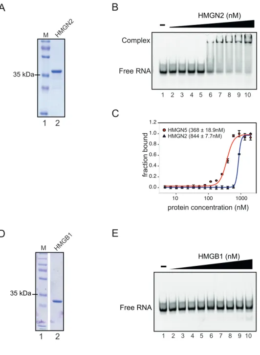
4.2 HMGN5 is a specific RNA binding protein ... 46
4.3 The nucleosome-‐binding domain is required but not sufficient for RNA binding . 51 4.3.1 Stabilization of RNA-‐complexes by intramolecular interaction of HMGN5 ... 54
4.4 The ability to bind RNA is a characteristic of the HMGN family ... 55
4.5 Establishment of inducible stable HMGN5 cell line ... 59
4.6 Effect of HMGN5 deregulation in the global transcriptome pattern ... 62
4.7 HMGN5 UV-‐crosslinking immunoprecipitation ... 69
4.8 HMGN5 binds RNA in vivo ... 72
4.9 HMGN5 forms distinct complexes either with nucleosomes or RNA ... 83
4.10 HMGN5 bind preferentially to regulatory regions and regulates RNA metabolic genes 85 4.11 HMGN5-‐dependent transcriptional changes at the DNA-‐binding sites ... 91
4.12 HMGN5 preferentially binds CTCF recognition motif genome-‐wide ... 95
4.13 Identification of HMGN5-‐interacting partners ... 97
4.14 HMGN5 binds CTCF in vivo and proteins regulating the pre-‐rRNA processing .... 99
5 Discussion ... 103
5.1 HMGN5 has a novel RNA binding activity ... 104
5.1.1 Intrinsic disorder of HMGN5 in RNA binding ... 105
5.2 HMGN5 binds RNA in vivo ... 108
5.3 HMGN5 is coupling global chromatin architecture and gene expression ... 109
5.4 A Regulatory HMGN5-‐CTCF network ... 113
6 Conclusion and Perspectives ... 118
7 Materials and methods ... 120
7.1 Materials ... 120
7.1.1 Equipment and consumables ... 120
7.1.2 Reagents ... 124
7.1.3 Cell lines ... 133
7.1.4 Plasmids ... 134
7.1.5 Oligonucleotides ... 138
7.1.6 Software and databases ... 141
7.1.7 High throughput datasets ... 143
7.2 Methods ... 144
7.2.1 Microbiological methods ... 144
7.2.2 Nucleic acids methods ... 145
7.2.3 Proteins ... 154
7.2.4 In vitro interactions ... 161
7.2.5 Mammalian Cell culture ... 164
7.2.6 Chromatin specific methods ... 169
7.2.7 High throughput sequencing ... 175
8 References ... 180
9 Appendix ... 197
9.1 Supplementary Figures ... 197
9.3 High throughput sequencing command lines ... 212
9.3.1 ChIP-‐seq analysis command lines ... 212
9.3.2 CLIP-‐seq analysis command lines. ... 268
10 Acknowledegments ... 290
List of figures
Figure 1. Structure of nucleosome core particle. _____________________________________________ 23 Figure 2. Electron micrograph of the chromatin “beads-‐on-‐a-‐string” structure. ___________ 24 Figure 3. Hierarchical higher-‐order compaction of chromatin. _____________________________ 26 Figure 4. Classical and new model of higher order chromatin folding. ______________________ 28 Figure 5. PTMs associated with different chromatin states. _________________________________ 30 Figure 6. Spatiotemporal organization of nuclear architecture. ____________________________ 37 Figure 7. Schematic diagram of HMGN family. _______________________________________________ 40 Figure 8. Representation of HMGN5 tethering to LacO array ________________________________ 44 Figure 9. HMGN5-‐mediated chromatin decondensation. _____________________________________ 45 Figure 10. PTMS tethering to the LacO array. ________________________________________________ 46 Figure 11. Purification of recombinant HMGN5 ______________________________________________ 48 Figure 12. HMGN5 interaction with nucleic acids. ___________________________________________ 50 Figure 13. Interaction of deletion and phosphomimetic mutants of HMGN5 with RNA. ___ 53 Figure 14. The RNA binding is stabilized by protein intramolecular interactions. _________ 55 Figure 15. Comparison between HMGN5 and HMGN2 protein features. ____________________ 56 Figure 16. HMGN2-‐RNA interaction. __________________________________________________________ 58 Figure 17. Establishment of HMGN5 FlpIn inducible cell line. _______________________________ 60 Figure 18. Time course of HMGN5 expression. ________________________________________________ 61 Figure 19. Modulation of global transcriptome profile. ______________________________________ 63 Figure 20. qPCR validation of 4 candidate genes. ____________________________________________ 64 Figure 21. Gene set enrichment analysis after HMGN5 overexpression. _____________________ 66 Figure 22. Gene set enrichment analysis after HMGN5 knockdown. _________________________ 67
Figure 23. Gene set enrichment analysis of the overlapped genes between overexpression and knockdown of HMGN5. ____________________________________________________________________ 68 Figure 24. HMGN5 CLIP standardization. _____________________________________________________ 70 Figure 25. Sonication and RNAse treatment for CLIP. ________________________________________ 71 Figure 26. Examples of HMGN5-‐bound RNAs. ________________________________________________ 75 Figure 27. Distribution of HMGN5 peaks in the SEC31B transcript. _________________________ 76 Figure 28. Distribution of HMGN5 peaks in an intronic region of gene NFIA. _______________ 79 Figure 29. HMGN5 global CLIP peaks distribution. ___________________________________________ 81 Figure 30. Significantly enriched HMGN5 de novo motifs. ___________________________________ 82 Figure 31. RNA-‐nucleosome competition assay. ______________________________________________ 84 Figure 32. UCSC genome browser tracks depicting the HMGN5 distribution at three
different example locus. ________________________________________________________________________ 86 Figure 33. Genome-‐wide HMGN5 occupancy. _________________________________________________ 88 Figure 34. Motif distribution around HMGN5 ChIP-‐seq peaks. ______________________________ 90 Figure 35. Correlation of HMGN5 dependent transcriptional changes and DNA-‐binding sites. _____________________________________________________________________________________________ 92 Figure 36. GO analysis of HMGN5-‐bound genes at regulatory regions. ______________________ 94 Figure 37. De novo motif analysis in HMGN5 ChIP-‐seq peaks. _______________________________ 95 Figure 38. CTCF binding site is the most enriched motif in HMGN5 ChIP-‐seq data. ________ 96 Figure 39. HMGN5 co-‐IP-‐standardization for mass spectrometry. __________________________ 98 Figure 40. Significant enriched proteins in HMGN5 Co-‐IP. _________________________________ 100 Figure 41. Intrinsic disorder prediction of HMGN5. ________________________________________ 105 Figure 42. Standardization of HMGN5 knockdown. ________________________________________ 197 Figure 43. Total RNA quality control. _______________________________________________________ 198
Figure 44. Electopherogram of the libraries for CLIP-‐seq run with the High sensitivity DNA chip. ___________________________________________________________________________________________ 199 Figure 45. In vitro mononucleosomes assembly. ____________________________________________ 200 Figure 46. MgCl2-‐dependent RNA fragmentation. _________________________________________ 201 Figure 47. Interaction of PTMS with RNA. __________________________________________________ 202
List of tables
Table 1. Single-‐stranded nucleic acids used in EMSA and MST. ______________________________ 49 Table 2. Demultiplexed reads from CLIP libraries sequencing. ______________________________ 72 Table 3. HMGN5-‐associated transcripts. ______________________________________________________ 74 Table 4. Top 30 HMGN5-‐associated intronic regions from RNAs ____________________________ 78 Table 5. Global distribution of HMGN5 in transcript-‐associated genomic features _________ 80 Table 6. Genome ontology of HMGN5 ChIP-‐seq peaks ________________________________________ 87 Table 7. List of identified HMGN5-‐binding partners ________________________________________ 102 Table 8. Barcodes for CLIP-‐seq ______________________________________________________________ 178 Table 9. Identified HMGN5-‐associated exons _______________________________________________ 203 Table 10. Identified HMGN5-‐associated introns ____________________________________________ 208
Abbreviations
List of abbreviations
°C Degree Celsius
∆ Deletion
6-FAM 6-carboxyfluorescein
Å Ångström
aa Amino acid
Ab Antibody
ADP Adenosine diphosphate
Amp Ampicillin
ANOVA Analysis of variance
APS Ammonium persulfate
AS Alternative splicing
ATP Adenosine triphosphate
BANF1 Barrier to autointegration factor 1 / BAF BLAST Basic Local Alignment Search Tool
bp basepairs
BSA Bovine serum albumin
caRNA Chromatin-associated RNA
cDNA Complementary DNA
CDS Coding sequence
ChIP Chromatin immunoprecipitation
CLIP UV-crosslinking RNA immunoprecipitation
cm Centimeters
CMV Cytomegalovirus
Co-IP Co-Immunoprecipitation
CpG Cytosine-phosphate-guanine
CPM Counts per million
Cryo-EM Cryogenic electron microscopy
CTCF CCCTC-binding factor
CTs Chromosome territories
Cy3 Cyanine 3
Cy5 Cyanine 5
DAPI 4',6-diamidino-2-phenylindole
Df31 Decondensation factor 31
DGE Differential gene expression
DHSs DNase I Hypersensitive Sites
DMSO Dimethyl sulfoxide
DNA Deoxyribonucleic acid
DNAme DNA methylation
DNMTs DNA methyltransferase
dNTP Deoxynucleotide triphosphate
DTT Dithiothreitol
E.coli Escherichia coli
EDTA Ethylenediaminetetraacetic acid
EGR1 Early Growth Response 1
EGTA Ethylene glycol-bis(β-aminoethyl ether)-N,N,N',N'- tetraacetic acid
EMSA Electrophoretic mobility shift assay
ENCODE Encyclopedia of DNA Elements
ER Estrogen receptor
EtBr Ethidium bromide
EZH2 Enhancer of Zeste homolog 2
FBS Fetal bovine serum
FC Fold change
FISH Fluorescent in situ hybridization
FRT Flp recombination target site
Fw Forward
g Relative centrifugal force (RCF)
GAPDH Glyceraldehyde-3-Phosphate Dehydrogenase
GFP Green fluorescent protein
GO Gene ontology
GOI Gene of interest
GST Glutathione S-Transferase
H1 Linker histone H1
H3K27ac Acetylation of lysine 27 on the histone H3 H3K4me3 Trimethylation of lysine 4 on the histone H3
HAS High-affinity sites
HATs Histone acetyltransferases
HDACs Histone deacetylases
HF High Fidelity
His Histidine
HMG High mobility group
HMGA High Mobility Group AT-Hook
HMGB High mobility group Box
HMGN High Mobility Group Nucleosome Binding
HMGN5 High Mobility Group Nucleosome Binding Domain 5
HMTs Histone methyltransferases
HRP Horseradish peroxidase
HS High sensitivity
iBAQ Intensity-based absolute quantification IDR Intrinsically disordered region
IF Immunofluorescence
IGF2/H19 Insulin Like Growth Factor 2/ H19, Imprinted Maternally Expressed Transcript
IP Immunoprecipitation
IPTG Isopropyl β-D-1-thiogalactopyranoside
Kb Kilobase
Kd Knockdown
kDa KiloDalton
LacI lac repressor
LacO lac operon
LADs Lamina-associated domains
LAP Lamina-associated protein
LAP2α Lamina-associated polypeptide 2-alpha
Lap2β Lamina-associated polypeptide 2, isoforms beta/gamma LAS1L LAS1 Like, Ribosome Biogenesis Factor
LB Luria-Bertani
LCMS/MS Liquid chromatography-tandem mass spectrometry
LCS Low-complexity sequences
lncRNA Long non-coding RNA
LOCK Large organized chromatin K modification
M Molar
mA Milliampere
MBD Methyl-CpG Binding Domain Protein
MeCP2 Methyl-CpG Binding Protein 2
MCP MS2 coat protein
min Minute
miRNA microRNA
mJ Millijoules
MM Master mix
MNase Micrococcal nuclease
mRNA Messenger RNA
MSL Male-specific lethal
MST Microscale thermophoresis
MW Molecular weight
MWCO Molecular weight cut-off
NBD Nucleosomal binding domain
NBP-45 Nucleosomal binding protein 45
ncRNA Non-coding RNA
NE Nuclear envelope
Ni-NTA Nickel-nitrilotriacetic acid
NL Nuclear lamina
NLS Nuclear localization signal
nm Nanometer
NOL9 Nucleolar Protein 9
NOR Nucleolar organizer region
NSBP1 Nucleosomal Binding Protein 1
nt Nucleotides
OD600 Optical density measured at 600 nm
OE Overexpression
ON Overnight
ORF Open reading frame
PAA Polyacrylamid
PAGE Polyacrylamide gel electrophoresis PARD3 Par-3 Family Cell Polarity Regulator
PARP1 Poly [ADP-ribose] polymerase 1
PBS Phosphate-buffered saline
Pc Polycomb
PCR Polymerase chain reaction
PELP1 Proline, Glutamate and Leucine Rich Protein 1
PFA Paraformaldehyde
PI Protease inhibitor mix
piRNA PIWI-interacting RNA
PML Promyelocytic leukaemia protein
PMSF Phenylmethylsulfonyl fluoride
PolII RNA polymerase II
PRC2 Polycomb repressive complex 2
PTMS Parathymosin
PTMs Posttranslational modifications
PVDF Polyvinylidene fluoride
qPCR Quantitative Real-Time PCR
Rb Rabbit
RBPs RNA-binding proteins
RD Regulatory domain
rDNA Ribosomal DNA
RNA Ribonucleic acid
RNP Ribonucleoprotein
roX RNA on the X
RPL30 Ribosomal Protein L30
rpm Revolutions per minute
rRNA Ribosomal RNA
RT Room temperature
Rv Reverse
SAXS small-angle X-ray scattering
SDM Site-directed mutagenesis
SDS Sodium dodecyl sulfate
sec Seconds
SENP3 SUMO1/Sentrin/SMT3 Specific Peptidase 3
Seq Sequencing
siRNA Small interfering RNA
Snf2H Snf2 homolog protein
snoRNA Small nucleolar RNA
snoRNP Small nucleolar ribonucleoprotein
SOC Super Optimal broth with Catabolite repression
ssDNA Single-stranded DNA
ssRNA Single-stranded RNA
SWI/SNF SWItch/Sucrose Non-Fermentable
TADs Topologically associated domains
Taq Thermus aquaticus
TARDBP TAR DNA Binding Protein
TBE Tris/Borate/EDTA
TCEP Tris(2-carboxyethyl)phosphine
TEMED Tetramethylethylendiamine
Tet Tetracycline
TEV Tobacco Etch Virus
TEX10 Testis-Expressed Protein 10
TF Transcription factor
TRIS Tris(hydroxymethyl)aminomethane
TSS Transcription start site
TTF-I Transcription Termination Factor I
U Units
UTR Untranslated region
UV Ultraviolet
V Volts
VGF VGF Nerve Growth Factor Inducible
WB Western blot
WCE Whole cell extract
WDR18 WD Repeat Domain 18
wt Wildtype
XCI X chromosome inactivation
XIST X-inactive specific transcript
ZCCHC12 Zinc Finger CCHC-Type Containing 12
1 Summary
The modulation of higher order structure of chromatin has profound implications in the regulation of nuclear processes, like transcription, replication, recombination or DNA repair. Those processes require accessible DNA to recruit large protein complexes to function.
In humans, the architectural proteins of the “high mobility group nucleosomal binding domain” (HMGN) family participate in the opening of chromatin structure and the regulation of gene expression. HMGN proteins bind the nucleosome particle through a conserved nucleosomal binding domain (NBD) and compete with H1 for the binding to chromatin.
Of all HMGN family members, HMGN5 has the biggest effect on transcriptional regulation in mouse and human cells. Moreover, HMGN5 is also able to induce large- scale chromatin decondensation in vivo.
In the present work we study the functional role of HMGN5 in the opening of higher- order structure of chromatin and gene expression by biochemical and genome-wide methods.
We identified a novel and specific RNA binding domain overlapping with the NBD of HMGN5. Moreover, by in vitro competition assays we demonstrated that HMGN5 exhibits exclusive binding to nucleosomes or to RNA. Furthermore, we showed that the RNA binding activity is a feature of other HMGN members as well, highlighting a novel function for those proteins.
The overexpression and knockdown of HMGN5 in human cell lines affect the expression of about 3000 genes respectively, with 1287 overlapping target genes.
ChIP-seq analysis of HMGN5 revealed that HMGN5 mainly associates with active regulatory genomic regions, like promoters and CpG islands, and it localizes to DNase I hypersensitive sites (DHSs). Moreover, we found that the actively regulated target genes belong to the group of genes involved in RNA metabolic processes.
HMGN5 binding overlaps with RNA polymerase II binding sites. CLIP-seq analysis of HMGN5-bound RNAs revealed that HMGN5 is able to bind nascent transcripts. In the light of the biochemical results, we propose that HMGN5 participates in the regulation of RNA metabolism by a dual mechanism that enables HMGN5 binding either
chromatin or RNA since the HMGN5-bound RNAs have no functional relationship with the chromatin function of HMGN5.
Interestingly, by using quantitative mass spectrometry we identified CTCF, BANF1 and seven proteins associated with the pre-ribosomal RNA processing.
Strikingly, our ChIP-seq data revealed that HMGN5 co-localizes with HMGN5. As CTCF constitutes the major organizer of chromatin architecture, the results presented here suggest a cooperative role of both proteins in the organization of higher-order structure of chromatin.
Further functional characterization of the potential HMGN5-CTCF complex, may shed light on the regulation of higher-order chromatin organization.
2 Introduction
2.1 Chromatin as functional organizer of DNA in the nucleus
In eukaryotic cells, the DNA molecule needs to be tightly packed to fit into the nucleus which has a diameter of 10µm, and yet be accessible to allow regulation of processes like transcription, replication or DNA repair. This is possible trough the highly specialized and dynamic packaging of DNA in the chromatin fiber. Chromatin was first discovered and named by Walther Flemming in the 19th century as a stainable fibrous structure within the nucleus of cells. Later, in 1928 the German botanist Emil Heitz made the first categorization of chromatin by discriminating the chromosomes in euchromatin (genetically active) and heterochromatin (genetically inactive) based on its staining properties under the light microscope (Passarge, 1979).
Besides the general categorization in euchromatin and heterochromatin, the chromatin is organized in specialized, spatiotemporal changing compartments that are required for the proper function of all DNA-dependent processes.
Several regulatory mechanisms like ATP-dependent chromatin remodelers, posttranslational modification of histone, DNA modification or regulatory RNAs have been described –globally termed epigenetic mechanisms-, that control the fate of gene expression, by modulating locally (at nucleosomal level) or globally (by long distance chromatin interactions) chromatin accessibility (Bartkuhn & Renkawitz, 2008). This highlights the crucial role of the coordinated and dynamic regulation of chromatin organization on the transcriptional regulation of genes.
2.2 DNA packaging and higher-order chromatin organization
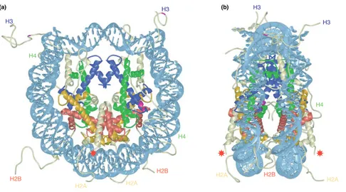
The nucleosome corresponds to the basic level of chromatin organization, and is composed of a nucleosome core particle (NCP) a linker DNA. The nucleosome core particle is composed of 147 bp of two tight 1.65 left handed superhelical turns of DNA wrapped around a histone octamer (Figure 1) which is composed of two copies of the histones H2A, H2B, H3 and H4 (Luger, 2003). In the NCP, the core histones are assembled into four heterodimers, two H2A/H2B and two H3–H4 dimers (Luger, Mäder, Richmond, Sargent, & Richmond, 1997; A. L. Olins & Olins, 1974; Woodcock, Safer, & Stanchfield, 1976). The core histones are evolutionary conserved basic small proteins (ranging between 11-16kDa), that have 2 characteristic functional domains; a histone fold domain mediating histone-DNA and histone-histone interactions required to form the nucleosome particle, and the N-terminal histone tails (and C-terminal tail in H2A and H2B) that are disordered, flexible and accessible structures protruding from the nucleosome core particle (Downs, Nussenzweig, &
Nussenzweig, 2007). The histone tails are hotspots of regulatory posttranslational modifications.
Figure 1. Structure of nucleosome core particle.
Scheme of the nucleosomal structure at the 2.8 Å resolution level obtained from X-ray crystal structure. A) Front view of the nucleosome particle, formed by 147bp of double- stranded DNA (in light blue) wrapped around a histone octamer composed of the histone H2A (yellow), H2B (red), H3 (blue) and H4 (green). The respective histone tails extensions are shown. Red star indicates site of ubiquitination in yeast. B) Side view of the nucleosome
H3
H3
H3 H3
H2A H2A H2A H2A
H2B H2B H2B
H4 H4
H4
(a) (b)
Histones interact with DNA through different mechanisms including salt bridges, hydrogen bonds with DNA, non-polar contacts with the deoxyribose, electrostatic interactions between the positively charged N-terminal tails with the DNA phosphates and base-specific contacts (Widom, 1998).
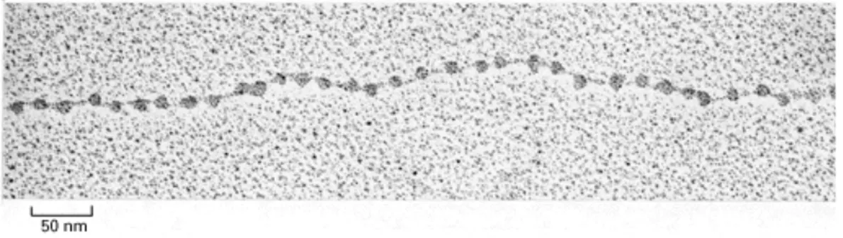
The nucleosome core are separated by a flexible DNA linker–with a variable length between 20-80bp depending on the species and the cell type (Felsenfeld &
Groudine, 2003)-. This array of nucleosomes forms the so called “10 nm fiber” that resembles a “beads-on-a-string” structure under electron microscopy (Figure 2) at low salt conditions (A. L. Olins & Olins, 1974; Woodcock et al., 1976). A diploid human cell that contains about 6x10⁹ nucleotide pairs (with a total average length of 2m), DNA is packaged on average into 30 millions nucleosome cores.
Figure 2. Electron micrograph of the chromatin “beads-on-a-string” structure.
Decondensed "beads-on-a-string" form of purified chromatin visualized by electron microscopy. Picture adapted from Molecular Biology of the Cell. 3rd edition. (Alberts et al., 1994)
2.3 Higher-order chromatin organization
Inside the cell nucleus, chromatin is not linear in structure, but it is rather organized into 3-dimentional higher-order structures. This higher-oder organzation plays critical roles for the regulation of nuclear functions.
At a first level, the binding of the linker histone (H1 and H5) helps the packaging of nucleosomes into the chromatosome core particle (Harshman, Young, Parthun, &
Freitas, 2013; Simpson, 1978). Linker histones are composed of a tripartite structure, consisting of a flexible and short N-terminal tail, a conserved central globular domain, and a long (100 amino acids) intrinsically disordered and basic C-terminal domain (Allan, Hartman, Crane-Robinson, & Aviles, 1980).
By using cryo-microscopy (cryo-EM) and crystal structures it was recently shown that the C-terminal domain of H1 associates primarily with a single linker in the nucleosome, while the globular domain interacts with both DNA linkers and the nucleosome dyad, resulting in a reduction of the flexibility of linker DNA and a more compact nucleosome conformation (Bednar et al., 2017). It has been shown that depletion of H1 is lethal in mice (Fan et al., 2003) and Drosophila (Lu et al., 2009).
Moreover, H1 depletion alters the proper folding of chromosmes during mitosis (Maresca, Freedman, & Heald, 2005) highliting its role in the higher-order chromatin conformation.
2.3.1 Classical view of chromatin higher-order organization
It has been described that chromatin is highly packaged into a hierarchical higher- order structure. In the classcial text-book view of chromatin organization (Figure 3), the nucleosomal array is folded into thought to fold into intermediate fibers of increasing diameter of 30, 120 300 and 700 nm diameter, to finally form the mitotic chromosome, eye-visible under the light microscope.
Two competing models have been proposed to explain the formation of the 30 nm fiber. The solenoid model, in which consecutive nucleosomes are located adjacent to one another in the fiber, and fold into a simple “one-start helix“ (Finch & Klug, 1976), and the zig-zag model, which assumes an arrangement of the nucleosomes in a
alternate nucleosomes interacts with the neighbor nucleosome of the other row (Dorigo et al., 2004; Horowitz et al., 1994; Worcel et al., 1981). Both models assume a selective internucleosomal interaction of close neigbors nucleosomes on the DNA strand (Maeshima, Imai, Tamura, & Nozaki, 2014).
Despite that for many years the hierarchical folding of chromatin has been widely accepted, the chromatin folding in vivo is still a controversial topic.
Figure 3. Hierarchical higher-order compaction of chromatin.
DNA is wrapped around histone octamers forming nucleosomes. Individual nucleosomes are separated by free linker DNA and associate with histone H1. The 10 nm fiber twists into a large coil, generating the condensed, supercoiled 30 nm fiber. The coils form loops (300- nm fiber) and the loops coil further, producing the metaphase chromosome as highest condensation level of DNA in eukaryotes (image and description taken from Alberts, 3rd edition.)
2.3.2 A new concept of higher-order chromatin structure
Over years, the formation of the 30 nm fiber has been supported by several biophisical studies, including X-ray crystallography, or small-angle X-ray scattering (SAXS), and the 30 nm chromatin conformation has been the reference for many studies in the chromatin field. However, despite all the efforts made to unravel the specific conformation of chromatin in vivo it is still not clear if the 30 nm fiber really exist in vivo (Fussner et al., 2011; Luger, Dechassa, & Tremethick, 2012; Maeshima, Hihara, & Eltsov, 2010; Nozaki et al., 2014). This is because the classical methods have technical limitations, as they are mainly based on in vitro structure of reconstituted chromatin from DNA and histones (P. J. J. Robinson., 2006; Schalch et al., 2005), or based on chromatin purified from permeabilized cells (Belmont & Bruce, 1994; Horowitz et al., 1994; Worcel et al., 1981), lacking many components of the physiological chromatin context.
By the use of cryo-electron microscopy (cryo-EM) analysis of vitrified chromosomes it was suggested, already three decades ago, that chromatin was irregularly folded in vivo (McDowall et al., 1986).
Furthermore, the latest evidence in the field strongly argues against the existence of the 30 nm fiber in vivo (Bouchet-Marquis, et al., 2006; Eltsov et al., 2008; Nishino et al., 2012; Ou et al., 2017).
By using a sofisticated method that combines electron microscopy tomography and labeling methods (ChromEMT), the 3D chromatin ultrastructure in the nucleus of living human cells was analyzed for the first time. In the study performed by Ou and colleagues it was revealed that chromatin is organized as irregular polymers with diameters of 5 to 24 nm in interphase and mitotic chromosomes (Ou et al., 2017), instead of the classically described higher-order structures.
Those results are in agreement with the recently proposed "polymer melt model"
(Figure 4), in which chromatin is organized as flexible and disordered dynamic folded 10 nm fiber similar to a “polymer melt” (Maeshima et al., 2010). In this model, at low nucleosome concentration, nucleosome fibers can form 30 nm fibers mediated by intra nucleosome interactions, which can explain the observed in vitro formation of the 30 nm fibers. However, as the nucleosome content increases nucleosomes form inter fiber associations (due to increased cation concentration or molecular crowding
"polymer melt" model represents several advantages in determining accessibility of the DNA to the recruiting of regulatory machineries and for the formation of functional chromatin domains through long-distance chromatin interactions.
Figure 4. Classical and new model of higher order chromatin folding.
Comparison between the classical view of a hierarchical chromatin folding (left), and the new model of irregularly folded nucleosome fibers (“polymer melt”). In the classical view, nucleosomes are organized in regular fibers of 30 nm, which are subsequently packaged to get a highly compacted mitotic chromosome inside the nucleus. In the novel model, chromatin is organized as irregular folded 10 nm fiber which implies a flexible and less constrained chromatin organization.
Picture taken from:
https://www.nig.ac.jp/labs/MacroMol/e-more_detailed_description.html (Maeshima et al., 2014)
2.4 Mechanisms regulating chromatin structure
Epigenetic regulatory mechanisms involve posttranslational modification of histones, canonical histone replacement by histone variants, DNA methylation, non-coding RNA and ATP dependent chromatin remodeling. Each mechanism has been extensively studied, revealing that they are highly coordinated, functionally interacting and influencing each other (Armstrong, 2013). The actors in the epigenetic marking system, have been classified as “writers”, “erasers”, and “readers” due to their ability to add, remove or recognize, respectively, histones or DNA chemical modifications (marks) to establish the gene expression program of the cell (Torres & Fujimori, 2015).
2.4.1 Histone posttranslational modification
Histones are marked by a large number of posttranslational modifications (PTMs), including acetylation, methylation, phosphorylation, ubiquitination, citrullination, sumoylation, ADP-ribosylation and proline isomerization (Rothbart & Strahl, 2014).
Most of the modifications are located in the unstructured N-terminal and C-terminal tails that protrude from the nucleosome particle. However, some modifications have been found in the histone-fold domain, required for nucleosome unwrapping and disassembly (Simon et al., 2011).
PTMs have been associated with specific functions, but they are mainly found in combinations, suggesting a “histone code” that finally define a specific chromatin state (Strahl & Allis, 2000) by the recruitment of modifying enzymes, such as histone acetyltransferases (HATs), histone methyltransferases (HMTs,) or histone deacetylases (HDACs), among others, that alter the structure of the surrounding chromatin environment.
Of the PTMs, the mostly described histone modification is lysine methylation. Lysine can be mono-, di- or tri-methylated, and these modifications can be associated with either gene activation or silencing depending on possition and the cross-talk with other PTMs (Torres & Fujimori, 2015). As an example, mono- and trimethylation of
(H3K36me3) are associated with transcription activity, whereas H3K27me3 and H3K9me2 have been associated with transcriptional repression (Woo, Ha, Lee, Buratowski, & Kim, 2017). Specific combinations of histone modifications are hallmarks of the active/inactive regulatory genomic elements like promoters, enhancers, or intron/exon boundaries (V. W. Zhou, Goren, & Bernstein, 2010). A summary of the different histone marks and their associated functions is depicted in (Figure 5).
.
Figure 5. PTMs associated with different chromatin states.
A) At promoters, PTMs contribute to fine-tune gene activity from active to poised to inactive states. B) At gene bodies, they discriminate between active and inactive conformations. C) At distal sites, histone marks correlate with levels of enhancer activity. D) On a global scale they may confer repression of varying stabilities and be associated with different genomic features. For example, lamina-associated domains (LADs) in the case of stable repression and Polycomb (Pc) bodies in the case of context-specific repression. DNAme, DNA methylation; LOCK, large organized chromatin K modification. Figure and description legend obtained after Zhou et al., 2011.
2.4.2 Histone variants
Additionally to the histone PTMs, chromatin can be regulated by the replacement of canonical histones by histone variants.
Histone variants are non-allelic isoforms of the canonical histones that have a specific expression, localization, specie specific distribution pattern (Kamakaka &
Biggins, 2005) and play determinant roles in regulating chromatin structure.
Different than canonical histones, that are produced during the DNA synthesis (S) phase of the cell cycle, histone variants are expressed throughout the cell cycle, and are incorporated to chromatin in a replication-independent manner by specific histone chaperones (Biterge & Schneider, 2014).
The genes coding for the (known) histone variants contain intronic sequences, the pre-mRNAs are polyadenylated and can undergo alternative splicing (Biterge &
Schneider, 2014).
It is believed that the deposition of histone variants alters the stability of the nucleosome, affecting the interactions between histones and with the DNA, thus helping to modify the chromatin conformation (Gautier et al., 2004; Park, Dyer, Tremethick, & Luger, 2004)
Histone variants can differ from their canonical counterparts by minor modifications (as one amino acid change in the H3 variants H3.1 and H3.2) to drastic structural modifications as in the H2A variant macroH2A (E. Bernstein & Hake, 2006).
To datel, all histones, with the exception of H4 have described histone variants. The most studied histone variants belong to the H2A family and they are involved in diverse cellular processes, associated with activation or repression of transcription, as DNA damage response and centromeres formation (E. Bernstein & Hake, 2006).
Among them, H2a.Bbd (associated with active transcription), H2A.X (involved in DNA repair and genome integrity), H2A.Z (activation and repression of transcription and chromosome segregation) and macroH2A which have been associated with X- chromosome inactivation and transcriptional repression (Biterge & Schneider, 2014;
Sarma & Reinberg, 2005).
The number of histone H3 variants differs among species. In mammals there are four main isoforms, H3.1, H3.2, H3.3 and CENPA (found at centromeric chromatin) (Filipescu, Mueller, & Almouzni, 2014). However, recent studies have revealed an
expression pattern. Those variants include the testis-specific histones H3.4, and H3.5, and the histone H3.Y which is conserved among primates (Filipescu et al., 2014).
For many of the histone variants, posttranslational modifications have been described, contributing to the modulation of gene expression (Biterge & Schneider, 2014).
2.4.3 DNA methylation
In vertebrates, DNA methylation is key for regulation of different processes, like gene expression, genomic imprinting, silencing of transposable elements or X- chromosome inactivation. This modification occurs extensively in CpG dinucleotides and is mediated by a group of enzymes called DNA methyltransferases (DNMTs) that catalyze the methylation at the carbon-5 position of cytosine residue at the CpG dinucleotide to form 5-methylcytosine (5-mC) (Prokhortchouk & Defossez, 2008).
Methylation of DNA is essential for development and viability in mammals (Jackson- Grusby et al., 2001; Okano, Bell, Haber, & Li, 1999). In humans the CpG methylation patterns can be categorized in two groups; the first group, covering the vast majority of the genome (98%), possesses low CpG frequency (1 each 100bp) but highly methylated. Those CpGs are generally associated with transcriptionally repressed chromatin. In the second group (covering 2% of the genome) the CpGs are highly concentrated in the so-called CpG islands (CGIs), found in a ratio of 1 CpG every 10bp in DNA stretched of about 1000bp (Illingworth et al., 2008). The human genome contains about ~30000 CGIs and most of them (around 21000) are found close to transcription start sites (TSSs) of genes and remain unmethylated which correlates with transcriptional activity. However, about 9000 CGIs are found at gene body, and the methylation at those sites have been associated with enhanced gene expression levels (Ball et al., 2009; Illingworth et al., 2008; Krinner et al., 2014) indicating that DNA methylation is a versatile mark with functions that depend on the genomic context. It has been described that the silencing mediated by DNA methylation can occur by two different mechanisms; DNA methylation can act by masking the
methylation readout can be performed by methyl-binding proteins (“readers”) like Methyl-CpG Binding Domain Proteins (MBD1, MBD2 and MBD4), and Methyl-CpG Binding Protein 2 (MeCP2) that can recruit chromatin remodeling complexes, DNA methyltransferases or histone deacetylases, that lead to transcriptional repression (Baubec, Ivanek, Lienert, & Schuebeler, 2013).
It has been lately shown that besides CpG, cytosines followed by adenine, thymine or another cytosine can also be methylated. The methylation of those cytosines is known as Non-CpG methylation and is prevalent in human embryonic stem cells (Ramsahoye et al., 2000) or brain development (Guo et al., 2014; Lister et al., 2013), however, the specific functions of those modifications are still unclear.
2.4.4 ATP dependent remodeling complexes
To allow the binding of regulatory machineries to specific genomic sites, the chromatin needs to be dynamically changed. This active process is mediated by chromatin remodeling complexes, which use ATP-hydrolysis to move, destabilize, eject or restructure nucleosomes (Clapier & Cairns, 2009; Erdel, Krug, Längst, &
Rippe, 2011), thereby regulating the accessibility of DNA regulatory factors.
The chromatin remodeling complexes have ATPases, that belong to the SF2 helicase superfamily, and accessory regulatory subunits which are required for targeting and regulation of nucleosome remodelling (Erdel et al., 2011; Längst &
Manelyte, 2015).
The remodelers can be classified into four families, the SWI/SNF (SWItch/Sucrose Non-Fermentable), CHD (chromodomain, helicase, DNA binding), ISWI (imitation switch) and INO80 (inositol requiring 80) family (Längst & Manelyte, 2015).
The activity of chromatin remodeling complexes and their targeting to specific genomic regions can be regulated by the interplay with different chromatin signals like DNA sequence, DNA structure, methylation, histone modifications, histone variants, and they can interact with chromatin associated proteins and complexes, as transcription factors (reviewed in Erdel et al., 2011), structural proteins that regulate chromatin compaction as H1 (Hill & Imbalzano, 2000) or HMG chromatin binding proteins (Bonaldi, et al., 2002; Heppet al., 2014; Rattner, Yusufzai, & Kadonaga,
(Längst & Manelyte, 2015). Those interactions will help to determine the accessibility of chromatin required for the regulation of gene expression.
2.4.5 Regulatory RNAs
In the last years there have been cumulative evidence showing that non-coding RNAs (ncRNAs) are essential for regulation of chromatin architecture and gene expression. ncRNA have been classified according to their size, biogenesis and function. According to size, they are commonly (and loosely) classified into two sub- categories, small ncRNA, with an average length of less than 200nt, and long non- coding RNAs (lncRNA) with a size of >200nt (Patil, Zhou, & Rana, 2013).
Non-coding RNAs play a role in the regulation of different processes, including gene regulation, translation, splicing, cell cycle control, genome defense and chromosome structure (Brown, Mitchell, & Neill, 2011).
The mechanisms by which ncRNA participate in gene regulation include interactions with different epigenetic regulatory machineries. As an example, they can guide and modulate the activity of transcription factors, direct DNA methylation, recruit histone modifying enzymes to either activate or silence gene expression (main mechanisms described in Mattick, Amaral, Dinger, Mercer, & Mehler, 2009) or they can directly interact with DNA to modulate recruitment of transcriptional regulatory machineries (Schmitz, Mayer, Postepska, & Grummt, 2010).
It was shown already four decades ago that RNA is stably associated with chromatin in different species (Holoubek, et al., 1983; R. C. Huang & Huang, 1969; Huang &
Bonner, 1965).
Furthermore it has been shown that RNA also plays a structural role contributing to the higher-order chromatin organization (Caudron-Herger et al., 2011; Rodriguez- Campos & Azorin, 2007; Schubert et al., 2012).
The group of Karsten Rippe demonstrated the existence of RNA transcripts associated with chromatin -which they called “chromatin-interlinking” RNAs or ciRNAs- that were responsible for maintaining chromatin in a decondensed and active state during interphase in human and mouse cell lines (Caudron-Herger et al., 2011).
Moreover, in our laboratory it was shown that the small nucleolar RNAs (snoRNAs) constitute the mayor fraction of chromatin-associated RNAs in Drosophila and human cells, and it was demonstrated that they are required to keep the higher-order structure of chromatin in an open state in Drosophila (Schubert et al., 2012), indicating that RNA is a key player in the global reorganization of chromatin architecture.
A well-studied example of ncRNAs involved in modulating chromatin conformation is the dosage compensation in mammas and in fly. In female mammals, the lncRNA Xist RNA controls the developmentally regulated chromatin-mediated X chromosome inactivation (XCI) by recruiting silencing complexes in cis, thus generating a compact heterocromatinized global chromatin architecture in one of the X-chromosomes (Pandya-Jones & Plath, 2016). In Drosophila melanogaster on the other hand, the roX RNAs (RNA on the X) are required for balancing dosage by increasing the transcription levels in the single male X-chromosome. This is mainly done by the recruitment of the male-specific lethal (MSL) complex that regulates nucleosome positioning at High-Affinity Sites (HAS) from topological associated domains (TADs), from which the complex is spread leading to the global activation of the male X- chromosome (Ramírez et al., 2015).
To date, a huge number of non-coding RNAs have been discovered –including short ncRNAs and lncRNAs- and many of them have been shown to be required for different nuclear processes -reviewed elsewhere-, however, for most of them, the mechanisms underlying their function still need to be elucidated.
Indeed the main concept that defines the functional borders between coding and non-coding RNA needs to be re-defined, as it has been shown that some non-coding RNAs have coding potential (Andrews & Rothnagel, 2014; Ruiz-Orera, Messeguer, Subirana, & Alba, 2014), and also some classically defined messenger RNAs (mRNAs) have been shown to possess non-coding functions (Kumari & Sampath, 2015; Poliseno et al., 2010; Sampath & Ephrussi, 2016) for which a dual coding/non- coding function have been proposed (Nam, Choi, & You, 2016).
2.5 Nuclear architecture and gene regulation
In the interphase nuclei the chromatin is dynamically compartmentalized. Beyond the global separation between euchromatin and heterochromatin, it has been shown that each chromosome occupies non-random territory (Bodnar & Spector, 2013) called
“chromosome territories” (CTs) (Figure 6A and B). By the use of dedicated microscopy methods, together with chromosome conformation capture techniques (from 3C to Hi-C), it was demonstrated that the chromosome territories are further organized into sub-domains called Topologically Associated Domains (TADs) that are formed by long-distance regulatory interactions between the promoters and their regulatory enhancers which are often located hundred of kilobases up to megabases away (Kaiser & Semple, 2017). TADs are delimited by sharp boundaries that are enriched in housekeeping genes and insulator sites that are bound by architectural proteins as the CCCTC-binding factor (CTCF) or cohesins that have been shown to be required for the establishment of TADs through promoter-enhancers interactions through the formation of intrachromosomic loops and in less extent interchromosomal interactions (Dixon et al., 2012; Gonzalez-Sandoval & Gasser, 2016).
This compartmentalization allows the functional clustering of gene-rich (associated with euchromatin) or gene-poor chromatin regions (heterochromatin), keeping them apart from each other, and thereby contributing to the coordinated regulation of transcription of specific set of genes (Meaburn & Misteli, 2007). The gene-rich chromatin is generally located in the nuclear interior while the heterochromatin (like centromeric and pericentromeric chromatin) is clustered in the nuclear periphery generally associated to the nuclear lamina (NL), forming the Lamina-Associated Domains (LADs). LADs comprise large chromatin domains, ranging from 100kb to 10Mb in length and covering around 40% of the genome, that are in close contact with the nuclear lamina in the inner membrane of the nucleus (Guelen et al., 2008;
Pickersgill et al., 2006).
TADs are highly conserved between species, and invariant between cell types. It has been shown that the disruption of the boundaries in engineered mouse models recapitulates human developmental disorders (Kaiser & Semple, 2017). Moreover in different limb genetic malformation in Human and Mouse, TADs appear disrupted with altered promoter-enhancer interactions and misexpression (Lupiáñez et al.,
2015) which highlights that gene positioning trough higher-order chromatin organization is crucial for the control of transcriptional fate of the cell.
Besides chromosomal territories, functional nuclear substructures have been characterized, including Cajal bodies, nuclear speckles, promyeolocytic leukemia nuclear bodies (PML NBs) and the nucleolus (Figure 6B); those self-organizing structures are formed by dynamic nucleoprotein complexes that play vital roles in the regulation of the nuclear homeostasis. Cajal bodies, for example, play a role in posttranscriptional modification of spliceosomal components, as the maturation of snRNPs (small nuclear ribonucleoproteins) and snRNA (small nuclear RNAs).
Nuclear speckles have been described as sites for splicing factor storage and modification (reviewed in Wood, Garza-Gongora, & Kosak, 2014).
Figure 6. Spatiotemporal organization of nuclear architecture.
A) 24-Color 3D FISH representation and classification of chromosomes in a human G0 fibroblst nucleus. All visible chromosomes in the section are represented with false colors after classification with the program goldFISH. Image modified after Bolzer et al., 2005. B) Diagram depicting the compartmentalization of fuctional nuclear components. Chromosome territories (CTs), nucleolus, nuclear speckles, nuclear pores, Cajal bodies, nuclear lamina, PML bodies and Nuclear envelope are indicated in the figure. Image after Lanctôt et al., 2007.
Nuclear
speckle Interchromatin compartment
Nuclear envelope
Nuclear lamina
Cajal body
Nuclear pore
Nucleolus
Chromosome territories Chromosome
territories
PML body
A B
Of all functional nuclear compartments, the best-characterized example is the nucleolus, where the rRNA transcription, processing and assembly of ribosomal particles take place. The nucleolus is built around the rRNA genes at specific chromosomal loci called “nucleolar organizer regions” (NOR) on the short arms of the acrocentric chromosomes (Nemeth & Längst, 2011). Inside the nucleolus there are specific chromatin conformations associated to the transcriptional activity of the rRNA genes. On the active rRNA genes there have been described specific long-range interactions between promoter and terminator sequences. It has been shown that those interactions are mediated by TTF-I (Transcription Termination Factor), subdividing the rRNA transcription unit into functional chromatin domains (Németh et al., 2008).
Nuclear transcription is carried out by specialized “transcription factories” in which the transcription machinery –like transcription factors, remodeling complexes, coregulators or RNA polymerases- is concentrated and fixed while the DNA template moves relative to the polymerization site (Papantonis & Cook, 2013). In the nucleolus, as an example, the RNA is transcribed by “factories” containing up to 30 active RNA polymerases each (Jackson, Iborra, Manders, & Cook, 1998).
Although in the last years many efforts have been made to understand the mechanisms that regulate chromatin dynamics, it is still not known how the functional organization of the genome into higher-order structures can orchestrate the regulation of transcription.
2.6 HMGN5 regulates higher-order structure of chromatin
The “High Mobility Group” (HMG) proteins constitute another group of players that participate in the regulation of chromatin structure. The HMG super family is divided into three non-related subfamilies, the HMGA (characterized by the AT-hook motif), HMGB (characterized by the HMG-box motif) and HMGN (High mobility group nucleosome-binding) which are non-histone architectural proteins that were shown play roles in the regulation of transcription, replication and DNA repair (Reeves, 2010).
The HMGN family -described so far only in vertebrates- participate in the modulation of higher-order structures of chromatin by direct interaction with the nucleosome through a highly conserved “nucleosomal binding domain” (NBD) (Postnikov &
Bustin, 2010).
The HMGN family consists of five members, HMGN1, HMGN2, HMGN3, HMGN4 and HMGN5 (Figure 7), all of them sharing the conserved N-terminal domain containing the Nuclear Localization Signal (NLS) and the NBD, including a conserved octapeptide “RSARLSA”. This motif is required for the recruitment of HMGN to the core nucleosome particle, to decompact chromatin architecture, locally and globally, by competing with the binding of linker histone H1 to chromatin (Kugler, Deng, &
Bustin, 2012).
Figure 7. Schematic diagram of HMGN family.
Cartoon depicting the HMGN functional domains; the bipartite nuclear localization signal (NLS) is depicted in red; the conserved Nucleosome Binding Domain (NBD) is indicated in Cyan; the NBD core octapeptide is indicated in blue and the C-terminal chromatin regulatory domain (RD) is indicated in orange. The regulatory serines from the NBD core are highlighted in red.
HMGN5, formerly known as NSBP1 or NBP-45, is the last discovered member of the HMGN family (Shirakawa, Landsman, Postnikov, & Bustin, 2000). It is a highly abundant protein and shares the functional domain NBD with the other members of the family, as well as the ability of counteracting the binding of H1 to chromatin, by a mechanism involving the interaction of the C-terminal tails of both proteins (Rochman et al., 2009). But differentIn contrast to the other HMGNs, HMGN5 is characterized by the presence of a very long negatively charged and unstructured C-terminal domain, of about 300 amino acids in mouse (Shirakawa et al., 2000) and 200 amino acids in humans (King & Francomano, 2001; Rochman et al., 2009). The C-terminal tail of both, mouse and human proteins is enriched in aspartic and glutamic acid and organized in repeated motifs. The acidic motif EDGKE is repeated 11 times in mouse HMGN5 and 4 times in the human protein, but its functional relevance is unknown so far. It was reported that the C-terminal tail is responsible for the specific chromatin location of HMGN5. In mouse, HMGN5 is tethered to euchromatin (Rochman et al., 2009) and in human cells it was shown associated to both, euchromatin and
Nuclear localization signal NBD core
Chromatin regulatory domain
RRSARLSA
Nucleosomal binding domain
NLS NBD NLS RD
NBD core
heterochromatin (Malicetet al., 2011). Moreover, deregulation of HMGN5 lead to changes in the expression of a large set of genes in cells and mouse models (Kugler et al., 2013; Malicet et al., 2011; Rochman et al., 2009; 2011) being the HMGN protein with the strongest effect on transcriptional regulation.
It has been shown that HMGN5 induces large-scale chromatin decondensation in vivo -(Rochman et al., 2009) and our own work presented in this dissertation-, and it was suggested that the global chromatin reorganization observed in vivo is mediated by the unstructured C-terminal tail. These results highlight the relevance of this protein in the regulation of higher-order structure of chromatin and the cellular transcriptional identity.
Interestingly, several studies have reported an oncogenic role of HMGN5 in various types of cancer (Chen et al., 2012; Yang et al., 2014). Moreover, HMGN5 has an important function in embryonal development, demonstrating that transcriptional changes during differentiation were a result of the induced changes in the global chromatin architecture mediated by HMGN5 (Shirakawa et al., 2009).
It is known that HMGN5, as all HMGNs, is a target of posttranslational modifications.
Phosphorylation and acetylation of the NBD control the cell-cycle dependent binding of the protein to chromatin in vivo (Moretti et al., 2015; Pogna, Clayton, &
Mahadevan, 2010). Furthermore, the recent evidences suggest a role of HMGN5 in the tethering of chromatin to the Nuclear Lamina (NL) (Zhang et al., 2013). However, the mechanism by which HMGN5 can regulate the opening of chromatin structure is far from been understood.
Interestingly, the functional homolog of HMGN5 in the Drosophila system, the decondensation factor 31 (Df31) which is also required for opening higher order- structures of chromatin, is tethered to chromatin in an RNA-dependent manner (Schubert et al., 2012; Schubert & Längst, 2013), hinting the possibility of a conserved RNA-dependent mechanism involved in the regulation of higher-order structures of chromatin in the human system.
Due to the role of HMGN5 in the organization of chromatin architecture and its impact on transcriptional regulation, studying the dynamics and function of HMGN5 may help to understand the contribution of chromatin architecture to the maintenance of cell homeostasis.
3 Objectives
Here we proposed to use the human HMGN5 protein as a model protein to study the dynamics of opening of higher-order structure of chromatin and its contribution to the regulation of transcription, by using integrative genome-wide analysis.
For that purpose, we will analyze the genomic distribution of HMGN5, and study the effects of HMGN5 deregulation on the global transcription pattern. The integration of this data will shed light on the influence of the binding of HMGN5 at specific genomic loci on the transcriptional levels of the associated genes. We will try to determine the functional role of HMGN5 by analyzing its genome-wide association with regulatory elements and characterized chromatin hallmarks, like the histone PTMs H3K27ac and H3K4me3, or RNA polymerase II distribution, and others.
To test the hypothesis of the involvement of RNA in the regulatory mechanism mediated by HMGN5, we will characterize the in vitro RNA binding properties of the protein, and we will try to identify the potential RNA interacting partners in vivo. The data obtained will be integrated with the genomic distribution of HMGN5 and the transcriptome profile to get more insight into a possible functional mechanism.
In parallel, we will perform quantitative mass spectrometry to analyze the global HMGN5-protein interactome with the aim to reveal potential physiological binding partners that may help to delineate the role of HMGN5 in the regulation of higher- order structure of chromatin.
4 Results
4.1 HMGN5 decompacts chromatin
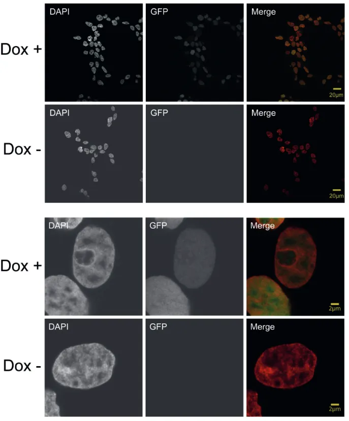
It was already shown that HMGN5 induces large-scale chromatin decompaction (Rochman et al., 2009). As proof of these results, we tested the global chromatin decompaction in vivo by using the LacI/LacO tethering system (Figure 8). This method allows the targeting of a protein to an array of a tandem repeat (256 copies) of the Lac operon (LacO) sequence which is inserted in a highly compacted telomeric region in U2OS cells (Jegou et al., 2009). The HMGN5 protein is fused with a GFP- LacI construct, in which the LacI repressor allows the recruitment to the LacO sequence, and GFP fluorescence is used for in vivo visualization. The hHMGN5- GFP-LacI construct (vector psV2_HMGN5-GFP-LacI) was used for transient transfection of U2OS cells containing the LacO array, and GFP fluorescence was monitored by confocal microscopy. When HMGN5 was tethered to the LacO array, large-scale chromatin decompaction induced by HMGN5 was observed, as the LacO array appears decondensed compared with the GFP control, in which a highly condensed chromatin structure is observed (Figure 9). We also tested the human Parathymosin (PTMS) protein (construct pSV2-PTMS-GFP-LacI), which, like HMGN5, is an acidic and unstructured protein with a nuclear and nucleolar localization and that was described to counteract the binding of H1 to chromatin (Martic, 2005) and to decompacts chromatin. As shown in Figure 10, the recruitment of PTMS to the LacO array does not decondense chromatin in our system, indicating the functional specificity of HMGN5 in the global chromatin reorganization.
Figure 8. Representation of HMGN5 tethering to LacO array
The diagram illustrates the LacI/LacO tethering system. The fusion protein containing HMGN5-GFP-LacI is tethered through the LacI repressor (depicted in blue in the left diagram) to a tandem array (256 copies) of the LacO sequence, which is stable integrated in a telomeric region is the U2OS cell line. Using confocal microscopy the effect of HMGN5 in LacO array decompaction is visualized by the distribution of GFP signal from the fusion protein. In the right drawing a cartoon of a cell containing the stable integration of the LacO array is indicated.
Image adapted from:
https://malone.bioquant.uni-heidelberg.de/methods/single_cell/single_cell.html.
LacO x256 LacO x256
LacI GFP LacI
GFP HMGN5HMGN5