epithelial organs
DISSERTATION ZUR ERLANGUNG DES DOKTORGRADES DER NATURWISSENSCHAFTEN (DR.RER.NAT.)
DER FAKULTÄT FÜR BIOLOGIE UND VORKLINISCHE MEDIZIN DER UNIVERSITÄT REGENSBURG
vorgelegt von
Inês Maria Santos Cabrita Bustorff Silva aus
Amadora, Portugal
im Juni 2020
DAS PROMOTIONSGESUCH WURDE EINGEREICHT AM:
12.06.2020
DIE ARBEIT WURDE ANGELEITET VON:
PROF. DR. KARL KUNZELMANN
UNTERSCHRIFT:
DIE ARBEIT WURDE ANGELEITET VON: PROF. DR. KARL KUNZELMANN
PRÜFUNGSAUSSCHUSS:
VORSITZENDER: PROF. DR. MED. ARMIN KURTZ
1.GUTACHTER: PROF. DR. MED. KARL KUNZELMANN
2.GUTACHTER: PROF. DR. RER. NAT. CHRISTIAN WETZEL
3. PRÜFER: PROF. DR.MED. FRANK SCHWEDA
ERSATZPERSON: PROF. DR. RER. NAT. JENS SCHLOSSMANN
SUMMARY
Autosomal dominant polycystic kidney disease (ADPKD) is characterized by the development of bilateral renal cysts that continuously expand, leading to impaired renal function. ADPKD is originates when mutations occur in the PKD1 or PKD2 genes, whose protein products are collectively called polycystins. Polycystin-1 (encoded by PKD1) is a membrane protein with large extracellular domains, probably working as a receptor for an unidentified ligand. Polycystin-2 (encoded by PKD2) is also a membrane-associated protein that works as a non-selective ion channel with high permeability for calcium ions (Ca2+).
Together, these proteins are thought to form a functional complex, which can regulate intracellular Ca2+ signaling. One of the hallmarks of ADPKD is the transepithelial Cl- secretion towards the cyst lumen. This secretion is thought to occur mainly through the cAMP- activated Cl- channel Cystic Fibrosis Transmembrane Conductance Regulator (CFTR).
CFTR is expressed in the apical membrane of epithelial cells of several organs, such as airways and kidney. Its function is proposed to be upregulated in cystic epithelial cells.
However, heterogeneity in CFTR expression suggests that this channel may not be the only Cl- channel responsible for transepithelial chloride (Cl-) secretion in ADPKD. In fact, Ca2+- activated Cl- channels (CaCCs) activated through purinergic receptors seem to play a fundamental role in this process. TMEM16A, TMEM16F and TMEM16K represent members of a family of CaCCs and phospholipid scramblases, which are also expressed in airways and renal epithelial cells. Together with CFTR, they could participate in Cl- secretion and may therefore contribute to cyst development. Also, aberrant Ca2+ signaling in ADPKD may further enhance Ca2+-activated Cl- secretion and cell proliferation.
In the present thesis I demonstrate that TMEM16 paralogs facilitate compartmentalized Ca2+
signaling in various tissues. TMEM16A and TMEM16K are shown to enhance Ca2+- dependent intestinal ion secretion. Apart from supporting ion secretion, basic cellular functions such as volume regulation and apoptosis are supported by TMEM16K. Mutations in TMEM16K cause cellular defects and genetic disorders through dysfunctional local Ca2+
signaling. It is concluded that intracellular Ca2+ signals augmented by TMEM16A and TMEM16F support ATP-dependent constitutive mucus secretion in airways and intestine. By knockdown of PKD1 or PKD2 in the mouse collecting duct cell line M1, a cystic phenotype could be induced in an organoid model in vitro. Kidneys of mice with a renal tubular knockdown of Pkd1 or Pkd2 developed a cystic phenotype similar to ADPKD. Cells lacking expression of PKD1 or PKD2 present increased constitutive and ATP-stimulated Cl- secretion, mainly through upregulation of TMEM16A. These cells also show an increase in proliferative activity. It is shown that the increase in TMEM16A-activity enhances endoplasmic reticulum Ca2+ store content and intracellular Ca2+ signaling.
In additional studies it is demonstrated that inhibition of TMEM16A by the FDA-approved drugs niclosamide or benzbromarone, reduces airway inflammation, attenuates mucus secretion, and promotes bronchorelaxation. The same inhibitors are shown to potently suppress cyst formation in a mouse model for ADPKD, essentially by inhibiting increased intracellular Ca2+ signals, Cl- secretion and cell proliferation. Thus, therapeutic inhibition of TMEM16 paralogs lays the groundwork for a novel treatment of both inflammatory airway disease and polycystic kidney disease. The results presented here may also establish a novel therapeutic concept for several additional diseases in which TMEM16A and other TMEM16 paralogs play a central role.
ZUSAMMENFASSUNG
Die autosomal dominante polyzystische Nierenerkrankung (ADPKD) ist durch die Entwicklung bilateraler Nierenzysten gekennzeichnet, die sich kontinuierlich ausdehnen und zu einer Beeinträchtigung der Nierenfunktion führen. ADPKD entsteht durch Mutationen in den Genen PKD1 oder PKD2, deren Proteine kollektiv als Polyzystine bezeichnet werden.
Polyzystin-1 (kodiert durch PKD1) ist ein Membranprotein mit großer extrazellulärer Domäne, das vermutlich als Rezeptor für einen bislang nicht identifizierten Liganden dient.
Polycystin-2 (kodiert durch PKD2) ist ebenfalls ein membranassoziiertes Protein, das als nicht-selektiver Ionenkanal mit hoher Permeabilität für Kalzium- (Ca2+) Ionen operiert. Man nimmt an, dass beide Proteine zusammen einen funktionellen Komplex bilden, der intrazelluläre Ca2+ -Signale reguliert. Eines der Kennzeichen der ADPKD ist die transepitheliale Chloridionensekretion in Richtung des Zystenlumens. Bisherige Untersuchungen ließen vermuten, dass die Sekretion hauptsächlich durch den cAMP- aktivierten Cl--Ionenkanal CFTR (Cystic Fibrosis Transmembrane Conductance Regulator) erfolgt. CFTR ist in der apikalen Membran von Epithelzellen verschiedener Organe wie z.B.
solchen in Luftwegen und der Niere exprimiert. Die Funktion von CFTR scheint in Epithelzellen, die das Zystenlumen auskleiden, hochreguliert zu sein. Heterogenitäten der CFTR-Expression legen jedoch nahe, dass dieser Kanal möglicherweise nicht alleinig für die transepitheliale Chloridsekretion bei ADPKD verantwortlich ist. Tatsächlich dient eine weitere Klasse von Chloridionenkanälen der Chloridsekretion, die sog. Ca2+ aktivierten Cl- Kanäle (CaCCs). Diese werden beispielsweise über purinerge Rezeptoren aktiviert.
TMEM16A, TMEM16F und TMEM16K gehören zu einer Familie von Proteinen, die CaCCs und Phospholipid-Scramblasen bilden. Diese sind ebenfalls in Epithelzellen der Luftwege und in Nierentubuli exprimiert. Wahrscheinlich nehmen diese TMEM16-Paraloge zusammen mit CFTR an der Cl- Sekretion teil und tragen so zur Zystenentwicklung bei. Die aberranten Ca2+-Signale bei ADPKD könnten die Ca2+-aktivierte Cl- Sekretion sowie die Zellproliferation weiter verstärken.
In der vorliegenden Arbeit zeige ich, dass TMEM16-Paraloge intrazelluläre submembranär lokalisierte Ca2+ -Signale in verschiedenen Geweben verstärken. Neben TMEM16A erhöht auch TMEM16K die Ca2+-abhängige intestinale Ionensekretion. Abgesehen von der Unterstützung der Anionensekretion werden auch grundlegende zelluläre Funktionen wie die Volumenregulation und die Apoptose durch TMEM16K unterstützt. Mutationen in TMEM16K verursachten zelluläre Defekte und genetische Störungen durch dysfunktionale lokale Ca2+-Signale. Weiterhin zeigen die Ergebnisse, dass intrazelluläre Ca2+-Signale, welche durch TMEM16A und TMEM16F verstärkt werden, die ATP-abhängige konstitutive Schleimsekretion in den Luftwegen und im Darm unterstützen. shRNA-Knockdown in der
Sammelrohr-Zelllinie M1, sowie Knockdown von Pkd1 oder Pkd2 in Tubuluszellen der Maus induzierten einen zystischen Phänotyp, ähnlich wie bei ADPKD. Zellen ohne Pkd1 oder Pkd2 weisen eine erhöhte konstitutive und ATP-stimulierte Cl- Sekretion auf, die wesentlich durch die Hochregulierung von CFTR und TMEM16A bedingt ist. Die betroffenen Zellen zeigen eine Zunahme der Proliferation. Wir konnten nachweisen, dass die Zunahme der TMEM16A-Aktivität die Ca2+ Füllung des endoplasmatischen Retikulums und die Rezeptor- vermittelte Ca2+-Freisetzung erhöht.
Die hier vorgestellten Ergebnisse könnten ein neuartiges therapeutisches Konzept darstellen, einerseits für die Behandlung von Luftwegserkrankungen wie auch zur Unterdrückung der Zystenbildung bei polyzystischer Nierendysplasie. Die Untersuchungen konnten zeigen, dass die Hemmung von TMEM16A durch die zugelassenen Medikamente Niklosamid und Benzbromaron, die Entzündung der Atemwege reduziert, die Schleimsekretion dämpft und die Weitstellung der Bronchien fördert. Die gleichen Inhibitoren unterdrücken ebenfalls die Zystenbildung in einem Mausmodell für ADPKD, indem sie pathologisch verstärkte Ca2+-Signale normalisierten und die Cl--Sekretion sowie die Zellproliferation hemmten. Die therapeutische Hemmung der TMEM16-Paraloge legt somit den Grundstein für eine neuartige Behandlung von entzündlichen Atemwegserkrankung und der polyzystischen Nierendysplasie.
PREFACE
The present thesis is composed of articles which were published as results of my work during my PhD project. Therefore, the reader may encounter some variations in the style of writing which are due to the different journal guidelines. The chapters of the present thesis are based on the following manuscripts:
Chapter 2: Cabrita I., Benedetto R., Fonseca A., Wanitchakool P., Sirianant L., Skryabin B.V., Schenk L.K., Pavenstadt H., Schreiber R., Kunzelmann K. Differential effects of anoctamins on intracellular calcium signals. FASEB J. 2017 May; 31(5): 2123-2134.
Chapter 3: Wanitchakool P., Ousingsawat J., Sirianant L., Cabrita I., Faria D., Schreiber R., Kunzelmann K. Cellular Defects by Deletion of ANO10 are due deregulated local Calcium Signaling. Cell Signal. 2016 Nov 30:41-49.
Chapter 4: Cabrita I., Benedetto R., Schreiber R., Kunzelmann K. Niclosamide repurposed for the treatment of inflammatory airway disease. JCI Insight. 2019 Aug 8;4(15).
Chapter 5: Cabrita I., Buchholz B., Schreiber R., Kunzelmann K. TMEM16A drives renal cyst growth by augmenting Ca2+ signaling in M1 cells. J Mol Med (Berl) 2020 Mar 18 98, 659–671.
Chapter 6: Cabrita I., Kraus A., Schreiber R., Kunzelmann K., Buchholz B. Cyst growth in ADPKD requires Ca2+ dependent chloride secretion and is prevented by pharmacological inhibition of TMEM16A in vivo. (under revision).
INDEX
SUMMARY ... I ZUSAMMENFASSUNG ... III PREFACE ... V INDEX ... VII LIST OF FIGURES ... IX LIST OF TABLES ... XII
CHAPTER 1 | INTRODUCTION ... 1
TMEM16 PROTEINS ... 1
Calcium-dependent chloride secretion ... 2
Crosstalk between TMEM16A and CFTR ... 3
Cellular Ca2+ signaling ... 5
Modulation of Calcium signaling by TMEM16 proteins ... 6
TMEM16A as tethering protein in lipid rafts ... 7
Modulation of Exocytosis by TMEM16 proteins ... 7
A novel function of TMEM16 proteins for mucus secretion ... 8
Increase in Cellular Proliferation via TMEM16 proteins ... 11
AUTOSOMAL DOMINANT POLYCYSTIC KIDNEY DISEASE ... 13
Regulation of Calcium signaling by Polycystins ... 14
Cyst development as a consequence of enhanced proliferation and fluid secretion ... 15
AIM OF THE STUDY ... 19
CHAPTER 2 | DIFFERENTIAL EFFECTS OF ANOCTAMINS ON INTRACELLULAR CALCIUM SIGNALS ... 21
CHAPTER 3 | CELLULAR DEFECTS BY DELETION OF ANO10 ARE DUE TO DEREGULATED LOCAL CALCIUM SIGNALING ... 43
CHAPTER 4 | NICLOSAMIDE REPURPOSED FOR THE TREATMENT OF INFLAMMATORY AIRWAY DISEASE ... 65
CHAPTER 5 | TMEM16A DRIVES RENAL CYST GROWTH BY AUGMENTING
CA
2+SIGNALING IN M1 CELLS ... 89
CHAPTER 6 | CYST GROWTH IN ADPKD REQUIRES CA
2+DEPENDENT
CHLORIDE SECRETION AND IS PREVENTED BY PHARMACOLOGICAL
INHIBITION OF TMEM16A IN VIVO ... 109
CHAPTER 7 | DISCUSSION ... 133
TMEM16 PROTEINS MODULATE CA2+ SIGNALING IN MICRODOMAINS ... 133
TMEM16A and TMEM16F regulate constitutive mucus secretion ... 136
TMEM16A modulates cyst development in the kidney ... 139
THERAPIES FOR AUTOSOMAL DOMINANT POLYCYSTIC KIDNEY DISEASE ... 142
CONCLUSIONS AND FURTHER PERSPECTIVES ... 143
ACKNOWLEDGEMENTS ... 145
ERKLÄRUNGEN ... 147
CURRICULUM VITAE ... 149
REFERENCE LIST ... 151
LIST OF FIGURES
Figure 1.1 | TMEM16 proteins operate as Ca2+-activated chloride channels (a) or as Ca2+-activated
phospholipid scramblases that also conduct ions (b). ... 2
Figure 1.2 | TMEM16A and CFTR are functionally related and interact through EPAC1 and ADCY1. ... 4
Figure 1.3 | Elements of the Ca2+ signaling toolkit. ... 6
Figure 1.4 | Regulated mucin secretion. ... 10
Figure 1.5 | TMEM16A mediates ATP-dependent constitutive exocytosis at the apical pole of goblet cells. ... 11
Figure 1.6 | Proposed schematic representation of TMEM16A and CaMKII proliferative activity. ... 12
Figure 1.7 | Model of the mechanisms involved in fluid accumulation in ADPKD. ... 17
Figure 2.1 | Receptor-mediated increase in intracellular Ca2+ is Cl- dependent. ... 26
Figure 2.2 | Anoctamins affect intracellular Ca2+ signals. ... 28
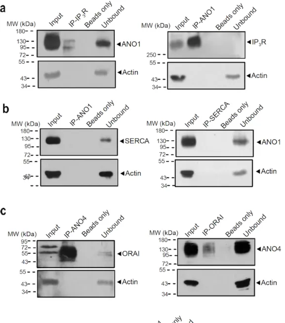
Figure 2.3 | Coimmunoprecipitation of ANO1 and ANO4 with IP3 receptor and SERCA in HeLa cells. ... 30
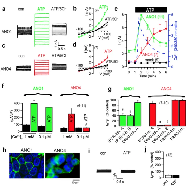
Figure 2.4 | ANO1 and ANO4 Cl- currents are activated by different Ca2+ sources. ... 32
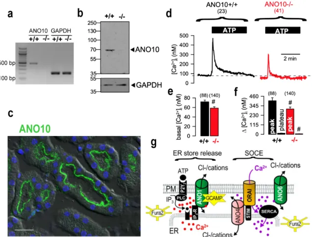
Figure 2.5 | Attenuated Ca2+ signals in isolated epithelial cells from Ano10-/- mice. ... 34
Supplementary Figure 2.1 | Role of ANO1, ANO4, and ANO6 for Ca2+ signaling and whole cell currents. ... 38
Supplementary Figure 2.2 | Anoctamins control activation of ion channels through regulation of Ca2+ levels. ... 40
Supplementary Figure 2.3 | Representative periodic acid-Schiff stainings of kidneys from Ano10+/+ and Ano10-/- animals. ... 41
Supplementary Figure 2.4 | Phenotypic characterization of mice with renal tubular specific Ano10 knockout. ... 41
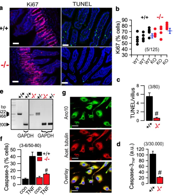
Figure 3.1 | Cellular abnormalities in jejunum of Ano10-/- mice. ... 51
Figure 3.2 | Ca2+ signals and ion transport in jejunum of Ano10-/- mice. ... 53
Figure 3.3 | Abnormal apoptosis in jejunal epithelial cells and macrophages of Ano10 knockout animals. ... 54
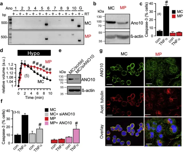
Figure 3.4 | Reduced expression of Ano10 in TPH1 macrophages attenuates regulatory volume decrease and caspase 3 activity. ... 55
Figure 3.5 | Cellular localization and role of Ano10 in FRT cells. ... 57
Supplementary Figure 3.1 | Activation of VRAC by expression of ANO10 in Xenopus oocytes. ... 61
Supplementary Figure 3.2 | Intracellular localization of ANO10. ... 62
Supplementary Figure 3.3 | Relationship between ANO10 and VRAC/LRRC8A. ... 62
Supplementary Figure 3.4 | ANO10 ataxia mutations may compromise functional interaction of ANO10 with the VRAC subunit LRRC8A. ... 63
Figure 4.1 | The TMEM16-inhibitor niflumic acid attenuates inflammatory airway disease. ... 71
Figure 4.2 | Inhibition of TMEM16A and TMEM16F by niclosamide. ... 72
Figure 4.3 | Niclosamide attenuates inflammatory airway disease. ... 74
Figure 4.4 | Niclosamide attenuates inflammatory airway response. ... 76
Figure 4.5 | TMEM16F controls mucus production. ... 77
Figure 4.6 | Production of mucus but not release is affected in TMEM16Fflox/floxCreVil1 intestine. ... 79
Figure 4.7 | Effect of niclosamide on intestinal mucus release. ... 80
Supplementary Figure 4.1 | Role of TMEM16A and TMEM16F for ionomycin-induced quenching in HT29 cells. ... 84
Supplementary Figure 4.2 | Inhibition of Ca2+ signaling by niclosamide in different cell types. ... 84
Supplementary Figure 4.3 | Inhibition of Ca2+ signaling by niclosamide in the presence and absence of extracellular Ca2+. ... 85
Supplementary Figure 4.4 | Role of TMEM16A, TMEM16F and TMEM16K for mucus production in Calu3 cells. ... 86
Supplementary Figure 4.5 | Inhibition of intestinal Ca2+ signals in the absence of TMEM16F, and by niclosamide. ... 87
Supplementary Figure 4.6 | Coupling of P2Y2 receptors but not muscarinic M3 receptors with TMEM16F. ... 87
Figure 5.1 | TMEM16A augments Ca2+ signaling and ion transport in MDCK cells. ... 95
Figure 5.2 | Role of TMEM16A in plasma membrane and primary cilium of MDCK cells. ... 96
Figure 5.3 | M1 renal organoid and cyst model. ... 97
Figure 5.4 | Increased expression of TMEM16A, proliferation and organoid growth by knockdown of PKD1 or PKD2. ... 99
Figure 5.5 | Induction of Cl- secretion by knockdown of PKD1 or PKD2. ... 100
Figure 5.6 | Upregulation of TMEM16A is essential for enhanced Ca2+ signaling upon knockdown of PKD1 and PKD2. ... 101
Figure 5.7 | TMEM16A is essential for enhanced Ca2+ store release by knockdown of PKD1 and PKD2. ... 103
Figure 5.8 | Enhanced Ca2+ signaling in primary renal tubular epithelial cells from PKD-/- mice. .. 104
Supplmentary Figure 5.1 | Inhibition of ATP-induced Ca2+ store release by Ani9. ... 107
Figure 6.1 | Cyst growth of Polycystin-1-deficient collecting duct cells depends on cAMP- and Ca2+- activated secretion. ... 116
Figure 6.2 | Tubule-specific deletion of TMEM16a reduces cyst progression in an ADPKD mouse model. ... 118
Figure 6.3 | Enhanced expression of CFTR and increased cell proliferation in PKD1-/- kidneys depends on overexpressed TMEM16A. ... 119
Figure 6.4 | Induction of Cl- secretion and enhanced Ca2+ signaling by knockdown of PKD1 in medullary primary epithelial cells. ... 121
Figure 6.5 | Niclosamide inhibits polycystic kidney disease in a mouse model for ADPKD. ... 122
Figure 6.6 | Benzbromarone inhibits polycystic kidney disease in a mouse model for ADPKD. .... 123
Supplementary Figure 6.1 | Deletion of Tmem16a inhibits expression of Cftr. ... 126
Supplementary Figure 6.2 | Expression of Tmem16a and Cftr in murine kidney. ... 127
Supplementary Figure 6.3 | Tmem16a is essential for upregulated chloride conductance in primary Pkd1-/- cells. ... 128
Supplementary Figure 6.4 | Inhibition of enhanced store operated Ca2+ entry (SOCE) by TRP and Orai channel inhibitors. ... 129 Supplementary Figure 6.5 | Tmem16a is essential for upregulated chloride conductance in cortical primary epithelial cells from Pkd1-/- mice. ... 130 Supplementary Figure 6.6 | Augmented Ca2+ signals in renal cortical primary epithelial cells. .... 131 Figure 7.1 | Compartmentalized Ca2+ signaling by TMEM16 proteins. ... 134 Figure 7.2 | TMEM16K regulates calcium signaling in the ER modulating apoptosis, proliferation and volume regulation. ... 135 Figure 7.3 | TMEM16A and TMEM16F regulate constitutive mucus and pro-secretory cytokine secretion. ... 137 Figure 7.4 | TMEM16A upregulation increases Ca2+ signaling, secretes chloride and increases proliferation activity in ADPKD. ... 141
LIST OF TABLES
Table 3.1 | RT-PCR mouse primer. ... 45
Table 3.2 | RT-PCR human primer. ... 46
Table 4.1 | Primers used for RT-PCR. ... 68
Table 4.2 | Upregulation of expression of TMEM16A,F,K in airways of asthmatic mice. ... 75
Table 5.1 | RT-PCR Primer (mouse). ... 91
Table 5.2 | Primers for real time PCR. ... 92
Table 6.1 | Genotyping primer sequences. ... 110
CHAPTER 1 | INTRODUCTION
TMEM16 PROTEINS
TMEM16 constitutes a family of ten homologous channels (TMEM16A-K; ANO1-10) characterized by their highly conserved sequence, mostly around the putative pore-forming region 1,2. Although these proteins are broadly expressed and although they are present in almost every cell type 3, their cellular functions are not well defined: some members are calcium-activated chloride channels (CaCCs), while others are described as phospholipid scramblases, that also conduct ions 2. These proteins fulfill various physiological functions such as maintaining hemostasis, chloride (Cl-) secretion by glands, muscle contraction, volume regulation, neuronal excitability, and many more 4. Defects in TMEM16 proteins cause a wide range of diverse health problems, like bleeding disorders, bone malformation, muscular dystrophy, or anxiety 1. Overexpression or enhanced function of TMEM16 proteins cause ataxia, cancer or asthma 1. Recently, the structure of TMEM16 proteins has been identified 1,5,6. The most accurate structure of TMEM16A (anoctamin 1; ANO1) was determined for mouse TMEM16A. mTMEM16A forms a stable dimer with both N- and C- termini located on the cytoplasmatic side of the plasma membrane. Each subunit contains ten transmembrane domains, an extracellular domain and an ion conductive pore with two calcium (Ca2+) binding sites 7. TMEM16A is activated by Ca2+ through stimulation of G- protein-coupled receptors, like those for angiotensin II or adenosine triphosphate (ATP) (Fig.
1.1A) 8-11. Paulino et al. 6 data showed that Ca2+ binding is the essential gating mechanism for TMEM16A. Ca2+ binding in the transmembrane domains α7 and α8 alters the electrostatic properties of the ion channel and triggers structural rearrangement of TMEM16A. This event leads to a conformational rearrangement of α6 domain with the bound ligands, directly coupling Ca2+ binding to pore opening. TMEM16A is upregulated in various types of cancer, such as head and neck squamous cell carcinoma (HNSCC) and colonic cancer. It is generally linked to cell hyperproliferation, migration, and apoptosis.
TMEM16F (anoctamin 6; ANO6) is a Ca2+-activated phospholipid scramblase of the plasma membrane lipid bilayer 12, and also a Ca2+-activated ion channel 13,14 (Fig. 1.1B). TMEM16F is expressed at high levels in bone cells, immune cells and platelets 15,16. During phospholipid scrambling, TMEM16F translocates phospholipids in both directions across the plasma membrane bilayer, which leads to a transient or, in case of apoptotic scrambling, to a sustained collapse of the membrane asymmetry 17,18. Ion transport and scrambling by TMEM16F are activated simultaneously upon a large increase in intracellular Ca2+19,20. The structural characterization of nhTMEM16, a TMEM16F homologue from the fungus Nectria hematococca, revealed the presence of a partially hydrophilic environment within the
conductive pore required for both phospholipid scrambling and ion transport 19. Subsequent structural determination by cryo-electron microscopy demonstrated that human TMEM16F operates as nhTMEM16 6,21. While TMEM16A forms a narrow pore and only conducts Cl- ions, TMEM16F has an open hydrophilic cleft that faces the lipid bilayer and therefore facilitates translocation of phospholipid between membrane leaflets together with ions 18,22.
Figure 1.1 | TMEM16 proteins operate as Ca2+-activated chloride channels (a) or as Ca2+-activated phospholipid scramblases that also conduct ions (b).
TMEM16K (anoctamin 10; ANO10) is an intracellular protein, which is found in various tissues, but is particularly highly expressed in brain frontal cortex, occipital cortex, and cerebellum 11,23,24. TMEM16K most likely also operates as Ca2+-regulated phospholipid scramblase and ion channel 23. TMEM16K is found in Purkinje cells of the cerebellum and mutations in TMEM16K are known to cause autosomal-recessive cerebellar ataxia 23,25-28, a rare neurodegenerative disorder related to coenzyme Q10 deficiency 31. Cerebellar degeneration was associated with dysfunctions in mitochondrial- and DNA-repair.
TMEM16K also has a role in schizophrenia 25 while a coding variant of TMEM16K (ANO10- R263H) was shown to affect the immune response against Borrelia27. TMEM16K is frequently found to be co-expressed with TMEM16A and TMEM16F, especially in neuronal and muscular tissues 23,29. Furthermore, the Drosophila ortholog of TMEM16K, „Axs‟, has been found to play a role in spindle formation and cell cycle progression, mediating meiotic spindle assembly and chromosome segregation 30.
Calcium-dependent chloride secretion
CaCCs exhibit slow activation, voltage dependence, outward rectification of the steady-state current voltage relationship and an anion permeability sequence of I- > Cl-. CaCCs are activated by an increase in intracellular Ca2+ released from the endoplasmic reticulum (ER)
Ca2+ store 31, and by Ca2+ entering the cell through store- or receptor-operated ion channels
32. CaCCs are abundantly expressed in most eukaryotic cells and are associated with many physiological functions, such as epithelial fluid secretion, sensory transduction, control of neuronal and cardiac excitability, regulation of smooth muscle contraction and nociception.
In addition, cell proliferation, phospholipid scrambling, and regulation of cell death is affected by TMEM16. The intestinal epithelium secretes Cl- transiently upon stimulation with carbachol, histamine, and nucleotides, due to transient activation of TMEM16A 33-35. TMEM16A is predominantly expressed in mouse ileal and colonic epithelium. Ca2+-activated Cl- transport was found to be absent in tracheal 36 and colonic epithelium 37, as well as hepatocytes and acinar cells from pancreatic and salivary glands of TMEM16A null mice 1,9. In the renal tubular epithelium, the function of Ca2+-activated chloride channels is to maintain fluid and electrolyte transport as well as acid-base balance 38. In human or mouse kidney, TMEM16A is mainly expressed in proximal tubular epithelial (PTE) cells, in podocytes, intercalated cells of collecting ducts and in the primary cilium of renal epithelial cells 38,39. Thus, TMEM16A is important for growth of the primary cilium 39,40. Faria et al. showed that lack of renal TMEM16A leads to proteinuria and accumulation of numerous large reabsorption vesicles in PTE cells 38. TMEM16A currents were activated by intracellular acidic pH, probably in order to support H+ secretion by the Vacuolar-type H+-ATPase (V- ATPase) 38. In TMEM16A-kidney specific knockout mice, proximal tubular proton secretion was decreased along with endosomal acidification. This caused a defect in proximal tubular albumin reabsorption. Taken together, these results point out that renal TMEM16A has a role in proximal tubular proton secretion and protein reabsorption.
TMEM16A has also been suggested to contribute to apical Cl- secretion in renal cysts in polycystic kidney disease 41-43. Later it was shown that TMEM16A has pro-proliferative function in the cyst lining epithelium 44,45. In mice, tubular-specific knockout of TMEM16A caused reduced numbers of nephrons, albuminuria and tubular damage 46. Regarding TMEM16F, it was found to contribute to volume-regulated Cl- currents and cellular volume regulation 13,47. In fact, upon high intracellular calcium TMEM16F is activated, leading to a decrease in cell volume due to Cl- loss and water efflux. Recent studies showed that reactive oxygen species (ROS) also activate TMEM16F 48. TMEM16F missense mutations have been reported in patients with Scott syndrome, a rare congenital bleeding disorder caused by a defect in platelet aggregation required for blood coagulation 12,49.
Crosstalk between TMEM16A and CFTR
Cystic Fibrosis Transmembrane Conductance Regulator (CFTR) is a Cl- channel that is regulated by cAMP, produced and inactivated by adenylate cyclase and phosphodiesterase, respectively 50. It is expressed in the apical membrane of epithelial cells of many organs,
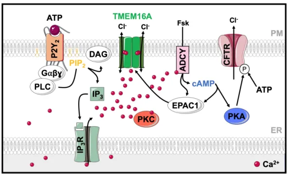
including airways, intestine and kidney. Interestingly, it controls the function of other ion channels such as epithelial sodium channels (ENaC) 51,52, outwardly rectifying Cl- channels (ORCC) 53 and CaCCs 54. Mutations in CFTR cause cystic fibrosis (CF), the most common severe autosomal recessive disease among Caucasians. It is characterized by reduced Cl- and bicarbonate secretion in airways, pancreas, and intestine. This leads to formation of a thick and sticky mucus and a compromised local barrier defense, resulting in frequent airways infections 55. Protein kinase A (PKA) activated by cAMP phosphorylates CFTR at a number of serine residues in the so-called regulatory (R) domain, which is accompanied by ATP-binding to the cytoplasmic nucleotide-binding domains (NBD). Both modifications lead to a conformational change and opening of the CFTR channel 56,57. In addition, CFTR is also Ca2+-regulated, as outlined below. In contrast to CFTR, TMEM16A supports Cl- secretion indirectly by facilitating intracellular Ca2+ signaling required for the activation of basolateral K+ channels and for activation of luminal CFTR 36,37,58 (Fig. 1.3). Due to this regulation, Ca2+- activated Cl- secretion significantly occurs through TMEM16A-mediated activation of apical CFTR and basolateral SK4. Moreover, there is also a functional coupling of CFTR and TMEM16A through exchange protein directly activated by cAMP (EPAC1) and Ca2+-sensitive adenylate cyclase type 1 (ADCY1) 59. Purinergic stimulation of Ca2+ release from the ER not only activates TMEM16A but also stimulates ADCY1, which has a high relevance for production of cAMP. Activation of ADCYs by forskolin (Fsk) generates cAMP which activates not only CFTR, but also TMEM16A via EPAC1 and protein kinase C 36.
Figure 1.2 | TMEM16A and CFTR are functionally related and interact through EPAC1 and ADCY1.
Purinergic stimulation increases Ca2+, not only activating TMEM16A but also stimulating ADCY1, which produces cAMP. The intracellular messenger cAMP activates CFTR via protein PKA. Activation of ADCYs by forskolin generates cAMP and activates EPAC1, activating both CFTR and TMEM16A via EPAC1.
It is known that purinergic P2Y receptors are coupled to intracellular second messengers, like Ca2+ and cAMP 60. Intracellular Ca2+ controls enzymes responsible for production and degradation of cAMP 61. Ca2+ activates CFTR through inhibition of phosphatases and activation of protein kinase C (PKC) 58,62. PKC dependent phosphorylation of CFTR enhances subsequent phosphorylation of the channel by PKA 63. We can conclude that a functional crosstalk between cAMP-activated CFTR and Ca2+-activated TMEM16A takes place 64. This functional relationship between CFTR and TMEM16A may also occur in the kidney. In patients with ADPKD this may contribute to Cl- secretion and cyst development.
Cellular Ca2+ signaling
Intracellular Ca2+ ions regulate a large number of cellular properties and functions. These processes include transcription, cell cycle regulation, differentiation, cell motility/migration, apoptotic cell death, muscle contraction/relaxation and neuronal excitability 65. Cells are equipped with a sophisticated machinery to precisely control intracellular Ca2+ signals, mostly in a spatial and time-dependent manner. Under resting conditions, cells maintain a [Ca2+]i of ~10-7 M, while the extracellular free Ca2+ concentration is around 1.2 mM and thus 10.000 times higher. Stimulation of cells, for example by excitation-induced depolarization, mechanical deformation or hormonal activation, increases the cytosolic Ca2+ concentration up to 1-2 µM 65. However, in subcellular compartments the cytosolic Ca2+ concentration may reach much higher values. Precise regulation of intracellular Ca2+ concentrations requires a sophisticated machinery, consisting of various types of Ca2+ stores such as the ER, acidic endolysosomal stores, and mitochondria, along with a number of transport proteins such as Ca2+-permeable ion channels, Ca2+ pumps and the Na+/Ca2+ exchanger (Fig. 1.2). Ca2+
signaling is triggered by hormonal or electrical stimuli, which generate Ca2+-mobilizing signals to release Ca2+ from internal stores and activate Ca2+ influx. Inositol-1,4,5- triphosphate receptors (IP3R) and ryanodine receptors (RyR) are Ca2+ release channels that allow exit of Ca2+ from the ER or from the sarcoplasmic reticulum (SR) in muscle cells. RyR are activated by entry of extracellular Ca2+ upon depolarization of the plasma membrane, a process called Ca2+-induced Ca2+ release, or by direct interaction of L-type Ca2+ channels with RyR. In contrast, IP3R are activated by inositol-1,4,5-triphosphate (IP3) 66. When agonists bind to G-protein-coupled receptors (GPCR), like adenosine triphosphate (ATP) binding to purinergic receptors (P2Y2), or acetylcholine (ACh) binding to muscarinic acetylcholine receptors (M1-5) 67, heterotrimeric G protein alpha subunits (Gα) bind to phospholipase C (PLC). PLC activation results in cleavage of phosphatidylinositol 4,5- bisphosphate (PIP2) into IP3 and diacylglycerol (DAG). IP3 binding to IP3R triggers release of Ca2+ from the ER into the cytoplasm 68.
There are different types of plasma membrane channels which allow increase of intracellular Ca2+ by entry of Ca2+ from the extracellular space. We can discriminate voltage-operated (VOCC) Ca2+ channels from receptor-operated (ROCC) and store-operated (SOCC) Ca2+
channels65. Store-operated Ca2+ entry (SOCE) results from depletion of Ca2+ within the ER.
This, in turn, activates Ca2+ channels in the plasma membrane such as calcium release- activated calcium channel protein 1 (Orai1) and transient receptor potential (TRP) channels.
Orai1 interacts with stromal-interacting molecule 1 (STIM1), which is the ER Ca2+ sensor, leading to opening of the plasma membrane Ca2+ channel 69. After increasing cytosolic Ca2+, its concentration rapidly returns to basal levels due to Ca2+ pumps and Na+/Ca2+-exchangers.
While Na+/Ca2+ exchangers and plasma membrane Ca2+ ATPases (PMCA) transport Ca2+
out of the cell into the extracellular space, the sarco-endoplasmic reticulum Ca2+ ATPase (SERCA) pumps Ca2+ back into the ER 70,71.
Figure 1.3 | Elements of the Ca2+ signaling toolkit.
Cells possess an extensive Ca2+ signaling toolkit, shown above are some examples relevant to the present project.
Modulation of Calcium signaling by TMEM16 proteins
Several studies suggest that TMEM16 proteins affect intracellular Ca2+ signaling, either by operating as Ca2+ channels or by controlling Ca2+ release from intracellular ER stores
14,37,38,72. Schreiber et al. showed that TMEM16A supports mouse intestinal Cl- secretion by three possible mechanisms 37: (1) TMEM16A may be localized in the ER and facilitate Ca2+
release through IP3R, by operating as counterion channels; (2) TMEM16A may control Ca2+
influx by operating as a plasma membrane localized Cl- bypass channel; (3) TMEM16A may tether the ER to the PM thereby enhancing local sub-membranous Ca2+ signaling. In case of intestinal epithelial TMEM16A, its predominant basolateral location will enhance
predominantly basolateral Ca2+ signals to facilitate activation of basolateral Ca2+-activated potassium (K+) channels. Activation of basolateral K+ channels provides the driving force for apical Cl- secretion by CFTR. Moreover, apical TMEM16A may contribute to activation of CFTR by increasing local Ca2+ concentrations, leading to Ca2+-dependent inhibition of protein phosphatases, Ca2+-dependent activation of protein kinase C (PKC), and stimulation of ADCY1 37. Similarly, overexpression of TMEM16K resulted in upregulated intracellular Ca2+ signaling which supports the function of volume-regulated Cl- channels 11,27. The Drosophila melanogaster ortholog of TMEM16H/K (Aberrant X segregation “Axs”), is an intracellular transmembrane protein located in the ER that was also shown to affect cytosolic Ca2+ signals 73,74. This suggests that Axs and TMEM16K may share similar properties 27. Remarkably, mutations in TMEM16K lead to impaired Ca2+ signaling in Purkinje cells 23,25,26,75. These properties of TMEM16K explain why tissue specific knockout of TMEM16K in intestinal epithelial cells leads to a compromised Ca2+-dependent intestinal Cl- secretion 37.
TMEM16A as tethering protein in lipid rafts
The yeast protein Ist2, a TMEM16A homologue, was described as tethering protein that maintains PM–ER junctions. Due to this property intracellular signaling components associate with the plasma membrane and are concentrated in lipid rafts. Lipid rafts are plasma membrane structures that are known as platforms for cellular signaling 76,77. Ist2 is located in the ER membrane extending its C-terminus to the cytosol. A poly-basic domain at the end of the Ist2 C-terminus acts as an anchor that attaches to the PM, forming a junction with the ER. Among the anoctamins, TMEM16K shows the highest degree of homology with Ist2. Jin et al. reported a similar role of TMEM16A in small nociceptive dorsal root ganglia neurons 78,79. According to their findings, TMEM16A is part of a signaling complex that also harbors GPCRs. They showed that TMEM16A is selectively activated by GPCR-induced release of Ca2+ from ER stores, but not by Ca2+ influx through voltage-gated Ca2+ channels.
Selective Ca2+ signaling is accomplished by direct interaction of TMEM16A with IP3R.
Moreover, TMEM16A was found to interact with ERLIN1, a protein found in the ER and in lipid rafts, that associates dynamically with IP3R. Taken together, these results along with other recent findings concerning the role of TMEM16A in neuronal ganglia 80, validate the role of TMEM16A in cellular Ca2+ signaling 80.
Modulation of Exocytosis by TMEM16 proteins
Exocytosis of intracellular vesicles is due to fusion of the vesicular membrane with the plasma membrane. This leads to release of the vesicular content into the extracellular space, which regulates various biological events such as neurotransmission, hormonal regulation, and inflammation. Moreover, exocytosis also delivers proteins and lipids to the plasma membrane, and is required for cell repair, growth, migration, and regulation of cell signaling
81-85. During exocytosis two membrane lipid bilayers merge into one by shielding the negative charge on the bilayer surface, thus diminishing the electrostatic repulsion force which overcomes the hydration barrier 86. TMEM16F-mediated lipid scrambling was shown to support membrane trafficking in several cell types. For example, TMEM16F is involved in microvesicular release from platelets and neutrophils 87. Human and mouse cells lacking expression of TMEM16F demonstrate a defect in generation of microvesicles 88,89,49,87,90. These are a subset of extracellular vesicles (EVs) that are also referred to as “microparticles”
or “ectosomes”. They shed from the plasma membrane in a Ca2+-dependent manner 91,92. Mice lacking expression of TMEM16F also show deficits in bone development 93, a process that requires release of microvesicles from osteoblasts 94. In macrophages, TMEM16F is necessary for phagocytosis stimulated by the purinergic receptor P2X7 16, while microglia lacking TMEM16F show defects in branch motility and phagocytosis 95. Moreover, TMEM16F-dependent release of EVs have also been implicated in the anti-inflammatory properties of neutrophils during joint inflammation 87. This vesiculation depends on TMEM16F-mediated lipid scrambling as vesicle formation begins after initiation of phosphatidylserine (PS) exposure 96.
A study conducted by Bricogne et al. provided initial evidence that TMEM16F-dependent plasma membrane expansion leads to extensive shedding of plasma membrane vesicles and production of ectosomes 97. Moreover, TMEM16F-null erythrocytes do not produce vesicles in response to intracellular Ca2+ signal 98. TMEM16F requires very high intracellular Ca2+ concentrations for its activation 14,18. Membrane trafficking triggered by high Ca2+ levels occur during the repair of the plasma membrane. Thus, an important function of TMEM16F and Ca2+-stimulated exocytosis could be repair of the plasma membrane. Repair of the plasma membrane occurs physiologically in skeletal muscle due to ongoing mechanical stress 99. Notably, deficiency in TMEM16E, a close paralog of TMEM16F, causes muscular dystrophy that is caused by defective membrane fusion and repair 100. It remains to be established whether TMEM16F is equally involved in membrane repair in other cell types.
A novel function of TMEM16 proteins for mucus secretion
Organs such as airways, stomach and intestine are covered by a mucus layer. In the stomach and colon, mucus is composed of two layers, an adherent layer that is impermeable to bacteria, and a non-adherent layer which traps bacteria 101,102. Similarly, in the airway mucus forms a barrier against bacteria, traps inhaled particles and prevents dehydration 103. Mucus is produced and secreted by specialized cells, called goblet cells. They acquired their name from their characteristic “wine glass goblet” shape, which is created by mucin granules accumulating in the apical region. The major component of intestinal mucus is the mucin MUC2 104,105, while the lungs produce MUC5AC and MUC5AB 103. MUCs are heavily
glycosylated large proteins that reach very high molecular weights and provide mucus with its elastic and gel-forming properties. Mucins consist of oligosaccharide chains, linked to a protein backbone with a high number of threonine/serine and proline-rich tandem repeats (TRs). The protein backbone of the mucins determines the class to which they belong 103. Mucins are synthesized and densely packed intracellularly in secretory vesicles of goblet cells 106. This condensed form is achieved due to the low vesicular pH and high calcium content 107. Upon exocytosis, the dense mucins expand more than 1000-fold. This process depends on HCO3-, required for precipitation of Ca2+, and an increase in pH 108,109. The negative core of mucins is then exposed and will force the compacted mucins into open, soluble and easily transportable strings 107,108.
In the early steps of exocytosis, the GTPase Rab3/27 localizes to secretory granules (SG) membranes and binds to myosin V via the anchoring protein melanophilin, thereby allowing the granule transition from microtubules to the cortical actin network 110-112. Upon stimulation of secretion, myristoylated alanine-rich C kinase substrate (MARCKS) binds to the mucin granules together with the Vesicle-associated membrane protein 8 (VAMP8) and the Cysteine String Protein (CSP) 113,114. This interaction helps to position granules for secretion, from microtubules-kinesin-dependent to actin-myosin-dependent transport at the cell periphery 115. Consecutively, the SGs are tethered and docked to the plasma membrane via interaction of the soluble N-ethylmaleimide sensitive factor attachment receptor (SNARE) proteins that are present on secretory vesicles (v-SNAREs, such as VAMP8) and their target membranes (t-SNAREs, such as Syntaxin and SNAP-23). The v- and t-SNAREs contain- helical domains that interact to form a tightly coiled four-helix bundle (the core complex) that brings together the opposing membranes 81,116. In addition, this event requires accessory components, such as the Munc proteins. In particular Munc18 and Munc13-2/4 promote the interaction of the core complex and syntaxins 81. Munc18 opens the Syntaxin structure and permits its interaction with the SNARE proteins. Munc13-2/4 interacts with Syntaxin in close proximity with Munc18 and dissociates their binding, thus allowing the assembly of the core complex 81,117.
Secretion of mucins can take place in at least two ways, as constitutive secretion or as compound exocytosis. Both events are Ca2+-dependent, following agonistic stimulation of P2Y purinoceptor 2 (P2Y2) receptors (via ATP or UTP), A3 adenosine receptors, muscarinic receptors (via carbachol or muscarinic acetylcholine) or protease-activated receptors 118. Constitutive exocytosis results from low-level stimulation of P2Y2 and A3 adenosine receptors, due to paracrine release of ATP and adenosine. This process includes the fusion of single vesicles with the plasma membrane, mediated by typical exocytic proteins like syntaxins, munc18, VAMP, and SNAP proteins 119. On the other hand, high levels of these
ligands or cholinergic agonists as well as inflammatory mediators lead to a dramatic form of mucus secretion, called compound exocytosis 120. Ca2+ signaling is involved in both the maturation of the vesicles prior to fusion 121 and in the fusion of the secretory vesicles with the plasma membrane 122 (Fig. 1.4).
Figure 1.4 | Regulated mucin secretion.
Left In the basal state, mucin granules become tethered to the plasma membrane by Rab proteins and effectors that have not yet been identified. They exist in the vicinity of components of the exocytic machinery. Center Activation of heptahelical receptors such as those for ATP (P2Y2), leads to activation of Gαβγ, and PLC, resulting in generation of the second messengers DAG and IP3. DAG activates the priming protein Munc13-2/4, and IP3 induces the release of Ca2+ from apical ER to activate Synaptotagmin-2 (Syt2). Munc13-2/4 also participates in granule priming, and an unknown high affinity calcium sensor likely functions in basal secretion rather than Syt2. Right Activation of the regulatory Munc13-2/4 and Syt2 proteins leads to full coiling of the SNARE proteins to induce fusion of the granule and plasma membranes. The interactions of the SNARE proteins take place on a scaffold provided by Munc18c. Exocytic syntaxins contain four hydrophobic coiled-coil domains that must be opened to initiate secretion (left panel), and during fusion the associated Munc18c protein remains associated only by an interaction at the Syntaxin N-terminus (right panel).
In small intestine of CF patients lacking functional CFTR channel 108, mucins remain anchored as levels of local bicarbonate is insufficient to remove calcium ions from the mucins to allow MUC2 unfolding 123. The immune modulator prostaglandin E2 (PGE2) has been shown to induce both fluid and bicarbonate secretion in the human colon 124. In mice and humans, a non-functional CFTR channel causes bacterial overgrowth, distal intestinal obstruction syndrome (DIOS) and sometimes meconium ileus. New insights in mucin
production and exocytosis could improve our efforts to reduce overproduction and hypersecretion of mucins in many diseases in the future. TMEM16A is expressed in intestinal goblet cells. Studies on intestinal goblet cells lacking expression of TMEM16A demonstrated a direct role for TMEM16A in exocytosis and mucus release (Fig. 1.5). We observed in an intestinal-specific TMEM16A knockout mouse enhanced intracellular mucus content in goblet cells 125. This mucus accumulation suggested a defect in mucus secretion. TMEM16A is also found in mucus producing cells of airways. TMEM16A is strongly upregulated under inflammatory conditions such as CF and asthma 125-130. Under normal conditions, mucins are continuously synthesized and released from goblet cells. In contrast, the airway-specific knockout for TMEM16A led to accumulation of mucus in goblet cells 125.
Figure 1.5 | TMEM16A mediates ATP-dependent constitutive exocytosis at the apical pole of goblet cells.
a Colinergic (carbachol; CCH) mediated compound exocytosis occurs independently of TMEM16A from the basolateral side, while b purinergic (ATP) constitutive exocytosis of mucus in single granules on the apical side is mediated by TMEM16A-localized Ca2+ increase.
Increase in Cellular Proliferation via TMEM16 proteins
Changes in [Ca2+]i are associated with progression through the cell cycle. Both extracellular
131,132 and intracellular Ca2+ signals 133 are required for cell proliferation. Ca2+ acts both as ubiquitous allosteric activator and inhibitor of intracellular enzymes in the cytosol, organelles, and nucleus. The best known Ca2+-binding protein is calmodulin (CaM). The complex Ca2+/calmodulin regulates numerous intracellular enzymes including phosphodiesterases, adenylyl cyclases, ion channels, protein kinases, and protein phosphatases (i.e., calcium-
calmodulin dependent kinase type II (CaMKII) and the CaM-dependent phosphatase calcineurin). The G1/S transition, the progression from G2 to M and the metaphase/anaphase transition are specific points of intervention of CaMKII 134,135. Activated CaM-kinase promotes phosphorylation of proteins, such as Ras (a cytosolic Ca2+ responsive transcription factor) and Ca2+/cAMP response element (CRE) binding protein (CREB, a nuclear Ca2+- responsive transcription factor), that are required for initiating and maintaining the cell cycle 136. Additionally, CaM-kinase and calcineurin-regulating nuclear factors are involved in the DNA division machinery, such as cyclin-dependent kinases (cdk4) and cyclins (cyclin D1) 134,135.
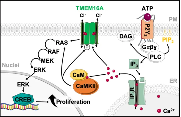
Figure 1.6 | Proposed schematic representation of TMEM16A and CaMKII proliferative activity.
As intracellular Ca2+ levels rise, TMEM16A is activated by Ca2+ and modulated by phosphorylation involving CaMKII. TMEM16A and CaMKII activate RAS-RAF-MEK-ERK pathway, which promotes the expression of genes for cell proliferation.
The function of TMEM16A as a regulator of proliferation and cancer growth is well known
137,138. Upregulation of TMEM16A was also found in polycystic kidney disease 44. TMEM16A is known to activate the RAS-RAF-MEK-ERK pathway 139. Moreover, CaMKII is reported to regulate Ca2+-activated Cl- currents in rabbit arterial and portal vein smooth muscle cells 140. Wang et al. 141 found that inhibiting CaMKII activity can lead to an increase of CaCC currents in basal smooth muscle cells (BASMCs) of the angiotensin II-induced hypertensive rats. This promotes proliferation of BASMCs and contributes to cerebral vascular remodeling. These and other data suggest that TMEM16A is modulated by CAMKII, thereby participating in cell proliferation (Fig. 1.6). Apart from TMEM16A, other members of the anoctamin family have also been implicated in regulation of cell proliferation and cancer development, like TMEM16E (ANO5), TMEM16G (ANO7) and TMEM16J (ANO9) 142. TMEM16E is now known
for its role in myoblast proliferation and muscle repair 143,144. Similar to TMEM16E, TMEM16F was reported to control myoblast proliferation 145. TMEM16G was shown to be a marker for prostate cancer 146,147. For TMEM16J an inverse correlation of expression and progression of colorectal cancer was described 148, while it may also promote pancreatic cancer 149.
AUTOSOMAL DOMINANT POLYCYSTIC KIDNEY DISEASE
Polycystic kidney diseases (PKDs) comprise a number of inherited disorders in which renal tubules become structurally and functionally abnormal, resulting in kidney cyst development and growth 150. The most common form, autosomal dominant polycystic kidney disease (ADPKD), affects 1 in 400-1000 individuals and is characterized by an acute and bilateral development of multiple renal cysts over time. This disease results in the displacement and destruction of the adjacent renal parenchyma, progressive organ enlargement and, ultimately, loss of renal function, being the cause of 10% of end-stage renal disease (ESRD) cases worldwide, which demands renal replacement therapy 151,152. Extrarenal manifestations also comprise cysts in other epithelial organs including the liver and pancreas
153.
ADPKD is a genetically heterogeneous disease caused by a germline mutation in one oftwo genes: Polycystic Kidney disease 1 (PKD1) or Polycystic Kidney disease 2 (PKD2) 154,155. Mutations in PKD1 (located on chromosome 16p13.3) lead to cysts development in 85% of the patients and cause a more serious clinical progression. The remaining 15% of the cases are due to PKD2 mutations (located on chromosome 4q 21-23) 156 and present a milder phenotype with later onset, longer renal survival and fewer complications 157. Even though ADPKD is a dominant disease, it behaves recessively at the cellular level. The human kidney is constituted of approximately a million nephrons. Nevertheless, microdissection studies indicated that cysts in ADPKD patients arise from about 1000 nephrons 158. All cells from the patient possess a germline mutation in either PKD1 or PKD2 but not all develop cysts, which indicates that the germline mutation alone cannot cause cystogenesis. A second-hit model was proposed to explain this phenomenon. This model states that every cyst emerges as a product of an individual somatic mutation event in addition to the germline mutation in either PKD1 or PKD2 alleles, leading to total loss of function of the polycystins 159,160. This hypothesis can explain the slow advance of the pathology over several decades. Recent studies show that late inactivation of the Pkd1 gene in adult mice kidneys resulted in a slow onset of cystogenesis 156,161,162. The authors reported that the slow onset of the disease could not be explained by the previous hypothesis and suggested a third hit theory. This hypothesis states that a cell is not necessarily cystogenic just by having a germline mutation (first hit) and a somatic inactivation of the second allele (second hit) 163,164, but by having an additional event that leads to cell proliferation and cyst growth (such as renal injury) 165. Although
disease-causing mutations are known, the complex mechanisms of cyst development and cyst growth are still poorly understood 166,167.
Regulation of Calcium signaling by Polycystins
Polycystin-1 (PC1), encoded by the PKD1 gene, is an integral membrane protein and is composed of eleven transmembrane domains, an extensive extracellular N-terminal domain, and a shorter C-terminal cytosolic domain 156. The N-terminus of PC1 contains well- recognized motifs involved in protein-protein, protein-saccharide, and protein-ligand interactions. Thus, PC1 is generally thought to function as a cell surface receptor and it is critical for cell-cell or cell-matrix interactions 152,168-172. It is mainly expressed in epithelial cells of developing and mature renal tubules. PC1 expression is regulated over time showing higher levels in foetal tissues and decreased presence in adult tissues 173. PC1 is localized on the plasma membrane, adhesion complexes in polarized epithelial cells and on the primary cilium, a single hair-like organelle that protrudes from the surface of most mammalian cells, implicated in many cystic diseases 174,175.
Polycystin-2 (PC2) or TRPP2, encoded by the PKD2 gene, is a membrane-associated protein that belongs to the TRP family 176. It has 6 transmembrane domains, two EF-hands (characteristic of Ca2+ binding domains that allow the protein to sense changes in Ca2+), an ER retention signal and cytoplasmic N- and C-terminal domains 155,177. PC2 forms homotetramers in the absence of PC1 178-180, which works as a nonselective cation channel with high affinity to Ca2+, being also permeable to Na+ and K+ ions 181-184. Although PC2 is found on the primary cilium and plasma membrane, where it mediates Ca2+ entry into the cytoplasm, most of its pool is located in intracellular compartments, such as the ER and the Golgi complex, acting as Ca2+ release channel183,185. PC2 regulates intracellular Ca2+ on the plasma membrane and primary cilium by interacting with other members of TRP superfamily, namely TRPC1 and TRPV4, to form an heteromeric channel 186-188.
The most significant partner of PC1 is PC2 189, interacting through their C-terminal coiled- coil domains 152,169. Biochemical and biophysical studies found that the PC1/PC2 complex contains one PC1 and three PC2 subunits 190-192. Several studies suggested that PC1 and PC2 reciprocally affect the cellular location of each other, and defects in one of the proteins lead to a mislocalization of the other one, however the precise mechanism of this interdependence has not yet been elucidated 193. It is thought that the PC1/PC2 interaction is critical for creation of a functional ion channel, yet, details remain unknown. It was shown that both PC1 and PC2 play a key role in intracellular Ca2+ homeostasis regulation, not only in the primary cilium, but also at the plasma membrane level and in intracellular compartments 194. In the ER, polycystins are known to interact with the IP3R 195,196. PC2 can
enhance Ca2+ release from the ER by stimulating the activity of this receptor 197, whilst PC1 suppresses this mechanism by a decrease on PC2-IP3R interaction involving STIM1 198. PC2 is also known to regulate the RyR 199. Consequently, it has been suggested that both PC1 and PC2 might regulate Ca2+ signaling in different intracellular compartments, and their dysregulation is responsible for the activation of aberrant pathways present in ADPKD, such as the abnormal fluid secretion events. However, the precise nature of these regulatory events remains to be fully elucidated.
Lack of PKD2 may also affect mitochondrial dynamics 200. Deficiency of PKD2 has been reported to be accompanied by changes in mitochondrial morphology and dynamics, with prolonged ER-mitochondria Ca2+ communication and increased Ca2+ mitochondrial uptake.
As mentioned before, the so-called primary cilium is a single antenna-like protrusion of the plasma membrane containing receptors, ion channels and signal proteins 201. PC1 and PC2 form a mechano- and/or chemosensory complex that transduces tubular stimuli to tubular epithelial cells 166. Primary cilia also form a signaling platform during vertebrate development, including hedgehog signaling. This requires proper distribution of phosphatidylinositol phospholipids 202. Mechanical, chemical and receptor-mediated signals increase intracellular Ca2+ within the cilium and possibly in the cytosol, although this is still a subject of discussion
203,204. Nevertheless, primary cilia can be regarded as specialized Ca2+ signaling compartments, with PC1 and PC2 forming a receptor/Ca2+ influx-channels complex 205. Notably, formation of the primary cilium is cell cycle-dependent, being disassembled upon re-entering the cycle 206,207. There is an accumulation of TMEM16A, as well as TMEM16F and TMEM16K, in the primary cilium 39,208,209 and TMEM16A was suggested to be essential for its maintenance. Thus, TMEM16A and hedgehog signaling are simultaneously active during development and in cancer cells 210.
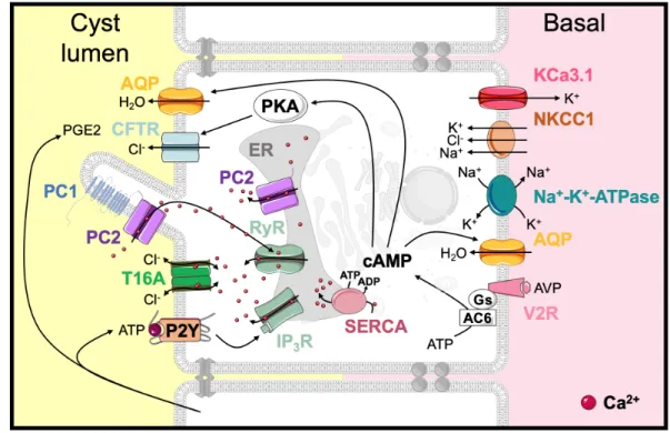
Cyst development as a consequence of enhanced proliferation and fluid secretion Continuous dilation of the tubules through increased cell proliferation, defects in the extracellular matrix, transepithelial fluid secretion and, ultimately, separation from the parental tubule lead to the formation of cysts in ADPKD 211,212. It is still unclear if cysts specifically arise from a determined nephron segment. Some studies report a generalized pattern 213. Chloride secretion by renal collecting ducts is regulated by circulating hormones and paracrine factors binding to GPCRs and modulation of intracellular cAMP and Ca2+
levels. Arginine Vasopressin (AVP) is the central hormone involved in the regulation of plasma osmolality by modulating water permeability of the collecting duct. AVP binding to V2 receptors (V2R) on the distal nephron and collecting ducts stimulates the production of intracellular cAMP by activation of adenylyl cyclase 6 (AC6) 214,215. In cystic epithelial cells, AVP maintains cAMP at elevated levels, activating pathways involved in cell proliferation