Role of prohibitins for proteolysis in yeast and murine mitochondria
I n a u g u r a l – D i s s e r t a t i o n
zur
Erlangung des Doktorgrades
der Mathematisch- Naturwissenschaftlichen Fakultät der Universität zu Köln
vorgelegt von
Metodi Dimitrov Metodiev
aus Targovishte, Bulgarien
Köln, 2005
1. Berichterstatter
Prof. Dr. Thomas Langer 2. Berichterstatter
Prof. Dr. R. Jürgen Dohmen
Tag der mündlichen Prüfung 10.01.2006
I. INTRODUCTION 5
1. The proteolytic system of mitochondria 5
1.1. ATP-dependent proteases in mitochondria 5
1.1.1. Lon- and Clp-proteases in the mitochondrial matrix 6
1.1.2. AAA proteases in the inner mitochondrial membrane 7
1.1.2.1. Domain structure of the AAA protease subunits 8
1.1.2.2. Function of the mAAA protease in yeast 11
1.1.2.3. Mammalian mAAA proteases 12
2. Prohibitins 13
2.1. Prohibitins are conserved SPFH-domain proteins 15
2.1.1. Functions of the SPFH domain 15
2.1.1.1. Role of the SPFH-domain for localization to the plasma membrane and lipid
rafts 15
2.1.1.2. Role of the SPFH-domain for protein-protein interactions and as a structural
scaffold 16
2.1.2. Functions of the proteins harboring SPFH-domains 17
2.1.2.1. Regulation of channels across the plasma membrane 17
2.1.2.2. Regulation of proteases 18
2.3. Expression of the prohibitin gene in mammals 18
2.4. Cellular localization of prohibitins 18
2.5. Prohibitins function as regulators of cell proliferation and are important for
normal development 19
2.5.1 Function of Prohibitin 1 as a regulator of cell proliferation and apoptosis 19
2.5.2. Role of Phb2 as a regulator of the estrogen signaling pathway 22 2.6. Mitochondrial localization and function of prohibitins 23
2.6.1. Assembly and structure of the yeast prohibitin complex in the inner mitochondrial
membrane 23
2.6.2. Role of prohibitins for mitochondrial morphology and inheritance 25
2.6.3 Role of prohibitins during proteolysis in mitochondria 26
II. AIMS OF THESIS 29
III MATERIALS AND METHODS 30
TABLE OF CONTENTS
1. Nomenclature of prohibitins 30
2. Materials 30
2.1. Yeast strains 30
2.2. Mouse strains 32
2.3. E. coli strains 32
2.4. Plasmids 33
2.5. Oligonucleotides 34
2.6. Enzymes and chemicals 34
2.7. Buffers 34
2.8. Yeast and E.coli growth media 35
3. Methods 36
3.1. Molecular biology and genetics methods 36
3.2. Biochemical methods 39
3.3. Immunological methods 46
3.4. Cell biology methods 51
3.5. Protein and DNA sequence analysis 53
IV. RESULTS 55
1. Function of Phb1 and Phb2 during proteolysis in yeast 55 1.1. The mAAA protease is quantitatively assembled with prohibitins 55 1.2. Role of prohibitins for the turnover of mitochondrial proteins 56
1.2.1. Degradation of non-assembled, mitochondrially encoded polypeptides 57
1.2.2. Stability of overexpressed model substrates 57
1.2.2.1. Stability of Oxa1ts 59
1.2.2.2. Stability of the membrane protein Yme2∆C 60
1.3. Synthetic interaction of prohibitins with oxa1ts 62 1.4. Physical interaction between prohibitins and the mAAA protease in the inner
mitochondrial membrane 63
1.4.1. A procedure for monitoring the assembly of the mAAA protease in a suprcomplex with prohibitins
1.4.2. Formation of the supercomplex between prohibitins and the mAAA protease is
nucleotide independent 65
1.4.3. Mutations in the Walker motifs of Yta10 and Yta12 inactivate the mAAA protease 67
1.4.3.1. Impaired assembly of the respiratory chain complexes in cells expressing
mutant Yta10 and Yta12 subunits 67
1.4.3.2. Processing of Ccp1 is inhibited by mutations in the Walker motifs of Yta10 and
Yta12 subunits of the mAAA protease 68
2. Identification of a supercomplex containing prohibitins and mAAA protease
subunits in murine mitochondria 70
2.1. The Phb1-Phb2 complex in murine mitochondria 70 2.2. Generation of antibodies specific for putative subunits of the murine mAAA
protease 71
2.3. Paraplegin, Afg3l1 and Afg3l2 co-migrate with Phb1 and Phb2 in BN-SDS PAGE
and sizing chromatography 72
2.4. Phb1 and Phb2 physically interact with paraplegin, Afg3l1 and Afg3l2 73 3. Subunit composition of murine mAAA proteases 75 3.1. Interactions between paraplegin, Afg3l1 and Afg3l2 75 3.2. Heterooligomeric Afg3l1-Afg3l2 complexes in mitochondria from a paraplegin-
deficient mouse strain 77
V. DISCUSSION 80
1. Role of prohibitins during proteolysis in yeast 80 2. Role of prohibitins in murine mitochondria 85 3. Murine mAAA proteases with variable subunit composition 85
VI. APPENDIXES 90
Appendix I 90
Appendix II 94
VII. REFERENCES 95
VIII. ABBREVIATIONS 107
LEBENSLAUF 110
EIDESSTATTLICHE ERKLÄRUNG 111
Zusammenfassung
Die Prohibitine Phb1 und Phb 2 sind evolutionär konservierte Proteine mit unterschiedlicher zellulärer Lokalisation und Funktion. In Hefe formen Prohibitine einen hochmolekularen Komplex in der inneren Mitochondrienmembran, der physikalisch mit der mAAA Protease interagiert und die Stabilität von nicht- assemblierten proteolytischen Substraten beeinflußt. In der vorliegenden Arbeit wurden zwei mögliche Funktionen von Prohibitinen bei der Proteolyse untersucht: 1) Prohibitine als negativer Regulator in der mAAA Protease (Steglich et al., 1999) und/oder 2) Prohibitine in der Funktion als molekulare Chaperone für die Assemblierung neu synthetisierter mitochondrialer Proteine (Nijtmans et al., 2000).
Um diese beiden Funktionen unterscheiden zu können, wurde die Stabilität der proteolytischen Substrate der mAAA Protease in Zellen untersucht, die unterschiedliche Mengen von Prohibitinen enthielten. Während die Überexpression von Prohibitinen die Stabilität von nicht-nativen Proteinen der inneren Membran nicht erhöhte, bewirkte die Abwesenheit von Prohibitinen ihre schnelle Proteolyse. Die Ergebnisse dieser Arbeit lassen also vermuten, daß Prohibitine bei der Stabilisierung von mitochondrialen Proteinen nicht als Chaperone fungieren. Es scheint eher, daß Prohibitine durch die Bindung an mAAA Protease in einer Nukleotid-unabhängigen Weise funktionieren und eventuell die Zugänglichkeit von Substraten für die Protease oder die Aktivität der Protease beeinflussen.
mAAA Protease ist ein entscheidender Faktor im mitochondrialen Qualitätskontrollsystem in Hefe und menschlichen Zellen (Arlt et al., 1996; Arlt et al., 1998; Nolden et al., 2005). In menschlichen Zellen besteht Protease aus Paraplegin und Afg3l2, die homolog zu den Hefeproteinen Yta10 und Yta12 sind (Atorino et al., 2003). In Mäusen wird eine dritte AAA Untereinheit, genannt Afg3l1 exprimiert (Kremmidiotis et al., 2001) und somit bleibt die Zusammensetzung der murinen mAAA Proteaseuntereinheit weiter unbekannt. In dieser Arbeit wurden die Komplexe aus Paraplegin, Afg3l1 und Afg3l2 in den Mitochondrien von Wildtyp- und SPG7-/- Mausstämmen untersucht. Immunpräzipitierungsexperimente zeigten, daß Paraplegin, Afg3l1 und Afg3l2 physikalisch interagieren und in einem hochmolekularen Komplex mit Prohibitinen enthalten sind. Dies deutet auf eine konservierte Rolle von Phb1 und Phb2 für die Proteolyse in murinen Mitochondrien
hin. Co-Immunpräzipitierung und Immunabbauexperimente mit Lebermitochondrien aus SPG7-/- Mausstämmen zeigten, daß Afg3l1 und Afg3l2 ebenfalls hochmolekulare Komplexe in Abwesenheit von Paraplegin bilden können. Darüberhinaus waren diese Komplexe in der Lage, mit Prohibitinen zu interagieren, was darauf hindeutet, daß Paraplegin für diese Interaktion nicht essentiell ist.
Offenbar gibt es in der inneren Membran muriner Mitochondrien mAAA Proteasen mit unterschiedlicher Zusammensetzung der Untereinheiten. Die physiologische Relevanz dieser mAAA Proteasen mit unterschiedlicher Zusammensetzung der Untereinheiten soll hinsichtlich ihrer Substratspezifität und/oder unterschiedlichen Gewebeverteilung diskutiert werden.
Abstract
Prohibitins, Phb1 and Phb2, are evolutionary conserved proteins with diverse cellular localization and different functions. In yeast, prohibitins form a high molecular weight complex in the inner mitochondrial membrane which physically interacts with the mAAA protease and affects the stability of non assembled proteolytic substrates. In the present work, I examined two possible roles of prohibitins in proteolysis: i) role as negative regulators of the mAAA protease (Steglich et al., 1999) and/or ii) function as molecular chaperones for the assembly of newly synthesized mitochondrial proteins (Nijtmans et al., 2000). To discriminate between both possibilities, the stability of proteolytic substrates of the mAAA protease was examined in cells harboring different levels of prohibitins. While overexpression of prohibitins did not stabilize non-native inner membrane proteins absence of prohibitins resulted in their rapid proteolysis. The results presented in this work suggest that prohibitins do not play a role as chaperones for the stabilization of mitochondrial proteins. Rather, prohibitins function by binding to the mAAA protease in a nucleotide independent manner and presumably modulating the accessibility of substrate to the protease or the activity of the protease.
The mAAA protease is recognized as a crucial component of the mitochondrial quality control system in yeast and in human (Arlt et al., 1996; Arlt et al., 1998;
Nolden et al., 2005). In human, the mAAA protease is built up of paraplegin and Afg3l2 which are homologous to the yeast Yta10 and Yta12 proteins (Atorino et al., 2003). In mice, a third AAA subunit called Afg3l1 is expressed (Kremmidiotis et al., 2001) and, therefore, the subunit composition of the murine mAAA protease is unknown. Here, the complexes between paraplegin, Afg3l1 and Afg3l2 were examined in mitochondria from wild type and in SPG7-/- mouse strains.
Immunoprecipitation experiments showed that paraplegin, Afg3l1 and Afg3l2 physically interact and are contained in a high molecular weight complex with prohibitins suggesting a conserved role of Phb1 and Phb2 for proteolysis in murine mitochondria. Co-immunoprecipitation and immunodepletion experiments using liver mitochondria from a SPG7-/- mouse strain showed that Afg3l1 and Afg3l2 can also form high molecular weight complexes in the absence of paraplegin. These complexes were additionally able to interact with prohibitins suggesting that
paraplegin is not essential for this interaction. Since paraplegin-specific antibodies are able to precipitate Phb1 and Phb2, it is conceivable that also paraplegin-Afg3l1 and paraplegin-Afg3l2 complexes will be able to bind prohibitins.
Thus, mAAA proteases with different subunit composition appear to exist in the inner membrane of murine mitochondria. The physiological relevance of mAAA proteases with different subunit composition is discussed in view of differences in their substrate specificities and/or different tissue distribution.
INTRODUCTION
I. INTRODUCTION
Prohibitins occur in a wide range of species and exhibit high similarity on both nucleotide and amino acid levels. Their strong conservation along with their abundance and ubiquitous expression point to an important role in the cell. To this moment, however, the molecular function of prohibitins has not been defined. In yeast mitochondria, prohibitins have been linked to the mitochondrial protein quality control system and demonstrated to directly interact with the mitochondrial ATP dependent mAAA protease (Steglich et al., 1999). Additional functions of prohibitins in mitochondria and in other cellular compartments have also been described and are discussed below. Since this work focuses on the function of prohibitins during proteolysis, a brief introduction to the mitochondrial proteolytic system is provided.
1. The proteolytic system of mitochondria
Many protein complexes in mitochondria are composed of subunits encoded by both the nuclear and the mitochondrial genome. Since imbalance of nuclearly or mitochondrially encoded subunits can lead to an accumulation of potentially harmful unassembled polypeptides, mitochondria like any other cellular compartment possess a protein degradation system (Kalnov et al., 1979; Goldberg et al., 1985).
Major components of this system are proteases which ensure removal of unassembled or damaged proteins and/or function during import of the nuclearly encoded proteins (Figure 1). During protein import, the matrix processing peptidase (MPP) (Brunner et al., 1994), the intermediate peptidase (MIP) (Kalousek et al., 1992; Isaya et al., 1994) and the innermembrane peptidase (IMP) (Behrens et al., 1991; Schneider et al., 1991; Nunnari et al., 1993; Esser et al., 1996) are responsible for the cleavage of targeting presequences of nuclearly encoded proteins.
1.1. ATP-dependent proteases in mitochondria
Improperly folded or unassembled proteins are removed by the action of ATP- dependent proteases within mitochondria (Figure 1) (Kalnov et al., 1979; Desautels and Goldberg, 1982). These proteases are ubiquitously present in prokaryotes and eukaryotes and belong to the AAA+ superfamily of P-loop ATPases (ATPase associated with a variety of cellular activities”) (Beyer, 1997; Neuwald et al., 1999;
Vale, 2000; Ogura and Wilkinson, 2001; Frickey and Lupas, 2004; Iyer et al., 2004).
ATP-dependent proteases are active in all subcompartments of mitochondria and fall into three subfamilies: Lon-, Clp- and AAA-proteases (Ogura and Wilkinson, 2001, Frickey, 2004 #1361; Nolden et al., 2005).
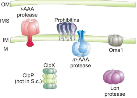
Figure 1. ATP-dependent proteases in the mitochondrial matrix and in the inner mitochondrial membrane. The Lon-protease and ClpXP (ClpP not present in yeast) are responsible for proteolytic breakdown of misfolded polypeptides in the mitochondrial matrix. Integral membrane and peripherally associated proteins are degraded by proteases of the inner mitochondrial membrane, the iAAA protease, active on the intermembrane side of the inner membrane; the mAAA protease, active in the mitochondrial matrix and the ATP-independent metallo-peptidase Oma1, which is thought to possess catalytic domains at the side of the inner membrane. Prohibitins built up a supercomplex with the mAAA protease. OM, outer membrane; IMS, intermembrane space; IM, inner membrane; M, mitochondrial matrix . Reprinted with modifications from (Nolden et al., 2005)
1.1.1. Lon- and Clp-proteases in the mitochondrial matrix
Lon proteases have been identified in the mitochondrial matrix of yeast and mammals (Figure 1) (Watabe and Kimura, 1985; Wang et al., 1993; Suzuki et al., 1994; Van Dyck et al., 1994; Wang et al., 1994). Lon-proteases are classified as serine proteases as they harbor a catalytic serine-lysine dyad and are presumably homooligomeric, ring-shaped complexes with hexameric or heptameric structure.
(Stahlberg et al., 1999; Botos et al., 2004; Rotanova et al., 2004). The Lon-protease in yeast, which is termed PIM1 (Van Dyck et al., 1994), mediates the proteolytic removal of aggregated proteins in collaboration with the chaperone mtHsp70, and its co-factors Mdj1 and Mge1 (Wang et al., 1993; Suzuki et al., 1994; Van Dyck et al.,
INTRODUCTION 1994; Wagner et al., 1994). Deletion of PIM1 is associated with inhibited growth on glycerol (Suzuki et al., 1994; Van Dyck et al., 1994). Mitochondrial matrix proteins are not degraded and tend to accumulate as electron dense inclusions in ∆pim1 cells (Suzuki et al., 1994). Similarly, downregulation of the human Lon protease leads to accumulation of protein inclusions, impaired mitochondrial function and apoptotic cell death (Bota et al., 2005). Pim1 is additionally required for maintenance of mtDNA in yeast by an unknown mechanism probably involving its ability to bind GT-rich DNA sequences (Suzuki et al., 1994; Van Dyck et al., 1994; Fu et al., 1997; Liu et al., 2004).
Clp (caseino-lytic protease) proteases are also found in the mitochondrial matrix space (Leonhardt et al., 1993; Van Dyck et al., 1998). In contrast to Lon- and AAA- proteases, their ATPase domain and the proteolytic domain are expressed as separate gene products. The ATPase domains (ClpX, ClpA) assemble into hexameric, ring complexes with ATPase and chaperone activity, while the proteolytic domains (ClpP) form heptameric, double ring complexes (Bochtler et al., 1997; Wang et al., 1997; Weber-Ban et al., 1999; Bochtler et al., 2000; Kang et al., 2002; Ortega et al., 2004). The ATPase domains determine the substrate specificity of the Clp- protease and also exert regulatory functions during proteolysis (Schmidt et al., 1999;
Weber-Ban et al., 1999; Flynn et al., 2001; Kang et al., 2002; Ortega et al., 2004).
While ClpX-like ATPase subunits are present in all organisms, ClpP-like proteins are present in mammals and plants, but have not been detected in yeast (Van Dyck et al., 1998; De Sagarra et al., 1999; Kang et al., 2002).
1.1.2. AAA proteases in the inner mitochondrial membrane
Two AAA proteases, active on both sides of the inner mitochondrial membrane have been identified in yeast and are presumably present in all organisms, the iAAA protease and the mAAA protease (Figure 1). The iAAA protease is presumably a homooligomeric complex of the Yme1 protein in yeast (Leonhard et al., 1996; Weber et al., 1996) and Yme1l/YME1L protein in mouse and human (Shah et al., 2000). In contrast, the mAAA protease is a heterooligomeric complex of Yta10 (Afg3) and Yta12 (Rca1) in yeast, and paraplegin and Afg3l2 in human (Arlt et al., 1996; Atorino et al., 2003). Three putative mAAA protease subunits have been identified in mice:
paraplegin, Afg3l1 and Afg3l2 (Kremmidiotis et al., 2001). Studies in yeast show that
both proteases have different topology into the inner mitochondrial membrane. While the iAAA protease exposes catalytic domains to the intermembrane space, catalytic domains of the mAAA protease are exposed to the matrix side of the inner mitochondrial membrane (Arlt et al., 1996; Leonhard et al., 1996).
1.1.2.1. Domain structure of the AAA protease subunits
The subunits of the iAAA and the mAAA protease share a common domain structure (Figure 2A). Each subunit is anchored to the inner mitochondrial membrane by one (Yme1) or two (Yta10 and Yta12) transmembrane (TM) domains at their N-terminal ends (Pajic et al., 1994; Arlt et al., 1996; Weber et al., 1996). The membrane spanning segments in the FtsH protease, a bacterial homologue of mitochondrial AAA-proteases, have been shown to play a crucial role for the oligomerization of the protease (Akiyama and Ito, 2000). Similarly, subunits of the mAAA protease lacking both TM-domains fail to assemble (Korbel et al., 2004). However, deletion of the TM- domain of one of the subunits does not inactivate the mAAA protease as cells expressing Yta10∆TM or Yta12∆TM can grow on glycerol (Korbel et al., 2004). The TM-domains are also dispensable for the degradation of peripheral membrane proteins (Atp7) and for the presequence removal of Ccp1 (Korbel et al., 2004).
Nevertheless, proteolysis of integral membrane proteins is impaired suggesting a specific role of the TM-domains of Yta10 and Yta12 during degradation of integral membrane proteins (Korbel et al., 2004).
The transmembrane domains are followed by the AAA domains with ATPase activity (Figure 2A). These domains contain conserved motifs which ensure binding (Walker A) and hydrolysis (Walker B and second region of homology, SRH) of ATP during proteolysis (Figure 2B) (Walker et al., 1982; Whiteheart et al., 1994; Hanson and Whiteheart, 2005). Homology modeling, site directed mutagenesis and crystallographic analysis have led to partial understanding of the mechanism of ATP hydrolysis in AAA+ proteins, including FtsH (Babst et al., 1998; Lenzen et al., 1998;
Yu et al., 1998; Bochtler et al., 2000; Sousa et al., 2000; Zhang et al., 2000; Wang et al., 2001; Krzywda et al., 2002). The Walker A motif contains the so called P-loop whose conserved lysine residue is crucial for binding of ATP. The lysine residue (K201) in the Walker A motif of FtsH (corresponding to K334 and K394 in Yta10
INTRODUCTION
Figure 2. Domain structure of the subunits of the iAAA and the mAAA protease. A. The nuclear- encoded subunits are imported into mitochondria via an N-terminal mitochondrial targeting sequence (MTS) which is removed after import. One (Yme1) or two (Yta10 Yta12, FtsH) transmembrane (TM) domains are present in each subunit. Towards the C-terminus are AAA-domains containing conserved motifs, characteristic for the AAA protein family; Walker A (WA) and Walker B (WB) motifs as well as the second region of homology (SRH). The AAA-domains are followed by proteolytic domains harboring the HEXGH metal binding motif. At the most C-terminal end is the coiled-coil region (CC).
Reprinted with modifications from (Nolden et al., 2005). B. A multisequence alignment of the amino acid regions containing the conserved Walker A, Walker B and the HEXGH motif from yeast, mammalian and bacterial AAA-protease subunits. Sequence coordinates of the amino acids are indicated. Alignment of the complete protein sequences is provided in Appendix I. m, mouse; h, human; ↑, amino acids crucial for the ATPase and the proteolytic activity of the subunits.
and Yta12, respectively) forms hydrogen bonds with oxygen atoms of the β− and γ−phosphates of ATP (Karata et al., 2001; Krzywda et al., 2002). Mutations of the corresponding lysine residue of other AAA+ proteins, including FtsH, are proposed to inhibit the protein via preventing binding of ATP (Karata et al., 1999; Hanson and Whiteheart, 2005). Together with the Walker A motif, aspartate (D254) and glutamate (E255) amino acids from the Walker B motif are crucial for the ATPase activity (Figure 2B) (Babst et al., 1998; Lenzen et al., 1998; Krzywda et al., 2002; Hanson and Whiteheart, 2005). The aspartate amino acid (D254) forms hydrogen bonds with
water molecules liganded to Mg2+ ions while the glutamate amino acid (E255 corresponding to E388 of Yta10 and E448 of Yta12) activates a water molecule for nucleophilic attack on ATP (Krzywda et al., 2002). Mutational exchange of the conserved glutamate renders the AAA+ proteins inactive probably due to defects in ATP hydrolysis but not ATP binding (Whiteheart et al., 1994; Babst et al., 1998;
Hanson and Whiteheart, 2005). Similarly, FtsH is inactivated by the mutational exchange of E255 in the Walker B motif (Karata et al., 2001). In addition to the Walker motifs, residues from the SRH function during ATP-hydrolysis as γ−phosphate groupsensors and arginine-fingers (Karata et al., 1999; Song et al., 2000; Karata et al., 2001; Krzywda et al., 2002; Ogura et al., 2004). Commonly, the AAA+ proteins assemble into oligomeric complexes with hexameric and sometimes heptameric structure (Lenzen et al., 1998; Yu et al., 1998; Vale, 2000; Hanson and Whiteheart, 2005). In these complexes ATP-binding sites are positioned in the interface between the subunits (Lenzen et al., 1998; Yu et al., 1998; Krzywda et al., 2002). Arginine side groups protrude into the ATP binding pocket of a neighboring subunit and contact the γ−group of an ATP bound there, thereby ensuring intersubunit cooperativity during ATP hydrolysis (Karata et al., 1999; Song et al., 2000; Karata et al., 2001; Krzywda et al., 2002; Ogura et al., 2004). Additional residues within the motifs interact to form a suitable environment for ATP hydrolysis (Krzywda et al., 2002). In addition to its ATPase function the AAA domain also binds unfolded membrane proteins (Leonhard et al., 2000).
The proteolytic domain and a conserved coiled-coil region are present C-terminally from the AAA-domain (Figure 2A) (Nolden et al., 2005). Central feature of the proteolytic domain is the metal binding HEXGH motif which is typical for the Zn2+- dependent metalloproteases (Figure 2B) (Vallee and Auld, 1990; Nolden et al., 2005;
Nolden et al., 2005a). In this motif a histidine amino acid coordinated the Zn2+ ion while the glutamate activates a water molecule for nucleophilic attack on the peptide bond of the substrate (Vallee and Auld, 1990; Hooper, 1994). Mutational exchange of the conserved glutamate abolishes the proteolytic activity of the protease but does not prevent substrate binding and therefore this mutation is often used for the creation of “substrate-trap” mutants (Arlt et al., 1996; Arlt et al., 1998; Nolden et al., 2005a).
Little is known about the function of the coiled-coil region (Figure 2A). In FtsH, the integrity of the coiled-coil region is essential for the activity of the protease (Shotland
INTRODUCTION
et al., 2000). Site directed mutagenesis shows that the coiled-coil region is an important structural element affecting the oligomerization and the ATP-dependant conformational changes in FtsH and other AAA proteins (Babst et al., 1998; Shotland et al., 2000). Additionally, recent studies in our and other laboratories have shown that the coiled-coil region of Yme1 is responsible for binding of substrates during proteolysis (Martin Graef, personal communication)(Shotland et al., 2000).
1.1.2.2. Function of the mAAA protease in yeast
Deletion of either Yta10 or Yta12 leads inhibited growth on glycerol (Tauer et al., 1994; Tzagoloff et al., 1994). Similarly, inactivation of Yme1 is associated with respiratory growth phenotypes and tendency to lose mtDNA at elevated temperatures (Thorsness and Fox, 1993; Thorsness et al., 1993). Combination of Yme1 deletion with Yta10 or Yta12 deletion is lethal, suggesting overlapping functions of both proteases. Indeed, both proteases have overlapping substrate specificities and integral membrane proteins are degraded by either protease depending on their accessibility from each side of the inner membrane (Leonhard et al., 2000). Mutations in the HEXGH motif of the proteolytic domains impair proteolysis without affecting the substrate binding activity of the protease (Arlt et al., 1996; Arlt et al., 1998). While proteolytic inactivation of a single subunit (Yta10 or Yta12) is not sufficient to completely inactivate the mAAA protease and cells can grow on glycerol, inactivation of both subunits is associated with an inhibited growth on glycerol (Guelin et al., 1994; Arlt et al., 1996). This suggests an essential proteolytic function of the mAAA protease for maintenance of respiratory growth (Arlt et al., 1996; Arlt et al., 1998).
Additionally, the assembly of the respiratory chain complexes and of the intermediate form of the F0 ATPase subunit 9 is impaired in cells expressing proteolytically inactive Yta10 and Yta12 (Tzagoloff et al., 1994; Arlt et al., 1998).
In addition to its role in protein quality control, the mAAA protease plays an essential role for the expression of the mitochondrial genome and the processing of intron containing mtDNA transcripts in yeast (Arlt et al., 1998; Nolden et al., 2005).
Proteolytic inactivation of both Yta10 and Yta12 leads to deficiencies in the processing or stability of the mitochondrially encoded cytochrome oxydase I (COX I) and cytochrome b (COB) transcripts, which can be suppressed by introduction of intronless mtDNA (Arlt et al., 1998). Recent findings revealed that mitochondrial
translation is drastically decreased in ∆yta10 or ∆yta12 yeast cells due to defective maturation and assembly of Mrpl32, a subunit of the large ribosomal particle (Nolden et al., 2005a). This function of the mAAA protease is sufficient to explain growth defects associated with inactivation of the mAAA protease (Nolden et al., 2005a).
1.1.2.3. Mammalian mAAA proteases
As previously mentioned the human mAAA protease is built up of paraplegin and Afg3l2 which are homologous to Yta10 and Yta12 from yeast (Casari et al., 1998;
Banfi et al., 1999; Atorino et al., 2003). Loss of paraplegin causes a recessive form of neuronal degeneration known as hereditary spastic paraplegia (HSP) (Casari et al., 1998; Casari and Rugarli, 2001). The disease is genetically heterogenous and is associated with progressive weakness and spasticity of the lower limbs which sometimes is complicated by the appearance of additional symptoms (pure and complicated HSP) (Harding, 1983; Harding, 1984; Harding, 1993; Fink et al., 1995;
Reid, 1997; Bross et al., 2004). Histologically, HSP is associated with axonal degeneration of the sensory neurons and neurons of the corticospinal tract (Schwarz and Liu, 1956). Notably, degeneration is restricted to neurons with the longest axons in the nervous system (Harding, 1984; Deluca et al., 2004; Ferreirinha et al., 2004).
Mouse models for HSP, caused by paraplegin deficiency, exhibit mitochondrial abnormalities in the longest axons suggesting a functional link of HSP to mitochondrial morphology (Ferreirinha et al., 2004). With age the number of abnormal mitochondria increases, but up to 8 months there is no or very small number of degenerating axons (Ferreirinha et al., 2004). Nevertheless, changed mitochondrial morphology does not correlate with changes in the activity of respiratory chain complexes as only senescent mice show decreased activity of complex I and moderately (~15%) reduced ATP synthesis (Ferreirinha et al., 2004).
Reduced activity of complex I, due to defective assembly, has also been reported for human HSP-patient fibroblasts (Atorino et al., 2003). These cells also exhibit increased sensitivity to reactive oxygen species (ROS) (Atorino et al., 2003), consistent with the previously observed role of ROS in the development of neurodegenerative diseases (Schapira et al., 1989; Taylor et al., 2003; Manton et al., 2004; Calabrese et al., 2005). Expression of paraplegin in HSP-fibroblasts is able to rescue both complex I assembly and resistance to ROS thereby confirming that
INTRODUCTION absence of paraplegin is the cause for the disease (Atorino et al., 2003).
Interestingly, the HSP-phenotype is also reverted by a proteolytic mutant of paraplegin (SPG7Q575) (Atorino et al., 2003). By analogy to yeast, however, the presence of SPG7Q575 is probably not sufficient to inactivate the protease. Similarly to yeast, mitochondrial translation in hepatocytes from SPG7-/- mice is decreased substantiating the functional conservation between the yeast and the human mAAA protease (Nolden et al., 2005). This poses the interesting possibility that the mammalian mAAA protease also plays a role in the expression of the mitochondrial genome.
These observations doubtlessly shed light on HSP caused by the absence of paraplegin, but they can not explain why only certain tissues are affected. The molecular explanation of HSP in mouse is additionally complicated by the fact that mice harbor a third putative mAAA protease subunit, termed Afg3l1 (Shah et al., 1998; Kremmidiotis et al., 2001). Although the AFG3L1 gene is expressed in humans it is not translated into a protein product (Kremmidiotis et al., 2001). Therefore, the subunit composition of the murine mAAA protease is neither clarified nor is the effect of the absence of either subunit on the assembly and function of the other two subunits. It is also conceivable that the putative AAA-protease subunits differ in their substrate specificity resulting in functional differences of the murine mAAA protease(s).
2. Prohibitins
Prohibitins comprise a family of highly conserved and ubiquitous proteins with two family members, Prohibitin 1 (Phb1) and Prohibitin 2 (Phb2) present in all eukaryotic cells. The proteins are highly conserved and have sequence identities between 40%
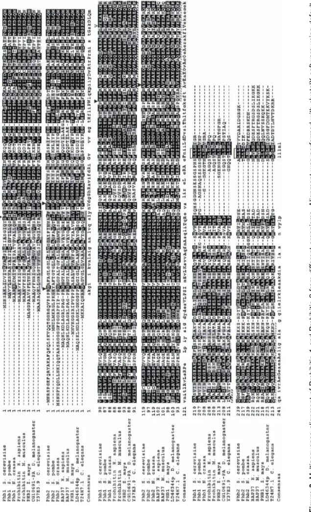
and 100% (Figure 4). Lower sequence similarity can be observed at the N-termini and C-termini of the proteins the most prominent being Prohibitin 1 in S. cerevisiae, which has an additional C-terminal amino acid stretch not present in other species. It is still obscure, however, if this structural divergence implicates a functional difference. The present knowledge on prohibitins is summarized below.
Figure 3. Multisequence alignment of Prohibitin 1 and Prohibitin 2 from different organisms. Alignment was performed with clustalX software using default settings. The accession numers are given in a table format inMaterials and Methods. Filled boxes show the span of the predictable transmembrane regions. Shadowed boxes show the position of the predictable coiled-coil region. V shows the position of a conserved Val residue which is predicted to be essential for the folding of the coiled-coil region. Short highly homologous stretches at the C-terminal end of both, Prohibitin 1 and Prohibitin 2, are also marked.
INTRODUCTION
2.1. Prohibitins are conserved SPFH-domain proteins
Based on their limited homology with stomatin and stomatin-like proteins, prohibitins are proposed to fall within the so called stomatin protein superfamily (Tavernarakis et al., 1999; Nadimpalli et al., 2000). This family is also designated PID after proliferation, ion, and death. In addition, proteins like the bacterial plasma membrane HflK and HflC, close bacterial homologues of prohibitins, as well as flotillins and plant disease response proteins (HIR) fall within this superfamily. A common feature of the aforementioned proteins is the so called SPFH domain (after Stomatin, Prohibitin, Flotillin and HflK/C) (Appendix II). The role of the SPFH-domain and the functions of the proteins carrying SPFH-domain are discussed bellow.
2.1.1. Functions of the SPFH domain
Most of the characterized SPFH family members are membrane proteins with their SPFH-domain exposed to a hydrophilic environment (Tavernarakis et al., 1999). In addition, stomatin and its homologues are associated with lipid microdomains called lipid rafts. These are tight associations between sphingolipids and cholesterol with distinct biophysical properties within the surrounding lipid bilayer (Brown and Rose, 1992; Simons and Ikonen, 1997; Brown and London, 2000; London and Brown, 2000). Association within membrane subcompartments makes sense in the light of protein sorting or concentrating molecules involved in processes like control of signal transduction, antigen representation and etc. (Anderson et al., 2000; Anderson and Jacobson, 2002; Bickel, 2002). Constant flotation, separation and fusion of such domains and their docked proteins allows for more flexible spatial and temporal regulation of signaling cascades according to the requirements of the cell (Simons and Toomre, 2000; Golub et al., 2004; Razzaq et al., 2004).
2.1.1.1. Role of the SPFH-domain for localization to the plasma membrane and lipid rafts
Proteins are targeted to lipid rafts via different mechanisms of which lipid modification (like palmitoylation) is the most common (Melkonian et al., 1999). In some stomatin homologues the SPFH domain contains sequences responsible for proper targeting to the plasma membrane and to lipid rafts (Salzer and Prohaska, 2001). Flotillin- 1/reggie-2, enriched in lipid rafts from erythrocytes (Salzer and Prohaska, 2001), is
targeted to the plasma membrane via a combination of two hydrophobic regions embedded within the SPFH domain (Liu et al., 2005). A cysteine residue (cys34) from the SPFH-domain of flotillin-1/reggie-2 is palmitoylated and functions together with the hydrophobic sequences in targeting to the plasma membrane (Morrow et al., 2002). Mutational exchange of cysteine at position 34 to alanine prevents the association of flotillin-1/reggie-2 with the plasma membrane suggesting a crucial role of palmitoylation for the proper localization (Morrow et al., 2002). Similarly to flotillin- 1/reggie-2, palmitoylation of cys29 targets stomatin to lipid rafts (Wang et al., 1991;
Snyers et al., 1999). Mutations, which prevent the proper localization to the plasma membrane have been detected in the SPFH-domain of podocin. In cells, podocin is normally localized to the plasma membrane and mutations in the SPFH domain cause retaining in the endoplasmic reticulum (Roselli et al., 2004).
2.1.1.2. Role of the SPFH-domain for protein-protein interactions and as a structural scaffold
The SPFH-domain functions as a mediator of protein-protein interactions in some stomatin homologues. The SPFH-domain of MEC-2, the C. elegans homologue of human stomatin, is involved in the interaction with MEC-4 and MEC-10 subunits of the degenerin channel in the plasma membrane of the touch-sensory neurons (Huang et al., 1995; Zhang et al., 2004). Mutations in the SPFH-domain of MEC-2 lead to loss of touch sensitivity due to an impaired interaction of MEC-2 with the degenerin channel (Zhang et al., 2004). In this case, the SPFH domain ensures proximity between the regulatory N- and C-terminal ends of MEC-2 and the channel subunits, but does not play a role in the proper distribution of MEC-2 along the sensory neurons (Zhang et al., 2004). Rather, this function is attributed to the MEC-2- specific N-terminal sequence (Huang et al., 1995).
In stomatin and podocin, the SPFH domain can form a hairpin conformation thereby properly localizing the N- and C- terminal ends to the cytoplasm where interaction with other molecules takes place (Snyers et al., 1999; Roselli et al., 2002). Thus, the SPFH domain not only provides proper targeting to the plasma membrane and lipid rafts, but also serves as a structural scaffold (Goodman et al., 2002; Zhang et al., 2004).
INTRODUCTION Prohibitins and bacterial HflK and HflC proteins were also proposed to possess SPFH-domains (Tavernarakis et al., 1999). HflK and HflC proteins form a high molecular weight complex which is anchored to the plasma membrane with the N- termini of the molecules and exposes C-terminal domains to the periplasmic side of the membrane (Kihara et al., 1997). However, no data exists whether HflK/C complexes are associated with lipid rafts or related subdomains in the bacterial plasma membrane. In contrast, mammalian prohibitins have been shown to localize to the plasma membrane in B-lymphocytes and several cancer cell lines (Terashima et al., 1994; Mengwasser et al., 2004). Prohibitins associate with the IgM antigen receptor in a TritonX-100 resistant manner (Terashima et al., 1994) in murine B-cell lines and purified lipid rafts are positive for Prohibitin 1 and Prohibitin 2 (Saeki et al., 2003; Mielenz et al., 2005) suggesting that prohibitins are associated with lipid rafts.
However, due to their different cellular localization it is difficult to assess whether association of prohibitins with lipid rafts is a rule or exception. In yeast, prohibitins are localized exclusively to mitochondria and no association with lipid rafts has been reported.
2.1.2. Functions of the proteins harboring SPFH-domains 2.1.2.1. Regulation of channels across the plasma membrane
SPFH-domain proteins of clinical importance are the close homologues stomatin and podocin. Podocin (NPHS2) is exclusively expressed in kidney podocytes and regulates the function of the glomerular filtration barrier (Boute et al., 2000; Roselli et al., 2002). Mutations in the NPHS2 gene cause the familial steroid-resistant nephric syndrome in which the filtration function of podocytes is disturbed and a condition known as protein-urea occurs.
The human stomatin protein (erythrocyte band 7 protein) has long been shown to be affected in overhydrated stomatocytosis (OHSt, spherocytosis of the erythrocytes and leakage of Na and K ions) (Lande et al., 1982; Stewart et al., 1992; Stewart et al., 1993). The protein is absent in the most severe cases of OHSt, but seems not to be the cause for the disease (Stewart et al., 1992; Fricke et al., 2003). Although the exact cause for OHSt is unknown, stomatin has been shown to interact with the actin cytoskeleton in erythrocytes (Stewart et al., 1992), with the glucose transporter GLUT-1 regulating the uptake of glucose (Zhang et al., 2001) and with ASIC-related
DEG/ENaC subunits in rat mechanosensory neurons (Price et al., 2004). Therefore, the interaction and regulation of channels across the plasma membrane is emerging as a common function of most of the so far characterized stomatin-like proteins.
2.1.2.2. Regulation of proteases
In contrast to stomatin-like proteins, HflK/C interacts with the membrane protease FtsH thereby modulating its proteolytic activity (Kihara et al., 1996; Saikawa et al., 2004). In the absence of HflK/C, the nonassembled subunit SecY of the membrane protein translocase (SecY-SecE) is rapidly degraded (Kihara et al., 1996). In contrast, the λ phage CII protein is stabilized under identical conditions (Kihara et al., 1997).
Similarly to HflK/C, prohibitins associate with the mAAA protease and were proposed to negatively regulate its activity in yeast [see chapter 2.6.3. and (Steglich et al., 1999)] .
Based on the involvement of HflK/C and prohibitins in proteolysis by direct interaction with proteases it was proposed that the region including the SPFH domain functions as an association factor for the formation of complexes with membrane associated proteases (Tavernarakis et al., 1999).
2.3. Expression of the prohibitin gene in mammals
Prohibitin genes are constitutively expressed in all mammalian cells and tissues with expression levels varying as a function of the cell cycle (Roskams et al., 1993). How the expression of prohibitins is regulated is presently unknown. Analysis of the promoter regions of rat and human PHB1 gene shows that the promoter has features similar to those of house keeping genes, e.g. lack of a clearly identifiable TATA box (McClung, 1995). Endogenous prohibitin mRNA and protein levels decrease in S- phase and rise again in the G2-phase. Interestingly, two forms of PHB1 mRNA, with molecular size of 1.2 kb and 1.9 kb, have been detected in human normal and immortalized cells and in rats (see below) (Nuell et al., 1991; Jupe et al., 1996).
2.4. Cellular localization of prohibitins
Prohibitins have been localized to various cellular compartments in different organisms. While in yeast Phb1 and Phb2 are localized exclusively to mitochondria, in mammalian cells they are also detectable in the nucleus (Wang et al., 2002;
INTRODUCTION Fusaro et al., 2003) and at the plasma membrane (Mengwasser et al., 2004). In mitochondria from yeast, C. elegans and mammals Phb1 and Phb2 form a high molecular weight complex (Steglich et al., 1999; Nijtmans et al., 2000; Coates et al., 2001; Artal-Sanz et al., 2003). Moreover, Phb1 and Phb2 are interdependent in yeast i.e. in the absence of Phb1 Phb2 is rapidly degraded and vice versa (Berger and Yaffe, 1998). Therefore, it is of interest to note that extramitochondrial localization is shown separately for Phb1 and Phb2 in mammalian cells. Since this thesis focuses on the mitochondrial localization and function of prohibitins, their localization and function in other cellular compartments will be discussed in less detail.
2.5. Prohibitins function as regulators of cell proliferation and are important for normal development
Both Phb1 and Phb2 are necessary for embryonic development in flies (Eveleth and Marsh, 1986), C. elegans (Artal-Sanz et al., 2003) and mice (Park et al., 2005).
Mutations in the Prohibitin 1 gene are recessive lethals in late larvae development or during the progression to pupae in Drosophila (Eveleth and Marsh, 1986). Similarly, down regulation of prohibitins by short interfering RNAs in C. elegans leads to embryonic arrest (Artal-Sanz et al., 2003). Furthermore, homozygous Phb2 knock-out mice die early in development (before E9.0) (Beal, 2003). Interestingly, Prohibitin 1 has also been recently shown to play a role during plant development (Chen et al., 2005). Although the exact role of prohibitins for development is not understood, it is conceivable that it is linked to their function as transcriptional regulators in the nucleus. Therefore, the role of both proteins in transcriptional regulation, cell proliferation and apoptosis is discussed below.
2.5.1 Function of Prohibitin 1 as a regulator of cell proliferation and apoptosis
PHB1 mRNA is capable to block proliferation in normal and HeLa human fibroblasts (McClung et al., 1989; Nuell et al., 1991; Liu et al., 1994). Transfected PHB1 mRNA has maximum antiproliferative activity in G0 to G1 transition phase by maintaining high prohibitin levels prior to entering in S-phase and blocking DNA synthesis (Roskams et al., 1993). The PHB1 gene gives rise to major (1.9 kb) and minor (1.2 kb) transcripts in human cells and in rats (McClung et al., 1989; Jupe et al., 1995;
Thompson et al., 2001). The level of the major transcript in immortalized cell lines is increased while the level of the 1.2 kb transcript remains constant (Jupe et al., 1995) suggesting functional role for the major transcript. Indeed, the antiproliferative effect of PHB1 mRNA is contained within the 3`-untranslated region (UTR) of the 1.9 kb transcript in a region absent in the minor transcript as indicated by two lines of evidence: i) the 3`-UTR alone is able to suppress cellular proliferation and ii) single nucleotide exchange (C/T) in this region or the removal of the 3`-UTR abolishes the antiproliferative effect of PHB1 mRNA. Moreover, the UTR/T allele is associated with increased risk of breast cancer ( Jupe et al., 1996; Manjeshwar et al., 2003).
The Phb1 protein can also affect proliferation (Wang et al., 1999; Wang et al., 1999a;
Wang et al., 2002; Wang et al., 2002a; Gamble et al., 2004; Wang et al., 2004). Phb1 binds to retinoblastoma-family members (Rb, p107 and p130) thereby repressing the activity of E2F-transcription factors (E2F1-5), which results in inhibition of cell proliferation (Wang et al., 1999; Wang et al., 1999a) (Figure 4A). To repress transcription, Phb1 also binds the co-repressor NcoR and recruits chromatin remodeling complexes, such as HDAC1 and Brg-1/Brm, to the promoter element (Wang et al., 2002; Wang et al., 2002а; Wang et al., 2004). Recruitment of HDAC1 and Brg-1/Brm is presumably independent of binding to Rb as Phb1/Brg-1/Brm complexes exist in the absence of Rb (Wang et al., 2002а). Binding of Phb1 to Rb is, however, compulsory for the activity of these complexes since Phb1 exerts no effect on E2F-mediated transcription in the absence of Rb (Wang et al., 1999; Wang et al., 2002а). Therefore, it is imaginable that binding of Phb1 to Rb is the rate limiting step which ensures recruitment to the promoter and specificity of the Phb1-mediated transcriptional repression.
Notably, stimulation of the IgM signaling network can reverse the Phb1-mediated suppression of cell proliferation (Figure 4B) (Wang et al., 1999а; Wang et al., 2002).
This effect is attributed to an activated Raf-1 kynase which translocates to the nucleus and binds to Phb1 thereby interfering with the formation of the Phb1/Rb or Phb1/E2F complexes (Wang et al., 1999а).
What is the role of Phb1 for development? E2F activity is essential for early development in Drosophila (Duronio et al., 1995; Myster et al., 2000) and Xenopus (Suzuki and Hemmati-Brivanlou, 2000). In mice, simultaneous deletions of E2F family members are lethal and deletion of a single E2F gene results in a specific developmental defect (Humbert et al., 2000; Rempel et al., 2000). In mammalian
INTRODUCTION cells, loss of E2Fs repression or deregulation of E2Fs protein levels have been shown to alter the expression of various genes involved in cell proliferation, apoptosis (APAF1, caspase3, caspase 7, Bcl-3), development (homeobox genes and genes from the TGFβ and Wnt pathways) and etc. (Hunt et al., 1997; Gaubatz et al., 2000;
Humbert et al., 2000а; Muller et al., 2001). It is conceivable that similarly to the inactivation of Rb (Knudsen and Wang, 1996) absence of Phb1 would result in higher transcription levels from E2F promoters thereby affecting the expression of E2F- responsive genes. Therefore, it is plausible that Phb1 balances the timely expression of E2F responsive genes which are necessary for normal development.
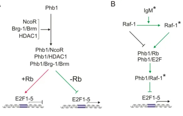
Figure 4. Phb1 functions as a negative regulator of cell proliferation by repressing the activity of E2F transcription factors. A. In the presence of Rb, Phb1 binds to E2F1-5 transcription factors and represses the expression from E2F responsive promoters. This is mediated by binding of Phb1 to the transcriptional co-repressor NcoR (Phb1/NcoR) and concomitant recruitment of histone deacetylase HDAC1 (Phb1/HDAC1). Alternatively, recruitment of the chromatin remodeling complexes Brg-1/Brm by Phb1 (Phb1/Brg-1/Brm) can repress E2F-mediated transcription. Although Phb1/Brg- 1/Brm and presumably Phb1/HDAC1 complexes are formed upstream of Rb binding (not shown), they can repress transcription only in the presence of Rb. Requirement of Rb is shown as +Rb. B. The antiproliferative effect of prohibitins is reverted in the presence of activated Raf-1 kinase. Stimulation of the IgM-receptor (IgM*) activates the Raf-1 kinase (Raf-1*), which can bind Phb1 (Phb1/Raf-1*) and thereby interferes with the formation of the Phb1/Rb of Phb1/E2F complexes. Phb1/Raf-1* complexes can not repress E2F-mediated transcription. green arrow or crossed line, pathway that allows E2F- mediated transcription, red arrow lines, pathway that represses E2F activity.
These studies, however, focus only on Phb1 and the role of Phb2 for the regulation of E2F promoters remains unclear.
2.5.2. Role of Phb2 as a regulator of the estrogen signaling pathway Independent studies have revealed that Phb2 can also function as a transcriptional regulator in the nucleus (Montano et al., 1999; Sun et al., 2004). Phb2 was identified in yeast two hybrid studies as a novel co-regulator of the nuclear estrogen receptor (ER) and was accordingly designated Repressor of Estrogen receptor Activity (REA) (Montano et al., 1999). When expressed in cell culture, Phb2 selectively represses the transcription from consensus and non-consensus ER responsive promoters (Delage-Mourroux et al., 2000). This is achieved by a direct binding of Phb2 to the liganded ER and recruitment of HDAC1 to the promoter element (Delage-Mourroux et al., 2000; Kurtev et al., 2004). A model was proposed according to which Phb2 represses transcription by mutually competing with the transcriptional co-activator SRC-1 for binding to the liganded ER (Figure 5) (Delage-Mourroux et al., 2000;
Kurtev et al., 2004).
The function of Phb2 in the estrogen signaling cascade is further substantiated in mice (Park et al., 2005). Although knock-out of Phb2 is embryonic lethal early in development, heterozygous mice lacking one PHB2 allele are viable. Notably, genes normally stimulated by estrogen (complement C3, PTα and etc.) are upregulated in the ovary from such animals (Park et al., 2005).
The similarities between the emerging functions of Prohibitin 1 and Prohibitin 2 as transcriptional regulators are interesting. Both proteins serve as transcriptional repressors and both can utilize HDAC1. It seems plausible that binding to transcription factors (E2F and ER) or repressors (Rb) ensures specificity of Phb1 or Phb2 mediated repression of transcription. Recent findings suggest a functional interaction between the ER pathway and the Phb1-mediated regulation of E2F (Wang et al., 2004). In particular, binding of anti-estrogen to the ER-receptor can target Phb1 and thereby induce repression the activity of E2F transcription factors (Wang et al., 2004). It remains unclear though, if both proteins act as a complex in this process.
INTRODUCTION
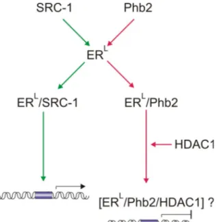
Figure 5. ER-mediated transcription is repressed by Phb2. A. Phb2 competes with the transcriptional activator SRC-1 for the liganded ER (ERL). ERL bound to SRC-1 (ERL/SRC-1) promotes transcription from ER-responsive elements. In contrast, binding of Phb2 to ERL (ERL/Phb2) inhibits the expression from ER-responsive promoters via recruitment of HDAC1. green green arrow lines, pathway that allows E2F-mediated transcription, red arrow lines, pathway that represses E2F activity,
?, unknown complex.
2.6. Mitochondrial localization and function of prohibitins
Phb1 and Phb2 were localized to mitochondria in plants (Snedden and Fromm, 1997;
Takahashi et al., 2003), yeast (Steglich et al., 1999; Nijtmans et al., 2000), mammals (Nijtmans et al., 2000) and C. elegans (Artal-Sanz et al., 2003). Their biogenesis, possible function and molecular interactions in yeast mitochondria are discussed below.
2.6.1. Assembly and structure of the yeast prohibitin complex in the inner mitochondrial membrane
Phb1 and Phb2 are nuclear-encoded and posttranslationally imported in mitochondria where they assemble into a high molecular weight complex in the inner mitochondria membrane (Steglich et al., 1999; Tatsuta et al., 2005). Targeting of both Phb1 and Phb2 to mitochondria in yeast occurs via non-cleavable N-terminal presequences. In Phb2, this presequence contains positively charged residues
capable of forming amphipathic α-helix, followed by a hydrophobic amino acid stretch which may serve as a membrane anchor (Ikonen et al., 1995; Tatsuta et al., 2005).
Deletion of each of both stretches drastically reduces the import of Phb2 (Tatsuta et al., 2005). Unlike Phb2, the N-terminal region of Phb1 lacks characteristics of mitochondrial presequences. Nevertheless, the first 28 amino acids have been shown to be essential and sufficient for mitochondrial import in yeast (Tatsuta et al., 2005). The stable insertion of Phb1 and Phb2 in the inner membrane depends on the Tim23-translocase, which is also responsible for the insertion of other presequence carrying proteins (Neupert, 1997; Pfanner and Geissler, 2001). The import of Phb1 and Phb2 is drastically reduced in mitochondria from cells harboring decreased levels of Tim23 (tim23ts) and depletion of Tim23 results in no detectable levels of Phb1 and Phb2 in vivo (Tatsuta et al., 2005).
The newly imported Phb1 and Phb2 subunits form assembly intermediates of ~120 kDa (Tatsuta et al., 2005). These intermediates are detectable on BN-SDS PAGE of imported radio labeled Phb1 or Phb2 in wild type cells but their exact composition is unclear. Along with Phb1 and Phb2, Tim13 can also be detected within such an intermediate although it is not essential for import of Phb1 and Phb2 in mitochondria (Tatsuta et al., 2005). The 120 kDa intermediates oligomerize into the mature Phb1- Phb2 complex. Within this complex, Phb1 and Phb2 are anchored to the inner mitochondrial membrane by their N-terminal ends and expose C-terminal ends to the intermembrane space (Figure 6C) (Ikonen et al., 1995; Berger and Yaffe, 1998;
Coates et al., 2001). As judged by gel filtration analysis and BN-SDS PAGE the complex has a molecular weigh between 1.000 kDa and 1.200 kDa (Steglich et al., 1999; Tatsuta et al., 2005). Therefore, it was proposed that the Phb1-Phb2 complex is built up of 12 to 18 subunits (Steglich et al., 1999; Nijtmans et al., 2000; Back et al., 2002). Additionally, crosslinking experiments show that Phb1 and Phb2 molecules are alternatingly assembled (Figure 6A and 6B) as no crosslinks can be observed between two Phb1 or two Phb2 molecules (Back et al., 2002). Most of the crosslinks are concentrated in the C-terminal region of Phb1 and Phb2 delineating it as the interaction site. Indeed, the C-terminal regions of Phb1 (amino acids 180–224) and Phb2 (amino acids 212–253) form computationally predictable coiled-coil regions whose disruption prevents formation of the mature Phb1-Phb2 complex, but do not affect the formation of the 120 KDa intermediates (Tatsuta et al., 2005). Thus, interactions between the coiled-coil regions of Phb1 and Phb2 are necessary for the
INTRODUCTION complex formation. Single particle electron microscopy revealed that the Phb1-Phb2 complex has a roughly a circular equatorial projection with a central cavity and dimensions ~260-170 Å (Tatsuta et al., 2005) (Figure 6D). The crystal structure of the Phb1-Phb2 complex as well as that of the subunits are currently unknown.
Figure 6. A representation of the superstructure of the prohibitin complex. A. Dimeric building block of Phb1 and Phb2 (top view). B. Proposed circular arrangement of the building blocks (top view).
C. A section of four building blocks and the mitochondrial inner membrane (side view). IM space, intermembrane space. reprinted from (Back et al., 2002). D Single particle EM-analysis of purified Phb1-Phb2 complex. Dataset is split into elliptical ring-like (top, 1-5) and rectangular-like (bottom, 6- 10) subsets. Bar, 200 Ǻ. reprinted from (Tatsuta et al., 2005).
2.6.2. Role of prohibitins for mitochondrial morphology and inheritance
The function of the Phb1-Phb2 complex in the inner mitochondrial membrane is only partially understood. In contrast to lethal phenotypes in C. elegans and mammals (Artal-Sanz et al., 2003; Park et al., 2005), yeast cells lacking prohibitins grow as wild type cells on different carbon sources (Berger and Yaffe, 1998; Steglich et al., 1999).
Mitochondrial morphology and inheritance are not affected in prohibitin null mutants (Berger and Yaffe, 1998). Only in aged cells, the absence of prohibitins leads to shortening of the replicative life span and an overall decrease in the mitochondrial membrane potential (Coates et al., 1997). These effects are attributed to a delayed, but not absent, segregation of mitochondria from old mother to daughter cells (Piper et al., 2002). However, deletion of prohibitins is lethal in combination with mutations