generation sequencing for the identification of lysine and arginine methyltransferase protein
interactions
D i s s e r t a t i o n
zur Erlangung des akademischen Grades d o c t o r r e r u m n a t u r a l i u m
( Dr. rer. nat.) im Fach Biologie eingereicht an der
Mathematisch-Naturwissenschaftlichen Fakultät I der Humboldt-Universität zu Berlin
von
Diplom-Biologin Mareike Weimann
Präsident der Humboldt-Universität zu Berlin Prof. Dr. Jan-Hendrik Olbertz
Dekan der Mathematisch-Naturwissenschaftlichen Fakultät I Prof. Dr. Andreas Herrmann
Gutachter: 1. Prof. Dr. Christian Spahn 2. Prof. Dr. Erich Wanker 3. Prof. Dr. Hans Lehrach
Tag der mündlichen Prüfung: 01. August 2012
Zusammenfassung
Proteinmethylierung spielt eine immer größere Rolle in der Regulierung zellulärer Prozesse. Die Entwicklung effizienter proteomweiter Methoden zur Detektion von Methylierung auf Proteinen ist limitiert und technisch schwierig. In dieser Arbeit haben wir einen neuen Hefe-Zwei-Hybrid-Ansatz (Y2H) entwickelt, der Proteine, die miteinander wechselwirken, mit Hilfe von Sequenzierungen der zweiten Generation identifiziert (Y2H-Seq). Der neue Y2H-Seq-Ansatz wurde systematisch mit dem Y2H-Seq-Ansatz verglichen. Dafür wurde ein Bait-Set von 8 Protein-Arginin-Methyltransferasen, 17 Protein-Lysin-Methyltransferasen und 10 Demethylasen gegen 14,268 Prey-Proteine getestet. Der Y2H-Seq-Ansatz ist weniger arbeitsintensiv, hat eine höhere Sensitivität als der Standard Y2H-Matrix-Ansatz und ist deshalb besonders geeignet, um schwache Interaktionen zwischen Substraten und Protein-Methyltransferasen zu detektieren. Insgesamt wurden 523 Wechselwirkungen zwischen 22 Bait-Proteinen und 324 Prey-Proteinen etabliert, darunter 11 bekannte Methyltransferasen-Substrate. Netzwerkanalysen zeigen, dass Methyltransferasen bevorzugt mit Transkriptionsregulatoren, DNA- und RNA-Bindeproteinen wechselwirken. Diese Daten repräsentieren das erste proteomweite Wechselwirkungsnetzwerk über Protein-Methyltransferasen und dienen als Ressource für neue potentielle Methylierungssubstrate. In einem in vitro Methylierungsassay wurden exemplarisch mit Hilfe massenspektrometrischer Analysen die methylierten Aminosäurereste einiger Kandidatenproteine bestimmt. Von neun getesteten Proteinen waren sieben methyliert, zu denen gehören SPIN2B, DNAJA3, QKI, SAMD3, OFCC1, SYNCRIP und WDR42A. Wahrscheinlich sind viele Methylierungssubstrate im Netzwerk vorhanden. Das vorgestellte Protein-Protein-Wechselwirkungsnetzwerk zeigt, dass Proteinmethylierung sehr unterschiedliche zelluläre Prozesse beeinflusst und ermöglicht die Aufstellung neuer Hypothesen über die Regulierung Molekularer Mechanismen durch Methylierung.
Schlagwörter: Proteinmethylierung, Protein-Arginin-Methyltransferase (PRMT), Protein-Lysin-Methyltransferase (PKMT), Demethylase, Protein-Protein-Wechselwirkung (PPI), Hefe-Zwei-Hybrid-System (Y2H), Sequenzierung der zweiten Generation, Protein-Wechselwirkungsnetzwerk
Abstract
Protein methylation on arginine and lysine residues is a largely unexplored posttranslational modification which regulates diverse cellular processes. The development of efficient proteome-wide approaches for detecting protein methylation is limited and technically challenging.
We developed a novel workload reduced yeast-two hybrid (Y2H) approach to detect protein-protein interactions utilizing second generation sequencing. The novel Y2H-seq approach was systematically evaluated against our state of the art Y2H-matrix screening approach and used to screen 8 protein arginine methyltransferases, 17 protein lysine methyltransferases and 10 demethylases against a set of 14,268 proteins. Comparison of the two approaches revealed a higher sensitivity of the new Y2H-seq approach. The increased sampling rate of the Y2H-seq approach is advantageous when assaying transient interactions between substrates and methyltransferases. Overall 523 interactions between 22 bait proteins and 324 prey proteins were identified including 11 proteins known to be methylated. Network analysis revealed enrichment of transcription regulator activity, DNA- and RNA-binding function of proteins interacting with protein methyltransferases. The dataset represents the first proteome-wide interaction network of enzymes involved in methylation and provides a comprehensively annotated resource of potential new methylation substrates. An in vitro methylation assay coupled to mass spectrometry revealed amino acid methylation of candidate proteins. Seven of nine proteins tested were methylated including SPIN2B, DNAJA3, QKI, SAMD3, OFCC1, SYNCRIP and WDR42A indicating that the interaction network is likely to contain many putative methyltransferase substrate pairs. The presented protein-protein interaction network demonstrates that protein methylation is involved in diverse cellular processes and can inform hypothesis driven investigation into molecular mechanisms regulated through methylation.
Keywords: Protein methylation, protein arginine methyltransferase (PRMT), protein lysine methyltransferase (PKMT), demethylase, protein-protein interaction (PPI), yeast-two hybrid system (Y2H), second/next generation sequencing, PPI network
Abbreviations
AD Activation domain
aDMA Asymmetrically dimethylated arginine
AdoMet S-adenosylmethionine
AdoHcy S-adenosylhomocysteine
Adox Periodate-oxidized adenosine
ALS6 Amyotrophic lateral sclerosis
AP-MS Affinity purification coupled to mass spectrometry
ATP Adenosintriphosphate
ATPase Adenosintriphosphatasen
bp Base pair
CoA Coenzyme A
co-IP Co-immunoprecipitation
CRL Cullin-RING finger ligase
CTD Carboxy-terminal domain
DBD DNA-binding domain
DMEM Dulbecco’s modified eagle’s medium
DNA Deoxyribonucleic acid
dNTPs Desoxyribonukleosidtriphosphate
DPBS Dulbecco’s phosphate buffered solution
ECL Enhanced chemiluminescence
EGF Epidermal growth factor
ES Embryonic stem
ER Estrogen receptor
FBS Fetal bovine serum
FL Full length
FNR False negative rate
FPR False positive rate
H Histone
HD1 Homology domain 1
HD2 Homology domain 2
HEK Human embryonic kidney
His Histidine
HIV Human immunodeficiency virus
hnRNPs Heterogeneous nuclear ribonucleoproteins
HRP Horseradish peroxidase
HTP High-throughput
IP Immunoprecipitation
JmjC Jumonji C
K Lysine
KH K homology
KRAB Krueppel-associated box
LB Lysogeny broth
LC-MS/MS Liquid chromatographie-massenspektometrie/massenspektometrie
MMA Monomethylated arginine
MPIMG Max Planck Institute for Molecular Genetics
mRNA Messenger RNA
MTP Microtiterplate
Myr Myristoylation morif
NCBI National Centre for Biotechnological information
NF?B Nuclear factor kappa-light-chain-enhancer of activated B cells
NHR Nuclear hormone receptor
NITE National Institute of Technology and Evaluation
NLS Nuclear localisation signal
OD600 Optical density at 600 nm
ORF Open reading frames
PA Protein A
PCR Polymerase chain reaction
PDeM Protein demethylase
PHD Plant homeo domain
PKMT Protein lysine methyltransferase
PMT Protein methyltransferase
PPIs Protein-protein interactions
PRMT Protein arginine methyltransferase
PTM Post-translational modification
PVDF Polyvinylidene fluoride
R Arginine
RG Arginine-glycine
RING Really interesting new gene
RLU Relative luciferase units
RNA Ribonucleic acid
RRM RNA recognition motifs
SAM S-adenosylmethionine
sDMA Symmetrically dimethylated arginine
SDS Sodium dodecyl sulfate
SDS-PAGE SDS-polyacrylamide gel electrophoresis
seq Sequencing
SET Su(var), Enhancer of zest, trithorax
SH3 Src homology 3
snRNP Small nuclear Ribonucleoprotein particles SOCS Suppressor of cytokines signalling
SR Serine arginine
SRPY SPla and the RYanodine Receptor
STAR Transduction and activation of RNA
TBE Tris Borate EDTA
TBS Tris-buffered saline
TBST TBS supplemented with Tween 20
TPR Tetratricopeptide repeats
TSS Transformation and storage solution
ub Ubiquitin
UV Ultra violet
Y2H Yeast-two hybrid
ZnF Zink finger
Zusammenfassung ... I Abstract ... III Abbreviations ... V
1 Introduction ... 1
1.1 Protein methylation ... 1
1.1.1 Protein arginine methylation ... 1
1.1.1.1 PRMT1, the predominant type I methyltransferase, is closely related to PRMT8 ... 4
1.1.1.2 CARM1 and PRMT6 are type I methyltransferases with pronounced substrate specificity 5 1.1.1.3 PRMT5 is the major type II methyltransferase ... 6
1.1.1.4 Additional members of the PRMT family are not characterized ... 6
1.1.2 Protein arginine demethylation ... 7
1.1.3 Protein lysine methylation ... 7
1.1.3.1 Chromatin state is regulated by histone lysine methylation ... 8
1.1.3.2 Non-histone protein functions are modulated by lysine methylation ... 9
1.1.4 Protein lysine demethylation ... 10
1.2 PMT substrate identification methods ... 10
1.2.1 Methods to detect methylated proteins ... 10
1.2.2 The Y2H system to find PMT interacting proteins and potential substrates ... 12
1.3 Aim of this study ... 14
2 Material ... 16
2.1 Chemicals ... 16
2.2 Lab ware ... 18
2.3 Enzyme, proteins, DNA, kits ... 19
2.4 Organism ... 20
2.4.1 Bacteria strains ... 20
2.4.2 Yeast strains ... 20
2.4.3 Mammalian cell lines ... 20
2.5 Reagents ... 20
2.5.1 E. coli and yeast miniprep ... 20
2.5.2 Yeast transformation ... 21
2.5.3 Agarose gel electrophoresis ... 21
2.5.4 SDS polyacrylamide gel electrophoresis and western blot... 22
2.5.5 Co-immunoprecipitation ... 23
2.5.6 E. coli protein expression and purification ... 24
2.6 Media ... 24
2.6.1 E. coli growth media ... 24
2.6.2 S. cerevisiae growth media ... 26
2.6.3 Mammalian cell culture media ... 26
2.7 Vectors ... 27
2.7.1 Gateway entry vectors ... 27
2.7.2 Expression vectors for E. coli ... 27
2.7.3 Expression vectors for S. cerevisiae ... 28
2.7.4 Expression vectors for mammalian cell lines ... 28
2.8 Oligonucleotides ... 29
2.9 Antibodies ... 29
2.10 Databases... 29
2.11 Software and tools... 30
3 Methods ... 32
3.1 Microbiology and molecular biology ... 32
3.1.1 Growth and storage of E. coli ... 32
3.1.2 Preparation of competent E. coli cells ... 32
3.1.2.1 Chemically competent cells ... 32
3.1.2.2 Electrocompetent cells ... 32
3.1.3 Transformation of competent E. coli cells ... 32
3.1.3.1 Chemical transformation of competent cells... 32
3.1.3.2 Electroporation of competent E. coli ... 33
3.1.4 Plasmid isolation of E. coli ... 33
3.1.5 Restriction digest of plasmid DNA ... 34
3.1.6 Separation of DNA fragments by agarose gel electrophoresis ... 34
3.1.7 Determination of DNA concentration ... 34
3.1.8 Polymerase chain reaction (PCR) ... 34
3.1.9 Gateway cloning technology ... 35
3.1.9.1 BP Reaction ... 35
3.1.9.2 LR Reaction ... 35
3.1.10 Expression of recombinant fusion proteins ... 35
3.2 Yeast specific molecular biology ... 36
3.2.1 Preparation of yeast media... 36
3.2.2 Growth and storage of S. cerevisiae ... 36
3.2.3 Transformation of S. cerevisiae ... 37
3.2.4 Autoactivation test of baits ... 37
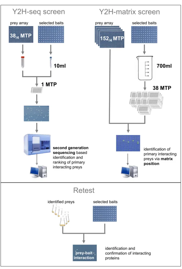
3.2.5 Screen of bait pools against a prey array (Y2H-matrix approach) ... 38
3.2.6 Screen of bait pool against a pooled prey array (Y2H-seq approach) ... 38
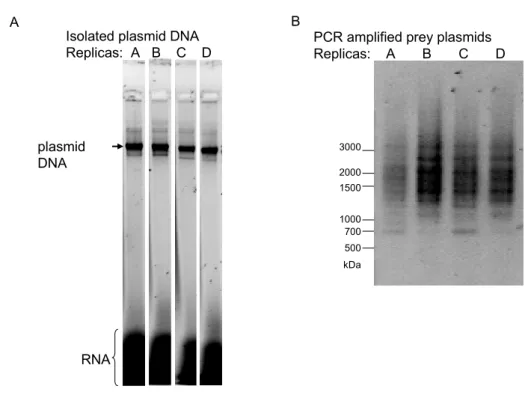
3.2.6.1 Preparation of plasmid DNA ... 39
3.2.6.2 Amplification of prey plasmid DNA ... 39
3.2.6.3 Computational analysis of second generation sequencing readout ... 40
3.3 Mammalian cell culture ... 41
3.3.1 Cell culture and transfection ... 41
3.3.2 Preparation of cell lysate for western blots ... 41
3.4 Protein biochemistry... 41
3.4.1 SDS-polyacrylamide gel electrophoresis ... 41
3.4.2 Protein gel stain ... 42
3.4.2.1 Coomassie blue ... 42
3.4.2.2 Blue silver... 42
3.4.3 Western blot ... 42
3.4.4 His-tag protein purification ... 43
3.4.5 Methylation assay ... 43
3.5 Protein interaction analysis ... 44
3.5.1 Database to store the data ... 44
3.5.2 R statistical software to generate a heatmap ... 44
3.5.3 Perl script to calculate RG-repeat score ... 44
3.5.4 Cytoscape to visualize obtained PPI data ... 44
3.5.5 DAVID to discover enriched functional-related gene groups ... 45
4 Results ... 47
4.1 Y2H screening as PPIs discovery tool ... 47
4.1.1 Development of a new Y2H second generation sequencing screen ... 47
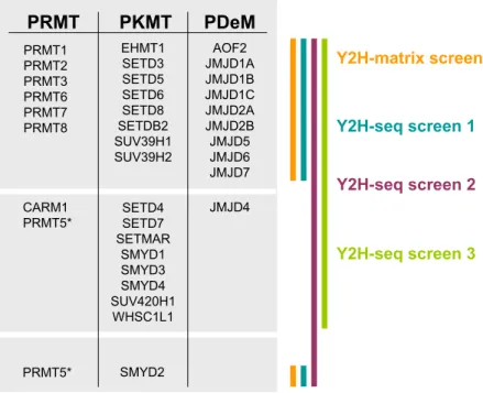
4.2 Y2H screens for protein methyltransferase (PMT) and protein demethylase (PDeM) interactions 49 4.2.1 Selection of PMTs and PDeMs to screen for interacting proteins ... 50
4.2.2 Preparation of PMTs and PDeMs for a Y2H PPI search ... 51
4.2.3 Overview of PMT- and PDeM-protein interaction screens ... 52
4.2.3.1 Proteome-wide screen of PMTs and PDeMs in a Y2H-matrix approach ... 53
4.2.3.2 Proteome-wide screen of PMTs and PDeMs applying Y2H-seq ... 54
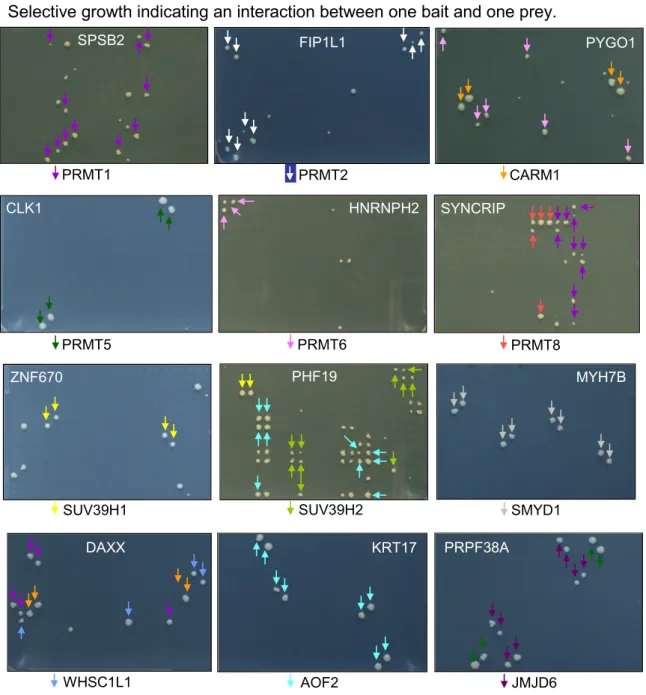
4.2.4 Verification of primary hits identified in the Y2H-matrix and Y2H-seq screens ... 56
4.3 Comparison of the Y2H-matrix and the Y2H-seq approaches ... 58
4.3.1 Comparison of the preys identified in the Y2H-matrix and the Y2H-seq screens ... 58
4.3.2 Comparison of the bait interactions in the Y2H-matrix and the Y2H-seq screens ... 59
4.3.3 Evaluation of the retest success rates in the Y2H-matrix and the Y2H-seq screens ... 60
4.3.4 Strong correlation of retest success rate and Y2H-seq prey rank ... 60
4.3.5 Increased sensitivity through Y2H-seq screening ... 62
4.4 Interactions of PMT and PDeM proteins ... 63
4.4.1 Distribution of interactions across PMTs and PDeMs ... 63
4.4.2 High interaction specificity of PMT and PDeM bait proteins ... 64
4.5 Analysis of PMT and PDeM interacting prey proteins ... 66
4.5.1 Known methylated proteins in the PMT and PDeM interaction dataset ... 66
4.5.2 Arginine methylation of PRMT interacting proteins containing RG-repeat motifs ... 69
4.5.3 Enrichment of proteins involved in transcriptional regulation and DNA-binding in the network 69 4.5.4 Enrichment of specific domains in the PMT- and PDeM interaction dataset ... 72
4.6 The PMT and PDeM interactome network ... 73
4.6.1 Subnetwork of common interaction partners between PRMT1 and PRMT8 ... 75
4.7 Identification of methylated residues in PMT interacting proteins ... 76
4.7.1 Detection of methylated proteins in a methylation assay using radioactive AdoMet ... 76
4.7.2 Mapping methylation sites using in vitro methylation coupled to mass spectrometry ... 78
5 Discussion ... 84
5.1 Quality estimates of Y2H protein interaction data ... 84
5.1.1 A new Y2H-seq approach reducing the workload and increasing the sensitivity ... 85
5.1.2 Y2H methods utilizing second generation sequencing ... 86
5.2 First proteome-wide interaction dataset of PMT and PDeM ... 87
5.2.1 Known methylated proteins detected in the PMT/PDeM network ... 88
5.2.2 New methylation substrates characterized in the methylation assay ... 89
5.3 Proteins interacting with PDeMs ... 91
5.3.1 AOF2 interacts with a variety of potential non-histone demethylation substrates... 91
5.3.2 JMJD6 interacts with spliceosomal proteins... 91
5.4 Proteins interacting with protein lysine methyltransferases ... 92
5.4.1 SUV39H1 preferentially interacts with zinc finger (ZnF) containing proteins and methylates the ZnF protein WIZ ... 92
5.5 Proteins interacting with protein arginine methyltransferases ... 95
5.5.1 CARM1 is the putative methyltransferase for the STAR protein quaking ... 95
5.5.2 Methylation of the SOCS box protein SPSB2 by PRMT1 maybe involved in regulating ubiquitination and degradation of target proteins ... 97
5.5.3 The E3 ligase complex associated WD40 domain containing protein WDR42A is methylated on multiple arginine sites by PRMT1 ... 99
5.6 Summary and further directions ... 100
References ... 102
Appendix ... 115
1 Introduction
1.1 Protein methylation
Protein methylation occurs predominantly on arginine, lysine and histidine residues and is catalyzed by S-adenosylmethionine (SAM, AdoMet) dependent enzymes that donate a methyl group to the side-chain nitrogen atom. Methylation reduces the charge and changes the structure of the side chain. Therefore it can alter function by increasing hybdrophobicity and may disrupt intra- and intermolecular hydrogen-bond interactions or provide a novel interface for proteins that bind preferentially to methylated proteins (Gary and Clarke, 1998; McBride and Silver, 2001). Enzymes catalyzing this reaction are called methyltransferases. Around 1 % of the human genome encode for methyltransferases with most of them presumably function as protein methyltransferases.
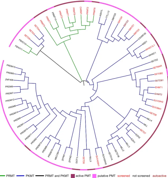
Beside proteins there is a variety of different methyltransferase substrates including RNA, DNA, lipids and small molecules. Thus, methyltransferases are involved in diverse biological pathways (Martin and McMillan, 2002; Schubert et al., 2003). There are structurally defined types of S-adenosylmethionine dependent methyltransferases. Class I enzymes have a common seven-ß-strand structure and are most abundant. Protein arginine methyltransferases (PRMTs) and the lysine methyltransferase DOT1L fall into Class I. Protein lysine methyltransferases (PKMTs) belong in general to the SET [Su(var), Enhancer of zest, trithorax] domain superfamily (Class II) and catalyze methylation of protein lysine residues (Dillon et al., 2005; Petrossian and Clarke, 2011). In Figure 1 protein methyltransferases (PMT) are illustrated in a dendrogram based on sequence similarity. The nine PRMTs have similar sequences and cluster together. The much larger set of 56 PKMTs are more diverse. For a number PKMTs no activity has been verified so far.
1.1.1 Protein arginine methylation
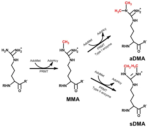
The PRMTs transfer the methyl group from the AdoMet donor molecule to the acceptor molecule the terminal nitrogen atom of the guanidinium side chain of an individual arginine residue in the target protein (Bedford and Clarke, 2009; Gary and Clarke, 1998). Three distinct types of methylated arginine residues occur in mammalian cells: monomethylated arginine (MMA), symmetrically dimethylated arginine (sDMA) and asymmetrically dimethylated arginine (aDMA) (Figure 2) which in contrast to phosphorylation and acetylation create a supplementary level of information. These three derivates are present on a multitude of distinct protein species in the cytoplasm, nucleus and organelles of mammalian cells (Bedford and Clarke, 2009). According to the type of methylation catalyzed by the enzyme, the PRMTs were classified into different groups.
While the type I PRMTs catalyze the formation of MMA and aDMA, the type II PRMTs form MMA and sDMA (Figure 2). PRMT1, PRMT3, CARM1 (PRMT4), PRMT6 and PRMT8 belong to the type I enzymes and PRMT5 and PRMT7 to the type II enzymes (Gary and Clarke, 1998;
Krause et al., 2007; Pahlich et al., 2006).
screened not screened autoactive active PMT putative PMT
PRMT PKMT PRMT and PKMT
Figure 1: Dendrogram of protein methyltransferases
Protein methyltransferases (PMTs) are ordered based on a basic primary protein sequence alignment in CLUSTALW. The figure is generated using ITOL. Branches represent PRMTs (green), PKMTs (blue) and PRMT/ PKMT (black). PMTs known to be active are indicated in dark purple and PMTs with unknown enzymatic activity are indicated in light purple. We will use a comprehensive set of PMTs to screen for protein-protein interactions. Label color indicates PMTs tested in our screens (red), not tested (black) or autoactive (dark red).
The seven PRMTs described above have been experimentally demonstrated to possess enzymatic activity while for PRMT2 and PRMT10 no activity has been demonstrated. PRMTs have a common catalytic methyltransferase domain which consists of a highly conserved core region and a subdomain important for binding of the methyldonor (Bachand, 2007). The individual PRMT family members differ in unique N-terminal regions of variable length and distinct domain motifs (Figure 3). PRMT2, PRMT3, PRMT8 and PRMT10 contain a Src homology 3 (SH3) domain, zinc finger (ZnF) domain, myristoylation (Myr) motif and tetratricopeptide repeats (TPR), respectively.
PRMT7 and PRMT10 have a second C-terminal catalytic domain.
RHN O
R`
NH N H2 NH2+
RHN O
R`
NH N H NH2+
CH3
RHN O
R`
NH N
H NH+ CH3H3C RHN
O R`
NH N NH2+ CH3 C H3
AdoMet AdoHcy
AdoM et
AdoHc y
AdoMet
AdoHc y PRMT
PRMT Type I
enzyme
PRMT Type I
I enz yme
asymmetric arginine
symmetric arginine monomethyl arginie
arginine
MMA
sDMA aDMA
Figure 2: Types of methylation on arginine residues
Type I and II PRMTs generate monomethylarginine (MMA) on one of the terminal guanidino nitrogen atoms. The subsequent generation of asymmetric dimethylarginine (aDMA) is catalyzed by type I enzymes and the production of symmetric dimethylarginine (sDMA) is catalyze by type II enzymes.
PRMTs use S-adenosylmethionine (AdoMet) as a methyldonor releasing S-adenosylhomocysteine (AdoHcy).
PRMT1 PRMT2 PRMT3 PRMT4/CARM1 PRMT5 PRMT6 PRMT7 PRMT8 PRMT10
SH3 ZnF
myr
core PRMT motif
TPR TPR
Figure 3: Protein arginine methyltransferases
The mammalian PRMT family currently contains nine highly related members. PRMTs have at least one core PRMT motif (blue). PRMT7 and PRMT10 have a duplication of this motif. PRMT2,
PRMT3, PRMT8 and PRMT10 contain a Src homology 3 (SH3) domain, zinc finger (ZnF) domain, myristoylation (Myr) motif and tetratricopeptide repeats (TPR), respectively.
1.1.1.1 PRMT1, the predominant type I methyltransferase, is closely related to PRMT8
PRMT1 was the first protein arginine methyltransferase in mammalian cells cloned and discovered independently by different groups (Abramovich et al., 1997; Lin et al., 1996). PRMT1 emerged as the predominant type I PRMT in mammalian cells contributing to over 85 % of type I protein arginine methyltransferase activity in cultured RAT1 fibroblast cells and mouse liver tissue (Pawlak et al., 2002; Tang et al., 2000a). PRMT1 knockout mice die shortly after implantation but embryonic stem cells generated from these mice are viable (Pawlak et al., 2000).
The first targets of PRMT1 identified were histones whose methylation is part of the histone code regulating gene expression (Abramovich et al., 1997; Lin et al., 1996). Later also non-histone proteins methylated by PRMT1 were identified. For example proteins involved in DNA damage response pathways as MRE11 and p53 binding protein 1 (53BP1) are methylated by PRMT1 in arginine-glycine (RG)-rich regions (Adams et al., 2005; Boisvert et al., 2005b). PRMT1 preferentially methylates RG-rich regions, a common feature of heterogeneous nuclear ribonucleoproteins (hnRNP) and other RNA binding proteins (RBP) that are the best known non-histone substrates of PRMT1 and are involved in various aspects of RNA processing and transport (Bedford and Richard, 2005; Liu and Dreyfuss, 1995). PRMT1 alters the subcellular localization of SYNCRIP, FUS and EWSR1 by methylation (Belyanskaya et al., 2001; Passos et al., 2006b; Rappsilber et al., 2003; Tradewell et al., 2012) but also affects protein-protein interactions (PPIs) (Bedford and Clarke, 2009; Bedford and Richard, 2005; McBride and Silver, 2001). For example, Sam68 contains proline-rich regions that interact with the SH3 and WW domains of several proteins. Interestingly, methylation decreases the binding of the SH3 domain but not WW domain allowing modulation of specific PPI (Bedford et al., 2000). Sam68 belongs to the family of signal transduction and activation of RNA (STAR) proteins. The STAR family proteins including SAM68, SLM-1, SLM-2, GRP33 and QKI-5 are known to be methylated.
SAM68, SLM-2 and GRP33 are methylated by PRMT1 whilst the modifying enzyme of SLM-1 and QKI-5 is unknown (Cote et al., 2003).
PRMT8 was identified through its high degree of sequence identity to PRMT1. Although PRMT8 is closely related to PRMT1 it is expressed in specific tissues, especially in the brain, and is attached to the plasma membrane via N-terminal myristoylation (Lee et al., 2005d). PRMT8 activity is much lower when compared to PRMT1. However, removal of the elongated N-terminal of PRMT8 results in an enhanced enzymatic activity of PRMT8 suggesting that the N-terminal domain regulates PRMT8 activity. The N-terminal region of PRMT8 was detected to be automethylated on arginine 58 and 73 (Sayegh et al., 2007). In an pull-down experiment 20
PRMT8 binding proteins were identified including hnRNPs, RNA-helicases (DEAD box proteins), FUS , EWSR1, TAF(II)68 and caprin (Pahlich et al., 2008). Some of these proteins are also known to bind to PRMT1, including EWSR1 which is methylated by PRMT1 and PRMT8 (Kim et al., 2008). Structural analysis of PRMT1 revealed that it forms a dimer (Zhang and Cheng, 2003).
PRMT8 was identified to interact with PRMT1. Hence, PRMT1 and PRMT8 form homo- and heterodimers (Kim et al., 2008; Lee et al., 2005b; Pahlich et al., 2008).
In summary, PRMT1 is involved in various processes including, signaling, DNA repair, transcriptional regulation, protein-protein interactions and localization. Most of the substrates identified are hnRNP proteins containing RG-rich regions but PRMT1 has been shown to methylated also non RG-rich proteins, suggesting that there are many more substrates which need to be identified.
1.1.1.2 CARM1 and PRMT6 are type I methyltransferases with pronounced substrate specificity
PRMT4 was discovered binding to the p160 family of nuclear receptor coactivators in a yeast-two hybrid (Y2H) approach. The association between PRMT4 and the p160 family enhances transcriptional activation as such PRMT4 is also called coactivator-associated arginine methyltransferase 1 (CARM1) (Chen et al., 1999). Embryos with a targeted distribution of CARM1 are small in size and die perinatally but embryonic stem cells generated from these CARM1-null embryos are viable (Kim et al., 2010; Yadav et al., 2003). In cells CARM1 forms a complex with ATP-remodeling (SWI/SNF) factors (Chi et al., 2004). CARM1 was identified to contribute to transcriptional regulation by methylation of histone 3 arginine 17 (Schurter et al., 2001). The first non-histone substrate identified was PABP1 a RNA binding protein (Lee and Bedford, 2002). Later on, substrates including transcriptional coactivators and a subset of splicing factors were identified (An et al., 2004; Cheng et al., 2007; Chevillard-Briet et al., 2002; Xu et al., 2001). The fact that CARM1 is a coactivator of nuclear receptors makes it a likely candidate for cancer (Bedford, 2007). In breast cancer tumors increased expression of CARM1 was shown (El Messaoudi et al., 2006). CARM1 was also involved in the development of prostate carcinomas (Hong et al., 2004).
Recently, it has been shown that the carboxy-terminal domain (CTD) of RNAPII is methylated by CARM1 and that phosphorylation of the CTD inhibits methylation (Sims et al., 2011). An automethylation site on CARM1 regulates cellular functions but does not affect enzymatic activity of CARM1. Phosphorylation instead abolishes AdoMet binding and inhibits the methyltransferase activity of CARM1 (Feng et al., 2009; Kuhn et al., 2011). In summary, CARM1 catalyzes methylation of distinct substrates as PRMT1 and is involved in regulating several cellular processes including transcription, splicing and protein-protein interactions.
PRMT6 was discovered by searching the human genome for protein arginine methyltransferases. It is a nuclear enzyme with a substrate specificity functionally distinct from PRMT1 and CARM1. Like CARM1, PRMT6 displays an automethylation activity (Frankel et al.,
2002). Specific substrates of PRMT6 include the nuclear scaffold protein HMGA1a (Miranda et al., 2005), DNA Polymerase ß (El-Andaloussi et al., 2006), histone H3/H4 (Lee et al., 2004) and the HIV Tat protein (Boulanger et al., 2005; Invernizzi et al., 2007; Invernizzi et al., 2006; Xie et al., 2007). Hence, only two human non-histone proteins are known to be methylated by PRMT6.
1.1.1.3 PRMT5 is the major type II methyltransferase
PRMT5 was identified in a Y2H approach interacting with Janus kinase 2 (Jak2) and appears to be the major type II mammalian arginine methyltransferase (Branscombe et al., 2001). PRMT5 is conserved in eukaryotes and widely expressed in human tissues (Pollack et al., 1999). Like CARM1, PRMT5 can complex with hSWI/SNF ATP dependent chromatin remodeling protein and in this context it functions as a transcriptional coactivator (Dacwag et al., 2007). PRMT5 forms also with plCln a complex called the methylosome. This methylates the RG-rich regions of SmD1, SmD3 and SmB which is a prerequisite for the survival of motor neuron (SMN) dependent assembly of the spliceosomal core-UsnRNP (Brahms et al., 2001; Brahms et al., 2000; Friesen et al., 2002; Meister et al., 2001; Meister and Fischer, 2002). Additionally, PRMT5 methylates SPT5, p53 and FEN1 (Guo et al., 2010; Kwak et al., 2003). Recently crosstalk between arginine methylation and tyrosine phosphorylation was described on the epidermal growth factor receptor (EGFR) (Hsu et al., 2011). Hence, PRMT5 methylation crosstalks with other posttranslational modifications, regulates transcription, degradation, splicing, DNA replication and signaling in the cell.
1.1.1.4 Additional members of the PRMT family are not characterized
PRMT2 was identified by sequence homology to PRMT1 (Katsanis et al., 1997). PRMT2 transcripts are detected in most human tissues and are predominantly localized in the nucleus.
PRMT2 null mice are viable and normal (Yoshimoto et al., 2006). A novel feature of PRMT2 is that it harbors a SH3 domain at its N-terminus (Scott et al., 1998) (Figure 3). Analysis using Y2H screening approaches identified PRMT2 interacting with the estrogens receptor alpha (Qi et al., 2002) and the androgen receptor (Meyer et al., 2007), both are nuclear hormone receptors.
Although PRMT2 does not have enzymatic activity it does function as a coactivator for the estrogen receptor (Qi et al., 2002) and therefore is of special interest.
PRMT3 belongs to the type I enzymes and is expressed widely in human tissues with subcellular localization in the cytoplasm (Tang et al., 1998). Mouse embryos with a target disruption of PRMT3 are small size but survive after birth and attain a normal size in adulthood (Swiercz et al., 2007). An important feature of PRMT3 is the C2H2-type zinc-finger domain (Frankel and Clarke, 2000) (Figure 3). The ZnF domain of PRMT3 appeared to be necessary and sufficient for binding of rpS2. RpS2 was shown to be a bona fide in vivo substrate of PRMT3 that can not be modified by other PRMTs (Swiercz et al., 2005). Recently rpS2 was reported to be a substrate of Peptidylarginine deiminase 4 (PAD4, PADI4). PAD4 removes methylation by
conversion of arginine to citrulline, a process known as deimination (see 1.1.2) (Guo et al., 2011).
PRMT3 has unique substrate specificity but only a handful of substrates are known.
PRMT7 was discovered by Miranda et al. and shown to monomethylate synthetic peptides (Miranda et al., 2004). Later it was shown that PRMT7 symmetrically dimethylates histones, myelin basic protein, fibrillarin and the spliceosomal protein SmB (Lee et al., 2005c). PRMT7 seems to be derived from a gene duplication event resulting in two putative substrate binding motifs (Figure 3). Both of these domains are required for the functionality of the enzyme as each separate domain was unable to function alone (Lee et al., 2005d). It is largely unknown which function PRMT7 has and no specific substrate is known.
PRMT10 was identified based on the homology to other PRMT family members as a product of a gene on the human chromosome 4y31 that is most closely related to PRMT7 (Cook et al., 2006; Krause et al., 2007; Lee and Stallcup, 2009). This gene is a candidate for PRMT10 and needs to be biochemically characterized. It contains two TPR which are known to mediate PPIs (Bedford et al., 2009).
The two F box-only family members, FBXO10 and FBXO11, have been suggested to be protein arginine methyltransferases but activity has not been clarified (Bedford and Clarke, 2009;
Cook et al., 2006; Krause et al., 2007; Petrossian and Clarke, 2011).
1.1.2 Protein arginine demethylation
The JmjC enzyme JMJD6 was the first arginine demethylase identified. Using specific antibodies Chang et al. demonstrated that dimethylation of histone H3 and histone H4 peptides is reduced after incubation with JMJD6 (Chang et al., 2007). However, recent data showed that JMJD6 does not demethylate histone H4 and H3 fragment peptides but hydroxylates U2AF65 by incorporation of oxygen atoms (Webby et al., 2009). Lysyl hydroxylation is the dominant oxidative catalytic activity of JMJD6. JMJD6 catalyzed arginine demethylation cannot be ruled out but would have to occur at a level below current levels of detection (Webby et al., 2009).
The Peptidylarginine deiminase 4 (PAD4, PADI4) removes methylation by conversion of arginines to citrulline, a process known as deimination. PAD4 deiminates not only methylated arginine residues but also unmethylated arginine residues. Citrullination and methylation modifications are antagonistic to each other suggesting a conserved posttranslational regulatory strategy (Cuthbert et al., 2004; Guo et al., 2011; Wang et al., 2004). Peptidylarginine deiminases are not true “demethylases” as they do not convert monomethylarginine back to arginine. Whether arginine methylation can be removed or is a more static posttranslational modification that is removed via protein degradation, is a pivotal question in the field awaiting clarification.
1.1.3 Protein lysine methylation
Other than arginine, lysine residues are methylated on proteins. Furthermore, acetylation, ubiquitination, sumolation and neddylation are common modification on lysine residues (Yang and
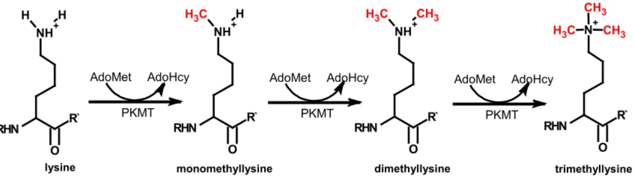
Seto, 2008). The fact that alternated modifications occur on lysines provides another level of regulation since the presence of one inhibits the attachment of the other modification. Methylation can exist in mono-, di- and trimethylated state (Figure 4).
AdoMet AdoHcy PKMT
AdoMet AdoHcy PKMT
AdoMet AdoHcy RHN PKMT
O R`
NH+ C
H3 CH3
RHN O
R`
N+ C H3
CH3 CH3
RHN O
R`
NH+
H H
RHN O
R`
NH+ C H3 H
lysine monomethyllysine dimethyllysine trimethyllysine
Figure 4: Types of methylation on lysine residues
PKMTs generate monomethyllysine, dimethyllysine and trimethllysine. PKMTs use S-adenosylmethionine (AdoMet) as a methyldonor releasing S-adenosylhomocysteine (AdoHcy).
Lysine methylation is accomplished by so called protein lysine methyltransferases (PKMT).
All PKMTs, except Dot1, share a SET domain that is responsible for catalysis and binding of the cofactor AdoMet (Dillon et al., 2005). There are 56 PKMTs present in the human proteome but enzymatic activity and substrate specificity of most of them is unknown (Figure 1) (Levy et al., 2011; Petrossian and Clarke, 2011). At first lysine methylated histones were identified and later also non-histone proteins (Huang and Berger, 2008).
1.1.3.1 Chromatin state is regulated by histone lysine methylation
Histones play a dynamic role in controlling chromatin structure and transcription. There are different states of chromatin. The more compact chromatin is called heterochromatin and euchromatin, which is believed to be accessible for transcription, has a more open structure.
Histone lysine methylation plays a major role in regulating the state of chromatin compaction.
Additionally, histone lysine methylation regulates activation and repression of gene transcription within the euchromatin (Hublitz et al., 2009). In general histone H3 methylation at lysine 4, 36 and 79 correlates with euchromatin and transcriptional activation whereas histone H3 methylation at lysine 9 and 27 and histone H4 at lysine 20 is associated with heterochromatin and transcriptional repression (Bannister et al., 2002). However, different extend of methylation and crosstalks between different modifications leads to different functions in the cell (Yang and Seto, 2008) and thus reveals a “histone code” that extends the information of the genetic code (Jenuwein and Allis, 2001).
The first PKMT with enzymatic activity identified was SUV39H1. SUV39H1 specifically target histone H3 lysine 9 (H3K9me) (Rea et al., 2000). EHMT2 (G9a) mono- and dimethylates euchromatin at H3K9 (Tachibana et al., 2005). SUV39H1 trimethylates H3K9 and that correlates with the formation of heterochromatin (Robin et al., 2007). The trimethylated H3K9 is recognized
by heterochromatin protein 1 (HP1) through its chromodomain, but not the mono- and dimethylated H3K9. HP1 is localized to heterochromatin sites where it mediates gene silencing (Bannister et al., 2001; Jacobs and Khorasanizadeh, 2002). This shows that different levels of methylation can exert different functional outcomes. Although, histones are by far the predominant substrates described for PKMTs a few non-histone substrates are also known (Huang and Berger, 2008).
1.1.3.2 Non-histone protein functions are modulated by lysine methylation
There are a few non-histone substrates identified including cytochrome c in plants and fungi (Kluck et al., 2000; Polevoda et al., 2000). Calmodulin (Sitaramayya et al., 1980), Rubisco in plants (Houtz et al., 1989) and several ribosomal proteins in yeast were also found to be lysine methylated (Kruiswijk et al., 1978; Porras-Yakushi et al., 2005). The first human non-histone substrate identified was the tumor suppressor p53 which is methylated by SETD7 (SET9, SET7, SET7/9, KMT7). Methylation of p53 positively affects its stability and regulates the expression of p53 target genes (Chuikov et al., 2004). P53 is also methylated by SMYD2 on lysine 370 close to the lysine 372 modified by SETD7 (Huang et al., 2006). Monomethylation by SMYD2 has repressory function whereas dimethylation of lysine 370 leads to activation by association of p53 with the coactivator 53BP1. It was shown that the mono- and dimethylation mediated by SMYD2 can be removed by AOF2 (Huang et al., 2007). AOF2 also removes SET8 mediated lysine 382 methylation of p53 which leads to inactivation of p53 (Shi et al., 2007). Additionally, SETD7 methylates DNA methyltransferase 1 (DNMT1) (Esteve et al., 2009), TBP-associated factor TAF10 (Kouskouti et al., 2004), estrogen receptor (ER) α (Subramanian et al., 2008) and MYPT1 (Cho et al., 2011). A study by Dhayalan et al. applied a peptide array to determine an optimized target sequence for SETD7. Based on this they identified 91 new peptide substrates derived from human proteins and verified methylation of nine non-histone proteins (Dhayalan et al., 2011). The NFкB subunits RelA and p65 are methylated by SETD7 (Ea and Baltimore, 2009; Yang et al., 2009). SETD6 methylates RelA, too. SETD6 mediated methylation is recognized by EHMT1 which mediates H3K9 methylation and repression of NFкB signaling. Serine phosphorylation blocks the binding of EHMT1 and relieves repression of the target genes (Levy et al., 2011). The Vascular Endothelial Growth Factors Receptor 1 (VEGFR1) has been identified as the only methylation target of SMYD3 (Kunizaki et al., 2007). EHMT2 methylates non-histone proteins including CDYL1, WIZ, ACINUS and EHMT2 (automethylation). HP1 binds methylation specific to the automethylation site of EHMT2 and to the trimethylated peptides of histone H3, CDYL1 and WIZ. In addition, it was shown that methylation of CDYL1 abolished the interaction of the CDYL1 chromodomain to H3K9me3 (Rathert et al., 2008; Sampath et al., 2007).
In summary, most non-histone substrates have been identified methylated by SETD7 but also other PKMTs methylate non-histone proteins. The discussed cases indicate that lysine methylation of non-histone proteins can recruit methyl recognition domains, regulate stability of proteins,
repress function of proteins and plays a role in assembling of complexes. This implies a broader function of lysine methylation as previously thought. For a number of PKMTs no substrates have been verified so far and it is possible that these may methylate non-histone proteins.
1.1.4 Protein lysine demethylation
Two kinds of lysine demethylases have been identified: The lysine (K)-specific demethylase 1A (KDM1A, AOF2, LSD1) and the Jumonji C (JmjC) domain family proteins. AOF2 can demethylate mono- and dimethyllysine, but not trimethyllysine. AOF2 functions as histone demethylase (Shi et al., 2004) but is also involved in demethylating non-histone proteins including p53, MYPT1 and DNMT1 (Cho et al., 2011; Esteve et al., 2011; Shi et al., 2007; Wang et al., 2009). It is suggests that there are further non-histone targets demethylated by AOF2.
In 2006, the JmjC family protein JHDM1 was purified and shown to catalyze demethylation of lysine residues (Tsukada et al., 2006). Some of the JmjC family proteins have been shown to act on histones including JHDM2A, JMJD2A, JMJD2B, JMJD2C and JMJD2D (Whetstine et al., 2006; Yamane et al., 2006). Whilst no non-histone substrate has currently been described for the JmjC protein family, lysine methylation can at least in part, be described as a dynamic posttranslational modification that is regulated by demethylase and methyltransferase activities (Huang et al., 2007; Ruthenburg et al., 2007).
1.2 PMT substrate identification methods
1.2.1 Methods to detect methylated proteins
Arginine protein methylation has been implicated in various functions like protein-protein and protein-RNA interactions, cellular localization, nuclear transport, RNA processing, ribosome assembly, maturation of hnRNPs, translation accuracy, protein metabolism and cell signaling.
(Choudhary et al., 2009; Kim et al., 2006a). There is a large family of protein methyltransferases which are expressed in a large variety of tissues and diverse subcellular localization, predominantly nuclear but also strictly cytoplasmatic, like PRMT5 (Herrmann et al., 2009). However, methyl binding domains containing proteins exist (Taverna et al., 2007) suggesting that protein methylation plays a ubiquitous role in many cellular processes other than epigenetic gene regulation. Progress to date has undoubtedly uncovered only a small portion of the roles of arginine methylation in regulation of protein function in biological processes (Lee and Stallcup, 2009).
Likewise, lysine methylation of proteins regulate diverse cellular processes and different publications suggest that there will be far more non-histone proteins discovered to be lysine methylated (Huang and Berger, 2008). Similar to protein acetylation that was initially characterized primarily on histones but later recognized as modification on many non-histone proteins (Gu and Roeder, 1997; Lee et al., 2005a; Sterner and Berger, 2000). Proteome-wide studies revealed that over 1700 proteins are acetylated. To understand the biological role of methylation, besides histone
methylation, proteome-wide screens are required to identify new lysine methylation substrates and the responsible enzymes. It is conceivable that there are more non-histone substrates and additional PKMTs methylating non-histone targets.
The detection of non-histone targets has proved technically challenging. Different immunopurification (IP) approaches coupled to mass spectrometry have been implicated to identify methylated proteins (Boisvert et al., 2003; Ong et al., 2004). Those studies are essentially limited by the quality of available antibodies. Many commercial available methyl antibodies crossreact with unmodified sequences or they fail to efficiently bind a large variety of methylated sequences (Levy et al., 2011). Further limitation of the IP approach is that the corresponding enzyme is not identified (Komyod et al., 2005; Pahlich et al., 2006).
Another approach to identify non-histone targets is to query the proteome for a linear amino acids sequence which surrounds the methylation sites. To do so, a specific profile of the protein methyltransferase methylation site is determined by methylation of peptide arrays. This specific profile is used in a proteome-wide searched for similar motifs. A proteome-wide search for EHMT2 specific profiles revealed 92 human proteins and showed methylation on eight in a direct in vitro methylation assay (Rathert et al., 2008). However, the motif search works only for enzymes recognizing linear amino acid sequences which might not be the case for all PKMTs and PRMTs.
There are two unbiased screening approaches to identify methylation substrates. First, hypomethylated cell extract of PC12 rat cells can be used to visualize methyl acceptor proteins. In this approach the hypermethylated cell extract is incubated with the radiolabel AdoMet in the presence of protein translation inhibitor. Proteins incorporate the radioactive labeled methyl group and were separated by 2D gel electrophoresis. More than 50 different methyl acceptor proteins were detected by fluorography. However, identification of these proteins was not possible (Liu and Dreyfuss, 1995; Najbauer and Aswad, 1990; Pahlich et al., 2006; Sampath et al., 2007). In a similar approach numerous heterogeneous nuclear ribonucleoproteins (hnRNPs) were discovered by immunopurification and 2D gel electrophoresis (Najbauer and Aswad, 1990; Pahlich et al., 2006).
Those approaches are limited by the detection and identification of low abundant proteins.
An alternative approach is the human protein microarray based platform which is incubated with the appropriate PKMT and AdoMet. Levy et al. identified 216 potential SETD6 substrates and showed methylation on six in a direct in vitro methylation assay (Levy et al., 2011). The readout was performed by methyl specific antibodies or radioactive labeled AdoMet. Antibodies are difficult to use for readout as discussed above. Radioactive labeling has inherent limitations in its signal to noise ratio and imaging of radioactivity exposed on film. In addition, the protein array system has several limitations. First, only one third of the human proteome is represented on the protein array. Second, many proteins on the protein array do not cover the full length sequence of the protein. Third, signal intensity of the array is low (Levy et al., 2011).
In summary, there are proteome-wide approaches to detect protein methylation but all have strong limitations in discovering methylated proteins.
1.2.2 The Y2H system to find PMT interacting proteins and potential substrates
A physical interaction is a requisite for PMT enzymes to methylate target proteins. Therefore, in principle substrates of PMTs ought to be identified by studying PMT-protein interactions (Passos et al., 2006a). The analysis of protein interactions is based on two main technologies: The Y2H system to detect binary interactions and affinity purification followed by mass spectrometry (AP-MS) which detects direct and indirect interactions. In an AP-MS approach stable protein complexes composed around the protein of interest are isolated from cells and then analyzed using mass spectrometry (Gingras et al., 2007). Both approaches have advantages and disadvantages. The analysis of human PPIs in the Y2H system provides a heterologous environment whereas the AP-MS approach enables to study complexes that assemble in a more natural cellular environment (Choudhary and Mann, 2010). Washing steps during affinity purification leads to the loss of weak or transiently bound proteins and hence these interactions are not detected in the mass spectrometer. Highly abundant proteins are predominantly identified in the AP-MS approach, often also as false positive interaction partners. The Y2H approach assays proteins independent of endogenous expression levels. Proteins are expressed at very low levels and tested systematically, thus can be statistically analyzed (Worseck et al., 2012). Recent studies proved that large scale Y2H analyses result in high-precision data. A study by Yu et al. demonstrated that Y2H and AP-MS data are of equal high-quality if taken into account the different and complementary nature of the interactions determined. They also showed that the binary interaction map in comparison to the co-complex interactome model is enriched for transient signaling interactions (Simonis et al., 2009; Venkatesan et al., 2009; Yu et al., 2008). It was also shown that direct human PPIs relevant for signal transduction are identified by systematic Y2H interaction screening (Vinayagam et al., 2011). Enzyme-substrate interactions are direct and often transient. Therefore, the Y2H system is an efficient method to detect PMT-protein interactions (Passos et al., 2006a) and additionally it is independent of affinity tools that are lacking for the analysis of protein methylation.
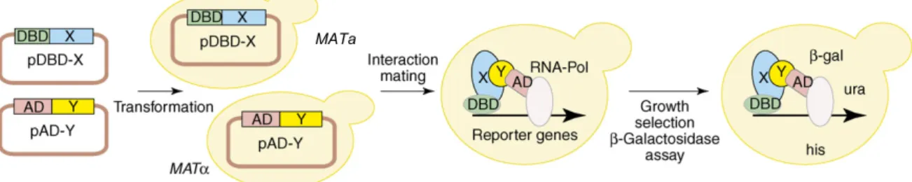
Y2H system to detect PPIs. The Y2H system was originally developed by Stanley Fields (Fields and Song, 1989). It is based on two separable domains of a transcription factor, the DNA-binding domain (DBD) that recruits the transcription factor to the DNA and the activation domain (AD) that initiates transcription of target genes. In the Y2H system, those two domains are separately fused to the proteins of interest resulting in DBD and AD fusion proteins referred to as bait and prey protein, respectively. An interaction between the fusion proteins in yeast cells expressing both hybrid proteins brings the DBD and the AD into close proximity and leads to reconstitution of the transcription factor (Figure 5). The functional transcription factor activates
the expression of one or more reporter genes. This enables the growth selection of yeast cells harboring an interacting protein pair (Figure 5).
MATa
Figure 5: Principle of the Y2H system for the detection of binary interactions
Coding sequences for a protein X and a protein Y are fused to a DNA-binding domain (DBD, i.e. bait plasmid) and a transcription activation domain (AD, i.e. prey plasmid). Two yeast strains of opposite mating type MATa and MATα are transformed with the bait and prey plasmids, respectively and mated. The diploid yeast is expressing both hybrid proteins. Upon interaction of protein X and protein Y, transcriptional activity of the DBD and the AD-domains is reconstituted leading to reporter gene activation. Reporter gene expression enables yeast growth in the absence of selected nutrients, such as histidine and uracil. In many Y2H systems, the lacZ gene is also utilized as reporter, so that an interaction of protein X and protein Y can be assayed via b-galactosidase activity (Stelzl and Wanker, 2006).
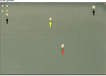
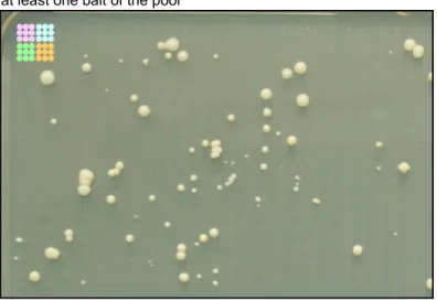
There are two Y2H high-throughput (HTP) methods: The matrix and the library approach. In the library approach, baits are screened separately against a prey pool (the prey library) containing random cDNA fragments or open reading frames (ORFs) (Chien et al., 1991). Yeast cells containing a positive interaction pair of proteins are selected based on the reporter gene activation and the resulting ability to grow on selective medium. In the Y2H library screen the plasmid DNA of the interacting prey proteins are separately isolated form the yeast colonies growing on the selective medium and are identified by Sanger sequencing (Chien et al., 1991). On the contrary, in the Y2H-matrix screen the prey clones are well characterized. In each position of array a particular prey is expressed. The baits are screened separately against the prey array and interacting protein pairs are identified based on the expression of the reporter gene resulting in growth on the selective medium in a specific position on the array. Hence, the identity of the prey protein can be identified via its position in the array (Worseck et al., 2012). The proteins encoded by the prey clones used in the Y2H-matirx are tested with the equal probability in contrast to the random protein encoding prey clones in the Y2H library screen which are not normalized. Hence, interactions are detected much more efficiently in the Y2H-matrix screen using a set of normalized preys (Reboul et al., 2003). Additionally, Y2H-matrix screens can be repeated (Worseck et al., 2012).
The relative low cost and the possibility for automation make HTP Y2H experiments to one of the most powerful tools to generate quality-controlled, proteome-wide, binary PPI maps, such as those generate for yeast (Ito et al., 2001; Uetz et al., 2000; Yu et al., 2008), fly (Giot et al., 2003), worm (Li et al., 2004; Simonis et al., 2009) and human (Bandyopadhyay et al., 2010; Rual et al., 2005; Stelzl et al., 2005; Vinayagam et al., 2011; Yu et al., 2011). Limitations of the Y2H-matrix
approach will be addressed in this study that describes a novel Y2H screening approach that utilizes second generation sequencing.
1.3 Aim of this study
Methylation is a post translational modification that can regulate diverse cellular processes and is subjected to increasing investigation. Several lines of evidence are suggesting that protein methylation plays a ubiquitous role in many cellular processes other than epigenetic regulation.
There are around 100 proteins known to be methylated on arginine residues but only for around 50 the responsible PMT is known. Even fewer lysine methylated proteins are known and so far there is only a handful of PKMTs known to methylate non-histone proteins. For a large set of PKMTs no methylation substrates has been identified so far. Despite the importance of methylation in maintaining cellular homeostasis, the development of proteome-wide approaches for detecting this modification has been limited and proven technically challenging. The aim of this study was to develop a proteome-wide methode to identify proteins interacting with PMTs or demethylases, assuming that a large fraction of identified proteins are methylation substrates of PMTs.
The HTP Y2H-matrix approach produces binary, high-quality PPI data and is an efficient method to detect the often transient enzyme-substrate interactions. However, the sensitivity of the screening method and thus low data coverage is a severe limitation. Hence, we developed a workload reduced Y2H approach with significantly increased sensitivity. The novel method utilized a second generation sequencing step and thus abbreviated Y2H-seq. As transient PPIs are efficiently detected if the sampling is increased the high sampling sensitivity of the Y2H-seq approach is of particular importance when detecting transient enzyme-substrate interactions, such as those between methyltransferases and their substrates.
The novel Y2H-seq approach was evaluated against our state of art Y2H-matrix screening approach and used to screen 8 PRMTs, 17 PKMTs and 10 PDeMs against a set of 14,268 proteins.
We generated a high-quality interaction network consisting of 523 interactions with 324 prey proteins. This is the first proteome-wide interaction dataset of enzymes involved in methylation.
Despite the few proteins known to be methylated and the incompleteness of the prey array we identified 11 prey protein substrates already known to be methylated. Whilst not all interacting proteins will be methyltransferase substrates it is clear methylation substrates of PMTs can be identified in the Y2H approach. Hence, methylation substrates of PMTs can be identified in the Y2H approach. Therefore, the interaction network will serve as resource to identify new methylation substrates. We developed a methylation assay using radioactive methyl donor and identified SYNCRIP to be methylated. To identify methylation sites an in vitro methylation assay was established and coupled to mass spectrometry analysis. Thus, new candidate methylation substrates were validated and the sites of methylation identified. On seven of nine proteins tested methylation sites were mapped including SPIN2B, DNAJA3, QKI, SAMD3, OFCC1, SYNCRIP
and WDR42A.
2 Material
2.1 Chemicals
4-(2-hydroxyethyl)-1-piperazineethanesulfonic acid (HEPES) (Sigma-Aldrich, Taufkirchen) 2-mercaptoethanol (Merck, Darmstadt)
Acetic acid (Merck, Darmstadt)
Acrylamid/Bisacrylamid 40 % (37,5:1) (Roth, Karlsruhe) Adenosine-5'-triphosphate (ATP) (Sigma-Aldrich, Taufkirchen)
Adenosyl-L-methionine, S-[methyl-3H]; (AdoMet[3H]) (PerkinElmer, USA) Agarose (Sigma-Aldrich, Taufkirchen)
Ammonium persulfate (APS) (Merck, Darmstadt) Ammonium sulfate (Merck, Darmstadt)
Ampicillin trihydrate (Sigma, Deisenhofen) Bacto Yeast Extract (BD BIOSCIENCES, USA) Bacto-Agar (BD BIOSCIENCES, USA)
Bacto-Pepton (BD BIOSCIENCES, USA) Bacto-Tryptone (BD BIOSCIENCES, USA) Betain (Sigma-Aldrich, Taufkirchen) Boric acid (Merck, Darmstadt)
Bovine serum albumin fraction V (BSA) (Roche, Mannheim) Bromphenol blue (Merck, Darmstadt)
Calcium chloride dihydrate (Merck, Darmstadt) Chloramphenicol (Boehringer, Mannheim) Chloroform (Merck, Darmstadt)
Coomassie Brilliant Blue G-250 (Biomol GmbH, Hamburg)
Deoxyribonucleic acid sodium salt from salmon testes (Sigma-Aldrich, Taufkirchen) Dimethyl sulfoxide 99.9 % (DMSO) (Sigma-Aldrich, Taufkirchen)
Dipotassium phosphate (Acros organics part of Thermo Fisher Scientific Inc., Geel, Belgium) Dithiothreitol (DTT) (Roth, Karlsruhe)
D-luciferin sodium lyophilized firefly (Sigma-Aldrich, Taufkirchen)
Dulbecco’s modified eagle medium (DMEM+GlutaMAX™-I) (Gibco BRL, Gaithersburg, USA) Dulbecco’s phosphate buffered saline (DPBS) (Gibco BRL, Gaithersburg, USA)
Ethanol (Merck, Darmstadt)
Ethylene glycol tetraacetic acid (EGTA) (Roth, Karlsruhe) Ethylenediaminetetraacetic acid (EDTA) (Roth, Karlsruhe)
Fetal bovine serum (FBS) (qualified FBS, south american) (Gibco BRL, Gaithersburg, USA) Glucose monohydrate (Merck, Darmstadt)
Glycerol (Merck, Darmstadt)
Glycine (MP Biochemicals, Aurora, USA) Glycogen (Roche, Mannheim)
Glycylglycine (Acros organics part of Thermo Fisher Scientific Inc., Geel, Belgium) Histidine (Sigma-Aldrich, Taufkirchen)
Imidazole (Sigma-Aldrich, Taufkirchen) Isopropanol (Merck, Darmstadt)
Isopropyl-ß-D-thiogalactopyranosid (IPTG), diaxone-free (Fermentas GmbH, St. Leon-Rot) Kanamycin sulfate (Sigma-Aldrich, Taufkirchen)
Leucin (Sigma-Aldrich, Taufkirchen)
Lithiumacetate (LiOAc) (Sigma-Aldrich, Taufkirchen) Magnesium chloride (Roth, Karlsruhe)
Magnesium sulfate (Roth, Karlsruhe) Methanol (Merck, Darmstadt)
Monopotassium phosphate (Roth, Karlsruhe) Opti-MEM I (Gibco BRL, Gaithersburg, USA)
Orthophosphoric acid (Alfa Aesar GmbH & Co KG, Karlsruhe) Paraformaldehyde (PFA) (Roth, Karlsruhe)
Phenol (Roth, Karlsruhe)
Phenylmethansulfonylfluorid (PMSF) (Roth, Karlsruhe) Polyethylene glycol (PEG) 3350 (Sigma-Aldrich, Taufkirchen) Polyethylene glycol (PEG) 8000 (Sigma-Aldrich, Taufkirchen) Potassium acetate (Merck, Darmstadt)
Potassium chloride (Roth, Karlsruhe) Protease inhibitor (Roche, Mannheim)
S-(5'-Adenosyl)-L-methionine chloride (AdoMet) (Sigma-Aldrich, Taufkirchen) Sodium carbonate (Merck, Darmstadt)
Sodium chloride (Roth, Karlsruhe) Sodium citrat (Roth, Karlsruhe)
Sodium dihydrogen phosphate (Merck, Darmstadt) Sodium dodecyl sulfate (SDS) (Roth, Karlsruhe) Sodium hydrogencarbonate (Merck, Darmstadt) Sodium hydroxide (Roth, Karlsruhe)
Sorbitol (Sigma-Aldrich, Taufkirchen)
Spectinomycin dihydrochloride pentahydrate (Sigma-Aldrich, Taufkirchen) Sucrose (Merck, Darmstadt)
SYBR Gold Nucleic Acid Gel Stain (Invitrogen, Darmstadt) Tetracycline hydrochloride (Sigma-Aldrich, Taufkirchen)
Tetramethylethylenediamine (TEMED) (Invitrogen, Darmstadt) Thiamine hydrochloride pure (AppliChem, Darmstadt)
Tris (hydroxymethyl) aminomethane (Tris Base) (Roth, Karlsruhe)
Tris (hydroxymethyl) aminomethane hydrochloride (Tris HCl) (Sigma-Aldrich, Taufkirchen) Triton X-100 (Sigma-Aldrich, Taufkirchen)
Tryptophan (Sigma-Aldrich, Taufkirchen) Tween 20 (Sigma-Aldrich, Taufkirchen) Uracil (Sigma-Aldrich, Taufkirchen)
Yeast nitrogen base (Difco part of BD Biosciences, USA) 2.2 Lab ware
384-well MTPs, PS, flat bottom, clear, sterile, with lid (Greiner Bio-One GmbH, 781186) 96-well deepwell plates (2000 µl/well) (Eppendorf, 0030 501.322)
96-well MTPs, PS, flat bottom, crystal clear (Greiner Bio-One GmbH, 655101)
96-well MTPs, PS, flat bottom, lumnitrac600, high binding, white, sterile (Greiner Bio-One GmbH, 655074)
96-well MTPs, PS, flat bottom, TC, white, sterile (Greiner Bio-One GmbH, 655073) 96-well PCR plate (Costar part of Corning Incorporated, 6511)
Agar-plates (241 x 241 x 20) (Nunc GmbH & Co. KG, 240845) BiomekNX (Beckman Coulter GmbH)
Biophotometer Plus (Eppendorf AG) Centrifuge 5810 R (Eppendorf AG)
E.A.S.Y 429k digital camera (Herolab GmbH Laborgeräte) Fixed-angle rotors F-45-30-11 (Eppendorf AG)
Glass beads, acid-washed 425-600 µm (Sigma-Aldrich, G8772) Incubator 1000 (Heidolph Instruments GmbH & Co. KG) InfiniteM200 multimode microplate reader (Tecan Group Ltd.) Innova44 shaker (New Brunswick Scientific)
Kby roboter (Cambridge, UK)
MicroPulser electroporation apparatus (Bio-Rad Laboratories)
Mini-PROTEAN tetra cell electrophoresis system (Bio-Rad Laboratories) NanoDrop ND-1000 (Thermo Fischer Scientific Inc.)
Ni-NTAagarose beads (Qiagen GmbH, Hilden)
Nitrocellulose Membrane (Bio-Rad Laboratories, 162-0115) Omnitrays (Nunc GmbH & Co. KG, 165218)
Pin tools with 96 and 384 pins. The steel pins are cylindrical with a diameter of 1.3 mm and the edge of the flat top that is touching the agar is bevelled 45°at 0.2 mm. Sterilize by heating the pins until they glow red. Let them cool in a sterile environment.