Genetic dissection of regulatory domains and signalling interactions of PRL1 WD-protein in
Arabidopsis
Inaugural-Dissertation zur
Erlangung des Doktorgrades
der Mathematisch-Naturwissenschaftlichen Fakultät der Universität zu Köln
vorgelegt von
Dóra Szakonyi
aus Kecskemét
KÖLN 2006
Berichterstatter: Prof. Dr. George Coupland Prof. Dr. Martin Hülskamp Prof. Dr. Karin Schnetz
Tag der mündlichen Prüfung: 06. 07. 2006
CONTENTS
FIGURES AND TABLES... IVI ABBREVIATIONS AND SYMBOLES... VIIII SUMMARY - ZUSAMMENFASSUNG... IX
1. INTRODUCTION ... 1
1.1. S
UGAR SIGNALLING IN PLANTS... 1
1.2. I
DENTIFICATION AND CHARACTERIZATION OF THE PRL1
MUTANT... 3
1.3. T
HEA
RABIDOPSISPRL1
GENE ANDPRL1
ORTHOLOGS... 4
1.4. C
ONSERVEDWD-
REPEAT PROTEINS... 5
1.5. PRL1
FAMILY MEMBERS ARE SUBUNITS OF CONSERVED SPLICING-
ASSOCIATED COMPLEXES.... 6
1.5.1. Prp19... 6
1.5.2. CDC5 ... 9
1.5.3. Crooked neck (CRN) proteins ... 10
1.6. F
UNCTION OF THEP
RP19-
ASSOCIATED COMPLEX IN SPLICING... 11
1.7. PRL1
INTERACTS WITHSNF1
RELATED PROTEIN KINASES OFAMP-
ACTIVATED(AMPK)
KINASE FAMILY... 13
1.8. A
RABIDOPSISS
NRK1
Α KINASES ARE FOUND IN ASSOCIATION WITH THE PROTEASOME... 14
1.9. A
IMS OF THE PRESENT WORK... 15
2.
MATERIALS AND METHODS... 17
2.1. M
ATERIALS... 17
2.1.1. Chemicals, enzymes and laboratory supplies ... 17
2.1.2. Bacterial Strains... 19
2.1.2.1. E. coli strains... 19
2.1.2.2. Agrobacterium tumefaciens strains... 20
2.1.2.3. Yeast strains... 20
2.1.3. Plant material ... 20
2.1.4. Plasmids vectors and constructs... 21
2.1.4.1. Plasmid vectors ... 21
2.1.4.2. Plasmid constructs ... 21
2.1.5. Arabidopsis yeast two hybrid cDNA library... 23
2.1.6. Oligonucleotides ... 24
2.1.6.1. Oligonucleotides for DNA sequencing ... 24
2.1.6.2. Oligonucleotides for cloning... 24
2.1.6.3. Oligonucleotides for site-directed mutagenesis ... 24
2.1.6.4. Oligonucleotides to screen for T-DNA insertion mutants in Arabidopsis... 25
2.2. G
ENERAL BUFFERS,
STOCK SOLUTIONS AND GROWTH MEDIA... 25
2.2.1.1. Solutions ... 25
2.2.2. Antibiotics... 26
2.2.3. Plant hormones ... 26
2.2.4. Culture media... 26
2.2.4.1. Bacterial media ... 26
2.2.4.2. Yeast media... 27
2.2.4.3. Plant media ... 27
2.2.5. Antibodies... 28
2.2.5.1. Primary antibodies ... 28
2.2.5.2. Secondary antibodies ... 29
2.2.6. Bioinformatic Resources... 29
2.2.6.1. Softwares ... 29
2.2.6.2. Databases ... 29
2.3. METHODS ... 30
2.3.1. General molecular biology methods ... 30
2.3.1.1. Preparation of plasmid DNA by alkaline lysis (modified from Birnboim and Doly, 1979). 30 2.3.1.2. DNA sequencing... 31
2.3.1.3. Phenol/Chloroform extraction ... 31
2.3.1.4. DNA precipitation... 31
2.3.1.5. Agarose gel electrophoresis ... 31
2.3.1.6. Purification of DNA fragments from agarose gels ... 32
2.3.1.7. Measurement of nucleic acid concentration... 32
2.3.1.8. Digestion with restriction endonucleases... 32
2.3.1.9. Generating blunt ends of digested DNA fragments ... 32
2.3.1.10. Dephosphorylation of DNA ends... 32
2.3.1.11. DNA ligation... 33
2.3.1.12. Gateway® LR cloning reaction ... 33
2.3.1.13. PCR amplification... 33
2.3.1.14. Site-directed mutagenesis of plasmids... 34
2.3.1.15. Isolation of plasmid DNA from Agrobacterium tumefaciens... 35
2.3.1.16. Transformation of bacterial cells ... 36
2.3.1.17. Transformation of E.coli cells by the heat-shock method... 36
2.3.1.18. Electroporation of bacterial cells ... 36
2.3.2. Protein biochemical methods... 36
2.3.2.1. Preparation of protein extracts from plant material ... 36
2.3.2.2. Determination of protein concentration ... 37
2.3.2.3. Electrophoretic separation of proteins ... 37
2.3.2.4. Western blotting... 38
2.3.2.5. Size separation of protein complexes on linear glycerol gradient ... 39
2.3.2.6. Immunoprecipitation of protein complexes ... 39
2.3.3. Yeast molecular biology methods... 40
2.3.3.1. Small scale transformation of yeast cells using the lithium acetate method... 40
2.3.3.2. Plasmid isolation from yeast... 40
2.3.3.3. β-galactosidase filter lift assay... 41
2.3.3.4. Yeast one-hybrid assay ... 41
2.3.4. Plant tissue culture and transformation... 41
2.3.4.1. Plant growth conditions in the greenhouse ... 41
2.3.4.2. In vitro cultivation of A. thaliana seedlings... 41
2.3.4.3. Maintenance of Arabidopsis cell suspensions ... 42
2.3.4.4. Sterilization of A. thaliana seeds ... 42
2.3.4.5. Agrobacterium-mediated transformation of A. thaliana plants by the floral dip method ... 42
2.3.4.6. Agrobacterium-mediated transformation of A. thaliana cell suspensions ... 42
2.3.4.7. Selecting for transformed plants ... 43
2.3.4.8. Crosses of Arabidopsis plants... 43
2.3.4.9. Measurement of flowering time... 43
2.3.4.10. DNA extraction from plant material ... 43
2.3.4.11. Histochemical assay of β-glucuronidase (uidA) reporter enzyme activity in planta ... 44
2.3.4.12. Isolation of plant cell nuclei... 44
2.3.5. Cell biological methods ... 45
2.3.5.1. Light microscopy ... 45
2.3.5.2. Indirect immunofluorescence microscopy ... 45
2.3.5.3. Confocal laser scanning microscopy ... 45
3.
RESULTS... 46
3.1. A
NALYSIS OFPRL1
GENE EXPRESSION PATTERN AND IDENTIFICATION OF ESSENTIAL TRANSCRIPTION REGULATORY REGIONS... 46
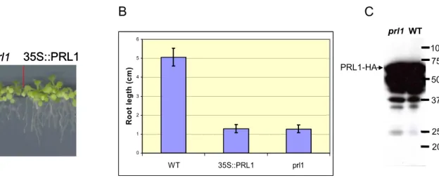
3.1.1. Overexpression of PRL1 cDNA... 46
3.1.2. Genetic complementation assay using an estradiol-inducible PRL1 genomic construct ... 47
3.1.3. Testing the stringency of estradiol induction of pER8 vector using a GUS reporter gene ... 48
3.1.4. Characterization of an estradiol-inducible PRL1 cDNA construct ... 49
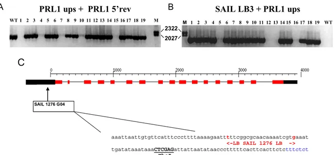
3.1.5. Characterization of the prl1-5 (SAIL_1276G04) mutant allele... 52
3.1.6. Search for putative regulatory elements in the PRL1 promoter sequence ... 53
3.1.7. Construction of a PRL1 promoter-driven GUS reporter gene... 54
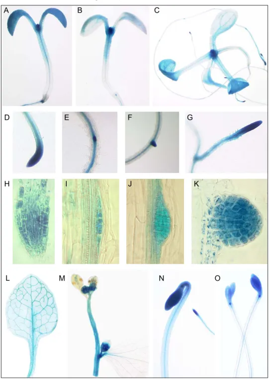
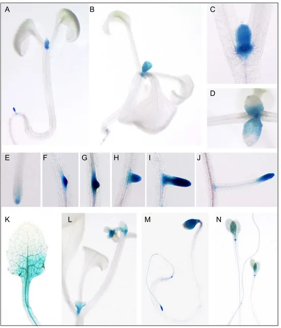
3.1.8. Characterization of PRL1 promoter activity in planta... 54
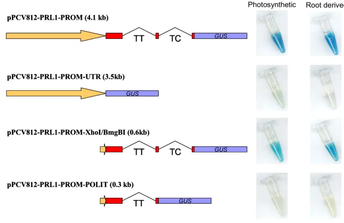
3.1.9. Construction of GUS reporter lines with PRL1 promoter deletions ... 56
3.1.10. Comparison of activity of PRL1 promoter-GUS constructs ... 56
3.1.11. Elimination of candidate regulatory regions from the second intron... 59
3.1.12. Confirmation of importance of intragenic PRL1 regulatory sequences by genetic complementation assays... 60
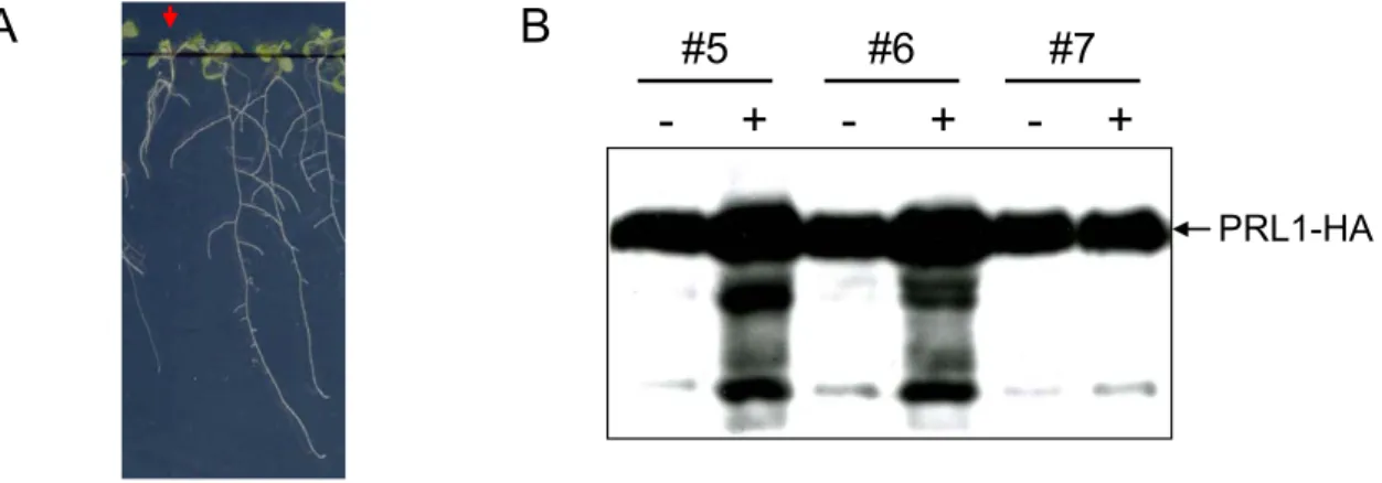
3.1.13. Western blot analysis of PRL1 complementation constructs... 62
3.1.14. PRL1-HA expression in different tissues of complemented lines ... 63
3.1.15. Complementation studies using heterologous promoters ... 64
3.1.16. Phenotypic analysis of PRL1 misexpressing plants... 65
3.2. Y
EAST-
ONE-
HYBRID SCREENING FOR TRANSCRIPTION FACTORS BINDING TO INTRAGENICPRL1
PROMOTER SEQUENCES... 67
3.2.1. Construction of promoter testing vectors and selection of test strains for yeast-one-hybrid assay... 67
3.2.2. Isolation of a cDNA encoding a PRL1 intron 2 binding factor... 68
3.3. A
NALYSIS OF EXPRESSION PATTERN AND CELLULAR LOCALIZATION OFPRL1
PROTEIN... 70
3.3.1. Construction of a vector for expression of a PRL1-GFP fusion protein... 70
3.3.2. Localization of PRL1-GFP fusion protein in planta... 70
3.3.3. Subcellular immunolocalization of PRL1-HA protein ... 70
3.3.4. Effects of plant hormone treatments on PRL1 protein level... 72
3.3.5. Regulation of PRL1 mRNA levels by hormones... 73
3.4. P
OSTTRANSCRIPTIONAL REGULATION OFPRL1 ... 74
3.4.1. Comparison of PRL1 transcript and protein levels in different plant organs ... 74
3.4.2. Analysis of PRL1 protein stability... 75
3.4.3. Degradation of PRL1 is inhibited by the proteasome inhibitor MG132... 76
3.4.4. Identification and site-specific mutagenesis of a putative destruction-box motif in PRL1 ... 77
3.4.4.1. Site-directed mutagenesis of the putative PRL1 D box ... 78
3.4.4.2. Assay of stability of D-box mutant PRL1-GFP protein in cell suspension ... 78
3.4.4.3. Immunodetection of stabilized PRL1-GFP in wild type plants ... 79
3.5. C
HARACTERIZATION OFPRL1
PROTEIN INTERACTIONS... 80
3.5.1. Yeast two hybrid assay with AtCDC5 ... 80
3.5.2. Detection of PRL1-AtCDC5 interaction in vivo ... 81
3.5.3. AtCDC5-HA is associated with the components of the protein degradation system... 82
3.5.4. Size fractionation of AtCDC5 complex on glycerol gradient... 83
3.5.5. Distribution of AtCDC5 in nuclear and cytoplasmic fractions... 84
3.5.6. Search for protein interactions with the N-terminal domain of PRL1 ... 85
3.6. G
ENETIC APPROACHES TO FUNCTIONAL CHARACTERIZATION OFPRL1
AND ITS PUTATIVE INTERACTING PARTNERS... 88
3.6.1. Isolation of new prl1 insertion mutant alleles... 88
3.6.1.1. PCR genotyping of the prl1-2 SALK_008466 line... 88
3.6.1.2. PCR genotyping of prl1-3 SALK_096289 and prl1-4 SALK_039427 lines ... 89
3.6.2. Isolation of prl2 T-DNA insertion mutations ... 92
3.6.2.1. PCR genotyping of the prl2-1 KONCZ16136 line ... 92
3.6.2.2. PCR genotyping of the prl-2-2 GABI 228D02 mutant line ... 93
3.6.2.3. Segregation analysis of prl2-1 and prl2-2 mutants ... 95
3.6.3. Generation of prl1 double mutants with mutations in genes coding for putative PRL1 interacting partners ... 95
3.6.4. Isolation of point mutations in the PRL1 coding region ... 97
3.6.4.1. Screening for EMS-induced point mutations by TILLING ... 97
3.6.4.2. Site-directed mutagenesis of the PRL1 gene ... 99
4.
DISCUSSION... 102
4.1. C
HARACTERIZATION OFPRL1
GENE EXPRESSION... 102
4.2. PRL1
GENE EXPRESSION IS REGULATED BY INTRAGENIC REGIONS... 103
4.3. PRL1
PROTEIN IS A POTENTIAL PROTEASOME SUBSTRATE... 106
4.4. PRL1
IS PRESENT IN ANA
TCDC5-
ASSOCIATED PROTEIN COMPLEX... 108
4.5. G
ENETIC APPROACHES TO FUNCTIONAL CHARACTERIZATION OFPRL1
INTERACTIONS... 109
5.
REFERENCES... 112
FIGURES
Figure 1. Simplified model of glucose signalling 2
Figure 2. Phenotype of the prl1 mutant. 3
Figure 3. Three dimensional structure of WD40 proteins 5
Figure 4. The Prp19-associated complex 8
Figure 5. Role of Ntc Prp19-complex in splicing 12
Figure 6. Proteasomal complexes of SnRK1α AMPK kinases 14
Figure 7. Genetic complementation assay with the CaMV35S:PRL1-HiA construct 46 Figure 8. Analysis of estradiol inducible PRL1-HA synthesis in transgenic plants carrying the
pER8(Km)-PRL1genomic-HA construct 48
Figure 9. Histochemical assay of GUS reporter enzyme activity of pER8-iGUS seedlings in the
absence and presence of estradiol 49
Figure 10. Analysis of plants carrying the estradiol-inducible pER8(Km)-PRL1-cDNA-HA
construct 50
Figure 11. Genetic complementation analysis with the estradiol-inducible pER8(Km)-PRL1-
cDNA- HA construct. 51
Figure 12. Immunodetection of PRL1-HA protein in seedlings displaying full or partial complememtation of prl1 root elongation defect by the eastradiol-inducible
pER8(Km)-PRL1-cDNA-HA construct in the presence of estradiol. 51 Figure 13. Localization of the T-DNA insertion in the prl1-5 (SAIL_1276 G04) mutant 52 Figure 14. Putative transcription regulatory sequences in the PRL1 promoter sequence 53 Figure 15. Histochemical analysis of PRL1::GUS expression pattern 55 Figure 16. GUS histochemical assay of activities of PRL1 promoter deletion constructs in cell
suspensions 57
Figure 17. Histochemical staining of GUS reporter enzyme activity controlled by the short
PRL1 promoter construct pPCV812-PRL1-PROM-XhoI-BmgBI 58 Figure 18. Histochemical assay of promoter-GUS constructs carrying deletions of TC-rich
repeat sequences. 59
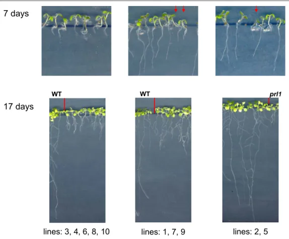
Figure 19. Phenotypic analysis of PRL1 complementation constructs 62 Figure 20. Immunodetection of PRL1-HA protein in prl1 lines complemented with different
PRL1 gene constructs 63
Figure 21. Immunodetection of PRL1-HA protein in various organs of complemented lines 64 Figure 22. Genetic complementation assays using misexpression of the PRL1 cDNA from
different heterologous promoters 66
Figure 23. Selection of pHISi-1 reporter strain 67
Figure 24. Yeast one-hybrid assay 68
Figure 25. Domain structure, binding site and regulation of expression of AtNAM 69 Figure 26. Analysis of PRL1-GFP expression pattern in planta by confocal laser scanning
microscopy 71
Figure 27. Cellular localization of PRL1-HA in a complemented prl1 mutant 72 Figure 28. Effects of hormone treatments on PRL1 protein levels 73 Figure 29. Immunodetection of PRL1 protein in seedlings treated with 50mM salicylic acid 73 Figure 30. Genevestigator microarray data on changes of PRL1 transcript levels in response
hormone treatments 74
Figure 31. Comparison of PRL1 mRNA and protein levels in plant organs 75
Figure 32. Analysis of PRL1 protein stability 76
Figure 33. Estradiol induced accumulation of PRL1 protein is enhanced by MG132 proteasome
inhibitor treatment 76
Figure 34. Inhibition of translation by cycloheximide does not affect the kinetics of PRL1
degradation 77
Figure 35. Identification of putative D-box motif in the PRL1 protein sequence 78 Figure 36. Assay of levels of wild type and D-box mutant PRL1-GFP proteins in cell suspension 79 Figure 37. Immunodetection of wild type and D-box mutant PRL1-GFP proteins in planta 80
Figure 38. Yeast two hybrid assays using AtCDC5 as prey 81
Figure 39. AtCDC5-HA immunoprecipitates PRL1 from protein extracts prepared from
Arabidopsis cell suspension. 82
Figure 40. Detection of subunits proteasome, SCF and COP9 complexes in the AtCDC5-HA
immunoprecipitated protein fraction. 83
Figure 41. Size fractionation of AtCDC5 complex on glycerol density gradient 84 Figure 42. Biochemical assay of subcellular localization of AtCDC5-HA protein. 84 Figure 43. Assay of PRL1 co-immunoprecipitation with AKIN11-HA, UFD1-HA and PAM1-
cMyc baits 85
Figure 44. Assay of co-immunoprecipitation of PRL1-HA with SnRK1α and ATAM1 amidase
proteins. 86
Figure 45. Size fractionation of PRL1-HA complexes from nuclear and whole cell protein
extracts 87
Figure 46. Size separation of protein complexes containing putative PRL1 interacting partner
proteins 87
Figure 47. PCR analysis of prl1-2 SALK_008466 allele 89
Figure 48. PCR genotyping of prl1-3 SALK_039427 lines 90
Figure 49. PCR genotyping of the prl1-4 SALK_096289 allele 91 Figure 50. Root elongation phenotype of seedlings carrying the prl1-2 to prl1-4 mutant alleles 91 Figure 51. Comparison of PRL1 and PRL2 gene expression patterns based on microarray data in
the Genevestigator database. 92
Figure 52. PCR analysis of the prl2-1 Koncz16136 (#4) mutant 93 Figure 53. PCR analysis of prl2-2 GABI 228D02 insertion line 94 Figure 54. Map positions of PIP genes in the Arabidopsis chromosomes 96 Figure 55. Genetic complementation assays with G77E mutation 99
Figure 56. Cytological analysis of prl1 root structure 100
Figure 57. Histochemical staining of prl1 and wild type plants carrying the CycB1;1::GUS reporter
gene. 101
TABLES
Table 1. Components of the yeast Prp19-associated protein complexes and their potential
orthologs in Arabidopsis 7
Table 2. Segregation analysis of prl2 mutant lines 94
Table 3. List of PIP genes, in which insertion mutations were isolated and used in crosses
with the prl1 mutant. 95
Table 4. List of crosses performed between prl1 and pip mutants 96 Table 5. Identification of EMS-induced mutations in the 5’ coding region of the PRL1 gene
by TILLING. 98
Table 6. Site-directed mutagenesis of the PRL1 gene 100
I ABBREVIATIONS AND SYMBOLES
A adenine
ABA abscisic acid
Ac acetate
ADP adenosine 5’-diphosphate
AMP adenosine 5’-monophosphate
APC Anaphase Promoting Complex/Cyclosome E3 ligase A. thaliana Arabidopsis thaliana
ATP adenosine 5’-triphosphate
ATPase adenosine 5’-triphosphatase
bp base pair
BSA bovine serum albumin
C cytosine
°C grad Celsius
CaMV Cauliflower Mosaic Virus
cDNA complementary DNA
CSM A. thaliana Cell Suspension culture Medium CS A. thaliana Cell Suspension culture
C-terminal carboxyterminal C-terminus carboxyl terminus
CTAB cetyltrimethylammonium bromide
2,4-D 2,4-Dichlorophenoxy acetic acid
DAPI 4,6-diamine-2-phenylindole dihydrochloride
DMSO dimethyl sulfoxide
DNA Deoxyribonucleic acid
dNTP deoxyribonucleotide triphosphate
DTT dithiotreitol
E. coli Escherichia coli
EDTA ethylenediaminetetraacetic acid
EtBr ethidium bromide
EtOH ethanol
G guanine
GA gibberellin
g gram
g relative centrifugal field unit
GST glutathione-S-transferase
GTC guanidium thiocyanate
h hour
HIS3 imidazole-glycerol-phosphate-dehydratase gene
IAA indole-3-acetic acid
IGEPAL (octylphenoxy)polyethoxyethanol
IgG immunoglobuline G
IP immunoprecipitation
kb kilobase
kDa kilo Dalton (1,000 Da)
l liter
lacZ E. coli β-galactosidase gene
M Molar
µg microgram
µl microliter
µM micromolar
mA milliampere
mg milligram
min. minute
ml milliliter
mM millimolar
mRNA messenger RNA
nt nucleotide
N-terminal amino terminal
N-terminus amino terminus
OD optical density
O/N overnight
PAGE polyacrylamide gel electrophoresis
POD peroxidase
PCR polymerase chain reaction
PEG polyethylene glycol
pH negative logarithm of the proton concentration
PMSF phenylmethylsulfonyl fluoride
PRL1 PLEIOTROPIC REGULATORY LOCUS 1
PVDF polyvinylidene difluoride
RNA ribonucleic acid
RNase ribonuclease
rpm revolution per minute
RT room temperature
S. cerevisiae Saccharomyces cerevisiae
SDS sodium dodecylsulfate
sec. second
T thymine
T Total
TAE Tris-acetate (40 mM); EDTA (1mM)
TCA trichloroacetic acid
TE Tris.HCl (10mM); EDTA (1mM)
TEMED N,N,N’,N’ tetramethylenethylendiamine
Tris Tris(hydroxymethyl) aminomethane
U unit
Ub ubiquitin
UTR untranslated region
UV ultraviolet light
V Volt
vol. volume
X-Gal 5-bromo-4-chloro-3-indolyl-β-D-galactopyranoside X-Gluc 5-bromo-4-chloro-3-indolyl-β-D-glucuronic acid
wt wild type
Aminoacids
A Ala Alanine
C Cys Cysteine
D Asp Aspartic acid
E Glu Glutamic acid
F Phe Phenylalanine
G Gly Glycine
H His Histidine
I Ile Isoleucine
K Lys Lysine
L Leu Leucine
M Met Methionine
N Asn Asparagine
P Pro Proline
Q Gln Glutamine
R Arg Arginine
S Ser Serine
T Thr Threonine
V Val Valine
W Trp Trptophan
Y Tyr Tyrosine
X any amino acid
II SUMMARY - ZUSAMMENFASSUNG
Der Arabidopsis PLEITROPIC LOCUS (PRL1) kodiert für ein nukleares WD40-Protein. Pflanzen, die eine PRL1-Insertionsmutation tragen, sind kleiner als der Wildtyp, haben kürzere Wurzeln, Blätter mit gesägten Blatträndern und kürzere Petiolen. In photosynthetisch aktiven Geweben der prl1-Mutante lassen sich erhöhte Mengen an Glukose, Saccharose, Fruktose, Stärke, Anthozyanin und Chlorophyll nachweisen. Der Verlust der PRL1-Funktion führt zur Hypersensitivität gegenüber Glukose, Saccharose und Pflanzenhormonen, einschliesslich Cytokinin, Ethylen, Abscisinsäure und Auxin.
Für die Analyse der regulatorischen Funktion von PRL1 entwickelten wir zunächst molekulare Werkzeuge um die PRL1-Genexpression zu charakterisieren. Ein Volllängen- (4.2 kb) PRL1- Promotor, der die Expression des β-Glukuronidase (GUS)-Reportergens als Fusion mit den ersten 69 Aminosäuren der kodierenden Sequenz des PRL1-Proteins steuert, liess sich in den meisten Geweben als exprimiert beobachten. Zur Identifizierung der regulatorischen Bereiche des PRL1-Gens wurde eine Anzahl von Promotordeletionen erzeugt und sowohl in Gewebekultur als auch in planta propagiert. Unsere Ergebnisse zeigten, dass intergenische Regionen entscheidend für eine korrekte Genexpression sind, da PRL1-Konstrukte, denen das zweite Intron fehlt, nur eine geringe Aktivität aufweisen. Ein kurzer Promotor, der eine alternative TATA-Box, 5´-UTR und einen 0.5 kb grossen kodierenden Bereich einschliesslich der ersten beiden Introns von PRL1 enthält, zeigt eine GUS- Aktivität ähnlich der des zuvor charakterisierten Volllängenpromotors. Während stromaufwärts gelegene regulatorische Sequenzen (~ 3.5 kb) allein nicht für eine korrekte mRNA-Expression ausreichend sind, konnten Konstrukte die das zweite Intron tragen, in genetischen Komplementationsversuchen vor dem prl1-Mutantenhintergrund als funktional nachgewiesen werden.
Die Sequenzanalyse legte die Bedeutung einer TC-reichen Region im zweiten Intron nahe. Die Deletion dieser Region führte jedoch nicht zu einer dramatischen Veränderung der Genexpression. Als PRL1 mit dem grünen fluoreszierenden Protein (GFP) markiert wurde, ließ es sich in den meisten Zelltypen als exprimiert und hauptsächlich als im Nukleus lokalisiert nachweisen. Zur Unterscheidung der Rolle des PRL1-Proteins in der Organentwicklung, wurden ein genomisches PRL1-Fragment und die cDNA hinter den heterologen Promotoren der AtKNAT1; AtSTM; AtUFO; AtAS1; AtSUC2 und TobRB7 Gene exprimiert. Während die genomischen Konstrukte in allen Fällen den prl1-Phänotyp komplementierten, konnten nur cDNA-Konstrukte, die von den AtAS- und AtSUC2-Promotoren angetrieben werden, den Blattphänotyp der prl1-Mutante komplementieren.
Aufgrund unseres Befundes, dass das Protein instabil ist, wurde eine posttranslationale
Regulation für PRL1 postuliert. Der reversible Proteasominhibitor MG132 stabilisiert PRL1 und so
liegt die Vermutung nahe, dass PRL1 in einer Proteasom-abhängigen Weise abgebaut wird. Im N-
terminalen Bereich von PRL1 konnte ein Zerstörungs- (destruction, D-) Boxmotiv identifiziert
werden, welches ein mögliches Degron für den „Anaphase Promoting Complex / Cyclosome E3
Ligase (APC) Komplex“ darstellt. Entsprechende Punktmutationen wurden eingeführt um die D-Box-
Konsensussequenz zu unterbrechen, mit der Zielsetzung so das PRL1-Protein zu stabilisieren. In
Zellsuspension konnte ein höheres Niveau von GFP-markiertem PRL1-Protein als mögliches Ergebnis der stabilisierenden Mutationen nachgewiesen werden, aber in drei Wochen alten Keimlingen war dies nicht der Fall. Aus diesem Grund sind weitere Untersuchungen für ein volles Verständnis der Kontrolle des PRL1-Abbaus erforderlich.
Biochemische Studien belegten die Anwesenheit von PRL1 in einem grossen nuklearen Proteinkomplex assoziiert mit dem Spliceosomkomponenten AtCDC5 Protein. Unsere Daten zeigen, dass AtCDC5 mit verschiedenen Komponenten des Proteindegradationssystems interagiert, wie z.B.
mit den 20S Kern- und den 19S Deckelpartikeln des Proteasoms, der CSN5 Untereinheit des COP9- Signalosoms und der SCF E3-Ubiquitinligaseuntereinheit Cullin1. Ausserdem lassen sich ubiquitinylierte Proteine in „pull-down“ Versuchen mit AtCDC5 nachweisen, was vermuten lässt, dass Substrate, die für das Proteasom bestimmt sind, möglicherweise mit diesem Komplex assoziiert sind.
Diese Ergebnisse deuten aufgrund seiner Interaktion mit AtCDC5 auf mögliche Rollen für PRL1 beim mRNA-Spleissen und beim Proteasom-abhängigen Proteinabbau hin. Fortlaufende Projekte mit der Zielsetzung eines erweiterten Verständnisses der PRL1-Funktion bedienen sich genetischer Ansätze und ortspezifischer Mutagenese.
The Arabidopsis PLEIOTROPIC REGULATORY LOCUS 1 (PRL1) encodes a nuclear WD40 protein.
Plants carrying a prl1 insertion mutation are smaller than wild type, have shorter roots, leaves with serrated leaf margins and shorter petioles. In photosynthetic tissues of the prl1 mutant elevated glucose, sucrose, fructose, starch, anthocyanin and chlorophyll levels are detected. Loss of the PRL1 function results in hypersensitivity to glucose, sucrose and plant hormones, including cytokinin, ethylene, abscisic acid and auxin.
***
To study the regulatory function of PRL1, we first developed molecular tools for characterization of
PRL1 gene expression. A full length (4.2 kb) PRL1 promoter driving the expression of β-
glucuronidase (GUS) reporter gene in fusion with coding sequences for the first 69 amino acids of
PRL1 protein was found to be expressed in most tissues. To identify regulatory regions in the PRL1
gene, a set of promoter deletions was generated and propagated in cell suspension and in planta. Our
results demonstrated that intragenic sequences are crucial for correct gene expression as PRL1
constructs lacking the second intron showed only low activity. A short promoter containing an
alternative TATA-box, 5’-UTR and 0.5 kb coding region with the first two introns of PRL1 showed
GUS activity similar to the previously characterized full-length promoter. Whereas upstream
regulatory sequences (~3.5 kb) alone were not sufficient for correct mRNA expression, constructs
containing the second intron proved to be fully functional in genetic complementation assays in the
prl1 mutant background. Sequence analysis suggested the importance of a TC-rich region in the
second intron. However, deletion of this sequence did not affect gene expression dramatically. When
PRL1 was tagged with green fluorescent protein (GFP), it was found to be expressed in most cell types
and localized mainly in the nucleus. To differentiate the role of PRL1 protein in organ development, a
PRL1 genomic fragment and cDNA were misexpressed using the heterologous promoters of AtKNAT1; AtSTM; AtUFO; AtAS1; AtSUC2, At4CL1 and TobRB7 genes. Whereas genomic constructs complemented the prl1 phenotype in all cases, only cDNA construct driven by the AtAS1 and AtSUC2 promoters were able to complement the prl1 mutant leaf phenotype.
Posttranslational regulation for PRL1 was suggested by our result indicating that the protein is unstable. The reversible proteasome inhibitor MG132 stabilized PRL1 suggesting that PRL1 is degraded in a proteasome-dependent manner. In the N-terminal region of PRL1, a destruction box (D box) motif was identified, which represents a putative degron for the Anaphase Promoting Complex/Cyclosome (APC) E3 ligase complex. Point mutations were introduced to disrupt the D box consensus sequence, in order to stabilize PRL1 protein. In cell suspension higher level of GFP-tagged PRL1 protein could be detected as potential result of stabilizing mutations, but this was not the case in three-week-old seedlings. Therefore, full understanding the control of PRL1 degradation requires further investigations.
Biochemical studies revealed that PRL1 is present in a large nuclear protein complex associated with the spliceosome component AtCDC5 protein. Our data indicate that AtCDC5 interacts with various elements of the protein degradation system, such as the 20S core and 19S lid particles of the proteasome, the CSN5 subunit of the COP9 signalosome and the SCF E3 ubiqutin ligase subunit CULLIN 1. In addition, ubiquitinated proteins were detected in pull-down assays with AtCDC5 suggesting that substrates targeted to the proteasome are potentially also associated to this complex.
These results indicate potential roles for PRL1 through its interaction with AtCDC5 in mRNA splicing and proteasome-dependent protein degradation. Ongoing projects aim at further understanding of the PRL1 function using genetic and site-specific mutagenesis approaches.
.
1. INTRODUCTION
1.1. Sugar signalling in plants
In plant research, a vastly growing interest focuses on the understanding of potential regulatory roles of sugar molecules. Sugars derived from glucose play well-defined roles in various metabolic reactions. For example, oxidation of sugars provides energy for all the life functions and sugar-derived metabolites are used as building blocks in amino acid and fatty acid synthesis and in biosynthetic pathways of secondary metabolites (Lea and Leagold, 1993). The excess of sugars is turned into storage compounds, such as starch, to build reserves in seeds or for starvation conditions. Plants possess a distinctive feature that after a short heterotrophic period can synthesize sugars through photosynthetic pathways. Mature leaf mesophyll cells function as source of photosynthesis-derived sugar compounds and carbohydrates are transported through the vascular system to sink organs, including roots, developing leaves, flowers and seeds that require energy import (Lemoine, 2000).
In plants, carbohydrates play also a pivotal role in development (for review see: Paul and Pellny, 2003; Gibson, 2005). High concentrations of externally provided glucose and sucrose are thus known to delay seed germination; but the germination rate remains apparently unaffected by internal increase of sugar concentrations (Dekkers et al., 2004). A contradictory role of carbohydrates was shown in combination with abscisic acid (ABA) as exogenously provided sugars alleviate the inhibitory effect of ABA on seed germination (Finkelstein and Lynch, 2000). During seedling development, 6%
glucose in combination with continuous light conditions inhibits greening and expansion of cotyledons, rosette leaf initiation, root and hypocotyl elongation (Jang et al., 1997). Higher sugar levels stimulate leaf expansion. The timing of developmental changes is also affected by carbohydrates. Several reports suggest that sugar treatment induces flowering (Corbesier et al., 1998;
Roldan et al., 1999). However, in other studies both high and low concentrations of exogenous sucrose inhibited the transition to flowering (Ohto et al., 2001). The intrinsic sugar content of plants also appears to control the regulation of onset of senescence as exogenous sugars can induce leaf senescence (Quirinho et al., 2000).
Physiological studies unravelled that carbohydrates act as important signalling molecules that modulate gene expression in connection with sugar metabolism, developmental processes and hormonal pathways (for review see: Smeekens, 2000; Rolland et al., 2002). Sugar signal perception is primarily connected to the functions of cell wall invertases catalyzing the cleavage of sucrose into glucose and fructose that are exported into the cell by specific hexose transporters (see Figure 1).
Invertases are also located in the cytoplasm and vacuolar membranes. Sucrose transporters are
responsible for sucrose translocation into the cytoplasm. The transported sucrose is subsequently
converted into fructose and UDP-glucose by sucrose synthases (for review see: Koch, 2004; Roitsch
and Gonzalez, 2004). The prominent sugar signalling cascade is connected to a hexokinase (HXK2),
which was proposed to be a major sugar sensor in plants (Jang et al., 1997). Hexokinase substrates,
such as glucose, mannose and 2-deoxyglucose inhibit the expression of photosynthetic genes
suggesting a feasible effect of hexokinase signalling on gene expression (Jang and Sheen, 1994). A disaccharide, trehalose was reported to affect plant development and biochemical pathways (for review see Eastmond et al., 2003). Trehalose-6-phosphate is synthesized in Arabidopsis by trehalose- 6-phosphate synthase and is thought to be an inhibitor of hexokinase transcription (Avonce et al., 2004). The downstream signalling pathways connected to hexokinases are still largely unknown.
Analysis of mutations causing sugar insensitivity and oversensitivity revealed that many of them are allelic to mutations previously isolated in ABA and ethylene signalling pathways. These results indicate a close cross-talk between sugar and hormone signalling (for review see: Gazzarrini and McCourt, 2001; Leon and Sheen, 2003). The glucose signalling pathway(s) likely involve also AMP- activated protein kinases that mediate the sensing of the cells’ energy status, modulate the activity and stability of metabolic enzymes, regulate gene expression and control the activity and stability of transcription factors (Hardie et al., 1998).
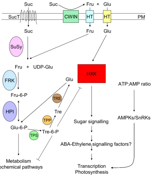
Figure 1. Simplified model of glucose signalling
Extracellular sucrose (Suc) is either directly transported into the cell by sucrose transporters (SucT) or hydrolyzed into fructose (Fru) and glucose (Glu) by cell wall invertases (CWIN). These monosaccharides are exported directly into the cell by hexose transporters (HT). Sucrose synthases (SuSy) catalyze the sucrose→fructose + UDP-glucose reaction. Subsequently, fructose is phosphorylated by fructokinases (FRK).
Fructose-6-phosphate (Fru-6-P) can be turned into glucose-6-phosphate (Glu-6-Pho) by hexose phosphate isomerases and in this form can enter into glycolysis, and further to fatty acid and amino acid biosynthesis.
Suc
Suc
SuSy
Fru + UDP-Glu
Fru-6-P
Metabolism Biochemical pathways
CWIN
Suc Fru + Glu
Fru Glu
FRK
HT HT
SucT
Glu-6-P HPI
HXK
Sugar signalling
Transcription Photosynthesis
ABA-Ethylene signalling factors?
Tre-6-P Tre
Glu
TPS TPP
TRE
ATP:AMP ratio
AMPKs/SnRKs
PM
Hexokinase (HXK2) phosphorylates glucose and is postulated to be a sugar sensor in plant cells. Downstream components of the sugar signalling pathway are not well known. It is suggested that ABA and ethylene signalling factors are also involved. Trehalose-6-phosphate (Tre-6-P), a negative regulator of hexokinase transcription, is synthesized from glucose-6-phosphate by trehalose phosphate synthases (TPSs). Trehalose-6- phosphate is subsequently converted to trehalose (Tre) by trehalose-6-phosphate phosphatases (TPP) and trehalose is hydrolyzed into glucose molecules by trehalase (TRE). AMP activated protein kinases (AMPKs) represent sensors of cellular energy status and also involved in glucose signalling.
1.2. Identification and characterization of the prl1 mutant
Classical genetic approaches provide powerful tools for identification of genes involved in biochemical or developmental pathways of interest (Koornneef, 1991). Moreover, mutant isolation allows further characterization of potential genetic interactions (e.g., suppressor screens, epistasis analysis etc.). In order to identify novel factors in the sugar signalling pathway, a T-DNA tagged population of Arabidopsis thaliana Col-0 wild type plants was germinated on MSAR plates supplemented with either glucose or sucrose (0.1, 0.5, 2, 4, 6, 8 and 10%). 1200 segregating M2 mutant families were screened to identify mutants showing germination defect and/or growth retardation in response to high sugar content (Nemeth et al., 1998). The life cycle of a candidate mutant was arrested on medium containing 6% sucrose or 5% glucose, and mutant plants died subsequently under these conditions (Figure 2 A).
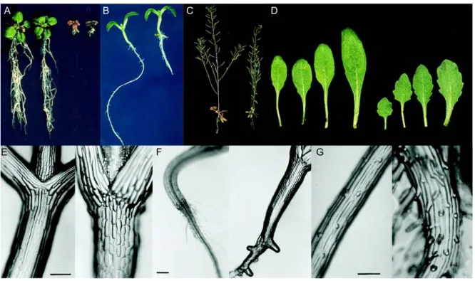
Figure 2. Phenotype of the prl1 mutant.
The effects of a T-DNA insertion in the PRL1 locus were analyzed in comparison to wild-type Col-0 plants (placed left on the pictures). The figure was adapted and modified from Nemeth et al. (1998). (A) Seedlings were grown on 6% sucrose. (B) Seedlings on MSAR plates containing 0.5% sucrose at light conditions. (C) Soil- grown plants. (D) Phenotype of rosette leaves. (E) Hypocotyls. (F) Roots five days after germination. (G) Root hair formation.
The identified T-DNA insertion event in the PLEIOTROPIC REGULATORY LOCUS 1 (PRL1) resulted in a recessive mutation. Soil-grown mutant plants were smaller then wild type plants, the
A B C D
E F G
leaves were smaller with shorter petioles and had serrated margins (Figure 2 C and D). Chlorophyll and anthocyanin accumulated in the mutant; hence these plants were darker than the wild type.
Glucose, fructose, sucrose and starch content of the leaves showed two- to five-fold increase in comparison to the wild type. The most characteristic phenotypic trait for the prl1 mutation was that the root elongation was reduced two- to three-fold both in light and dark conditions (Figure 2 B). Analysis of the root structure revealed that prl1 mutant seedlings developed early on side roots and showed ectopic root hair formation (Figure 2 F and G). In addition, elongation of the epidermal cells was inhibited and their number was duplicated in the hypocotyls (Figure 2 E). Hormone responses of the prl1 mutant were tested on MSAR plates containing auxins (2,4-dichlorophenoxyacetic acid- 2,4-D or 1-naphtaleneacetic acid-NAA), cytokinins (N6-(isopentenyl)adenosine riboside or N6-benzyladenine), abscisic acid, salicylic acid, methyl jasmonate, brassinosteroids, gibberellins and ethephone (i.e.
ethylene generating agent). In response to auxin treatment roots of the prl1 plants were converted into proliferating callus, while wild type roots developed numerous lateral roots densely covered by root hairs. In response to ABA treatment the prl1 mutant displayed hypersensitive seed germination and bleaching of seedlings at higher ABA concentrations. Combined cytokinin and sucrose treatment of wild type seedlings grown in the light was observed to phenocopy developmental defects of the prl1 mutation. Etiolated plants treated with ethylene exhibited about 20 to 30% reduction of hypocotyl elongation. In addition, the prl1 mutant showed growth reduction at 14°C.
1.3. The Arabidopsis PRL1 gene and PRL1 orthologs
Molecular characterization of the prl1 mutation revealed that the T-DNA insertion was located in the fourth chromosome disrupting sequences of the gene At4g15900 between exons 15 and 17 (Nemeth et al., 1998). The transcribed region of the PRL1 gene is approximately 3.5 kb, covering a 38 bp 5’ UTR, 17 exons and a 3’ UTR of 222 bp. The PRL1 cDNA of 1461 bp encodes a protein of 54 kDa carrying seven WD40 repeats in its C-terminal region. The PRL gene family is small; only one homolog of PRL1 was identified in the Arabidopsis genome. PRL2 (At3g16650) shows 83% amino acid identity with PRL1. The N-terminal region of PRL2 differs considerably from PRL1 (65% identity) as compared to homology between the C-terminal domains of these proteins (89% identity).
A potential ortholog of PRL1, Prp5, was identified in a mutant screen for pre-mRNA splicing
defects in fission yeast (Schizosaccharomyces pombe, Potashkin et al., 1998). The temperature
sensitive prp5-1 (precursor mRNA processing 5-1) mutant was shown to accumulate unspliced U6
snRNA precursor. The phenotype of prp5-1 yeast cells was analyzed under restrictive conditions. The
mutant displays conventional cell division cycle (cdc) phenotype: the yeast cells are elongated and the
chromatin is condensed into thin U-shaped structures. Flow-cytometry analysis revealed that the cells
were arrested with 2C DNA content. This result indicated that the life cycle of prp5-1 mutant proceeds
through the S phase of cell cycle and stops in G2. Prp46p, another ortholog of PRL1 in
Saccharomyces cerevisiae, was identified in an extensive two-hybrid screen designed for isolation of
mutations in new splicing factors that show interaction with the Prp22p DEAH-box RNA helicase
(Albers et al., 2003). Subsequently, Prp46p was found to interact with Prp45p, a direct binding partner of Prp22p. Deletion of the PRP46 gene results in non-viable haploid spores. Construction of a conditional mutant strain demonstrated that PRP46 is essential in budding yeast.
PRL1-related proteins are highly conserved also in other eukaryotes. Fission yeast Prp5p shares 69% identity with Arabidopsis PRL1, whereas budding yeast Prp46p shares 63% amino acid identity with PRL1. Analogously, sequence identity is 62% with a C. elegans PRL1 homologue and 59% with the human homologue PLRG1 (Nemeth et al., 1998).
1.4. Conserved WD-repeat proteins
The family of WD-repeat (also known as Trp-Asp, WD40 or β-transducin motif) proteins contains factors with diverse cellular functions, biochemical activity and subcellular localization. A unique feature of these proteins is that they share short, about 40 amino acid long tandem repeats starting with Gly-His and terminating mainly in Trp-Asp dipeptide motives (van der Voorn and Ploegh, 1992).
Notably, sequence identity is low among the members of the WD-repeat family. Rather, they share a common three dimensional structure, which was first described in the case of the Gβ subunit of a heterotrimeric GTPase (Lambright et al., 1996). The β propeller structure of WD-40 proteins consists of seven blades formed by four antiparallel β sheets of WD-repeats (Figure 3). It was reported that one blade is not produced by a single repeat but the C-terminus of the first repeat interlocks with the N- terminus of the next repeat, which might stabilize the three dimensional structure of these proteins (Jawad and Paoli, 2002). The overall structure is a hollow truncated cone, where the small hollow is completely occupied by water molecules (Madrona and Wilson, 2004). This arrangement is known to provide a solid platform for interacting proteins to bind.
Figure 3. Three dimensional structure of WD40 proteins
The structure of ScBub3p was chosen as a representative of WD-repeat proteins (Larsen and Harrison, 2004).
WD40 repeats form four antiparallel β sheets organized into a seven-bladed β propeller structure.
Comparative analysis of WD-40 proteins in the Arabidopsis genome was performed by van Nocker and Ludwig (2003). This investigation revealed that 269 Arabidopsis proteins contain at least a single WD40-motif, but the majority of the proteins (237) carry four or more tandem repeats.
CONSTITUTIVE PHOTOMORPHOGENIC 1 (COP1) is one of the well-characterized members of this family. COP1 carries three distinct domains: a RING-finger motif followed by a coiled coil domain and the WD40 domain (for review see: Yi and Deng, 2005). COP1 was identified as negative regulator of light-dependent photomorphogenic development, since cop1 mutants show constitutive photomorphogenic phenotype in the darkness (Deng et al., 1991). COP1 functions as E3 ubiquitin ligase that controls the degradation of transcription factors HY5, HYH, LAF1 and HFR1 by the 26S proteasome complex (Saijo et al., 2003; Jang et al., 2005).
1.5. PRL1 family members are subunits of conserved splicing-associated complexes Biochemical studies revealed that PRL1 orthologs are subunits of evolutionary conserved spliceosome-associated protein complexes. Mass spectrometric analyses have identified components of such complexes in Schizosaccharomyces pombe (McDonald et al., 1999; Ohi et al., 2002), Saccharomyces cerevisiae (Tsai et al., 1999; Ohi et al., 2002; Hazbun et al., 2003) and human cell cultures (Ajuh et al., 2000). Some of these experiments used the budding yeast SpCdc5p, fission yeast ScCef1p and human HsCDC5L proteins as baits and therefore the associated factors were termed as Cwfs or Cwcs (complexed with Cef1p/Cdc5p) or CCAPs (CDC5L complex associated proteins).
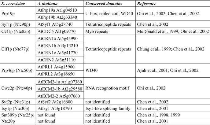
Sequence analysis of spliceosome-associated proteins isolated by immunopurification of CDC5 complexes exposed that the Cwf/CCAP complexes (Tarn et al., 1994, Chen et al., 2001, 2002) share common components with the exhaustively studied Prp19p-associated complex (Ntcstands for Prp nineteen complex; Tsai et al., 1999, Ohi and Gould, 2002). The Ntc complex contains at least 11 proteins, whereas 27 distinct factors were identified in the Cwf/Cwc complexes. The common core of Prp and Cwf/Cwc complexes is composed of the Prp19p, Syf1p (Ntc90p), Cef1p (Ntc85p), Clf1p (Ntc77p), Prp46p (Ntc50p), Cwc2p (Ntc40p), Syf2p (Ntc31p), Isy1p (Ntc30p), Snt309p (Ntc25p) and Ntc20p protein subunits. Elements of Prp and Cwf/Cwc complexes are not yet characterized in Arabidopsis thaliana (Table 1).
1.5.1. Prp19
Budding yeast Prp19 was identified in a screen searching for temperature sensitive mutants with impaired pre-mRNA splicing (Vijayraghavan et al., 1989). Prp19 was found to carry three canonical domains: a U-box, a coiled coil and a WD40 domain. Members of the U-box protein family were recently identified to function as novel E3 ubiquitin ligases. Comparative sequence analyses and protein modelling studies suggest that the U-box is a derivative of the RING-domain. However, the zinc chelating cysteine residues are not conserved in the U-box proteins (Aravind and Koonin, 2000).
The structure of Prp19p was defined using NMR spectroscopy (Ohi et al., 2003) and x-ray
crystallography with multi-wavelength anomalous diffraction experiments (Vander Kooi et al., 2006).
The Prp19p U-box domain folds into a similar tertiary structure like RING-fingers; a central α-helix is surrounded by four β-strands and a hydrophobic core. Instead of Zn
2+ions, hydrogen bonds and salt bridges stabilize the U-box structure (Ohi et al., 2003). Prp19p forms homotetramers in vitro through its coiled coil domain (Figure 4). The U-box domains are located at close proximity of the central coiled coil bundle, whereas the WD40-repeats are flexibly attached (Ohi et al., 2005, Vander Kooi et al., 2006). It is suggested that the U-box is responsible for binding of an E2 ubiquitin conjugating enzyme and that the WD40-repeats can bind the target proteins. Point mutations in the coiled coil region inhibiting tetramerization of Prp19p cannot complement the lethal phenotype of budding yeast prp19 mutant suggesting that the tetramer formation is essential for proper function of the Ntc complex in vivo (Ohi et al., 2005). The Prp19 U-box protein displays ubiquitin ligase activity in vitro (Hatakeyama and Nakayama, 2003, Ohi et al., 2003). Surprisingly, the prp19-1 splicing mutant suffered a V14I substitution in the U-box domain resulting in splicing deficiency. This mutation disrupted the three dimensional structure of the central hydrophobic core region, which might be responsible for interaction with an E2 ubiquitin conjugating enzyme (Ohi et al., 2003). Point mutations disrupting the predicted E2 interface cannot complement the prp19-1 mutant phenotype; however, the protein folding is not affected (Ohi et al., 2003). Human HsPrp19 interacts directly with the β7 subunit of the 20S proteasome (Loscher et al., 2005). There are only two U-box genes, UFD2 and PRP19 in budding yeast, and six genes UFD2a, UFD2b, CHIP, UIP5, CYC4 and PRP19 in mammals. In Arabidopsis, 37 genes are predicted to encode U-box-like factors, two of them represent homologues of Prp19p (At1g04510 and At2g33340; for review see Azevedo et al., 2001).
S. cerevisiae A.thaliana Conserved domains Reference AtPrp19a At1g04510
Prp19p
AtPrp19b At2g33340 U-box, coiled coil, WD40 Ohi et al., 2002; Chen et al., 2002 Syf1p (Ntc90p) AtSyf1 At5g28740 Tetratricopeptide repeats Chen et al., 2002
Cef1p (Ntc85p) AtCDC5 At1g09770 Myb repeats McDonald et al., 1999; Ohi et al., 2002 AtCRN1a At5g45990
AtCRN1b At3g13210 AtCRN1c At5g41770 Clf1p (Ntc77p)
AtCRN2 At3g51110
Tetratricopeptide repeats Chung et al., 1999; Chen et al., 2002
AtPRL1 At4g15900 Prp46p (Ntc50p)
AtPRL2 At3g16650 WD40 Ajuh et al., 2001; Ohi et al., 2002 AtECM2-1a At1g07360
AtECM2-1b At2g29580 Cwc2p (Ntc40p)
AtECM2-2 At5g07060
RNA recognotion motif Ohi et al., 2002 Syf2p (Ntc31p) AtSyf2 At2g16680 not identified Chen et al., 2002 Isy1p (Ntc30p) AtIsy1 At3g18790 Isy1-like splicing family Chen et al., 2001 Snt309p (Ntc25p) not found not identified Chen et al., 1998; 1999
Ntc20p not found not identified Chen et al., 2001
Table 1. Components of the yeast Prp19-associated protein complexes and their potential orthologs in Arabidopsis
The table was adapted and modified from Wang and Brendel (2004).
Figure 4. The Prp19-associated complex
(A) Prp19 forms a homotetrameric complex through its coiled coil domains. The U-box domains (U) probably interact with E2 ubiquitin conjugating enzymes, whereas the WD40-repeats are likely involved in substrate binding. (B) Identified protein interactions in the Prp19- Ntc- complex. The figure was adapted from Ohi et al.
(2005).
A weak thermo-conditional mutant allele of prp19 was isolated in a mutant screen searching for photoactivated psoralen-sensitive yeast mutants (pso mutants, for review see: Brendel et al., 2003).
These chemicals are successfully used in treatment of skin disorders; however, the risk of skin cancer among the patients increased significantly due to induced DNA lesions. Mutations in the PSO4 gene were found to cause pleiotropic defects, including increased sensitivity to DNA damaging agents (chemicals and irradiation), low frequency of spontaneous and induced recombination (i.e., gene conversion, crossing over and intrachromosomal recombination). The pso4 mutation, which entirely blocks pre-meiotic DNA synthesis and sporulation, proved to be allelic with the prp19 mutation indicating involvement of PRP19 in error-prone DNA repair (Grey et al., 1996). The core Ntc complex (Prp19, Cdc5, Prlg1 and Spf27) is associated with the WRN protein, a RecQ-type DNA helicase and involved in inter-strand cross-link repair (Zhang et al., 2005). In other studies, Prp19 was shown to interact with the terminal deoxynucleotidyl transferase and to play an important role in double-strand DNA repair (Mahajan and Mitchell, 2003). Prp19 was also found in a survey for differentially expressed genes in senescent human cells (SNEV= Senescence Evasion Factor; Grillari et al., 2000).
SNEV transcription is repressed in senescent human cells, whereas higher SNEV mRNA level is detected in tumour cell lines (Voglauer et al., 2006). Overexpression of SNEV results in an extended life span of endothelial cell lines in vitro. This phenomenon is not connected to increased telomerase activity, but the cells show elevated resistance to genotoxic and oxidative stress factors causing double-stranded DNA breaks. The natural occurrence of DNA damage was also significantly lower in the SNEV overexpression lines. SNEV mRNA is upregulated in malignant breast cancer cells.
WD40
U
WD40
U
WD40
U
WD40
U
WD40
U
WD40
U
WD40
U
WD40
U
E2
Ub conjugating enzyme
Substrate
A B
Coiled coil
Prp19 Prp19
Snt309p
Cef1p Cwc2p
Prp46p
Syf1p
Clf1p
Ntc20p Isy1p
However, patients expressing enhanced level of SNEV had better survival rate and the formation of lymph node metastases was significantly decreased.
Proteome analysis of nuclear matrix proteins with increased capability of reassembling resulted in the isolation of human nuclear matrix protein 200 (hNMP 200), which was later shown to be identical to Prp19 (Gotzmann et al., 2000). Study of subcellular localization of GFP-tagged hNMP 200 indicated that this protein is mainly located in the nucleus, but not in the nucleolus. At the nuclear periphery, speckle formation was detected. In prophase, hNMP 200 is uniformly distributed and not associated with the chromosomes. In metaphase, an enhanced hNMP 200 signal is detected around the aligning chromosomes. During anaphase, hNMP 200 is localized to the mitotic spindles throughout chromosome segregation.
1.5.2. CDC5
Cdc5p is highly conserved both structurally and evolutionally through various model organisms (Ohi et al., 1998; Hirayama and Shinozaki, 1996). Initially, cdc5
+was identified as a cell division cycle mutant in S. pombe. The cdc5-120 mutation arrests cells in the G2 phase of cell cycle before mitosis (Ohi et al., 1994). The N-terminal region of Cdc5 carries three helix-turn-helix DNA-binding Myb domains referred to as R1, R2 and R3. However, the third repeat is imperfect as it possesses a Val-Leu substitution at a critical position (Carr et al., 1996; Ogata et al., 1996). In the Arabidopsis genome, three groups of Myb proteins are encoded: Myb1R factors with one, R2R3-type Myb factors with two, and Myb3R proteins with three Myb repeats (for review see Jin and Martin, 1999; Stracke et al., 2001). Two well-characterized members of the MybR1 family are the circadian clock-related transcription factors CCA1 (Circadian Clock Associated 1) and LHY1 (Late elongated Hypocotyl 1).
The majority of plant Myb-like regulators carries two repeats and is involved in diverse regulatory processes (e.g., Transparent testa 2-TT2, Production of anthocyanin pigment 1-PAP1, Glabrous 1- GL1, Assymetric leaves 1-AS1, Werewolf-WER). The Myb3R factors are involved in cell cycle regulation.
Arabidopsis CDC5 binds double-stranded DNA in a sequence specific manner. CDC5 recognizes the CTCAGCG (complementary CGCTGAG) consensus sequence (Hirayama and Shinozaki, 1996). Human HsCdc5 binds a consensus sequence of 12 bp (GATTTAACATAA). The core ANCA motif is a typical target for Myb-like helix-turn-helix transcription factors. The flanking symmetrical TTA/TAA sequence increases the binding affinity of Cdc5 (Lei et al., 2000). A CDC5 ortholog in the basidiomycete mushroom Lentinula edodes specifically binds to the sequence GCAATGT (Miyazaki et al., 2004). However, budding yeast ScCef1p, which can complement the fission yeast cdc5-120 mutation, was not observed to have any DNA binding activity (Ohi et al., 1998).
Cdc5 is phosphorylated by a cAMP-dependent protein kinase in Lentinula (Miyazaki et al.,
2004). PCDC5RP immunopurified from COS-7 cells is also phosphorylated (Bernstein and Coughlin,
1997), whereas rat Cdc5 is suggested to be a substrate for protein kinase CK2 (Engemann et al., 2002).
Human CDC5L is known as a mitotic phosphoprotein; it is phosphorylated in vitro by cyclinB-cdc2 and cyclin E-cdc2 (Stukenberg et al., 1997; Boudrez et al., 2000). CDC5 was found to interact with a DAP-like serine/threonine-specific protein kinase in the yeast two-hybrid system and this result was verified by in vitro binding experiments (Engemann et al., 2002). Co-localization studies further support direct interaction between these proteins, since the DAP-kinase and CDC5 are both localized to nuclear speckles. However, Cdc5 is not phosphorylated by the DAP-like kinase in vitro. Cdc5 is also a binding partner of NIPP1, a member of PP1 serine/threonine protein phosphatase regulator factors that shows co-purification and co-localization with PP1C and CDC5L (Boudrez et al., 2000).
This interaction depends on the phosphorylation state of CDC5L, only phosphorylated CDC5L is able to bind to NIPP1.
In Arabidopsis a single gene encodes a CDC5 ortholog. Arabidopsis AtCDC5 (At1g09770) can complement the S. pombe cdc5-120 mutation suggesting functional similarity of CDC5 orthologs between different organisms (Hirayama and Shinozaki, 1996). Expression of the AtCDC5 gene was analyzed by in situ hybridization and Northern blot experiments. The AtCDC5 mRNA shows a strong localization to shoot and root apical meristems and leaf primordia. AtCDC5 is expressed in most tissues studied; the highest transcript levels are detected in roots.
1.5.3. Crooked neck (CRN) proteins
The Crooked neck (crn) locus was originally identified in a genetic screen for Drosophila embryo
lethal mutations and mapped into the X chromosome. Crn mutant embryos display serious disorders
during the development of nervous system, midgut and muscles (Perrimon et al., 1984). Yeast crn
knockout mutant shows a cell cycle phenotype, the dividing cells are arrested between the G2 and M
phase (Russell et al., 2000; Zhu et al., 2002) The CRN protein belongs to the tetratricopeptide repeat
(TPR) family. The TPR motif is involved in mediation of protein-protein interactions (for review see
Blatch and Lassle, 1999). Orthologs of CRN (Clf1p/Syf3p/Ntc77p) protein were characterized in
connection with the spliceosome complex in yeast (Chung et al., 1999; Wang et al., 2003), in
Drosophila (Raisin-Tani and Leopold, 2002) and in humans (Chung et al., 2002). In yeast, however,
Clf1p (Crooked neck-like factor 1) is considered as one of the moonlighting proteins with a novel role
in DNA replication (Jeffery, 2003). Temperature sensitive clf1 mutants show cell cycle arrest between
the G2/M transition (Zhu et al., 2002). However, when cells are blocked in G1 with α-factor, they
display a delay in DNA accumulation and fail to enter the S phase. Physical interaction between Clf1p
and the replication origin binding ORC complex was detected by yeast two-hybrid analysis and co-
immunoprecipitation. A possible link between Clf1 and vesicular transport proteins is also suggested
(Vincent et al., 2003). The Arabidopsis genome encodes four Crn orthologs: AtCRN1a (At5g45990),
AtCRN1b (At3g13210), AtCRN1c (At5g41770) and AtCRN2 (At3g51110), but these genes are yet
not characterized.
1.6. Function of the Prp19-associated complex in splicing
Transcription of eukaryotic genes that carry protein coding exons and non-coding intervening sequences, called introns, results in the synthesis of precursor RNA molecules. The intron sequences are excised from the pre-mRNAs in the nucleus and the messenger RNAs are transported to the cytoplasm for protein translation. Pre-mRNAs carry short consensus sequence elements at the 5’ (GU) and 3’ (AG) ends of intron boundaries flanking an internal branch site (UACUAAC).These sequences are recognized by the splicing machinery. The splicing reaction occurs by two trans-esterification reactions, in which covalent bonds are transferred from one location to another. This reaction does not require ATP hydrolysis. A free 2’-hydroxyl group of the branch site attacks the phosphate at 5’-end, and the reaction yields a looped lariat intermediate with the intron sequence and a cut mRNA. In the second step, the 3’-hydroxyl group of 5’-exon attacks the 3’ splice site resulting in a spliced and ligated mRNA molecule and a lariat form of intron sequence.
The large holoenzyme complex catalyzing the intron excision is named spliceosome that carries small nuclear ribonucleoprotein particles (snRNPs; U1, U2, U4, U5 and U6). The assembly of spliceosome and the splicing reaction are highly organized sequential process (for reviews see: Staley and Guthrie, 1998; Murray and Jarrell, 1999; Lorkovic et al., 2000) (Figure 5). The first step is the formation of the E (early presplicing) complex, in which the U1 snRNP binds to the 5’ splice junction in a sequence specific manner aided by complementary snRNA of this ribonucleoprotein complex.
Subsequently, U2 binds to the branch site and the A complex of spliceosome, which is also called a pre-spliceosome, is assembled. The B complex is formed in two steps. First, the U5/U4/U6 tri-snRNPs bind to the precursor RNA-splicing complex, which undergoes a structural rearrangement by releasing the U1 and U4 particles. This step is followed by formation of base pair interactions between U6 and U2, and between U6 and the 5’ splice site. During these stages, an enzymatically active spliceosome C complex is assembled that can catalyze intron excision and exon ligation by two trans-esterification reactions.
With the discovery of AT-AC type introns, which represent a minor class of intron regions defined by evolutionally conserved but distinct consensus sequences at the 5’ and 3’ splicing junctions, the existence of an alternative spliceosome became evident (for reviews see Kreivi and Lamond, 1996; Tarn and Steitz, 1997; Lorkovic et al., 2000). This alternative splicing complex shares only the U5 snRNP component with the canonical spliceosomal complex and contains unconventional U11, U12, U4atac and U6atac particles.
The Prp19/Cdc5 related complex is associated with the U2/U5/U6 tri-snRNP complex (Tarn et
al. 1994; Ohi et al., 2002). The assembly of spliceosome and formation of intermediates can be
analyzed using different ATP concentrations in vitro. At low ATP concentration inactive pre-splicing
forms of spliceosome (Complex A and B) accumulate, while increasing the ATP concentration
induces the assembly and rearrangement of spliceosome as these steps requires ATP hydrolysis. The
analysis of distinct snRNP complexes revealed that the Ntc complex is associated to the spliceosome
after the release of the U4 particle (Chan et al., 2003). After dissociation of U4 snRNP, the splicing
complex is reorganized and the spliceosome becomes activated. Three complementary binding sites were identified for the U6 snRNP at the 5’-splice junction by cross-linking experiments (Chan et al., 2003). One was predominantly found in inactive spliceosome complexes, whereas the two others were identified during the splicing reaction suggesting that a switch in binding happens during spliceosome activation. The Ntc-depleted U6 snRNP is exclusively found in inactive binding regions (Chan and Cheng, 2005). Similarly, the U5 snRNP occupies only two inactive binding sites in Ntc-depleted splicing reactions. These data suggest that the Ntc complex is required for the stable association of U5/U6 snRNPs to the mRNA complementary sequences, and that the Ntc helps to lock the spliceosome at the active position.
Figure 5. Role of Ntc Prp19-complex in splicing
Intron excision and exon ligation are highly organized sequential processes. Splicing starts with the recognition of 5’-splice site by the U1 snRNP (E complex). Subsequently, the U2 particle binds to the branch site of the intron (A complex). The B complex, carrying all five snRNPs on the precursor RNA, is still inactive. After the releasing of U1 and U4 snRNPs the spliceosome becomes activated by the Ntc complex (C complex). The active spliceosome catalyzes two trans-esterification reactions that result in the removal of intervening intron and ligation of adjacent exons.
3`EXON 5`EXON
GU UACUAAC AG
U1
3`EXON 5`EXON
GU UACUAAC AG
E complex U1
3`EXON 5`EXON
GU AG
U1
UACUAAC
U2
3`EXON 5`EXON
GU AG
U1
UACUAAC
U2
UACUAACA complex U2
U5
3`EXON 5`EXON
GU AG
U1 U6 U4
UACUAAC
U2 U5
3`EXON 5`EXON
GU AG
U1 U6 U4
UACUAAC
U2
UACUAACB complex U2
3`EXON 5`EXON
U5 U6
GUUACUAAC
U2
AG
3`EXON 5`EXON
U5 U6
GUUACUAAC
U2
UACUAACU2
AG
C complex
Ntc
3`EXON 5`EXON 3`EXON 5`EXON
U5 U6
GUUACUAAC
U2
UACUAACU2
AG