Targeting SUMO conjugates for degradation: The human RING finger RNF4 as a specialized ubiquitin ligase
I n a u g u r a l - D i s s e r t a t i o n
zur
Erlangung des Doktorgrades
der Mathematisch-Naturwissenschaftlichen Fakultät der Universität zu Köln
vorgelegt von
Kirstin Keusekotten aus Köln
Köln, 2010
Berichterstatter: Prof. Dr. Thomas Langer
Institut für Genetik, Zülpicher Str. 47, 50674 Köln Prof. Dr. Jürgen Dohmen
Institut für Genetik, Zülpicher Str. 47, 50674 Köln
Tag der letzten mündlichen Prüfung: 07.06.2010
TABLE OF CONTENTS
ABBREVIATIONS ... I
1. INTRODUCTION ... 1
1.1 Relevance of posttranslational modifications in living cells ... 1
1.2 Ubiquitin modification of proteins ... 1
1.2.1 Mechanistic aspects of Ubiquitin conjugation ... 2
1.2.1.1 E1 enzymes ... 3
1.2.1.2 E2 enzymes ... 3
1.2.1.3 Ubiquitin E3 ligases ... 4
1.2.1.3.1 RING/U-Box ligases ... 4
1.2.1.4 Deubiquitylating enzymes ... 5
1.2.1.5 Ubiquitin binding domains ... 5
1.2.2 Mono-ubiquitylation and polymeric ubiquitin chains ... 6
1.2.2.1 Mono-ubiquitylation ... 6
1.2.2.2 Poly-ubiquitin chains ... 7
1.2.2.2.1 K48-linked chains and the Ubiquitin-Proteasome system ... 8
1.3 Ubiquitin-like modifiers ... 9
1.4 SUMO modification of proteins ... 10
1.4.1 SUMO proteins ... 11
1.4.1.1 Yeast SUMOs: Smt3 and Pmt3 ... 11
1.4.1.2 Mammalian SUMO isoforms ... 12
1.4.2 Mechanistic aspects of SUMO conjugation ... 13
1.4.2.1 SUMO E1 enzymes ... 13
1.4.2.2 E2 enzyme Ubc9... 14
1.4.2.3 SUMO E3 ligases ... 14
1.4.2.3.1 PIAS proteins ... 15
1.4.2.3.2 RanBP2 ... 15
1.4.2.4 SUMO proteases ... 15
1.4.3 SUMO interaction motifs ... 17
1.4.4 Cellular roles of SUMO modification ... 18
1.5 ULS proteins ... 19
1.5.1 S. cerevisiae Slx5-Slx8 and Uls1 ... 20
1.5.2 S. pombe Rfp1/Rfp2-Slx8 ... 20
1.5.3 RNF4, a putative human ULS ... 21
1.6 The promyelocytic leukemia protein PML ... 22
1.6.1 Posttranslational modifications of PML ... 23
TABLE OF CONTENTS
1.6.2 PML functions ... 24
1.7 PML Nuclear Bodies ... 25
1.7.1 PML nuclear body formation ... 25
1.7.2 Functions of PML nuclear bodies ... 26
1.8 Acute Promyelocytic Leukemia ... 28
1.8.1 Aberrant functions of the PML-RARα fusion protein ... 28
1.8.2 Treatment of APL ... 28
1.9 Aims of this thesis ... 30
2. MATERIALS & METHODS ...31
2.1 Materials ... 31
2.1.1 Chemicals & Solutions ... 31
2.1.2 Kits ... 31
2.1.3 Plastic ware ... 31
2.1.4 Standards ... 31
2.1.5 Enzymes ... 31
2.1.6 Antibodies ... 32
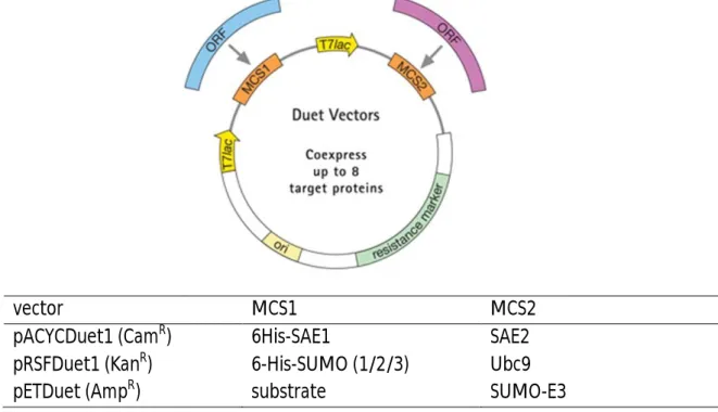
2.1.7 Plasmid vectors ... 33
2.1.8 Oligonucleotides... 36
2.1.9 E. coli strains and human cell lines ... 36
2.1.10 Sorbents and FPLC columns ... 36
2.1.11 Instruments ... 37
2.2 Methods ... 38
2.2.1 Buffers, Solutions and Media ... 38
2.2.1.1 Buffers & Solutions ... 38
2.2.1.2 Culture media for E. coli ... 40
2.2.1.3 Culture media for human cells ... 41
2.2.2 Biomolecular methods... 41
2.2.2.1 Standard techniques ... 41
2.2.2.2 Generation of artificial poly-SUMO chains ... 41
2.2.3 Cell biological methods ... 42
2.2.3.1 Cell cultivation... 42
2.2.3.2 Cell lysis ... 42
2.2.3.3 Cell transfection ... 42
2.2.3.4 Preparation of cells for fluorescent microscopy ... 42
2.2.4 Biochemical methods ... 43
2.2.4.1 SDS-PAGE and Western Blotting ... 43
2.2.4.2 Immunological detection of proteins on PVDF membranes ... 43
2.2.4.3 Protein purifications ... 44
TABLE OF CONTENTS
2.2.4.3.1 Affinity purification of GST-tagged proteins ... 44
2.2.4.3.2 Affinity purification of His
6-tagged proteins ... 45
2.2.4.3.3 Size exclusion chromatography... 45
2.2.4.3.4 UbcH5b purification ... 45
2.2.4.4 Expression and purification of SUMOylated proteins from E. coli... 46
2.2.4.5 PML (50-179) and RNF4 antibodies ... 47
2.2.4.6 DnaK contamination and DnaK depletion ... 48
2.2.4.7 In vitro ubiquitylation assays ... 48
2.2.4.8 Isolation of SUMO-ubiquitin-PML hybrid conjugates after in vitro ubiquitylation reactions ... 49
2.2.4.9 Assessing the ubiquitin attachment sites in His
6-SUMO-3 modified PML (50-179) ... 49
2.2.4.10 Analytical gel filtrations of RNF4 (1-105) and artificial SUMO chains ... 50
2.2.4.11 ITC ... 50
2.2.4.12 Interaction of GST-SIM containing proteins with individual SUMO paralogs.... 51
2.2.4.13 Isolation of multi-/polySUMOylated proteins from cells treated with different stress stimuli by GST-RNF4 (1-105) ... 51
2.2.4.14 Surface Plasmon Resonance ... 52
3. RESULTS ... 53
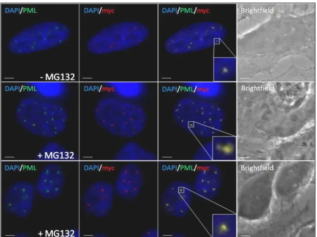
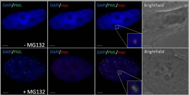
3.1 Proteasome inhibition induces accumulation of SUMO-2/3 conjugates at PML-NBs ... 53
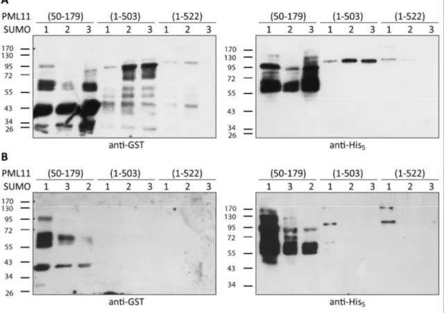
3.2 Establishment of an efficient method to produce and purify SUMO-modified proteins from E. coli ... 55
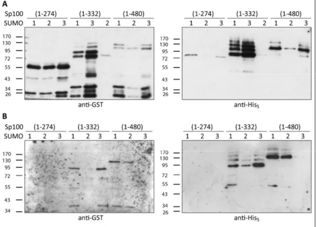
3.2.1 PML & Sp100 ... 59
3.2.2 HIP1α ... 61
3.2.3 Analysis of different SUMO-E3 ligases on substrate SUMOylation... 61
3.3 Establishment of an in vitro ubiquitylation assay ... 63
3.3.1 E2 purification & activity test ... 63
3.3.2 Identification of putative ULS proteins for PML ... 64
3.3.3 Activity test for putative ULS proteins ... 66
3.4 In vitro ubiquitylation of SUMOylated PML demonstrates a function of RNF4 as a mammalian ULS ... 67
3.4.1 ULS assay development ... 67
3.4.2 RNF4 is a ULS and prefers SUMO3-modified PML as substrate ... 71
3.4.3 The SUMOylation site of the PML substrate is not important for recognition by RNF4 in vitro ... 73
3.4.4 RNF4 preferentially ubiquitylates the SUMO moiety of its substrate... 75
3.4.5 Sp100 - a second substrate of RNF4-dependent ubiquitylation? ... 77
3.5 The RNF4 SIMs bind preferentially to SUMO chains ... 78
3.6 Mammalian SUMOs might discriminate between different SIM types ... 83
TABLE OF CONTENTS
3.7 Possible RNF4 substrates upon diverse cellular stresses ... 86
4. DISCUSSION ...92
4.1 PML might act as a SUMO-E3 ligase ... 92
4.2 Specificities in SUMO/SIM interactions ... 94
4.3 RNF4 is a ubiquitin ligase for SUMO conjugates ... 96
4.3.1 PML-NBs - a place for SUMO-dependent degradation in mammalian cells ... 96
4.3.2 RNF4 ubiquitylates specifically SUMOylated PML species ... 97
4.3.3 RNF4 recruitment involves defined SUMO binding properties ... 100
4.3.4 Outlook: Are ULS proteins master regulators of stress signaling? ... 102
APPENDIX ... 105
REFERENCES... 133
ABSTRACT ... 152
ZUSAMMENFASSUNG ... 153
ACKNOWLEDGEMENTS ... 154
ERKLÄRUNG ... 156
CURRICULUM VITAE ... 157
ABBREVIATIONS
AML Acute myeloid leukemia
Amp
RAmpicillin resistence
AOS Activation of Smt3p
APL Acute promyelocytic leukemia
APS Ammonium peroxydisulfate
ARNTL1 Aryl hydrocarbon receptor nuclear translocator-like protein 1 ATO, As
2O
3Arsenic trioxide
ATP Adenosine triphosphate
ATRA All-trans retinoic acid
B B-Box
B1 B-Box 1
BB Binding buffer
BSA Bovine serum albumin
Cam
RChloramphenicol resistence
CC Coiled coil
CENP-I Centromere protein I
CK2 Casein kinase 2
Coomassie Coomassie Brilliant Blue
DAPI 4’,6-Diamidine-2’-phenylindole dihydrochloride
Daxx Death associated protein 6
DMEM Dulbecco´s Modified Eagle Medium
DMSO Dimethylsulfoxide
DTT Dithiotreithol
DUB Deubiquitylase
EB Elution buffer
ECL Enhanced Chemiluminescence
EMEM Eagle's Minimal Essential Medium ERK Extracellular signal-regulated kinase FAT10 F-locus associated transcript 10
FCS Fetal calf serum
FPLC Fast Protein Liquid Chromatography
FT Flow through
GMP1 GAP modifying protein 1
GSH Glutathione
GST Glutathione-S-transferase
HDAC Class IIa histone deacetylase
HECT Homologous to E6AP C-terminus
HIF1 α Hypoxia inducible transcription factor alpha HIP1α Huntingtin interacting protein 1 alpha HMW-SC High Molecular Weight SUMO Conjugate
HP1 Heterochromatin protein 1
HRP Horse-radish peroxidase
HSV-1 Herpes simpex virus 1
ICP0 Infected Cell Polypeptide 0
IFN Interferon
IKK I kappa B kinase
IPI International Protein Index
ABBREVIATIONS
IPTG Isopropyl-β-D-thiogalactopyranosid ITC Isothermal titration calorimetry ITC Isothermal Titration Calorimetry IκBα Inhibitor of NFκBα
K
AAssociation constant
Kan
RKanamycine resistence
K
DDissociation constant
KSHV Kaposi’s sarcoma associated herpesvirus
LB Luria-Bertani
LIC Leukemia initiating cells
MCAF1 MBD1-containing chromatin associated factor
MCS Multiple cloning site
MS Mass spectrometry
M
WMolecular weight
N Stoichiometry
NAA Non-essential amino acids
NEM N-ethylmaleimide
NEMO NFκB essential modulator
NFκB Nuclear factor κB
NLS Nuclear localization signal
Nmi N-myc-interactor
NMR Nuclear magnetic resonance
NPC Nuclear pore complex
o/n Over night
ORF Open reading frame
ori Origin of replication
P Pellet
PBS Phosphate buffered saline
Pc2 Polycomb protein 2
PCNA proliferating cell nuclear antigen
PCR Polymerase chain reaction
PD Pulldown
PDSM Phosphorylation-dependent sumoylation motif
PFA Paraformaldehyde
PIAS Protein inhibitors of activated STAT
PIC1 PML-interacting clone 1
PML Promyelocytic leukemia protein
PML-NB PML nuclear body
polyUb Poly-ubiquitin
PROX1 Prospero-like protein
PVDF Polyvinylidene fluoride
R RING
RA Retinoid acid
RanBP2 Ran binding protein 2
RanGAP1 Ran-GTPase activating protein
RARE RA response elements
RAR retinoid acid receptor alpha RBCC motif RING, B-Box(es), coiled-coil domain
Rfp RING finger protein
RING really interesting new gene
RL Raw lysate
ABBREVIATIONS
RNF4 RING finger 4
RT Room temperature
RU Response units
S Supernatant
S200 Superdex 200
S75 Superdex 75
SAE SUMO activating enzyme
SBM SUMO binding motif
SDS Sodium dodecyl sulfate
SDS-PA SDS polyacrylamide
SDS-PAGE SDS polyacrylamide gel electrophoresis SENP Sentrin specific protease
SIM SUMO interaction motif
SMT3, smt3 Suppressor of Mif Two 3, gene, protein
SPR Surface Plasmon Resonance
SP-RING Siz/PIAS-RING
STAT Signal transducer and activator of transcription StUbL SUMO-targeted ubiquitin ligase
SUMO Small ubiquitin related modifier SUMOc./conj. SUMO conjugated
SUMOylation SUMO conjugation to substrates
SUSP SUMO specific protease
TEMED N,N,N‘,N‘-Tetra-methylethylendiamine
TEV Tobacco etch virus protease
TGF Transforming growth factor
TOPORS Topoisomerase I and p53 binding protein
TRIM Tripartite motif
TUBEs Tandem-repeated ubiquitin-binding entities
UBA Ubiquitin-associated
Uba/Ube1 Ubiquitin activating enzyme/Ubiquitin enzyme 1 Ubc/Ube2 Ubiquitin-conjugating enzyme/Ubiquitin enzyme 2
UBD Ubiquitin binding domain
UbE2 Ubiquitin E2
UBL Ubiquitin-like modifier
Ulp Ubiquitin-like protease
ULS Ubiquitin ligase for SUMO conjugates
UPS Ubiquitin proteasome system
Usp25 Ubiquitin specific protease 25
VHL Von Hippel-Lindau factor
wt Wildtype
ZNF Zinc finger
1. INTRODUCTION
1.1 Relevance of posttranslational modifications in living cells
Cells need to face and react to influences in their environment irrespective of being a protozoon or part of a higher organism to ensure their viability or to fulfill specialized tasks of a united cell-structure in a higher organism.
Therefore highly organized interconnected signaling networks evolved sensing extra- or intracellular stimuli and induce diverse changes ranging from altered transcription to the activation or repression of protein functions. The bases of these signaling networks are small molecules that can be attached to other molecules such as proteins, RNA or lipids to induce altered affinities or binding properties for other molecules as well as to regulate functional aspects such as enzymatic activity or cellular localization. These so- called posttranslational modifications permit much faster signaling and reaction than for example any altered transcription/translation modules could (Walsh, 2007).
Posttranslational modifications of proteins also greatly extend the functional diversity and dynamics of the proteome. The usually transient attachment of small molecules such as phosphate, methyl or acetyl groups to certain proteins is well established (Cheung, 2000; Cohen, 2001; Walsh, 2007; Yang and Seto, 2008; Ng, 2009). Additionally, there are small proteins in eukaryotic cells that can be covalently attached to other proteins. The most prominent member of these modifiers is ubiquitin, after which this group is named. Other Ubiquitin-like modifiers (UBLs) are SUMO, NEDD8, ISG15, Fat10, Atg8 or Atg12 (Kerscher, 2006; Hochstrasser, 2009).
1.2 Ubiquitin modification of proteins
Since ubiquitin was discovered in 1975, it became apparent that posttranslational modification of proteins by ubiquitin plays an important role in diverse cellular processes, including cell division, differentiation, signal transduction, protein trafficking, and quality control (Schlesinger, 1975; Hershko, 1983; Ciechanover, 2005;
Mukhopadhyay and Riezman, 2007). It is a highly conserved protein ubiquitously expressed in all eukaryotes with a size of 8,5 kDa and can be covalently attached to proteins by free lysine, cysteine, or N-terminal residues by specific enzymatic cascades.
These cascades typically involve E1, E2 and E3 enzymes (see section 1.2.1). The most
abundant attachment form of ubiquitin is the conjugation to lysine (K) residues in its
substrate molecules (Glickman and Ciechanover, 2002; Bloom, 2003; Cadwell and
Coscoy, 2005). Mono-ubiquitylation, the addition of a single ubiquitin moiety per
targeting site, was shown to modulate protein endocytosis, intracellular transport, and
DNA repair (see section 1.2.2.1; Haglund, 2003; Ulrich, 2005; Acconcia, 2009; Thompson
INTRODUCTION
2
and Hinz, 2009). Attached mono-ubiquitin can be further modified to generate poly- ubiquitin (polyUb) chains via linkages between several ubiquitin molecules, of which the most prominent is the K48-linked chain that targets the conjugated substrate for proteasomal degradation (see section 1.2.2.2; Hochstrasser, 2006).
1.2.1 Mechanistic aspects of Ubiquitin conjugation
Ubiquitin forms a stable β-grasp fold which is shared by all UBLs despite low sequence homologies. This suggests a common ancestry and indeed, the β-grasp fold may have arisen as an RNA-binding module in a primitive protein-translation system (Burroughs, 2007). Furthermore, the UBL conjugation pathway shows some similarity to bacterial sulphur transfer enzymes within the biosynthesis pathways of the molybdenum cofactor and thiamine (Iyer, 2006; Hochstrasser, 2009).
Four ubiquitin genes in humans encode a total of 14 copies of ubiquitin, so that it is synthesized as a linear poly-ubiquitin chain (Wiborg, 1985). To generate free cellular mono-ubiquitin molecules, theses chains require proteolytic processing by deubiquitylases (DUBs; Komander, 2009b). The single ubiquitin molecules then terminate with the typical Gly-Gly motif, which is common to all UBLs and which is indispensible for substrate modification (Hershko, 1981; Dye and Schulman, 2007).
Mechanistically, the covalent attachment of ubiquitin to substrate proteins involves several enzymatic steps, carried out by three types of enzymes – E1, E2 and E3 (see Figure 1.1; Hershko, 1983). The E1 enzyme (Ubiquitin activating enzyme, e.g. Ube1) activates ubiquitin for transfer by adenylating its C-terminus in an ATP-dependent step.
Ubiquitin is then coupled to the E1 active site cysteine, forming a reactive thioester between E1 and the C-terminal glycine of ubiquitin (Ciechanover, 1981; Hershko, 1981;
Ciechanover, 1982). The activated ubiquitin is subsequently transferred to one of the distinct ubiquitin-conjugating enzymes (Ubc’s; E2s) by transthiolation to a conserved cysteine of the E2 (Pickart and Rose, 1985; Haas and Bright, 1988). The E2 proteins catalyze substrate ubiquitylation in conjunction with an ubiquitin-protein ligase (E3). The concerted action of different E2 and E3 enzymes provide substrate specificity to the ubiquitin conjugation reaction (Reiss, 1989; Sung, 1991; Pickart and Eddins, 2004;
Hochstrasser, 2006; Christensen, 2007). During this last step of the enzymatic cascade, the ubiquitin molecule is usually transferred to an ε-amino group of a lysine side chain in the substrate, thereby forming a peptide-like amide bond (isopeptide bond; Goldknopf and Busch, 1977; Hershko, 1983; Pickart and Eddins, 2004). By adding activated ubiquitin moieties to internal lysine residues on the previously conjugated ubiquitin molecule, poly-ubiquitin chains are synthesized (see section 1.2.2.2; Chau, 1989; Hershko and Ciechanover, 1998).
Whether ubiquitin chain assembly is mediated via a simple sequential addition of
ubiquitin moieties to the nascent chain on the substrate or via pre-assembly on E2/E3
active sites and subsequent transfer onto the substrate is currently under debate
(Hochstrasser, 2006; Li, 2007; Ye and Rape, 2009).
INTRODUCTION
3 Figure 1.1: ubiquitin conjugation cascade
Ubiquitin is expressed as polyprotein; the C-terminal di-glycine motif of individual ubiquitin moieties is exposed after cleavage mediated by deubiquitylating enzymes (DUBs). Ubiquitin is activated by E1 enzymes under ATP consumption and forms a thioester to the E1 active cysteine. Activated ubiquitin is then transferred to an E2 enzyme. E2 and E3 enzymes ubiquitylate the substrate in a concerted action.
Attachment to other ubiquitin molecules forms poly-ubiquitin chains. Some types of ubiquitin chains are recognized by the proteasome that degrades the substrate. Modified from (Miteva, 2010).
1.2.1.1 E1 enzymes
There are two different E1 enzymes for ubiquitin activation in vertebrates, Ube1/UBA1 and Ube1L2/UBA6. They share 40 % of sequence homology to each other (Ciechanover, 1981; Ciechanover, 1982; Chiu, 2007; Jin, 2007; Pelzer, 2007). Ube1L2 was identified as an ubiquitin E1 in 2007, so before it was assumed that Ube1 is the only ubiquitin activating enzyme in vertebrates as it is in lower eukaryotes (Schulman and Harper, 2009). Ube1 charges most known ubiquitin E2 enzymes whereas Ube1L2 has a selective ubiquitin E2 enzyme, Ube2Z/Use1 (Jin, 2007). Furthermore, Ube1L2 is capable to activate F-locus associated transcript 10 (FAT10), a UBL, in vivo and in vitro (Chiu, 2007).
1.2.1.2 E2 enzymes
Family members of the ubiquitin conjugating enzymes (E2s) all possess a highly conserved ubiquitin-conjugating (Ubc) catalytic fold (van Wijk and Timmers, 2009).
These 14-16 kDa domains have an approx. 35 % sequence conservation among different family members and provide a binding platform for E1s, E3s, and the activated ubiquitin or UBL (Burroughs, 2008). The catalytic cysteine, which accepts the activated ubiquitin molecule, is embedded within this domain (van Wijk and Timmers, 2009).
Until now, 38 E2s have been identified in humans and some of them promote
specifically the conjugation of ubiquitin-like modifiers other than ubiquitin (Ye and Rape,
2009). Together with the respective E1 enzymes, this specificity results in parallel
conjugation pathways of ubiquitin and UBLs, although there are some cases of crosstalk
(e.g. the ubiquitin E2 Ube2L6/UbcH8 is shared by ISG15 or the before mentioned use of
Ube1L2 by ubiquitin as well as by FAT10; Kim, 2004; Zhao, 2004; Chiu, 2007). Several E2s
play distinct roles in the ubiquitylation of a substrate. Although most E2s ubiquitylate
substrates without any intrinsic selectivity for a specific acceptor lysine, some stimulate
ubiquitin chain assembly through a defined lysine in ubiquitin. In these cases, the next
INTRODUCTION
4
ubiquitin molecule being assembled is positioned in such a way that only the respective acceptor lysine side chain can attack the thioester bond to the E2 (Ye and Rape, 2009).
Examples are the E2 MMS2/Ubc13 complex in the assembly of K63-linked chains (Hofmann and Pickart, 1999; Deng, 2000; VanDemark, 2001) or the UbcH10/Ube2S complex in the assembly of K11-linked chains (Williamson, 2009).
Other E2s, such as UbcH5, catalyze the formation of ubiquitin chains that lack specificity for any lysine residue of ubiquitin (Brzovic and Klevit, 2006).
1.2.1.3 Ubiquitin E3 ligases
Approximately 600-1000 E3s exist in the human genome, some of which belong to a special class like the RING (really interesting new gene), the HECT (homologous to E6AP C-terminus) or the U-Box domain family (Hatakeyama and Nakayama, 2003; Li and Ye, 2008; Deshaies and Joazeiro, 2009; Rotin and Kumar, 2009).
For the HECT-domain family of E3s, ubiquitin is first transferred to a conserved cysteine of the E3 before it is finally transferred to a substrate group (Huibregtse, 1995; Rotin and Kumar, 2009). For most other ubiquitylation reactions, the E3 rather functions as an adaptor that positions the substrate in close proximity to the reactive E2~ubiquitin thioester. The majority of such E3s belong to the RING-based domain family (~616 proteins; Freemont, 1991; Deshaies and Joazeiro, 2009). In addition to substrate recognition, E3s might have other roles in the catalytic cycle, such as allosteric activation of the E2 as well as mediating linkage-specific poly-ubiquitin chain assembly together with the respective E2 enzymes (Huang, 2004; Pickart and Eddins, 2004; Deshaies and Joazeiro, 2009).
1.2.1.3.1 RING/U-Box ligases
The RING domain was first described in 1991 and was thought to mediate DNA binding or protein dimerization (Freemont, 1991; Joazeiro and Weissman, 2000; Deshaies and Joazeiro, 2009). Only later it became clear, that many RING domain containing proteins serve as ubiquitin ligases and bind to E2 enzymes (Bailly, 1997; Zachariae, 1998;
Joazeiro, 1999; Kamura, 1999; Lorick, 1999; Deshaies and Joazeiro, 2009). The consensus sequence of a RING domain is
C-X2-C-X(9-39)-C-X(1-3)-H-X(2-3)-C-X2-C-X(4-48)-C-X2-C
(where X is any amino acid). The conserved cysteine and histidine residues complex two zinc atoms while forming an interleaved globular structure, the RING domain (see Figure 1.2; Barlow, 1994; Borden, 1995). RING domain E3 ligases are either part of a multi- subunit complex or act as homo- or heterodimers to mediate substrate ubiquitylation (Sharp, 1999; Hashizume, 2001; Xia, 2003; Thornton and Toczyski, 2006; Kawai, 2007).
Other domains such as the B-Box of the tripartite motif (TRIM) subfamily of RING
proteins (see section 1.6; Tao, 2008) and the U-Box are structurally related to the RING
(Aravind and Koonin, 2000).
INTRODUCTION
5 Figure 1.2: RING domain – Dimer and cross-brace structure
A) Structure of the RING heterodimer BARD1/BRCA1. The dimer interface is further stabilized through α- helical interactions (Brzovic, 2001).
B) Schematic representation of a RING domain. The primary sequence organization of the RING domain is folded in a cross-brace structure to coordinate the two zinc atoms. The first cysteine that coordinates zinc is labeled as C1, and so on. H1 denotes the coordinating histidine. Xn refers to the number of amino acid residues in the spacer regions between the zinc coordinating residues (modified from Deshaies and Joazeiro, 2009).
1.2.1.4 Deubiquitylating enzymes
Deubiquitylases (DUBs) exhibit three major functions: They generate the pool of free ubiquitin molecules by cleaving the newly translated linear ubiquitin chains into single molecules. Then, they can remove ubiquitin (chains) from modified substrates.
Additionally, DUBs associated with the proteasome are responsible for ubiquitin recycling from proteins prone for degradation (Lam, 1997; Park, 1997; Borodovsky, 2001; Verma, 2002; Komander, 2009b). Furthermore, some DUBs can edit the form of ubiquitin modification by trimming ubiquitin chains (Wertz, 2004). In the human genome, approx. 79 genes encode for deubiquitylases of which nearly all family members are cysteine proteases while the members of one subgroup are zinc metalloproteases (Nijman, 2005).
1.2.1.5 Ubiquitin binding domains
Diverse proteins in the ubiquitin signaling pathway need to recognize and distinguish ubiquitin and its diverse chained forms from each other and from other UBLs. Therefore, a range of ubiquitin binding domains (UBDs) exist (UIMs, UBXs, UBAs, etc.), most of which recognize a specific hydrophobic patch around ubiquitin’s Ile44. The diverse UBDs differ in their structure and do not necessarily share a common motif (Dikic, 2009).
Furhtermore, some UBDs have been shown to act as a cis-E3 mono-ubiquitin ligase, promoting self-ubiquitylation of the protein they are part of (Hoeller, 2007).
A B
INTRODUCTION
6
1.2.2 Mono-ubiquitylation and polymeric ubiquitin chains Ubiquitin can be attached to the substrates in multiple ways.
The following scheme (Figure 1.3) gives an impression of the variety of signals achieved by different ubiquitylation types.
Figure 1.3: Different forms of ubiquitylation
A) The three general forms of ubiquitylation: mono-ubiquitylation, multiple mono-ubiquitylation and poly- ubiquitylation. B) Forms of homotypic poly-ubiquitylation. Each ubiquitin chain contains a single linkage type which may lead to distinct ubiquitin chain conformations. Multiple homotypic ubiquitin chains on the same substrate are possible. C) Forms of heterotypic poly-ubiquitylation. In mixed linkages, a ubiquitin chain has alternating linkage types. In branched or forked poly-ubiquitin chains, a single ubiquitin is extended at two or more lysine residues. The scheme was modified from (Komander, 2009a).
1.2.2.1 Mono-ubiquitylation
Mono-ubiquitylation in general serves as a signaling module to confer additional protein-protein interaction properties to its substrate proteins. In the case of receptor endocytosis, it regulates the internalization and sorting of the modified receptor that is either recycled or further transported to the endo-lysosomal compartment for degradation (Levkowitz, 1998; Levkowitz, 1999; Joazeiro, 1999; Acconcia, 2009).
In the case of the processivity factor “Proliferating cell nuclear antigen” (PCNA)
monoubiquitylation results in the recruitment of a damage-tolerant polymerase and
translesion synthesis while the modification of the same residue with a K63-linked chain
results in an error-free damage avoidance pathway. Its modification with the small
ubiquitin related modifier (SUMO) facilitates DNA damage tolerance upon replication
fork stalling by recruitment of Srs2 (Hoege, 2002; Stelter and Ulrich, 2003; Kannouche,
2004; Pfander, 2005; Ulrich, 2005).
INTRODUCTION
7 1.2.2.2 Poly-ubiquitin chains
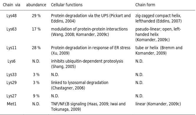
Ubiquitin possesses seven lysine residues on its surface, namely K6, K11, K27, K29, K33, K48 and K63 that can all serve as acceptor sites for additional ubiquitin molecules in vitro and in vivo, thereby forming poly-ubiquitin chains (see Table 1.1; Arnason and Ellison, 1994; Johnson, 1995; Baboshina and Haas, 1996; Peng, 2003). Poly-ubiquitin chains consisting of four ubiquitin moieties attached through K48 linkages typically mark a protein for proteasomal degradation (Hershko, 1983; Chau, 1989; Hershko and Ciechanover, 1998; Pickart and Fushman, 2004). Chain formation through other lysine residues of various lengths and shapes results in various chain conformations and create a range of molecular signals in the cell (Kim, 2007; Ikeda and Dikic, 2008). In addition, linear chains are also assembled in vivo by head-to-tail arrangement of ubiquitin moieties through the α-amino group at the N-terminus (Kirisako, 2006). Recently, it has been postulated, that all non-K63-linked ubiquitin chains target proteins for degradation (Xu, 2009).
Table 1.1: Relative cellular abundance and form of homotypic poly-ubiquitin chains
Chain via abundance Cellular functions Chain form
Lys48 29 % Protein degradation via the UPS (Pickart and Eddins, 2004)
zig-zagged compact helix, lefthanded (Eddins, 2007) Lys63 17 % modulation of protein-protein interactions
(Wang, 2008; Komander, 2009c)
pseudo-linear; open, left- handed helix
(Komander, 2009c) Lys11 28 % Protein degradation in response of ER stress
(Xu, 2009)
tube or helix (Bremm and Komander, 2009)
Lys6 N.D. inhibits ubiquitin-dependent proteolysis (Shang, 2005)
N.D.
Lys33 3 % N.D. N.D.
Lys29 3 % linked to lysosomal degradation (Chastagner, 2006)
N.D.
Lys27 9 % N.D. N.D.
Met1 N.D. TNF/NFκB signaling (Haas, 2009; Iwai and Tokunaga, 2009)
linear (Komander, 2009c)
INTRODUCTION
8
1.2.2.2.1 K48-linked chains and the Ubiquitin-Proteasome system
The majority of intracellular proteins are degraded via the ubiquitin proteasome system (UPS) (Lee and Goldberg, 1998). The ‘housekeeping’ 26S proteasomes are ATP-driven, multi-subunit proteolytic complexes that preferentially degrade proteins tagged with K48-linked poly-ubiquitin chains (Hershko, 1980; Chau, 1989; Hershko, 1991; Seufert and Jentsch, 1992; Hershko and Ciechanover, 1998; Voges, 1999; Elsasser and Finley, 2005).
26S proteasomes are comprised of a 20S core component and two flanking 19S regulatory complexes that regulate substrate specificity and access to the catalytic chamber of the 20S core. The 20S core is highly conserved from yeast to humans;
simpler prototypes are also found in prokaryotes (Löwe, 1995). Four α- and β-rings surround a barrel-shaped cavity in the 20S core (Groll, 1997). The two inner β-rings form a central chamber that harbors the proteolytic centers containing chymotryptic-, tryptic- and caspase-like activities (Marques, 2009). Tetra-ubiquitin is the minimum signal for efficient proteasomal targeting. The mechanism of targeting involves an increase in substrate affinity that is brought about by autonomous binding of the K48-linked tetra- ubiquitin chain. Binding is mediated by the UBDs (see section 1.2.1.5) of the 19S subunits Rpn10/S5a and Rpn13 (Deveraux, 1994; Lam, 2002; Groll, 1997; Husnjak, 2008;
Schreiner, 2008). Recognition of this signal is followed by substrate unfolding and
translocation into the degradation chamber while ubiquitin itself is recycled. After
degradation, the resultant peptides are released through the entry channel (Finley,
2002; Hutschenreiter, 2004; Komander, 2009b).
INTRODUCTION
9
1.3 Ubiquitin-like modifiers
Ubiquitin is the most prominent and most abundant member of a diverse group of evolutionarily conserved small proteins that are covalently conjugated to target proteins, the ubiquitin-like modifiers (UBLs). Although the other members vary in their degree of sequence similarity to ubiquitin, they all share the typical β-grasp fold and seem to be conjugated through an analogous enzymatic cascade of specific E1, E2 and E3 enzymes (see table A.1; Kerscher, 2006). Each UBL contains one or two glycines at the C-terminus that is used to form the isopeptide bond with target proteins. Most UBLs (with the exception of FAT10, ATG12 and URM1) are synthesized as precursors whose conjugation requires C-terminal cleavage at this glycine through specific processing proteases (Groettrup, 2008). Barring ubiquitin, the so far best studied UBLs are the family members of small ubiquitin related modifiers (SUMO) which have a wide range of substrates and functions (Hochstrasser, 2009). As this thesis is mainly dealing with a new function for the SUMO modification of proteins, this group is described in more detail (see section 1.4). Functions of other UBL family members are summarized in Appendix table A.1; a phylogenetic tree for all human UBLs is displayed in Figure 1.4.
Figure 1.4: Phylogenetic tree of human Ubiquitin-like modifiers
The tree was derived through human UBL sequence comparison using MAFFT and virtualized with its
associated software ArchaeopterixA (Katoh and Toh, 2008). Urm1 represents the closest relative to the
ancestors of UBLs; the human Atg8 homologues as well as the human SUMOs are sub grouped.
INTRODUCTION
10
1.4 SUMO modification of proteins
An increasing number of proteins have been shown to be SUMO modified and many of them are tumor suppressor proteins, transcription factors or nuclear body proteins.
SUMO is an ubiquitin-like protein and its conjugation to substrates (SUMOylation) is thus a reversible process including the typical enzymatic cascade with a SUMO-specific set of E1, E2 and E3 enzymes and a special set of proteases (see section 1.4.2 and Figure 1.7 therein; Xu, 2009).
SUMO is highly conserved in all eukaryotes, but lower eukaryotes like the yeasts S.cerevisiae and S. pombe as well as the nematode C. elegans possess only a single gene, while in mammalian cells four SUMO paralogs exist (Xu, 2009). Plants even have up to eight SUMOs (Kurepa, 2003; Lois, 2003). All SUMO paralogs are translated as precursors that need to be processed to produce the mature di-glycine motif at the C-terminus (Xu, 2009). Compared to ubiquitin, all SUMOs possess a flexible N-terminal extension (see Figure 1.5; Bayer, 1998; Huang, 2004).
Figure 1.5: Comparison of the three-dimensional structures of ubiquitin, SUMO-1 and SUMO-2
NMR structures of human ubiquitin (PDB ID: 1D3Z), SUMO-1 (PDB ID: 1A5R) and SUMO-2 (PDB ID: 2AWT) as ribbon diagrams. Despite their low sequence homology, ubiquitin and the SUMO paralogues share the highly conserved three-dimensional structure
of the β-grasp fold (α-helices are depicted in green, β-strands in yellow). Structures were displayed using Cn3D 4.1.
Out of the four mammalian SUMOs, SUMO-4 is probably not able to be conjugated to
substrates (Owerbach, 2005), whereas SUMOs 1-3 are conjugated to lysines in their
substrate proteins forming a covalent amide bond (SUMOylation; Johnson and Blobel,
1997; Johnson, 1997; Sampson, 2001). SUMO-1 shares 44 % sequence identity with
SUMO-2 and SUMO-3 which are 97 % identical in their processed and conjugatable form
(see Figure 1.6). Therefore, they are commonly seen as entity (Lapenta, 1997; Chen,
INTRODUCTION
11 1998; Maticvan Hagen, 2008). Yeast SUMOs (Bencsath, 2002; Bylebyl, 2003; Skilton, 2009) as well as SUMO-2/3 harbor a consensus SUMOylation motif ΨKxE/D (where Ψ represents a hydrophobic and x any amino acid) and are able to form polymeric chains via a lysine residue in their N-terminus (K11) (Tatham, 2001). For SUMO-1 this has not been observed in vivo.
Ubiquitin ---MQIFVKTLTGKTITLEVEPSD----21 Smt3- MSDS---EVNQEAKPEVK--PEVKPETHINLKV-SDGSSEIFFKIKKTT----43 Pmt3p MSESPSANISDADKSAITPTTGDTSQQDVKPST---EHINLKVVGQDNNEVFFKIKKTT----56 SUMO-1 MSD---QEAKPSTEDLGDKKEGEYIKLKVIGQDSSEIHFKVKMTT----42 SUMO-2 MAD---EKPKEG--VKTENNDHINLKVAGQDGSVVQFKIKRHT----38 SUMO-3 MSE---EKPKEG--VKTE-NDHINLKVAGQDGSVVQFKIKRHT----37 SUMO-4 MAN---EKPTEE--VKTENNNHINLKVAGQDGSVVQFKIKRQT----38
Ubiquitin TIENVKAKIQDKEGIPPDQQRLIFAGKQLEDGRTLSDYNIQKESTLHLVLRLRGG---76 Smt3p PLRRLMEAFAKRQGKEMDSLRFLYDGIRIQADQTPEDLDMEDNDIIEAHREQIGGATY---101 Pmt3p EFSKLMKIYCARQGKSMNSLRFLVDGERIRPDQTPAELDMEDGDQIEAVLEQLGGCTHLCL---117 SUMO-1 HLKKLKESYCQRQGVPMNSLRFLFEGQRIADNHTPKELGMEEEDVIEVYQEQTGGHSTV---101 SUMO-2 PLSKLMKAYCERQGLSMRQIRFRFDGQPINETDTPAQLEMEDEDTIDVFQQQTGGVY---95 SUMO-3 PLSKLMKAYCERQGLSMRQIRFRFDGQPINETDTPAQLEMEDEDTIDVFQQQTGGVPESSLAGHSF--103 SUMO-4 PLSKLMKAYCEPRGLSVKQIRFRFGGQPISGTDKPAQLEMEDEDTIDVFQQPTGGVY---95
Figure 1.6: Multiple sequence alignment of human and yeast SUMO isoforms compared to ubiquitin All SUMOs display an N-terminal extension missing in ubiquitin. Additional residues C-terminally of the di- glycine motif (which is highlighted in grey) have to be processed to form mature SUMOs. SUMO-4 cannot be processed to its mature form due to a proline residue (displayed in red) which prevents access to the catalytic center of the SUMO proteases (Owerbach, 2005). The SUMO-4 MV substitution at position 55 associated with a higher susceptibility for type 1 diabetes is depicted in magenta. Conjugatable lysine and methionine residues are highlighted in green, boxes represent the main attachment sites for polymeric chain assembly. The K42, K41 and K54 attachment sites in SUMO-2, SUMO-3 and Smt3, correspondingly, as well as the respective SUMO1 lysines have been shown in vitro only (Pedrioli, 2006; Jeram, 2010).
SUMO-1 was shown to be phosphorylated in vivo at S2 (depicted in blue). Sequences were aligned using MAFFT version 6 (Katoh and Toh, 2008).
Non-covalent interactions of proteins with SUMO or SUMOylated proteins, respectively, are conferred via SUMO interaction motifs (SIMs) (Minty, 2000; Song, 2004; Hannich, 2005; Song, 2005; Hecker, 2006; see section 1.4.3). The majority of SUMO targets is modified at very low steady state levels in vivo. However, SUMOylation-deficient mutants can have striking effects, probably due to dynamic SUMOylation/deSUMOylation cycles (Johnson, 2004).
1.4.1 SUMO proteins
1.4.1.1 Yeast SUMOs: Smt3 and Pmt3
In S.cerevisiae, SUMO is encoded by a single gene that was originally isolated as a
suppressor of mutations in a centromeric protein and was therefore named suppressor
of Mif Two 3 (SMT3) (Meluh and Koshland, 1995; Mannen, 1996). The SMT3 gene is
essential for viability while its orthologue pmt3 (for S. pombe homologue of SMT3) in
S. pombe is not, although mutants lacking pmt3 grow poorly and display severe defects
in genome maintenance (Tanaka, 1999).
INTRODUCTION
12
1.4.1.2 Mammalian SUMO isoforms
Human SUMO-1 was identified as a homologue to the S.c. Smt3 protein and shares only 18 % sequence identity to ubiquitin. It was the first SUMO paralog discovered - independently from several groups - and therefore was also aliased as hSmt3, Ubiquitin- like modifier 1 (UBL1), PML-interacting clone 1 (PIC1), Sentrin or GAP modifying protein 1 (GMP1) (Boddy, 1996; Mannen, 1996; Matunis, 1996; Okura, 1996; Shen, 1996).
It is expressed in many different cell types, predominantly localized to the nucleus, where it generally remains in the conjugated form (Kamitani, 1997; Saitoh and Hinchey, 2000). The first identified and best characterized substrate is the Ran-GTPase activating protein (RanGAP1). Conjugation with SUMO-1 localizes RanGAP1 to the nuclear pore complex (NPC) where it is involved in nuclear import/export (Matunis, 1996; Mahajan, 1997). Another prominent substrate in mammalians is the promyelocytic leukemia protein (PML), the scaffold protein for PML nuclear bodies (PML-NBs) which are large nuclear protein complexes (Sternsdorf, 1997; Müller, 1998; Ishov, 1999). SUMOylation of PML is a prerequisite for the formation of PML-NBs and many proteins that are localized to PML-NBs are also SUMOylated (Zhong, 2000; Matunis, 2006; Bernardi and Pandolfi, 2007). A more detailed description of PML-NBs is given in section 1.7.
Additionally, many transcription factors and chromatin-associated proteins are transient SUMO substrates (as, for example the before mentioned modification of PCNA in DNA repair pathways), so that SUMOylation is required for a variety of cellular processes (Zhao, 2007; see also section 1.4.4). In some cases, SUMO-1 modification is thought to counteract ubiquitylation and subsequent degradation of the substrate, as the modifications occur on the same lysine residue and exclude each other. An example for that is the Inhibitor of NFκB alpha (IκBα) whose SUMOylation stabilizes the protein, thereby inhibiting nuclear factor kappa B (NFκB) transcriptional activity (Desterro, 1998).
SUMO modification often occurs on specific lysine residues within the before mentioned consensus motif, ΨKxE/D, which can be directly recognized by the SUMO conjugating enzyme Ubc9/Ube2I (Sampson, 2001). SUMO-1 has also been shown to be phosphorylated in vivo at the very N-terminus (serine 2), but a function linked to that modification has not been identified yet (Matic, 2008).
SUMO-1 deficient mice are viable despite a first observation in which SUMO-1 deletion was lethal (Alkuraya, 2006). They lack any apparent phenotype, due to a compensatory utilization of SUMO-2/3 for SUMOylation of SUMO1 targets (Evdokimov, 2008; Zhang, 2008). However, on the cellular level, a decreased localization of SUMO-2/3 modified RanGAP1 to the nuclear pore was observed and PML nuclear body formation was reduced. Interestingly, the amount of PML protein as well as its modification by SUMO- 2/3 was significantly reduced (Evdokimov, 2008).
Generally, SUMO-2/3 supply the main reservoir of free SUMO that is used for
conjugation in response to certain stress stimuli like heat and ethanol as well as
oxidative and osmotic stress, resulting in high molecular weight SUMO conjugates
INTRODUCTION
13 (HMW-SCs). This response involves the modification of many proteins as well as SUMO chain formation and is reversed upon recovery; partly due to the action of SUMO- specific proteases (see section 1.4.2.4; Saitoh and Hinchey, 2000; Haindl, 2008;
Golebiowski, 2009). SUMO chain formation through the consensus lysine 11 in both SUMO-2 and SUMO-3 results in mixed chains in vivo (Matic, 2008). The exact roles of SUMO conjugation in the cellular stress response is not fully understood; it might help to eliminate otherwise toxic proteins as indicated by a newly discovered pathway (see section 1.5).
The non-conjugatable SUMO-4 isoform was originally characterized as a susceptibility gene for type 1 diabetes (Guo, 2004; Owerbach, 2005). This susceptibility is somehow limited to Asian populations as it is not found in Caucasians (Ikegami, 2008). Correlated to that was a polymorphism in SUMO-4 at position 55, with an amino acid substitution from methionine to valine (as depicted in Figure 1.6).
1.4.2 Mechanistic aspects of SUMO conjugation
Figure 1.7: SUMOylation cascade
Before SUMO can be conjugated it needs to be processed to its mature form by SUMO proteases (SENPs/Ulps). The SUMO-E1 heterodimer SAE1/SAE2 activates SUMO and transfers it from the active site cysteine in SAE2 to the SUMO-E2 enzyme Ubc9. Ubc9 either directly recognize substrates with the SUMO
consensus site ΨKxE (Ψ, hydrophobic; x any amino acid) or in conjunction with a SUMO-E3 ligase. Incontrast to SUMO-1, SUMO-2, SUMO-3 and yeast SUMOs are able to form chains that can be reversed by SENP/Ulp activity. Modified from (Miteva, 2010).
1.4.2.1 SUMO E1 enzymes
SUMO conjugation follows in principle the same mechanism as ubiquitin conjugation.
The SUMO E1 enzyme is a heterodimer composed of the proteins activation of Smt3p
and the ubiquitin activating protein 2, short AOS1/UBA2, in yeasts or its mammalian
homologues SUMO activating enzymes 1 and 2 (SAE1/SAE2). AOS1/SAE1 as well as
UBA2/SAE2 display sequence similarities to the N- and C-terminal parts, respectively, of
ubiquitin-activating enzymes (Dohmen, 1995; Johnson, 1997; Azuma, 2001). Both AOS1
INTRODUCTION
14
and UBA2 are essential for viability in S. c., consistent with an essential function for SUMO modification (Dohmen, 1995; Johnson, 1997).
A specific binding of the SUMO E2 enzyme Ubc9 is mediated by a ubiquitin-like domain in UBA2/SAE2 and that this binding involves a folding-upon-binding process of unstructured parts within the E1 enzyme (Lois and Lima, 2005; Wang, 2009). UBE1DC1, the E1 enzyme for Ufm1 modification, has been reported to also activate SUMO-2 in vitro and to colocalize with overexpressed SUMO-2 in vivo (Zheng, 2008).
1.4.2.2 E2 enzyme Ubc9
The E2 enzyme Ube2I/Ubc9 is the specific conjugation enzyme for SUMO (Desterro, 1997; Johnson and Blobel, 1997; Schwarz, 1998; Knipscheer, 2007).
Ubc9 is able to directly recognize substrates harboring the SUMOylation consensus site ΨKxE/D, to which it can bind without the help of an additional E3 enzyme (Sampson, 2001). An extended motif, ΨKxExxS/T(P) induces phosphorylation-dependent SUMOylation of substrates harboring this motif, also termed the phospho-SUMOyl- switch or for phosphorylation-dependent sumoylation motif (PDSM) (Hietakangas, 2003;
Hietakangas, 2006; Yang and Gregoire, 2006).
To promote chain formation, Ubc9 has a non-covalent binding site for SUMO (around residue H20) that is used to recruit another Ubc9-SUMO thioester intermediate (Tatham, 2003; Knipscheer, 2007). Ubc9 is essential for viability in most species, probably because Ubc9 deficiency leads to a complete abrogation of SUMOylation (Seufert, 1995; Jones, 2002; Johnson, 2004). An exception is S. pombe where SUMOylation itself is also not essential (Ho and Watts, 2003).
1.4.2.3 SUMO E3 ligases
Only a few SUMO E3 ligases have been identified so far. Substrate specificity is partly
linked to the distinct intracellular localization of the ligases and of the substrates,
respectively. Ran binding protein 2 (RanBP2), for example is associated with the NPC
(Wu, 1995; Yokoyama, 1995); the Polycomb protein 2 (Pc2) is mainly found in large
complexes on chromatin (Wotton and Merrill, 2007). Some SUMO E3 ligases have dual
functions, for instance the Class IIa histone deacetylases (HDACs). HDAC7 has been
shown to promote SUMOylation of PML independently of its deacetylase activity
(Gregoire and Yang, 2005; Zhao, 2005; Gao, 2008; Martin, 2009). TOPORS, a
Topoisomerase I and p53 binding protein (Haluska, 1999; Zhou, 1999), is the first
example of a protein that possesses both ubiquitin and SUMO E3 ligase activity. The
ubiquitination activity maps to a conserved RING domain in the N-terminal region of the
protein and is regulated by phosphorylation, both of which are not required for
SUMOylation activity (Rajendra, 2004; Weger, 2005; Park, 2008).
INTRODUCTION
15 1.4.2.3.1 PIAS proteins
The protein inhibitors of activated STAT (PIAS) were initially named for their ability to interact with and inhibit the signal transducer and activator of transcription (STAT) factors (Chung, 1997; Liu, 1998). PIAS proteins are evolutionarily conserved in eukaryotes with mammals encoding four PIAS genes, PIAS1, PIAS2 (PIASx), PIAS3 and PIAS4 (PIASy). Homologues of mammalian PIAS proteins are found in non-vertebrates, plants and yeast, including the S. cerevisiae proteins Siz1 and Siz2/Nfi1 (Johnson and Gupta, 2001; Palvimo, 2007).
The Siz/PIAS (SP)-RING displays structural similarity to RING and U-Box domains and is, together with the C-terminal SIM, required for activation of the Ubc9~SUMO thioester for conjugation to substrates (Yunus and Lima, 2009). Thus, similarly to the function of RING-type ubiquitin E3 ligases, PIAS proteins are likely to act as adaptors between the Ubc9~SUMO intermediate and the SUMO substrate.
1.4.2.3.2 RanBP2
In vertebrate cells, the nucleoporin 358 (Nup358)/Ran binding protein 2 (RanBP2) is a major component of the nuclear pore complex (Wu, 1995; Yokoyama, 1995). RanBP2 forms a complex with SUMOylated RanGAP1, the GTPase activating protein for Ran, thereby tethering it to the cytoplasmic filaments of the NPC (Matunis, 1996; Mahajan, 1997). Despite being equally well modified by SUMO-1 and SUMO-2 in vitro, RanGAP1 is primarily modified by SUMO-1 in vivo. This paralog-selective modification is due to a more stable, higher affinity complex of RanBP2 and SUMO-1-modified RanGAP1 that preferentially protects it from SUMO isopeptidases (Zhu, 2009). Moreover, RanBP2 functions as SUMO E3 ligase for RanGAP1 as well as for many other proteins in vitro and in vivo and is itself SUMOylated (Saitoh, 1998; Pichler, 2002; Kirsh, 2002; Dawlaty, 2008).
A C-terminal domain, which is characterized by the presence of two internal repeats, is sufficient to efficiently promote SUMOylation in the presence of E1, E2, SUMO-1 and ATP in vitro (Pichler, 2002; Kirsh, 2002).
1.4.2.4 SUMO proteases
The first SUMO-specific proteases, the distantly related Ubiquitin-like proteases Ulp1
and Ulp2, were discovered in S. cerevisiae (Li and Hochstrasser, 1999; Li and
Hochstrasser, 2000; Schwienhorst, 2000). Ulp1 is essential for the G2/M transition in the
cell cycle and its inactivation as well as overexpression of its catalytic domain is lethal,
pointing towards the essential function of a balanced SUMOylation/deSUMOylation (Li
and Hochstrasser, 1999; Takahashi, 2000; Mossessova and Lima, 2000). Ulp1 functions
both in SUMO maturation as well as in substrate deconjugation and is located at the
NPC (Li and Hochstrasser, 1999; Takahashi, 2000). Ulp2 instead is localized
predominantly in the nucleoplasm and functions in the disassembly of SUMO chains (Li
and Hochstrasser, 2000; Schwienhorst, 2000; Bylebyl, 2003) with implications for the
restart of the cell cycle after DNA repair and the checkpoint-induced metaphase arrest
INTRODUCTION
16
(Schwartz, 2007). Ulp2 deletion strains are viable despite showing an abnormal phenotype including chromosome mis-segragation and hypersensitivity to DNA damaging agents (Li and Hochstrasser, 2000; Schwienhorst, 2000).
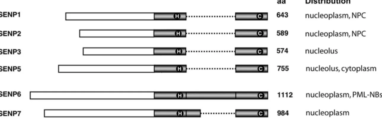
To date, six human SUMO specific proteases (SUSPs) or Sentrin specific proteases (SENPs) - SENP1, 2, 3, 5, 6 and 7 - have been discovered with diverse activities and localizations within the cell (listed in Figure 1.8; Xu, 2009). An additional family member is SENP8, which has been shown to be a deNEDDylating enzyme (Gan-Erdene, 2003). All SUMO proteases share a C-terminal ~ 250 amino acid long catalytic domain and differ in their N-termini which probably confer substrate specificity and intracellular localization.
Figure 1.8: Schematic representation of human SUMO proteases and their cellular distribution
The conserved SENP catalytic domain is represented in grey with catalytic residues in circles. The dotted line illustrates insertions within the catalytic domains of SENP6 and SENP7 (aa, amino acids; NPC, nuclear pore complex). Modified from (Mikolajczyk, 2007 and Xu, 2009).
SENP1 has been shown to mediate particularly the deconjugation of SUMO-1 modified substrates within the nucleus, while SUMO-2/3 substrates remain largely unaffected (Yamaguchi, 2005). Moreover, it exhibits the highest endopeptidase activity among the SENPs with its processing activity during SUMO maturation follows the order SUMO-1 >
SUMO-2 > SUMO-3 (Xu and Au, 2005). SENP2 instead has the highest maturation efficiency for SUMO-2 (Reverter and Lima, 2004). Both SENP1 and SENP2 have been located to the nucleoplasm and to the NPC (Gong, 2000; Hang and Dasso, 2002; Bailey and O'Hare, 2004). SENP3 and SENP5 are localized in the nucleolus (Nishida, 2000; Di Bacco, 2006; Gong and Yeh, 2006), though SENP5 might also function within the cytosol, where it has been shown to deSUMOylate a dynamin related protein, DRP1 that is involved in mitochondrial fission (Zunino, 2007). SENP3 and SENP5 show a preference for SUMO-2/3 over SUMO-1 maturation and deconjugation (Di Bacco, 2006; Gong and Yeh, 2006).
SENP6 and SENP7 both contain an insertion within the conserved catalytic domain and have been found to disassemble specifically SUMO-2/3 chains analogous to Ulp2 cleaving SUMO chains in S. cerevisiae. Partly deletions of the insertion in SENP7 reduce the affinity of SENP7 to bind SUMO-2/3 chains (Lima and Reverter, 2008).
Endopeptidase activity towards the SUMO precursors has not been detected for any of
the both enzymes (Mikolajczyk, 2007; Lima and Reverter, 2008). SENP6 and SENP7 have
INTRODUCTION
17 been both located to the nucleoplasm where they might exert overlapping functions.
RNAi mediated knockdown of SENP6 resulted in a drastic increase of PML-NB size and number, suggesting a function for SENP6 in the disassembly of SUMO-2/3 chains in these structures (Mukhopadhyay, 2006).
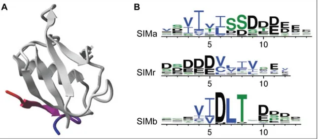
1.4.3 SUMO interaction motifs
Similar to the ubiquitin system, SUMO binding proteins display a common motif recognizing SUMO non-covalently. Unlike the interaction of ubiquitin and UBDs, binding to SUMO is mediated through a short motif, the SUMO interaction motif (SIM) or SUMO binding motif (SBM) (see Figure 1.9.A; Minty, 2000; Song, 2004; Hannich, 2005; Song, 2005; Hecker, 2006).
Three types of SIMs have been identified so far (see Figure 1.9.B). SIMa is characterized by a core consisting of four hydrophobic residues, immediately followed by a mixed cluster of S/D/E residues, the so-called acidic stretch. The third hydrophobic position in SIMa motifs is less conserved than the other hydrophobic positions and instead, also non-hydrophobic, even acidic residues may be present (Song, 2004; Hannich, 2005;
Miteva, 2010).
In a reversed orientation of that motif, SIMr, the acidic stretch precedes the hydrophobic core (Song, 2005; Hecker, 2006).
The type b SIM usually sticks to the consensus sequence V-I-D-L-T, with some variations in the first two hydrophobic amino acids (Uzunova, 2007; Miteva, 2010).
Mixed SIM types are also present, the PIAS SIMs, for example are of type b, followed by an acidic stretch. Whether these types of SIMs have different binding capacities towards different SUMO isoforms remains to be elucidated, although there is a hint that SIM type b binds SUMO-2/3 with a higher affinity than SUMO-1 (Sekiyama, 2008).
Binding occurs upon insertion of the hydrophobic SIM core into a hydrophobic cleft between the β2-strand and helix α1 of the SUMO molecule, forming a parallel β-sheet together with the β2-strand (Reverter and Lima, 2005; Song, 2005; Hecker, 2006;
Sekiyama, 2008). An example is depicted in Figure 1.9.A; the SIMb from PIAS2 bound to
SUMO-1.
INTRODUCTION
18
Figure 1.9: SUMO interaction motifs (SIMs)
A) Ribbon diagram of the SIMb from PIAS2 bound to human SUMO-1 (based on structure PDB ID: 2ASQ).
B) Residue conservation of the three SIM types is shown in a sequence logo representation. Overall height of a position indicates its information content; height of individual residues indicates their frequency at that position (charged amino acids are displayed in black, polar in green and hydrophobic in blue).
Pictures were taken from (Miteva, 2010).
Furthermore, SIMs often contain serine and threonine residues that are able to be acidified by phosphorylation (Hecker, 2006). A recent publication showed that binding to SUMO by the SIM of PIAS1 is enhanced after Casein kinase II (CK2) phosphorylation of serine residues adjacent to the SIM core domain (Stehmeier and Müller, 2009). Serine residues in similar SIMs, for example within the PML protein, were also shown to be phosphorylated by CK2 after osmotic stress induction (Scaglioni, 2008; Stehmeier and Müller, 2009). This indicates an integrative crosstalk function for SIM motifs between the phospho- and SUMO-regulated signaling pathways within mammalian cells.
In analogy to the activity of ubiquitin/substrate non-covalent interactions leading to mono-ubiquitylation (Hoeller, 2007), SIMs have also been shown to non-covalently recruit Ubc9~SUMO thioesters and serve as a cis-regulatory SUMO-E3 ligase modules (Lin, 2006; Knipscheer, 2008; Meulmeester, 2008; Cho, 2009).
1.4.4 Cellular roles of SUMO modification
Roles of SUMO modification in the cellular context are as multifaceted as the proteins subjected to SUMOylation. Some SUMO substrates are modified in the cytosol such as the inhibitor of NFκB, IκBα, thereby inhibiting cytokine or innate immune receptor signaling, or the mitochondrial fission GTPase DRP1 whose activity is linked to its SUMOylation (Figueroa-Romero, 2009).
However, the vast majority of substrates are nuclear proteins, highlighting the primary
nuclear functions of SUMOylation. As already mentioned above, SUMOylation is
involved in DNA repair, transcriptional regulation, chromosome maintenance and
mitosis, but also in inflammation, nuclear organization, protein localization and stress
pathways, as reviewed in Gill, 2005; Ulrich, 2005; Matunis, 2006; Heun, 2007; Palvimo,
INTRODUCTION
19 2007; Dasso, 2008; Tempe, 2008; Bergink and Jentsch, 2009; Garcia-Dominguez and Reyes, 2009. Deregulated SUMOylation may lead to cancer and is implicated in neurodegenerative diseases (Kim and Baek, 2006; Martin, 2007; Sarge and Park-Sarge, 2009).
Generally, SUMOylation is important for nuclear organization as many substructures as the nuclear envelope, Nucleoli and PML nuclear bodies are disrupted when the SUMO pathway is defective (Nacerddine, 2005; Heun, 2007).
Proper protein localization upon SUMOylation is not only known for RanGAP1 and PML, but for many nuclear proteins and is mainly achieved by mono-SUMOylation or multiple mono-SUMOylation of a given protein and its interaction with SIM-containing partners (Matunis, 2006). Many transcription factors, for instance, relocalize to PML-NBs when SUMOylated (Johnson, 2004; Palvimo, 2007). Transcriptional regulation is also mediated through SUMOylation of transcriptional co-repressors or transcriptional activators (Palvimo, 2007; Garcia-Dominguez and Reyes, 2009).
Dynamic SUMOylation/deSUMOylation events are also indispensable for sister chromatid separation in mitosis. The process is not fully understood but many centromeric proteins are SUMOylated, including yeast mif-2, whose mutant phenotype led to the discovery of SUMO (Dasso, 2008). Furthermore, many factors involved in DNA replication, telomere elongation and DNA repair are SUMOylation targets, thereby revealing also emerging connections between the SUMO and the ubiquitin pathways (Bergink and Jentsch, 2009; Galanty, 2009; Morris, 2009).
A connection of the pathways was also discovered in the cellular response to stress.
As already mentioned for SUMO-2/3 in humans (see section 1.4.1.2; Saitoh and Hinchey, 2000), diverse stress stimuli upregulate SUMO-modification, generating HMW-SCs (Zhou, 2004). SUMO-targeted ubiquitin ligases (StUbLs) - or else - ubiquitin ligases for SUMO conjugates (ULS) recognize these HMW-SCs and target them for degradation via the proteasome. The next section will describe the discovery of these ligases in yeasts and their functional requirements.
1.5 ULS proteins
During the initial phase of this work, ULS proteins have been just identified in S. cerevisiae and in S. pombe.
ULS proteins target SUMO-modified proteins for ubiquitylation and subsequent
degradation. They contain a RING domain and one or several SIMs (as shown
schematically in Figure 1.10).
INTRODUCTION
20
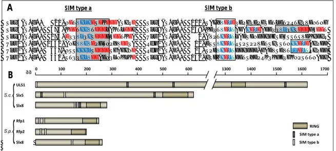
Slx5(S.c.)-- 20-NETVILIDSDKEEDASIRE Slx5(S.c.) 111-SAHYVDLDQEPGQEPGSETLETPRTIQVDNTNG Slx5(S.c.) 473-KEETIIVTDDDLAKTLEDI Slx8(S.c.) 158-KEQTVDLTADAIDLDAEEQQVLQISDDDFQEET Slx8(S.c.) 174-EQQVLQISDDDFQEETKEA Uls1(S.c.) --2-AVPTIDLTLADSDNEDIFHSFSSSTSVDKIDIR Uls1(S.c.) 1532-ANHVVIVDPFWNPYVEEQA Rfp1(S.p.) -10-ESSVIDLTRSPSPPVETSISSTNIIDLDAIPDD Uls1(S.c.) -367-NSSIIILSDEDESGAGIND Rfp2(S.p.) -15-SPEVIDLTEDIEDD-GADVSEVTLLDLTRIPEF Uls1(S.c.) -540-STQQILVDEAENQLNKLKE Slx8(S.p.) 151-ISDMIDLTDETSYDPRKQKFEQGKNPSTTNAEI
Figure 1.10: Yeast ULS proteins
A) Alignment of proved and putative SUMO interaction motifs type a (boxed in dark grey) and type b (boxed in light grey) in ULS proteins from S. cerevisiae and S. pombe (hydrophobic residues within the SIM are displayed in blue, acidic in red and threonines in green).
B) Schematic representation of yeast ULS proteins. Domains/Motifs are depicted relative to the total protein size (ULS, ubiquitin ligase for SUMO conjugates; SIM, SUMO interaction motif; aa, amino acids).
1.5.1 S. cerevisiae Slx5-Slx8 and Uls1
The S. cerevisiae ULS proteins, the RING containing proteins Slx5 (alias Hex3) and Uls1 (alias Ris1 or Dis1) have been identified as non-covalent SUMO (Smt3) interacting proteins with each harboring several SIMs (for SIM type refer to Figure 1.10; Uzunova, 2007). Slx5 was also isolated as a high-copy suppressor of a temperature-sensitive Ulp1 mutant (Xie, 2007). Slx5 forms a RING dimer with Slx8 and the complex was shown to be an active ubiquitin ligase involved in genome stability (Mullen, 2001; Ii, 2007). Slx5-Slx8 and ULS1 bind especially to HMW-SCs (Uzunova, 2007). Both Slx5-Slx8 and ULS1 target HMW-SCs for proteasomal degradation by a concerted action together with the redundant ubiquitin E2s Ubc4/Ubc5 (Uzunova, 2007). Further, an in vitro model substrate, a Rad52-SUMO fusion, was shown to be preferentially modified with ubiquitin by Slx5-Slx8 compared to Rad52 alone (Xie, 2007).
1.5.2 S. pombe Rfp1/Rfp2-Slx8
ULS proteins were also identified in S. pombe, where Slx8 forms a dimer with the redundant RING finger proteins Rfp1 and Rfp2. These are not related to Slx5 but also harbor several SIMs (see
Figure 1.10) and were found to interact with S. pombe SUMO (Pmt3; Kosoy, 2007;
Prudden, 2007; Sun, 2007). As Slx5, Rfp1 and Rfp2 lack intrinsic ubiquitin ligase activity but in a complex with Slx8 they form an active ubiquitin ligase targeting HMW-SCs and thus serve as functional homologues for Slx5. Cells lacking these complexes are sensitive to genotoxic stress and show genomic instability (Kosoy, 2007; Prudden, 2007; Sun, 2007). Rfp1-Slx8 and Rfp2-Slx8 stimulate both the in vitro ubiquitylation of a GST-SUMO
SIM type a SIM type b