A bioinformatical approach for a reliable determination of short motifs for SUMO and Atg8 interaction in Saccharomyces cerevisiae
Inaugural–Dissertation zur
Erlangung des Doktorgrades
der Mathematisch-Naturwissenschaftlichen Fakult¨at der Universit¨ at zu K¨ oln
vorgelegt von
Diplom-Chemiker Benjamin Vogt
aus Bonn
April 2014
1. Berichterstatter: Professor Dr. Kay Hofmann 2. Berichterstatter: Professor Dr. R. J¨ urgen Dohmen
Tag der m¨ undlichen Pr¨ ufung: 10. April 2014
Zusammenfassung
Regulatorische Prozesse werden durch posttranslationale Proteinmodifikation eingeleitet, die seine Aktivit¨ at, seine Stabilit¨ at, seine Lokalisation innerhalb der Zelle oder seine Interaktion mit anderen Proteinen ver¨ andern. Neben vielen anderer solcher Prozesse stellen die Dekodierung des SUMOylierungssignals oder die Erkennung von lipidgebun- denem Atg8 Ausgangspunkte f¨ ur e↵ektive Verarbeitungsrouten dar, die durch SUMO-In- teraktionsmotive (SIMs) und Atg8-Interaktionsmotive (AIMs) in Proteinsequenzen ein- geleitet werden. Ihr geringer Informationsgehalt verhindert jedoch ihre Unterscheidung von willk¨ urlich auftretenden Sequenzen. Diese Dissertation handelt von einem Detek- tionsansatz mit bioinformatischen Methoden f¨ ur bisher unbekannte SIMs und AIMs.
Der erste Teil dieser Arbeit beschreibt die bioinformatische SIM-Detektionsmethodik.
Diese ist f¨ ur alle drei SIM-Typen gleich, jedoch sind die einzelnen Suchl¨aufe auf SIMa-, SIMb- und SIMr-Charakteristiken zugeschnitten. Als Maß f¨ ur die Konservierung wurde eine informationstheoretische Metrik gew¨ ahlt und auf jeweils zwei unterschiedlich weit gefasste phylogenetische Verwandtschaftsbereiche angewendet. Eine Kombination bioin- formatischer Methoden dient zur Absch¨ atzung, ob ein Proteinbereich unstrukturiert, nicht-globul¨ ar oder globul¨ ar ist. Der vorgestellte bioinformatische Ansatz verwendet Eigenschaften zu Konservierung und strukturellem Kontext f¨ ur eine Wertungsmethodik zur Funktionalit¨ at neuer, noch unbekannter SIM-Instanzen.
Experimente zeigen SUMO-Interaktion f¨ ur Dbp10, Drs1, Rfc1, Rad18 und Tdp1 aus den bioinformatischen SIM-Detektionsdurchl¨ aufen. Dbp10 und Drs1 treten in der ri- bosomalen Biogenese im Nukleolus auf. Ein SIM ist in diesem biologischen Kontext noch nicht bekannt, obwohl SUMOylierung hier im Zusammenhang mit dem ¨ Ubergang pr¨ aribosomaler Partikel ins Nukleoplasma auftritt. Tdp1, Rfc1 und Rad18 treten im Zusammenhang mit DNS-Replikation und in der DNS-Schadensreparatur auf, in de- nen SUMOylierung als wichtiger Aktivit¨ atsfaktor f¨ ur andere Proteine bekannt ist. F¨ ur das Motiv in Rfc1 konnte im Rahmen dieser Arbeit eine Verantwortlichkeit f¨ ur SUMO- Bindung in Mutationsstudien gezeigt werden. Es verursacht auch einen beobachtbaren Wachstumsph¨ anotyp unter chemisch-induziertem DNS-Schadensstress. Das Motiv in Rad18 wurde in der Zwischenzeit auch von Parker und Ulrich identifiziert.
Im zweiten Teil dieser Arbeit wird eine Anwendung analoger Methoden f¨ ur einen bioinfor-
Abstract
Regulatory processes are initiated by posttranslational modifications of proteins which alter their activity, stability, localization or their interaction with other proteins. Among many other processes, decoding the SUMOylation signal or the recognition of lipidated Atg8 represent starting points to e↵ective downstream signalling pathways, initiated by SUMO interacting motifs (SIMs) and Atg8 interacting motifs (AIMs) in protein se- quences. The low information contents of SIMs and AIMs prevent their detection from spurious sequences. This thesis is about a detection approach for so far unknown SIMs and AIMs with bioinformatical methods.
The first part of this thesis describes the bioinformatical SIM detection method. The overall method is common for all SIM types, whereas the single SIM detection screens ap- ply to the characteristics for SIMa, SIMb and SIMr. Two sets of phylogenetic distances from budding yeast in combination with information theoretical approaches and sliding averages serve as a conservation measure. A combination of bioinformatical tools is used for an estimation whether a protein segment is unstructured, non-globular or globular.
The bioinformatical approach in this thesis uses these conservation and structural fea- tures for the evolution of a functionality scoring measure for unknown SIM instances.
Experimental interaction studies show so far unknown SUMO interaction for Dbp10, Drs1, Rfc1, Rad18 and Tdp1 from the bioinformatical SIM detection screens. Dbp10 and Drs1 are involved in ribosomal biogenesis in the nucleolus. A SIM in this biological context has not yet been reported, whereas SUMOylation is involved in the release of pre-ribosomal particles into the nucleoplasm. Tdp1, Rfc1 and Rad18 are involved in DNA replication and damage repair, where SUMOylation is a crucial activity factor for other proteins. The motif in Rfc1 identified from the bioinformatical detection screen is shown responsible for SUMO interaction in mutation studies. This motif also causes observable growth phenotype under chemically induced DNA damage stress. The motif in Rad18 was meanwhile identified by Parker and Ulrich.
The second part of this study describes the application of analogous methods for a
bioinformatical AIM detection approach. The conservation characteristics of established
AIM are found similar to those for SIMs, whereas the structural context is harder to be
represented with bioinformatical methods.
Contents
1. Introduction 1
1.1. Posttranslational protein modification . . . . 1
1.2. The ubiquitin conjugation machinery . . . . 2
1.3. The Small Ubiquitin-like Modifier in Saccharomyces cerevisiae (SUMO) . 4 1.3.1. SUMO conjugation . . . . 4
1.3.2. SUMO ligases . . . . 5
1.3.3. The SUMO interacting motif . . . . 6
1.3.4. SUMO-targeted ubiquitin ligases . . . . 9
1.3.5. DeSUMOylation . . . 11
1.4. Autophagy . . . 12
1.4.1. The Atg8 conjugation cascade . . . 13
1.4.2. The Atg8 family proteins share a common fold . . . 13
1.4.3. Substrate recognition and the Atg8 interacting motif . . . 14
1.5. Comparison between Ubiquitin, SUMO and Atg8 interaction . . . 18
1.6. Bioinformatics . . . 18
1.6.1. Short linear motifs . . . 19
2. Materials 21 2.1. Strains . . . 21
2.2. Plasmids . . . 22
2.3. Oligonucleotides . . . 23
2.4. Chemicals . . . 28
2.5. Miscellaneous equipment . . . 30
2.6. Enzymes . . . 31
2.7. Antibodies . . . 31
2.8. Media . . . 32
2.8.1. Growth media for yeast . . . 32
2.8.2. Growth media for bacteria . . . 33
2.9. Antibiotics . . . 34
Contents
2.10. Protocol . . . 34
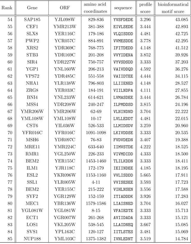
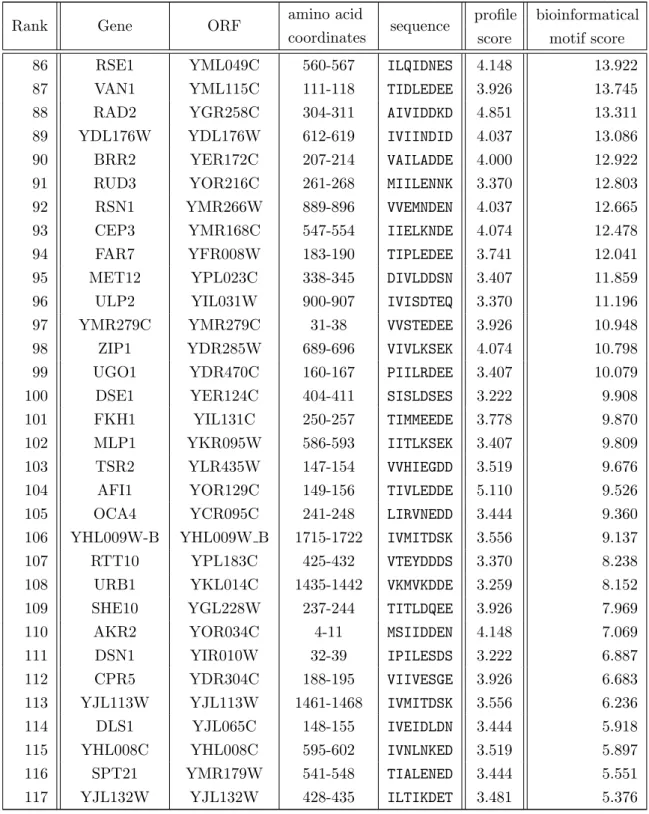
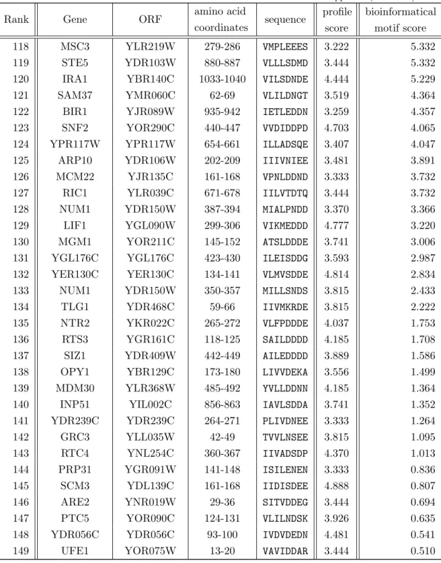
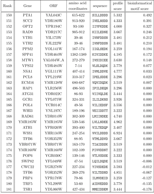
3. Methods 35 3.1. Bioinformatical Methods . . . 35
3.1.1. Protein disorder prediction . . . 35
3.1.1.1. The GlobPlot method . . . 36
3.1.1.2. The DisEMBL method . . . 37
3.1.1.3. The IUPred method . . . 38
3.1.1.4. The RONN method . . . 38
3.1.1.5. The FoldIndex method . . . 39
3.1.1.6. The FoldUnfold method . . . 39
3.1.2. Multiple sequence alignments . . . 39
3.1.2.1. Multiple sequence alignment construction and substitu- tion matrices . . . 40
3.1.2.2. Progressive alignment tools: MUSCLE . . . 41
3.1.2.3. Progressive alignment tools: MAFFT . . . 41
3.1.2.4. Dialign-TX . . . 42
3.1.2.5. ProbCons . . . 42
3.1.3. Computing sequence conservation from multiple alignments . . . . 43
3.1.3.1. Computing sequence conservation from multiple align- ments . . . 43
3.1.3.2. Information theory as mathematical background of the SeqLogo approach . . . 44
3.1.3.3. The sliding window average technique . . . 44
3.2. Genetic Techniques . . . 45
3.2.1. Preparation of plasmid DNA from Escherichia coli . . . 45
3.2.2. Quantitation of nucleic acids . . . 45
3.2.3. Generation and transformation of competent Escherichia coli cells 45 3.2.4. DNA manipulations . . . 46
3.2.5. DNA gel electrophoresis . . . 47
3.2.6. Generation and transformation of competent Saccharomyces cere- visiae . . . 47
3.2.7. Preparation of genomic DNA from Saccharomyces cerevisiae . . . 48
3.2.8. Sanger DNA sequencing . . . 48
3.2.9. The PCR epitope tagging technique . . . 48
3.2.10. The site-directed mutagenesis technique . . . 49
Contents
3.2.11. The Gateway
™cloning technique . . . 50
3.2.12. Preparation of crude extracts from Saccharomyces cerevisiae . . . 51
3.2.13. Preparation of crude extracts from Escherichia coli . . . 52
3.2.14. SDS polyacrylamide gel electrophoresis . . . 52
3.2.15. Western blot analysis . . . 54
3.2.16. The Yeast Two-Hybrid assay . . . 54
3.2.17. The GST-Pulldown assay . . . 55
3.2.18. Two-step gene replacement . . . 56
4. Results 57 4.1. General aspects for a bioinformatical prediction of SUMO interacting motifs 57 4.1.1. SUMO interacting motifs belong to one of the three consensus patterns . . . 57
4.1.2. Four criteria for evaluation of database screen results for new SUMO interacting motifs . . . 59
4.1.3. Comparison of di↵erent disorder prediction tools for a bioinfor- matical SIM detection procedure . . . 59
4.2. Generation of multiple sequence alignments . . . 61
4.2.1. Two phylogenetic ranges of species for the identification of orthologs 61 4.2.2. Choice of a suitable multiple sequence alignment tool . . . 62
4.3. Conservation criteria from multiple sequence alignments . . . 62
4.3.1. A suitable metric for assessing residue conservation . . . 62
4.3.2. Smoothing of information raw data as conservation measures . . . 65
4.4. The bioinformatical SIM detection approach . . . 68
4.4.1. Basic steps of the bioinformatical approach . . . 68
4.4.2. SIMa . . . 68
4.4.2.1. Consensus pattern and profile from established SIMa for a bioinformatical SIMa screen . . . 68
4.4.2.2. Conservation scores to discriminate between SIM-like se- quences . . . 70
4.4.2.3. GlobPlot and IUPred disorder/globularity prediction . . 71
4.4.2.4. Combination of sequence consensus, conservation thresh- olds and disorder/globularity prediction data . . . 71
4.4.3. SIMb . . . 74
4.4.3.1. Consensus pattern and profile from established motifs for
a bioinformatical SIMb screen . . . 74
Contents
4.4.3.2. Combination of sequence consensus, conservation thresh- olds and disorder/globularity prediction data . . . 76 4.4.4. SIMr . . . 78
4.4.4.1. Consensus pattern from established SIMr for a bioinfor- matical SIMr screen . . . 78 4.4.5. Selection of possible SUMO interacting proteins for validation . . . 81 4.5. Application of analogous methods for a genome-wide AIM screen in Sac-
charomyces cerevisiae . . . 82 4.6. Experimental validation of SUMO interacting motifs in Saccharomyces
cerevisiae . . . 89 4.6.1. Verification of SUMO/SIM interaction in Rfc1 using the yeast two-
hybrid technique . . . 90 4.6.2. Verification of SUMO/SIM interaction in GST-SUMO pulldown
assays . . . 94 4.6.2.1. Comparison of various lysis bu↵er compositions for best
↵-HA signal depletion in GST-SUMO pulldown assays . . 95 4.6.2.2. GST-SUMO pulldown depletes ↵-HA signals of Dbp10
and Drs1 . . . 97 4.6.2.3. GST-SUMO pulldown shows SUMO interaction for Rfc1
and Rad18 . . . 98 4.6.2.4. Strategy for SIMa mutation in Rfc1 . . . 99 4.6.3. Rfc1 SIM mutation may induce growth phenotype upon DNA
damage . . . 102 4.6.4. Verification of Rfc1 SIM/Smt3 interaction . . . 102
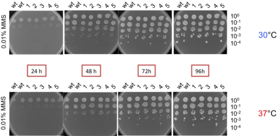
4.6.4.1. The SIMa mutation is integrated into RFC1 via two-step gene replacement . . . 102 4.6.5. Relevance of Rfc1 SIMa/SUMO interaction . . . 105 4.6.5.1. No growth phenotype upon thermal stress . . . 105 4.6.5.2. No observable growth phenotype under chemical stress:
methyl methanesulfonate . . . 106 4.6.5.3. Observable growth phenotype under chemical stress: hy-
droxy urea . . . 107 4.6.5.4. Observable growth phenotype under chemical stress: Eb-
selen . . . 109
Contents
5. Discussion 110
5.1. General aspects of bioinformatical motif discovery . . . 110
5.2. Experimental findings from the bioinformatical results . . . 111
5.3. Discussion of exemplary results . . . 112
5.3.1. Dbp10/SUMO and Drs1/SUMO interaction . . . 112
5.3.1.1. Biological context of Dbp10 and Drs1 . . . 112
5.3.1.2. Experimental validation of SUMO interaction for Dbp10 and Drs1 . . . 112
5.3.2. Tdp1/SUMO interaction . . . 113
5.3.2.1. Biological context of Tdp1 and Top1 . . . 113
5.3.2.2. Experimental validation of Tdp1/SUMO interaction . . . 114
5.3.3. Rfc1/SUMO interaction . . . 115
5.3.3.1. Biological context of Rfc1 . . . 115
5.3.3.2. Experimental validation of Rfc1/SUMO interaction . . . 115
5.3.4. Rad18/SUMO interaction . . . 117
5.3.4.1. Biological context of Rad18 . . . 117
5.3.4.2. Experimental validation of Rad18/SUMO interaction . . 118
5.4. The application of analogous methods for a bioinformatical AIM detection approach . . . 118
5.5. General aspects of the applied bioinformatical approach . . . 119
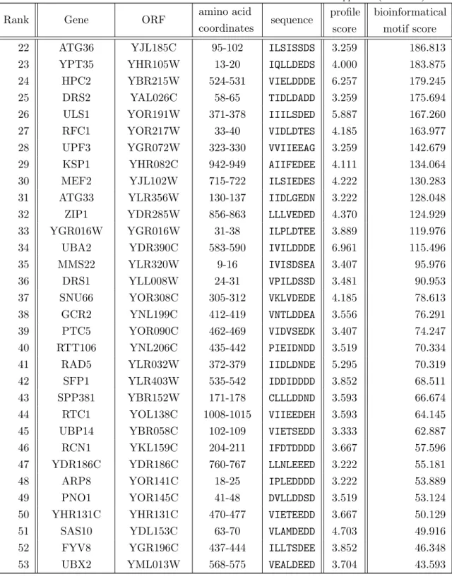
5.6. General aspects of experimental validations . . . 119 A. Appendix: Motif list from the bioinformatical SIMa detection approach 121 B. Appendix: Motif list from the bioinformatical SIMb detection approach 131 C. Appendix: Motif list from the bioinformatical SIMr detection approach 133 D. Appendix: Motif list from the bioinformatical AIM detection approach 136
References 151
Acknowledgement 199
Eidesstattliche Erkl¨ arung 200
Lebenslauf 201
List of Figures
1.1. The ubiquitin fold as a structural element for ubiquitin-like modifiers. . . 2 1.2. NMR solution structures show a functional binding site for noncovalent
SUMO/SIM interaction. . . . 7 1.3. Solution structures of the Atg8 homologs show the characteristic -grasp
fold. . . 14 1.4. Solution structures of AIM (LIR in mammals) binding to di↵erent Atg8
homologs show a common binding site. . . 16 1.5. Information theoretical SeqLogo representation and corresponding con-
sensus pattern of the Atg8 family interaction motif. . . 17 4.1. Siz1: MSA as a representation of the three-dimensional domain architec-
ture of a protein. . . 64 4.2. Gnuplot graphical representations of conservation scores from Siz1 ‘Sac-
charomycetales’ MSA. . . 65 4.3. Conservation thresholds t1, t2 and t3 allow to describe an established
SIMa. Gnuplot graphical representation of Siz1 conservation scores with employed sliding average technique. . . 66 4.4. Schematic derivation of a consensus pattern and a sequence profile from
established SIMa. . . 69 4.5. SIMs are highly conserved and lie in more variant MSA regions. . . 74 4.6. Schematic derivation of a consensus pattern and a sequence profile from
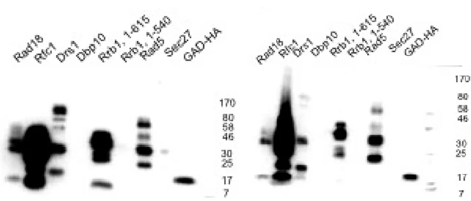
established AIM. . . 83 4.7. ↵-HA signal intensity (3F10) as a measure for protein expression level. . . 92 4.8. Rad18, Rfc1 and Tdp1 show SUMO interaction in yeast two-hybrid ex-
periments. . . 93 4.9. Glass bead lysis gives crude extracts with more specific ↵-HA signal in
western blots than from the direct boiling protocol. . . 95 4.10. Di↵erent lysis bu↵er compositions for best lysis performance for ↵-HA in
western blots. . . 96
List of Figures
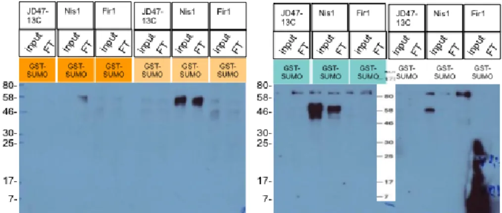
4.11. Nis1 crude extract from glass bead lysis is a good positive control. Best
↵-HA signal depletion is observed for bu↵er “green”. . . 96 4.12. Dbp10 and Drs1 show ↵-HA signal depletion upon SUMO interaction. . . 97 4.13. ↵-HA immunoblotting shows SUMO interaction for Rfc1 from ↵-HA sig-
nal depletion and for Rad18 from elution. . . 98 4.14. Three SIM-like sequences in the Rfc1 amino terminal region including the
SIMa from the bioinformatical SIMa and SIMb detection screens. . . 99 4.15. Nucleotide exchanges for selective mutation of the Rfc1 SIM-like sequences
in Saccharomyces cerevisiae. . . 100 4.16. “SIM2” VIDLDT in Rfc1 from the bioinformatical SIMa and SIMb screens
is responsible for SUMO interaction. . . 101 4.17. Strategy for a SIMa mutagenesis for RFC1 from the bioinformatical screens.
The motif is changed from VIDLDT to VIDAA. . . . 103 4.18. First step in the two-step gene replacement cloning strategy. . . 103 4.19. Vector map for the integratable shuttle vector rfc1 :pRS306. . . 104 4.20. Amplification and subsequent SalI restriction helped identify a correct
SIM mutant integrant. . . 105 4.21. No observable growth phenotype of rfc1 SIM mutant strain upon thermal
stress. . . 106 4.22. No observable growth phenotype of rfc1 SIM mutant strain upon MMS
exposure. . . 107 4.23. Observable growth phenotype of rfc1 SIM mutant strain upon exposure
to hydroxy urea. . . 108 4.24. Observable growth phenotype of rfc1 SIM mutant strain upon Ebselen
exposure. . . 109 5.1. An excerpt from the Saccharomycetales MSA of Rfc1 shows three SIM-like
sequences. . . 116
List of Tables
1.1. Sequence characteristics of established SUMO interacting proteins. . . . . 8
1.1. Sequence characteristics of established SUMO interacting proteins (con- tinued). . . . 9
1.2. SeqLogo representations and corresponding consensus patterns of the three SUMO interacting motif groups. . . . 9
1.3. Characteristics of ubiquitin-like modifiers (Ubl) and respective enzymes. . 15
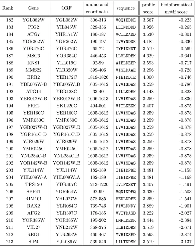
1.4. Sequence characteristics of established Atg8 interacting proteins. . . 17
2.1. Yeast strains used in this study. . . 21
2.2. Bacterial strains used in this study. . . 21
2.3. Plasmids used in this study. . . 22
2.4. Oligonucleotides used in this study. . . 23
2.4. Oligonucleotides used in this study (continued). . . 24
2.4. Oligonucleotides used in this study (continued). . . 25
2.4. Oligonucleotides used in this study (continued). . . 26
2.4. Oligonucleotides used in this study (continued). . . 27
2.4. Oligonucleotides used in this study (continued). . . 28
2.5. Chemicals used in this study. . . 28
2.5. Chemicals used in this study (continued). . . 29
2.5. Chemicals used in this study (continued). . . 30
2.6. Miscellaneous equipment used in this study. . . 30
2.6. Miscellaneous equipment used in this study (continued). . . 31
2.7. Enzymes used in this study. . . 31
2.8. Antibodies used in this study. . . 31
2.8. Antibodies used in this study (continued). . . 32
3.1. Bioinformatical web based methods used in this study for the globularity and disorder prediction of proteins. . . 36
3.2. Bioinformatical methods used in this study (miscellaneous). . . . 40
List of Tables
3.3. Web based multiple sequence alignment software used in this study. . . . 43 3.4. Typical program set-up for a Polymerase Chain Reaction (PCR). . . 46 3.5. Sanger sequencing PCR protocol using the “BigDye Terminator V.3.1”
kit from Becton-Dickinson. . . 48 3.6. BigDye
®Terminator reaction mix composition (a) and Exo/SAP clean-
up protocol (b). . . 49 3.7. The Gateway
™BP reaction mix used in this study. . . 51 3.8. The Gateway
™LR reaction mix used in this study. . . 51 3.9. Resolving gel compositions, with respect to di↵erent acrylamide contents. 53 3.10. Stacking gel, 3 % (v/v) acrylamide. . . 53 4.1. SIM sequence characteristics as criterion in a bioinformatical prediction
approach. . . 58 4.2. Consensus patterns for SIMa, SIMb and SIMr for pattern-based Saccha-
romyces cerevisiae protein sequence screens. . . 58 4.3. Four criteria for a reliable detection of relevant SUMO interacting motifs. 59 4.4. Motifs in Daxx, Prrg3 and Wss1 proteins for a performance test of bioin-
formatical disorder and globularity prediction tools. . . 60 4.5. Two sets of MSA for a characterization of SIM conservation. . . 61 4.6. The level of SIMa conservation can be calculated with substitution matrix
and SeqLogo approaches. . . 63 4.7. The sliding average technique with di↵erent window sizes (w1,w2) deter-
mines a distinct conservation threshold triplet (t1,t2,t3) for established SIMa. . . 67 4.8. Profile scores from a SIMa profile-based protein database screen returns
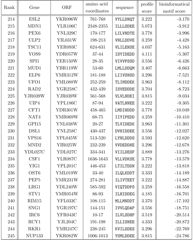
established SIMa. . . 69 4.9. E↵ects of di↵erent combinations of sequence and conservation restrictions
on the results of a genome-wide sequence search. . . 70 4.10. Final results from a genome-wide bioinformatical SIMa screen. . . 73 4.11. Final results from a genome-wide bioinformatical SIMa screen. . . 73 4.12. The sliding average technique with di↵erent window sizes (w1, w2) deter-
mines a distinct conservation threshold triplet (t1, t2, t3) for SIMb. . . 75 4.13. Comparison of results from SIMb pattern and profile scans in a Saccha-
romyces cerevisiae protein sequence collection. . . 75
4.14. Final results from a genome-wide bioinformatical SIMb screen (part I). . . 77
4.15. Final results from a genome-wide bioinformatical SIMb screen (part II). . 77
List of Tables
4.16. Genome-wide pattern-based sequence scans using three sets of conserva- tion thresholds in synergy with SIM consensus patterns. . . 78 4.17. Final results of a genome-wide bioinformatical SIMr pattern-based screen
in Saccharomyces cerevisiae (part I). . . 80 4.18. Final results of a genome-wide bioinformatical SIMr screen in Saccharo-
myces cerevisiae (part II). . . 80 4.19. Final selection of putative motifs for experimental validation from the
bioinformatical genome-wide SIM screens. . . 82 4.20. The level of AIM conservation can be calculated the same way as for SIMs
using substitution matrix and information theoretical approaches. . . 84 4.21. The sliding average technique with di↵erent window sizes (w1, w2) deter-
mines a distinct conservation threshold triplet for AIM (t1, t2, t3). . . 85 4.22. Derivation of conservation thresholds for AIM detection. . . 86 4.23. Profile scores of established AIM. A profile was derived from the estab-
lished AIM in Atg1, Atg3, Atg4, Atg19, Atg32 and Atg34. . . 86 4.24. Profile scores of established AIM. A profile was derived from established
AIM in Atg3, Atg4, Atg19, Atg32 and Atg34. . . 87 4.25. AIM consensus pattern pattern search and a AIM-based profile search. . . 87 4.26. Final results from the bioinformatical AIM screen (part I). The results
were compared to the values of established AIM. . . 88 4.27. Final results from the bioinformatical AIM screen (part II). The results
were compared to the values of established AIM. . . 88 4.28. Selection of putative SIM sequences from the previous bioinformatical
screens for experimental validation. . . 89 4.29. GAD protein fusions with their di↵erent insert sizes used in this study. . . 91 4.30. Epitope tagged proteins in full-length comprising the SIM sequence from
the bioinformatical screen used for GST pulldown assays. . . 94 A.1. List of SIMa instances from the bioinformatical SIMa detection approach. 121 A.1. List of SIMa instances from the bioinformatical SIMa detection approach
(continued). . . 122 A.1. List of SIMa instances from the bioinformatical SIMa detection approach
(continued). . . 123 A.1. List of SIMa instances from the bioinformatical SIMa detection approach
(continued). . . 124
List of Tables
A.1. List of SIMa instances from the bioinformatical SIMa detection approach (continued). . . 125 A.1. List of SIMa instances from the bioinformatical SIMa detection approach
(continued). . . 126 A.1. List of SIMa instances from the bioinformatical SIMa detection approach
(continued). . . 127 A.1. List of SIMa instances from the bioinformatical SIMa detection approach
(continued). . . 128 A.1. List of SIMa instances from the bioinformatical SIMa detection approach
(continued). . . 129 A.1. List of SIMa instances from the bioinformatical SIMa detection approach
(continued). . . 130 B.1. List of SIMb instances from the bioinformatical SIMb detection approach 131 B.1. List of SIMb instances from the bioinformatical SIMb detection approach
(continued). . . 132 C.1. List of SIMr instances from the bioinformatical SIMr detection approach. 133 C.1. List of SIMr instances from the bioinformatical SIMr detection approach
(continued). . . 134 C.1. List of SIMr instances from the bioinformatical SIMr detection approach
(continued). . . 135 D.1. List of AIM instances from the bioinformatical AIM detection approach. . 136 D.1. List of AIM instances from the bioinformatical AIM detection approach
(continued). . . 137 D.1. List of AIM instances from the bioinformatical AIM detection approach
(continued). . . 138 D.1. List of AIM instances from the bioinformatical AIM detection approach
(continued). . . 139 D.1. List of AIM instances from the bioinformatical AIM detection approach
(continued). . . 140 D.1. List of AIM instances from the bioinformatical AIM detection approach
(continued). . . 141 D.1. List of AIM instances from the bioinformatical AIM detection approach
(continued). . . 142
List of Tables
D.1. List of AIM instances from the bioinformatical AIM detection approach (continued). . . 143 D.1. List of AIM instances from the bioinformatical AIM detection approach
(continued). . . 144 D.1. List of AIM instances from the bioinformatical AIM detection approach
(continued). . . 145 D.1. List of AIM instances from the bioinformatical AIM detection approach
(continued). . . 146 D.1. List of AIM instances from the bioinformatical AIM detection approach
(continued). . . 147 D.1. List of AIM instances from the bioinformatical AIM detection approach
(continued). . . 148 D.1. List of AIM instances from the bioinformatical AIM detection approach
(continued). . . 149 D.1. List of AIM instances from the bioinformatical AIM detection approach
(continued). . . 150 D.1. List of AIM instances from the bioinformatical AIM detection approach
(continued). . . 151
1. Introduction
Cells constantly have to overcome diverse situations that endanger cellular homeostasis:
Starvation, damage to cell compartments, or just any disturbance in the sensitive bal- ance of protein levels have to be detected and responded to by the cell.
The ubiquitin proteasome system (UPS) and ubiquitin-like (Ubl) protein conjugation pathways are integral to regulated proteolysis in the cell, but they also have essential multiple nondegradative biological roles. The UPS regulates location and activity of cel- lular proteins. Their function and malfunction are important factors in various human diseases such as cancer or neurodegenerative disorders [Bedford et al., 2011, review].
Breakdown of bulk proteins, carbohydrates and lipids constantly replenishes the cell’s supply for carbon and nitrogen under starvation conditions. The detection of nonfunc- tional substrates and subsequent increase in repair mechanisms may be sufficient for the adaptation to a stress situation. Several repair pathways exist. But depending on the kind and severity of damage to the cell more drastic measures like apoptosis or senescence can be triggered to protect the tissue or the whole organism.
1.1. Posttranslational protein modification
Posttranslational modifications of proteins (PTMs) drive a variety of cellular processes in eukaryotes like regulation of cell growth, cell division, or adaptive and developmen- tal processes. Protein modifications are covalent attachments which alter its proper- ties. Most modifications make major structural contributions to their target prote- ins, as it is the case for phosphorylation, acetylation or methylation [Sartorelli et al., 1999, Ubersax and Ferrell, 2007, Polevoda and Sherman, 2007, Webster et al., 2014].
These modifications a↵ect the activity, stability, localization or the interaction of pro-
teins with other proteins. There are also other types of PTMs such as modification with
ubiquitin-like proteins (Ubls) such as ubiquitin (Ub), the small ubiquitin-like modifier
(SUMO) or the neural precursor cell-expressed, developmentally downregulated gene 8
(NEDD8). Modifications by Ubls introduce a new interaction site for proteins. Ubls
share a three-dimensional structure that resembles ubiquitin in its characteristic tightly
1. Introduction
packed globular ↵ -fold, also termed as “ -grasp”, with -sheets wrapped around an ↵-helix, whereas both primary sequence and charge surface distributions are highly diverse [Hochstrasser, 2000, Burroughs et al., 2007]. The respective Ubl structures are virtually superimposable (figure 1.1) [Bayer et al., 1998]. The evolutionary relationship of di↵erent Ubls suggest functional similarities, as all Ubls described so far are conju- gated via similar conjugation machineries. These machineries employ E1, E2 and E3 enzymes on their way onto their respective target.
(a) Ubiquitin (b) Atg8 (c) Smt3 (d) NEDD8
Figure 1.1. The ubiquitin fold as a structural element for ubiquitin-like modifiers. The - grasp fold of ubiquitin (a) is a common structural core element for Ubls such as Atg8 [1UBI], Smt3 [2KQ7]
and NEDD8 [2KO3]. Pictures were taken from the Protein Data Bank with the assigned identifiers. All structures were directly processed on the PDB web page using the “Jmol” application and the “screen shot” application. The structures were orientated in a similar conformation with the ↵-helix (bright purple) of the characteristic ubiquitin-like fold in front of the -strands (yellow).
1.2. The ubiquitin conjugation machinery
Ubiquitin is a 76-residue protein, highly conserved within eukaryotes and found in all tissues of eukaryotic organisms, only with a sequence di↵erence of three amino acids be- tween plants, yeast and mammals [Hershko and Ciechanover, 1998]. It shares an almost identical conjugation process with its homologs in other species [Kerscher et al., 2006].
Ubiquitin is first expressed in an inactive form and needs processing at its carboxy ter-
minus to expose a double glycine motif. After maturation of its C-terminus, ubiquitin is
activated by an activating enzyme (E1) which catalyzes the formation of a high-energy
thioester bond between its cysteine group and the carboxy terminus of ubiquitin [Haas
et al., 1982]. The E1 acts as a catalyst and passes the modifier via transthiolation to the
active cysteine side chain of a conjugating enzyme (E2) [Hershko et al., 1983, Hershko
and Ciechanover, 1998]. Together with a substrate-specific E3 enzyme, the E2 cataly-
ses ubiquitin transfer onto the target protein. The complexity of this posttranslational
modification through ubiquitin is even increased by the fact that ubiquitin can form dis-
tinct polyubiquitin chains via seven lysine residues (K6, K11, K27, K29, K33, K48, K63)
1. Introduction
or the N-terminus [Yeh, 2009]. This allows fine control of numerous cellular pathways:
K48-polyubiquitinated proteins are generally degraded by the 26S proteasome, while K63-polyubiquitinated proteins can target membrane proteins to the lysosomal degra- dation pathway. Whereas the chain topology is determined by the E2 enzyme, substrate specificity comes with the E3 ligases [Yeh, 2009]. In this final step, the E3 ligase pro- motes the transfer of ubiquitin onto a lysine of its substrate. There are three classes of ubiquitin E3 ligases with regard to sequence and mechanism: E3 enzymes either contain a HECT-type (homologous to E6AP carboxyl terminus), a RING-type (really interest- ing new gene) or U-box domain [Barlow et al., 1994, Huibregtse, 1995, Borden et al., 1995, Hatakeyama et al., 2001, Jiang et al., 2001, Pringa et al., 2001]. The conserved HECT domain comprises ⇠ 350 amino acids with a conserved active site cysteine and is located at the E3 carboxy terminus. RING domains do not carry a covalently bound ubiquitin but rather serve as a sca↵old to bring E2 and substrate in close proximity for transthiolation. A U-box domain forms a similar E2 binding surface as the RING do- main. It lacks zinc ions, but is able to form stabilizing hydrogen bonds and salt bridges.
The UPS pathway is initiated by the covalent attachment of ubiquitin to a target pro- tein’s amino group via the conjugation pathway [Pickart, 2001,Pickart and Eddins, 2004].
Ubiquitin is recognized by a number of pre-folded ubiquitin-binding domains (UBD)
for substrate recognition. They are classified as UBA (ubiquitin-associated motif),
UIM (ubiquitin interacting motif), UMI (UIM- and MIU-related UBD), DUIM (double-
sided UIM), MIU (motif interacting with ubiquitin, or reversely orientated UIM), CUE
(coupling of ubiquitin conjugation to endoplasmatic reticulum degradation), UEV (ubi-
quitin E2 variant), UBZ (ubiquitin-binding zinc finger) and UBM [Wilkinson et al.,
2001, Mueller and Feigon, 2002, Hofmann and Falquet, 2001, Oldham et al., 2002, Pornil-
los et al., 2002, Swanson et al., 2003, Donaldson et al., 2003, Kang et al., 2003, Prag
et al., 2003, Shih et al., 2003, Sundquist et al., 2004, Bienko et al., 2005, Penengo et al.,
2006, Hirano et al., 2006, Pinato et al., 2011, Burschowsky et al., 2011]. They all have
in common that they are composed of one or more ↵-helices binding to a hydrophobic
patch on ubiquitin around Ile44. The general process of how ubiquitin and other Ubls
get conjugated onto a substrate is well understood, however, the characteristics of their
respective recognition sites in specific substrates and therefore the target specificity of a
given modifier are still not known.
1. Introduction
1.3. The Small Ubiquitin-like Modifier in Saccharomyces cerevisiae (SUMO)
SUMO proteins are expressed in all eukaryotes, but while Saccharomyces cerevisiae, Caenorhabditis elegans and Drosophila melanogaster have one SUMO gene, organisms such as plants and vertebrates have several genes. It encodes a 101 amino acid polypep- tide with 18% sequence identity to ubiquitin. The human genome encodes four SUMOs with SUMO1-3 expressed ubiquitously and SUMO4 mainly expressed in kidney, lymph node and spleen [Guo et al., 2004]. SUMO was shown to be a reversible modifier of proteins. Its attachment, called SUMOylation, can alter the protein localization by al- tering protein interactions. SUMOylation is a modification with important functions ranging from DNA damage control to regulation of mitochondrial dynamics [Livnat- Levanon and Glickman, 2011]. The Saccharomyces cerevisiae orthologue SMT3 of the mammalian SUMO was discovered in a genetic suppressor screen for the centromeric protein Mif2 [Meluh and Koshland, 1995, Mannen et al., 1996]. Additionally, SUMO was found to be covalently attached to the RanGTPase activating protein RanGAP1 and as binding partner for human Rad51 and Rad52 [Shen et al., 1996, Matunis et al., 1996, Mahajan et al., 1997].
1.3.1. SUMO conjugation
In yeast, the activation of SUMO is carried out by the heterodimeric protein complex Aos1-Uba2, after maturation of the otherwise unfunctional SUMO to expose the C- terminal GG motif [Johnson et al., 1997, Desterro et al., 1999, Gong et al., 1999, Okuma et al., 1999, Johnson, 2004]. Aos1 (activation of Smt3) resembles the N-terminus of Uba1, the ubiquitin activating enzyme (E1), whereas Uba2 shows structural homology to the C-terminus of Uba1. This close structure/function relationship shows how strongly related the conjugation machineries of ubiquitin and SUMO are. Similar to ubiquitin, the activation of SUMO is ATP-dependent and leads to a SUMO-adenylate conjugate which is an intermediate species in the thioester formation between SUMO and the active site cysteine in Uba2 within the Aos1–Uba2 complex [Dohmen et al., 1995, Johnson et al., 1997]. Upon activation, SUMO is transferred from the Aos1–Uba2 complex to a cysteine of the essential E2 conjugating enzyme Ubc9 [Johnson et al., 1997, Desterro et al., 1997, Schwarz et al., 1998]. In this step, Ubc9 binds both SUMO and the Aos1–
Uba2 enzyme complex which places the two active cysteine residues into close proximity
for SUMO transfer [Desterro et al., 1999,Gong et al., 1999,Lee et al., 1998,Saitoh et al.,
1998, Wang et al., 2007]. Structural data suggest that the multi-protein binding surface
1. Introduction
of Ubc9 establishes contact to the KxE motif of a given substrate (with a large hydrophobic amino acid, K the SUMO target lysine residue, x a wild-card amino acid and E a glutamic acid residue) [Sampson et al., 2001, Bernier-Villamor et al., 2002].
1.3.2. SUMO ligases
Like ubiquitin, SUMO also engages E3 ligases to assist in the final step of conjugation and to promote the transfer of SUMO to a lysine residue in the substrate. The number of SUMO E3 enzymes is smaller than the number of E3s of ubiquitin. As a characteristic, the largest group of SUMO E3 ligases bears a SP–RING motif, which resembles the RING domain in ubiquitin E3 ligases [Hochstrasser, 2001]. SP–RING E3 ligases bind both their substrates and Ubc9 directly, and then position SUMO via a noncovalent SUMO interaction motif for a favorable SUMO-to-target orientation. The SP–RING ligases can be subdivided into di↵erent classes: The PIAS (protein inhibitor of activated STAT) protein family comprises a group of proteins which were initially described as negative regulators of cytokine signaling that inhibits STAT-transcription factors [Chung et al., 1997,Liu et al., 1998]. The PIAS-family with its yeast members Siz1 and Siz2 (SAP and miz-finger domain protein) and mammalian PIAS1, PIAS3 with splice variants PIASx↵, PIASx and PIASy [Johnson and Gupta, 2001, Kahyo et al., 2001, Kotaja et al., 2002, Nakagawa and Yokosawa, 2002, Nishida and Yasuda, 2002, Sachdev et al., 2001, Schmidt and M¨ uller, 2002]. Other SP–RING E3 ligases are Mms21 (methyl methanesulfonate sensitivity protein 21) and Zip3 [Roeder and Agarwal, 2000, Takahashi et al., 2001, Zhao and Blobel, 2005, Reindle et al., 2006]. Mms21 is part of an octametric Smc5–Smc6 complex essential for vegetative growth and DNA repair [Zhao and Blobel, 2005,Andrews et al., 2005, Potts and Yu, 2005]. Zip3 is part of the synapse-initiation complex [Cheng et al., 2006]. Human RanBP2 (Ran binding protein 2) is another SUMO E3 ligase, but without an SP–RING [Mahajan et al., 1997, Mahajan et al., 1998]. Its catalytic domain is located in a natively unfolded protein region assuming that the domain structure is established just upon binding to Ubc9 [Pichler et al., 2004, Reverter and Lima, 2005].
Further SUMO E3 ligases are the human Polycomb group member Pc2 (polycomb 2) [Kagey et al., 2003, Kagey et al., 2005], HDAC4 (Histone deacetylase 4) [Kirsh et al., 2002, David et al., 2002] and TOPORS (DNA topoisomerase I binding protein) [Weger et al., 2003, Weger et al., 2005].
A SUMO moiety attached to a target protein alters the protein shape and its surface charge distribution. The final e↵ects of SUMOylation on a substrate may be numerous.
SUMOylation may act as an ON-switch for interaction: only SUMOylated RanGAP1
interacts with RanBP2 and SUMOylated PCNA recruits yeast DNA helicase Srs2 to
1. Introduction
replication forks [Matunis et al., 1996,Mahajan et al., 1997,Papouli et al., 2005,Pfander et al., 2005]. SUMOylation can also be an OFF-switch for interaction as it is the case for the transcription repressor ZNF76: Its SUMO-acceptor site interferes with its binding site for the TATA-binding protein and therefore, only one interaction is possible at a time [Zheng and Yang, 2004].
1.3.3. The SUMO interacting motif
SUMOylation proceeds via covalent modification of the target protein, whereas decoding of the SUMO signal depends on noncovalent interactions. Hecker et al. suggested a - sheet of the SUMO-fold as SUMO/SIM interaction site [Hecker et al., 2006]. Minty and co-workers were the first to suggest a specific consensus pattern for SUMO interacting motifs (SIM) [Minty et al., 2000]. Minty et al. derived an 11-amino-acid stretch from sequence comparisons of the isolated proteins as an assumed SUMO-1 interaction site:
P ro Ile Ile Leu Ser Asp Ser Glu Glu Glu Glu
An “Ser–X–Ser” motif is preceded by hydrophobic residues and followed by acidic residues at its carboxy terminus. NMR studies by Song et al. drew the attention away from the serine residues in Minty’s “Ser–X–Ser” motif as core element in SUMO-1 binding. They evolved a consensus pattern focused on a hydrophobic core [Song et al., 2004]:
V al Ile
!
X V al
Ile
! V al Ile
!
This motif has been found in several proteins like the SUMO ligases PIASX and RanBP2 and acts as binding site [Song et al., 2004, Song et al., 2005]. Solution structures of a SIM peptide in M–IR2 binding to SUMO1 (figure 1.2(a)) and a SIM peptide in MCAF1 binding to SUMO3 (figure 1.2(b)) indicate the same SUMO surface for SUMO/SIM interaction [Namanja et al., 2012, Sekiyama et al., 2008]. The same surface of the - grasp fold is employed for binding in both structures.
Their structures and the works of Hannich et al. showed new SUMO interacting proteins like yeast Fir1, Nis1, Ris1 and Sap1 for a refined SIM consensus [Hannich et al., 2005]:
Lys X
3 5V al Ile
! Leu Ile
! Leu Ile
! X
30 B B B B
@ Asp Glu Asn Gln
1 C C C C A
Asp Glu
! Asp Glu
!
1. Introduction
(a) SUMO1/M-IR2 (b) SUMO3/MCAF1
Figure 1.2. NMR solution structures show a functional binding site for noncovalent SUMO/SIM interaction. SIMs bind to a surface between ↵-helix (purple) and -strand of the ubiquitin-like fold and extends to the -sheet (both yellow). Depicted here are the following published structures: (a) A structural NMR study of SUMO1 in complex of a peptide comprising the SUMO1- specific M–IR2 SUMO interacting motif (SIM)[2LAS] [Namanja et al., 2012]. (b) A solution structure derived from NMR spectroscopy shows that the MCAF1 SIM employs the same surface of the SUMO fold [2RPQ] [Sekiyama et al., 2008]. The figures show the solution structures placed in a similar orienta- tion to the observer to indicate common structural and binding characteristics. The SIM approaches the ubiquitin-like fold from a bottom right direction in a superimposable way. White arrows in the figures are pointed at the SIM. The arrow in figure (b) is drawn shorter not to cover the protein fold. All structures were taken from the Protein Database (PDB) web page using the “Jmol” application on that page. The images were taken by screen-shot from that application.
Additionally, it was reported that binding to SUMO can be performed in both sequence
orientations, giving more variability in protein topology [Reverter and Lima, 2005, Song
et al., 2005, Uzunova et al., 2007]. In 2007, Uzunova et al. performed yeast two-hybrid
experiments with Smt3 as bait, in which they confirmed Siz1, Nis1, Fir1, Slx5 and Ris1
as SUMO interacting proteins (SIPs). They established a classification of SIMs into three
types, ‘SIMa’, ‘SIMb’ and ‘SIMr’ [Uzunova et al., 2007]: A SIMa consensus sequence
is composed of four hydrophobic amino acids followed by several acidic residues. The
SIMr consensus sequence is like the one for SIMa, but with a reverse orientation of
the sequence. The SIMb consensus sequence is composed of a shorter stretch of less
variant hydrophobic residues. An acidic stretch at its carboxy terminus seems to be
conserved, but less than for the other SIM consensus sequences. Experimental data
helped to make a list of established SIMs (table 1.1). All motifs show close similarity to
one of the three consensus types. Grouping these motifs in one of the three SIM types
allows their information theoretical representation [Schneider et al., 1986, Schneider and
Stephens, 1990]. These representations are based on information theory and display the
residue variation in alignments of protein or nucleic sequences. A web-based application
is provided by SeqLogo [Crooks et al., 2004]. The information theoretical SeqLogo
approach can be applied to the list of established SIMs as another SIM representation
aside from classical consensus pattern representations to each of the three SIM types
SIMa, SIMb and SIMr (figure 1.2).
1. Introduction
Table 1.1. Sequence characteristics of established SUMO interacting proteins.
Protein Sequence motif Reference(s)
Slx5 VILIDSDK YVDLD
[Ii et al., 2007b, Xie et al., 2010]
Fir1 VILLDEDE [Hannich et al., 2005, Uzunova et al., 2007]
Nis1 IIIPDSQD [Hannich et al., 2005, Uzunova et al., 2007]
Uls1 IIILSDED TIDLT
[Hannich et al., 2005, Uzunova et al., 2007]
Sap1 LIDLT [Hannich et al., 2005]
Siz1 IIINLDSD [Johnson and Gupta, 2001, Pichler et al., 2002, Uzunova et al., 2007]
Slx8 VLQISDD [Uzunova et al., 2007, Sun et al., 2007]
Uba2 IVILDD [Johnson et al., 1997]
Rad18 DDDLQIV [Parker and Ulrich, 2012]
Elg1 QITIDD [Parnas et al., 2010]
DDLIVI DDISII
Srs2 IIVID [Pfander et al., 2005, Kolesar et al., 2012], [Armstrong et al., 2012]
RanBP2 KKPEDSPSDDDVL [Pichler et al., 2002, Hecker et al., 2006]
DDVLIV [Song et al., 2004]
PIASX VIDLT [Song et al., 2004]
PIAS1 VIDLT [Hecker et al., 2006]
PIAS2 VIDLT [Hecker et al., 2006]
PIAS3 VIDLT [Hecker et al., 2006]
PIAS4 VVDLT [Hecker et al., 2006]
TOPORS VITIDS [Weger et al., 2003]
Daxx IIVLSD [Lin et al., 2006]
MCAF1 VIDLT [Sekiyama et al., 2008]
RNF4 IELVET [Sun et al., 2007]
VVDLT VVIVDE
IVDLT
Pc2 VILLSD [Merrill et al., 2010, Yang and Sharrocks, 2010]
PML VVVISS [Minty et al., 2000]
1. Introduction
Table 1.1. Sequence characteristics of established SUMO interacting proteins (continued).
Protein Sequence motif Reference(s)
Wss1 VVILDD
VIDLT
[Biggins et al., 2001, Uzunova et al., 2007, Mullen et al., 2010]
Rfp1 VIDLT
IIDLD
[Sun et al., 2007]
Rfp2 VIDLT
LLDLT
[Sun et al., 2007]
Table 1.2. SeqLogo representations (middle) and corresponding consensus patterns (right) of the three SUMO interacting motif types a, b and r.
SIMa
0 B B B B
@ P ro
Ile Leu V al M et
1 C C C C A
0 B B
@ Ile Leu V al M et
1 C C A X
0 B B
@ Ile Leu V al M et
1 C C A
0
@ Asp Glu Ser
1 A
3
SIMb
0 B B B B
@ P ro
Ile Leu V al M et
1 C C C C A
0 B B
@ Ile Leu V al M et
1 C C
A Asp Leu T hr
SIMr
0
@ Asp Glu Ser
1 A
3
0 B B
@ Ile Leu V al M et
1 C C A X
0 B B B B
@ Ile Leu V al M et P ro
1 C C C C A
0 B B B B
@ Ile Leu V al M et P ro
1 C C C C A
1.3.4. SUMO-targeted ubiquitin ligases
Not only single SUMO moieties serve as targeting signals. Protein modification by
SUMO chains represents recognition signals to a novel class of SUMO-targeted ubiqui-
tin ligases (STUbLs) or ubiquitin ligases for SUMOylated proteins (ULS, E3-S). One
function of this class of proteases is the STUbL-mediated ubiquitination of SUMOylated
proteins as a signal for proteasomal degradation. For this purpose, STUbLs show two
1. Introduction
characteristics: a RING domain for interaction with an E2 ubiquitin-conjugating enzyme and multiple SUMO interacting motifs for SUMO binding [Uzunova et al., 2007,Tatham et al., 2008].
The human Rnf4 protein (RING finger protein 4) is so far probably the best described STUbL [Prudden et al., 2007,Sun et al., 2007,Lallemand-Breitenbach et al., 2008,Tatham et al., 2008, Weisshaar et al., 2008]. Its architecture with at least three SUMO interact- ing motifs shows clear preference for binding of SUMO chains [Tatham et al., 2008]. A RING domain allows formation of a Rnf4 homodimer and binding of a single ubiquitin- charged E2 [Liew et al., 2010, Plechanovova et al., 2011, Plechanovova et al., 2012].
Several RNF4 substrates were found: Its ubiquitin ligase activity plays a crucial role in the ubiquitination of SUMOylated PML (promyelocytic leukemia protein) and in the disruption of PML nuclear bodies (PML–NB) in cells treated with arsenic trioxide (ATO) [Lallemand-Breitenbach et al., 2008, Tatham et al., 2008, Weisshaar et al., 2008].
ATO induces PML oligomerization and an increased affinity for Ubc9 [Zhang et al., 2010]. Poly-ubiquitination directs SUMOylated PML to the 26S proteasome for degra- dation [Tatham et al., 2008, Lallemand-Breitenbach et al., 2008].
In yeast, Slx5 is part of the DNA binding heterodimer Slx5–Slx8, also known as Uls2
[Wang et al., 2006, Yang et al., 2006, Ii et al., 2007a, Xie et al., 2007b, Uzunova et al.,
2007]. This complex was also identified as STUbL from yeast two-hybrid data [Hannich
et al., 2005,Uzunova et al., 2007,Xie et al., 2007b]. Both subunits contain a RING finger
domain which was shown essential for dimer formation [Tatsuya Ii and Brill, 2007]. The
Slx5 RING finger allows binding to Slx8, the Slx8 RING finger is the active ubiqui-
tin ligase. Slx5 comprises several SIMs and is responsible for binding of SUMOylated
proteins [Tatsuya Ii and Brill, 2007]. But Slx5–Slx8 can target substrates in a SUMO-
independent manner [Xie et al., 2010]. The heterodimer is a ubiquitin ligase that links
SUMOylation to recombinational DNA repair [Ii et al., 2007b,Xie et al., 2007b]. Slx5 and
Slx8 are required for the viability of yeast cells lacking the Sgs1 DNA helicase [Kaliraman
et al., 2001, Mullen et al., 2001]. Slx5 and slx8 null mutants show slow growth, sen-
sitivity to hydroxy urea (HU) and increased rates of gross chromosomal rearrangements
and mitotic recombination [Mullen et al., 2000, Ii et al., 2007a, Xie et al., 2007b]. Slx5
and Slx8 mutations are synthetically lethal when combined with mutations in the SUMO
pathway [Wang et al., 2006]. SUMO interacting activity of Uls1 was found in a yeast
two-hybrid screen [Hannich et al., 2005, Uzunova et al., 2007]. Uls1 is a ubiquitin ligase
for SUMO conjugates. The Uls1 protein architecture comprises four SIMs in the amino
terminal half, a Swi2/Snf2-like translocase motif and a RING domain in the carboxy ter-
minal half [Dresser et al., 1997,Zhang and Buchman, 1997,Hannich et al., 2005,Uzunova
1. Introduction
et al., 2007, Cal-Bakowska et al., 2011]. The combination of SIMs and RING domain suggests putative STUbL activity. Uls1 also binds to SUMO and SUMOylated pro- teins and shows interaction with Ubc4 ubiquitin ligase E2 in pulldown assays [Uzunova et al., 2007, Tan et al., 2013]. Uls1 mutant strains accumulate high molecular weight SUMO conjugates (HMW) and display synthetic growth e↵ects [Uzunova et al., 2007].
These e↵ects are increased in uls1 slx5 or uls1 slx8 double mutants [Pan et al., 2006, Uzunova et al., 2007]. Nevertheless, ubiquitin ligase activity of Uls1 has not yet been reported.
1.3.5. DeSUMOylation
For cell cycle progression and regulation of many processes, it is essential for a cell to be
competent to remove the covalently attached SUMO from a given substrate at a given
point of time. The de-modification is carried out by SUMO-specific proteases. This class
of enzymes has two distinct functions: they C-terminally cleave the newly synthesized
SUMO precursors to expose the di-glycine motif prior to conjugation, acting as so-called
peptidases. They may also reverse SUMOylation by dissolving the SUMO isopeptide
bond to its substrate and are therefore called isopeptidases. Both peptidases and isopep-
tidases are subclasses of hydrolases cleaving amide bonds by hydrolysis. Among these
deSUMOylating enzymes are yeast Ulp1 and Ulp2 (Ubl specific protease) with a char-
acteristic conserved approximate 200 amino acid C-terminal catalytic domain which is
essential for the deSUMOylation activity [Li and Hochstrasser, 1999,Schwienhorst et al.,
2000, Mossessova and Lima, 2000]. They have six human homologs SENP1–3, SENP5–7
(sentrin-specific proteases) [Mukhopadhyay and Dasso, 2007]. Ulp1 possesses cysteinyl
proteinase activity. Additionally, Ulp1 deconjugates single SUMO moieties or SUMO
chains from the "-amino residue of the substrate. These functions are required for cell cy-
cle progression in Saccharomyces cerevisiae [Li and Hochstrasser, 1999]. Ulp1 is SUMO
specific. It lacks any sequence similarity to known ubiquitin deconjugating enzymes and
is unable to deconjugate ubiquitin-targeted substrates. Ulp2, a second deSUMOylat-
ing proteinase, was found together with SUMO in the same screen for suppressors of
a Mif2 mutation [Li and Hochstrasser, 2000]. Ulp2 was also found to have deSUMOy-
lating activity and is located in the nucleus. It is not involved in SUMO maturation,
but is required for chromosomal stability and for recovery from cell cycle checkpoint
arrest [Li and Hochstrasser, 2000, Strunnikov et al., 2001, Bachant et al., 2002, Bylebyl
et al., 2003, Felberbaum and Hochstrasser, 2008, Lee et al., 2011].
1. Introduction
1.4. Autophagy
Autophagy plays a crucial role in the maintenance of a positive energy balance upon starvation stress [Kroemer et al., 2010, Ravikumar et al., 2010]. Dysfunctions in au- tophagic pathways are therefore associated with several diseases [Levine and Kroemer, 2008, Levine et al., 2011, Mizushima et al., 2008, Mizushima and Komatsu, 2011]. Au- tophagic pathways selectively remove aggregated proteins, surplus, damaged organelles and bacterial cells [Mizushima et al., 2008, Kirkin et al., 2009b, Noda and Yoshimori, 2009]. A crucial step is the formation of internal membranes in the cytoplasm which in turn form unique organelles, called autophagosomes [Baba et al., 1994, Kirisako et al., 1999, Suzuki et al., 2001, Mizushima and Komatsu, 2011]. Autophagosomes have been suggested to emerge from ER membranes [Hayashi-Nishino et al., 2009, Hayashi- Nishino et al., 2010]. More than 30 autophagy-related genes (Atgs) are involved in autophagy [Suzuki et al., 2007, Mizushima et al., 2011]. Most Atgs are involved in the following processes:
(i) The Atg1 kinase complex and its regulators [Matsuura et al., 1997, Cheong et al., 2008, Yeh et al., 2010, Kijanska and Peter, 2013]. Complex activity is enhanced upon starvation and plays a crucial role in autophagosome formation [Kamada et al., 2000]. It is composed of Atg1, Atg11, Atg13, Atg17, Atg20, Atg24, Atg29 and Atg31. Atg1 serves as a Ser/Thr kinase. It is directly connected to the Tor signaling pathway. Starvation induces dephosphorylation of Atg13. Atg13 now has a larger binding affinity to Atg1 in- creasing Atg1 kinase complex activity [Kamada et al., 2000,Funakoshi et al., 1997,Scott et al., 2000, Cheong et al., 2008].
(ii) A ternary Atg17–Atg29–Atg31 complex associates with Atg1. This also enhances Atg1 kinase complex activity. In a second step, the Atg17–Atg29–Atg31 complex recruits Atg proteins to the preautophagosomal structure (PAS) [Suzuki et al., 2001, Kawamata et al., 2008, Cheong et al., 2008]. Whereas Atg11 and Atg17 serve as a sca↵old for PAS under nutrient-rich conditions, the Atg1–Atg13 complex associates with the Atg17–
Atg29–Atg31 complex upon starvation [Shintani et al., 2002, Kawamata et al., 2008].
(iii) The phosphatidyl-inositol 3-kinase complex (PtdIns3) in Saccharomyces cerevisiae
is Vps34 [Schu et al., 1993]. It forms a ternary Vps34–Atg6–Atg14 complex which is
targeted to the PAS, whereas the Vps34–Atg11-Vps38 complex is required for vacuole
protein sorting by the endosome [Kihara et al., 2001, Obara et al., 2006, Dove et al.,
2004]. Atg14 recruits PtdIns3 binding proteins to the PAS, including Atg18. Atg18 in
turn forms a Atg18–Atg2 complex which is responsible for Atg9 cycling between periph-
eral structures and the PAS [Noda et al., 2000, Reggiori et al., 2004, Suzuki et al., 2007].
1. Introduction
(iv) Immunoelectron microscopy data suggest a central role for Atg8 in the autophagic pathway [Kirisako et al., 1999,Kirisako et al., 2000]. Atg8 is lipidated after ubiquitin-like conjugation to phosphatidylethanolamine into the autophagic membrane, a key process in autophagosome formation [Tanida et al., 2003, Kabeya et al., 2000, Kabeya et al., 2004].
1.4.1. The Atg8 conjugation cascade
Atg8 conjugation to phosphatidylethanolamine is dependent on two ubiquitin-like con- jugation systems: Atg8 is attached to the lipid phosphatidylethanolamine (PE) via a ubiquitin-like conjugation mechanism. After proteolytic maturation cleavage by the pro- tease Atg4, modified Atg8 is activated by the E1 enzyme Atg7 [Kirisako et al., 2000,Kim et al., 2001a]. Subsequently, activated Atg8 is transferred to the E2 enzyme Atg3 and finally conjugated to the amino group of the target lipid PE. This last step is catalyzed by an unusual E3 enzyme, which is a complex of Atg16 and Atg5, covalently modified by another ubiquitin-like modifier called Atg12 [Hanada et al., 2009]. Atg12 is activated by the E1 enzyme Atg7 and its specific E2 enzyme Atg10 for its covalent conjugation to Atg5. The Atg12-modified Atg5 associates with Atg16, thus forming the E3 for the transfer of Atg8 onto lipid–PE [Hanada and Ohsumi, 2005, Hanada et al., 2007, Kuma et al., 2002].
1.4.2. The Atg8 family proteins share a common fold
Atg8 with its central role in the autophagic system is the most prominent member of the Atg. Whereas only one ATG8 gene is known in yeast, there are six Atg8 homologs in mammals: LC3A, LC3B, LC3C, GABARAP, GABARAPL1, GATE-16/GABARAPL2 [Mann and Hammarback, 1994, Wang et al., 1999, Sagiv et al., 2000, Xin et al., 2001, He et al., 2003]. Their expressed proteins are here referred to as the Atg8 family proteins.
The three-dimensional structures of Atg8 family proteins show a C-terminal ubiquitin
fold and an N-terminal helical extension (figure 1.3). [Paz et al., 2000, Stangler et al.,
2002, Sugawara et al., 2004, Noda et al., 2008, Duszenko et al., 2011]. The N-terminal
region consists of two ↵-helices and is a unique feature of Atg8 that distinguishes it from
other Ubls. All Atg8 family proteins have exposed -strands, which are responsible for
their interaction upon intermolecular -sheet expansion. They all have two hydrophobic
pockets, termed W-site (E17, I32, K48, L50, F104) and L-site (Y49, V51, P52, L55,
F60, V63) [Noda et al., 2008]. The interaction between autophagic receptors and Atg8
contributes to specific cargo selection, whereas the AIM anchors the cargo with its
1. Introduction
receptor to lipidated Atg8 [Ichimura et al., 2008, Kirkin et al., 2009a, Noda et al., 2008, Okamoto et al., 2009].
(a) Atg8 (b) LC3 (c) GABARAP
Figure 1.3. The solution structures of the Atg8 homologs show the characteristic -grasp fold. The structures were taken from the Protein Database. Yeast Atg8 (a): 2KQ7 [Schwarten et al., 2010]. Mammalian LC3 (b): 1V49 [Kouno et al., 2005]. Mammalian GABARAP (c): 1KOT [Stangler et al., 2002]. All tertiary structures are placed in a similar orientation to display the common -grasp fold with its -strands (yellow) to the back and its characteristic↵-helix (purple) to the front.
1.4.3. Substrate recognition and the Atg8 interacting motif
In autophagy, specific adaptor proteins link selective autophagy cargo to lipidated Atg8 family members and thus to the autophagy machinery. In yeast, such adaptors addi- tionally bind Atg11 for both selective autophagy and the cytoplasm-to-vacuole targeting (CVT) pathway [Yorimitsu and Klionsky, 2005]. The CVT pathway is closely related to autophagy but serves as non-conventional targeting of Ams1 (↵-mannosidase 1) and Ape1 (aminopeptidase 1) to the vacuole [Baba et al., 1997, Scott et al., 1997, Scott et al., 2001]. Atg19 and Atg34 mediate the incorporation of oligomerized structures of Ams1 and Ape1 in CVT vesicles [Watanabe et al., 2010]. Under both nutrient-rich and starvation conditions, Atg19 mediates the association of the CVT complex with the PAS via interaction with Atg8 and Atg11 [Hutchins and Klionsky, 2001, Shintani et al., 2002, Chang and Huang, 2007]. Under starvation conditions, Atg19 binding to Ams1 is no longer possible, which is then accomplished by the cargo receptor Atg34 [Watanabe et al., 2010, Suzuki et al., 2010]. The first Atg8 interacting motif (AIM) was described in the protein Atg19, which is the substrate recognition factor of the yeast CVT [Scott et al., 2001, Kim et al., 2001b]. The Atg19/Atg8 interaction could be attributed to a WEEL motif in Atg19. The Atg19 AIM adopts an extended -conformation and an intermolecular parallel -sheet with 2 of Atg8 (figure 1.4(a)) [Watanabe et al., 2010].
A similar site was found in Atg34 as an WEEI motif [Watanabe et al., 2010]. Atg11
binds cargo-receptors in an AIM-independent manner, implying that Atg8 and Atg11
use distinct binding sites in cargo receptors Atg19 and Atg34 [Okamoto et al., 2009].
1. Introduction
Table1.3.Characteristicsofubiquitin-likemodifiers(Ubl)andrespectiveenzymes.
Ub l m at u rat ion b y p rot eas e? E1 E2 E3 con ju gat ion pro duct ubiquitin by E 1 en zy m e Ub a1
a, Ub a6
bUb c1–8, Ub c10, Ub c11 HE C T
c/U- B ox
d/R ING
ed om ai n p rot ei n s Ub -t ar ge t S UM O Ul p 1, Ul p 2
fAos 1/Ub a2
gUb c9
hS iz 1, S iz 2
iS UM O -t ar ge t A tg8 A tg4
jA tg7
kA tg3
kA tg12–A tg5 · A tg16
lA tg8–P E A tg12 (n on e) A tg7
mA tg10
nA tg5
oA tg12–A tg5
p a[McGrathetal.,1991] b [Jinetal.,2007] c[Huibregtse,1995] d [Hatakeyamaetal.,2001,Jiangetal.,2001,Pringaetal.,2001,Murataetal.,2001] e[Freemontetal.,1991,Barlowetal.,1994,Bordenetal.,1995] f [MossessovaandLima,2000] g[Johnsonetal.,1997,Gongetal.,1999] h[JohnsonandBlobel,1997,Tathametal.,2005] i[JohnsonandGupta,2001,Strunnikovetal.,2001] j[Kirisakoetal.,2000] k[Ichimuraetal.,2000] l [Mizushimaetal.,1999,Mizushimaetal.,2003,Fujitaetal.,2008] m[Tanidaetal.,1999] n[Shintanietal.,1999] o [Mizushimaetal.,1998] p [Mizushimaetal.,1999,Kumaetal.,2002]1. Introduction
(a) Atg8/Atg19 (b) LC3/p62 (c) LC3B/OPTN (d) GABARAP1/NBR1
Figure 1.4. Solution structures of AIM (LIR in mammals) binding to di↵erent Atg8 ho- mologs show a common binding site. The AIM (LIR) binding to a Atg8 homolog is structurally comparable to SIM binding to SUMO. It involves the same structural element with respect to the -grasp fold, as can be seen from published structural data: Shown here are solution structures representing the interaction between Atg8/Atg19 (a, PDB 2ZPN), LC3/p62 (b, PDB 2K6Q), LC3B/OPTN (c, PDB 2LUE) and GABARAP1/NBR1 (d, PDB 2L8J). The respective structures are aligned to each other with respect to the ubiquitin-like structural core in a similar orientation with the -strands to the back (yellow) and the characteristic ↵-helix to the front. The AIM approaches that fold at a cleft between that helix and the -strands from a bottom right direction.
The Atg8 interaction of Atg19 is enhanced by concomitant binding to Atg11. There are other proteins bearing AIM. Atg32 is an outer-membrane, mitochondria-anchored recep- tor protein and bears an AIM for direct Atg8 binding [Kanki and Klionsky, 2008, Kanki et al., 2009, Okamoto et al., 2009, Aoki et al., 2011]. In mammals, a similar function is ascribed to Nix (mammalian Nip3-like protein, also known as BNIP3-like, BNIP3L) [No- vak et al., 2010,Schwarten et al., 2009]. The pexophagy receptor Atg36 is shown to bind to Atg8 and the peroxisome via Pex3 [Motley et al., 2012,Farre et al., 2013]. Motley et al.
suggested eight putative AIMs in Atg36 from sequence similarities to other AIMs, how- ever the functional AIM could not be detected so far. A FDDI motif is better conserved than the other AIM candidates and thus might be functionally most important [Motley et al., 2012].
Other autophagic receptors responsible for the recognition of specific cargo have been
described in mammals, including p62 [Bjørkøy et al., 2005, Komatsu et al., 2007, Pankiv
et al., 2007, Ichimura et al., 2008] and NBR1 in mammals [Kirkin et al., 2009a, Waters
et al., 2009]. p62 interacts with ubiquitinated proteins via its C-terminal UBA domain
and oligomerizes with other “loaded” p62 via its N-terminal PB1 domain [Wilson et al.,
2003, Moscat et al., 2007, Seibenhener et al., 2007, Saio et al., 2009, Nakamura et al.,
2010, Isogai et al., 2011]. Docking to nucleating autophagosomes is mediated by the
LC3 interacting motif (LIR in mammals, AIM in yeast) [Pankiv et al., 2007, Ichimura
et al., 2008]. NDP52 was found as an receptor for ubiquitin-coated Salmonella enter-
ica serovar Typhimurium [von Muhlinen et al., 2010, Thurston et al., 2009]. It has a
carboxy-terminal zinc finger for ubiquitin binding and an LC3 interacting motif, char-
1. Introduction
acteristics NDP52 shares with p62 and NRB1 [von Muhlinen et al., 2012]. Optineurin (OPTN) is an adaptor protein in the pathogen-induced autophagy pathway. OPTN has been shown to be an autophagy adaptor for ubiquitin-coated Salmonella enterica [Per- rin et al., 2004, Wild et al., 2011]. It interacts with LC3/GABARAP via a LIR/AIM motif (figure 1.4(c)). Interestingly, Ser-170 within this LIR is phosphorylated by TBK1, making it more acidic and thus increasing its affinity for LC3/GABARAP [Kirkin et al., 2009a, Novak et al., 2010, Behrends et al., 2010]. A consensus pattern for AIM was evolved with experimental data (table 1.4, figure 1.5) [Alemu et al., 2012].
Table 1.4. Sequence characteristics of established Atg8 interacting proteins. TheSaccharo- myces cerevisiaeprotein sequences were used for bioinformatical pattern-based and profile-based screens.
Protein Sequence Reference(s)
Atg1 YVVV [Yeh et al., 2010, Kraft et al., 2012]
Atg3 WEDL [Hanada et al., 2009, Yamaguchi et al., 2010]
Atg4 YVDI [Satoo et al., 2007, Fass et al., 2007]
Atg19 WEEL [Shintani et al., 2002, Noda et al., 2008]
Atg32 WQAI [Okamoto et al., 2009]
Atg34 WEEI [Suzuki et al., 2010]
Nix WVEL [Schwarten et al., 2009, Novak et al., 2010]
NBR1 YIII [Lamark et al., 2009, Rozenknop et al., 2011]
p62 WTHL [Pankiv et al., 2007, Ichimura et al., 2008]
Figure 1.5. Information theoretical SeqLogo representation (left) and corresponding consensus pattern of the Atg8 family interaction motif.