A role for Sum1 in HML silencing and replication initiation in Saccharomyces cerevisiae
D
ISSERTATIONzur Erlangung des akademischen Grades Doktor rerum naturalium
(Dr. rer. nat.)
im Fach Biologie eingereicht an der
Mathematisch-Naturwissenschaftlichen Fakultät I der Humboldt-Universität zu Berlin
von Diplom-Biologe
Horst Irlbacher
geb. 11.05.1971 in Schwandorf/Bay.
Präsident der Humboldt-Universität zu Berlin Prof. Dr. Jürgen Mlynek
Dekan der Mathematisch-Naturwissenschaftlichen Fakultät I Prof. Thomas Buckhout, PhD
Gutachter/-innen: 1. Prof. Dr. Harald Saumweber 2. Prof. Dr. Ann Ehrenhofer-Murray 3. Prof. Dr. Jörn Walter
Zusammenfassung Zusammenfassung
In der eukaryotischen Ontogenese ist die Etablierung differenzierungsspezifischer Genexpression eng an die Unterteilung des Genoms in funktionell getrennte Domänen gekoppelt. Solche Domänen lassen entweder erhöhte transkriptionelle Aktivität zu oder unterdrücken sie und werden Eu- bzw. Heterochromatin genannt. Heterochromatin enthält spezielle Proteine, die zur Ausbildung dieser repressiven Chromatinstruktur beitragen. Eine der Hauptfragen in der Heterochromatinbiologie ist, wie solche Proteine rekrutiert werden.
Dieser Prozess ist entscheidend damit einzelnde Regionen im Genom koordiniert zeit- und ortsabhängig reprimiert werden können. In Saccharomyces cerevisiae entsteht Hetero- chromatin an den silent-mating-type Loci HMRa und HMLα durch die zielgerichtete Rekrutierung des Sir-Komplexes über eine Gruppe von Proteinen, die an sogenannte silencer- DNA Sequenzen binden. In diese Arbeit wird gezeigt, daß das Protein Sum1, bisher bekannt als Repressor meiotischer Gene im vegetativen Zellzyklus, als Heterochromatin- Rekrutierungsfaktor für HMLα fungiert. Sum1 konnte in vitro und in vivo an HMLα über ein funktionelles Element innerhalb des HML-E silencers binden und die Deletion von SUM1 verursachte einen Verlust von Repression an HMLα.
SUM1 beeinflußte außerdem die Fähigkeit von HML-E als Replikationsstartpunkt (origin) zu agieren, was eine Rolle von Sum1 in der Replikation nahelegt. Die Beobachtung, daß orc2-1 und orc5-1 mit sum1∆ synthetisch lethal waren und daß cdc6-1, cdc7-1 oder cdc45-1 mit sum1∆ einen synthetischen Wachstumsdefekt aufwiesen unterstützt die Vermutung, daß SUM1 eine globale Rolle in der Replikationsinitiation besitzt. In einer genomweiten Suche wurden ARS Elemente gefunden, die sowohl Sum1 als auch ORC rekrutieren. Dabei konnte gezeigt werden, daß die Replikationsaktivität dieser ARS Elemente von Sum1 bzw. Sum1 Bindungsstellen abhängig war. Als Repressor von meiosespezifischen Genen interagiert Sum1 oft mit der Histondeacetylase Hst1. In diesem Zusammenhang konnte gezeigt werden, daß SUM1-regulierte origins ebenfalls HST1 zur vollen Aktivität benötigten.
Zusammenfassend schlagen wir Sum1 als neuartigen Modulator für die Replikationsinitiation an einer Untergruppe chromosomaler Replikationsstartpunkte vor.
Schlagworte:
Chromatin, Silencing, Replikationsstartpunkt, Replikation, ARS, Sum1, Hst1
Abstract
Abstract
The division of eukaryotic chromatin into functionally distinct domains is critical to implement gene expression programs that drive the development of multicellular organisms.
Regions termed euchromatin exist in the genome that are generally conducive to transcription, whereas heterochromatin contains specialized chromatin binding proteins that repress transcription in these regions. A central question in heterochromatin biology is how the heterochromatin factors are targeted to specific genomic regions, a process that is crucial to ensure that the designated domains, and only they, are repressed in the appropriate spatial and temporal fashion. In Saccharomyces cerevisiae heterochromatinization at the silent mating- type loci HMRa and HMLα is achieved by targeting the Sir complex to these regions via a set of anchor proteins that bind to the silencers. Here, we have identified a novel heterochromatin targeting factor for HMLα, the protein Sum1, a repressor of meiotic genes during vegetative growth. Sum1 bound both in vitro and in vivo to HMLα via a functional element within the HML-E silencer, and deletion of SUM1 caused HMLα derepression. Significantly SUM1 was also required for origin activity of HML-E, suggesting a role of Sum1 in replication initiation.
Our observations of a synthetic lethality between orc2-1 or orc5-1 and sum1∆ as well as a synthetic growth defect of cdc6-1, cdc7-1 and cdc45-1 with sum1∆ support the notion that SUM1 has a global role in replication initiation. In a genome-wide search for Sum1-regulated origins, we identified a set of autonomous replicative sequences (ARS elements) that bound both the origin recognition complex and Sum1. Full initiation activity of these origins required Sum1, and their origin activity was decreased upon removal of the Sum1 binding site. In its role as a repressor of meiosis specific genes, Sum1 often works in concert with the histone deacetylase Hst1. We found that SUM1-regulated origins also required HST1 for full activity. Taken together we propose that Sum1 is a novel replication initiation modulator for a subset of chromosomal origins.
Keywords:
Chromatin, silencing, origin, replication, ARS, Sum1, Hst1
Contents
Contents
1 Introduction ... 3
1.1 Epigenetics and regulation of gene expression ... 3
1.2 Chromatin and gene expression ... 4
1.3 Chromatin composition ... 5
1.4 Mechanisms that alter chromatin properties ... 6
1.4.1 Chromatin remodeling... 6
1.4.2 Chromatin modifications... 7
1.5 Silencing in Saccharomyces cerevisiae... 9
1.5.1 Silencing at the HM loci... 10
1.5.2 Silencing at the telomeres and the rDNA locus ... 13
1.6 Replication initiation ... 14
1.6.1 Origins of replication ... 15
1.6.2 Events during replication initiation ... 17
1.7 Regulation of replication initiation ... 19
1.8 Proteins investigated in this work ... 22
1.8.1 Sum1... 22
1.8.2 Hst1 and Rfm1 ... 25
1.9 Outline of this thesis... 27
2 Materials and Methods ... 29
2.1 E.coli strains ... 29
2.2 Growth conditions and media ... 29
2.3 Yeast strain construction ... 29
2.4 Yeast strains ... 30
2.5 Plasmid construction ... 32
2.6 Silencing assays... 34
2.7 Plasmid loss assay ... 34
2.8 Yeast extracts for Western blotting... 35
2.9 SDS PAGE and Immunoblotting ... 35
2.10 Co-Immunoprecipitation ... 35
2.11 Electrophoretic mobility shift assays (EMSA)... 36
2.12 Chromatin immunoprecipitations... 37
2.13 In vivo replication-origin assay ... 37
Contents
3 Results ... 39
3.1 Definition of a core region within the D element... 39
3.2 Genetic interaction of SUM1 and the D element... 41
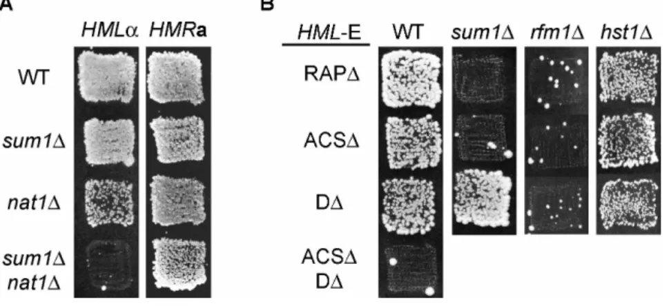
3.2.1 sum1∆ caused HMLα derepression... 41
3.2.2 sum1∆ was epistatic to the D element... 42
3.2.3 SUM1 dependent HMLα silencing was independent of HST1 and RFM1... 43
3.3 Sum1 bound specifically to the D element within HML-E ... 44
3.3.1 In vitro binding of Sum1 to HML-E... 44
3.3.2 In vivo localization of Sum1 at HML-E ... 46
3.4 sum1∆ decreased origin function of HML-E... 48
3.5 sum1∆ interacted genetically with orc mutations, cdc6-1, cdc7-1 and cdc45-1... 49
3.6 Sum1 was a replication initiation factor for several origins of replication ... 51
3.6.1 Identification of genomic sites of combined Sum1 and ORC binding ... 51
3.6.2 Sum1 influenced origin function of ARS1013... 54
3.6.3 Sum1 binding sites controlled origin function of ARS1013 ... 55
3.6.4 Sum1 affected the chromosomal origin activity of ARS1013 ... 56
3.6.5 Sum1 influenced origin function of selected origins ... 57
3.6.6 Sum1 affected origin function at chromosomes... 58
3.7 Hst1 affected Sum1-modulated replication origins... 60
4 Discussion... 63
4.1 Sum1 in silencing ... 63
4.2 Sum1 in replication initiation... 65
4.3 Sum1 as a cell programm-dependent replication initiator?... 69
5 Appendix ... 73
The D-element silencer screen in the “silencing cassette”- plasmid... 73
6 References ... 76
Abbreviations... 94
Curriculum Vitae ... 95
Publications... 96
Acknowledgements... 97
Introduction
1 Introduction
The following work addresses a molecular link between two major cellular processes:
heterochromatin formation and replication initiation. Since these processes are connected to chromatin organisation, an introductory chapter will focus on chromatin biology. Moreover, the establishment of heterochromatin will be reviewed, with a particular focus on current knowledge of silencing in Saccharomyces cerevisiae. The connection of heterochromatin formation and replication initiation is reflected by the fact that numerous proteins are involved in both processes. Therefore, a second part of this introduction discusses principles of replication and factors involved in this process.
1.1 Epigenetics and regulation of gene expression
Tight control of gene expression is of pivotal importance during the life of a cell. Besides housekeeping genes that are constantly expressed, various genes are only used at specific stages of the cell cycle or under particular environmental conditions. Also, during the process of differentiation in higher organisms, expression and repression of only the appropriate subset of genes at a given time or cell type is fundamental. To accomplish this, mechanisms have evolved to switch genes on and off. Organisms have developed a large set of proteins that bind to DNA in a sequence-specific manner and positively or negatively influence transcription. These proteins are called transcription factors or transcriptional repressors.
However, besides the dynamic properties of transcriptional regulation, daughter cells of a specific cell type can also “remember” the expression pattern of their mother cells. This is of particular importance in highly specialized cell types. The process of inheriting a specific gene expression pattern without changing the genomic sequence is called epigenetics (reviewed in (Hendrich and Willard, 1995)). Epigenetic mechanisms influence a broad spectrum of cellular processes ranging from gene expression control to replication origin choice in metazoa or gene silencing in eukaryotes. Gene silencing can be distinguished from promoter-specific gene repression in that it acts in a regional and gene independent manner. It leads to transcriptional inactivation of whole chromosomal areas by densely packing chromatin to inhibit access for DNA binding proteins or factors of the transcriptional
Introduction machinery. Therefore chromatin plays an important role in determining the transcriptional status of a gene.
1.2 Chromatin and gene expression
Chromatin has traditionally been divided into two main classes based on structural and functional criteria. Euchromatin contains the majority of the genes, both actively transcribed and quiescent. Heterochomatin is transcriptionally silent and contains large regions of repetitive DNA sequence (reviewed in (Grewal and Moazed, 2003)). It was first described in light microscopic studies of moss nuclei as the part of chromatin that remains condensed throughout the cell cycle (Heitz, 1928). Coexistence of heterochromatin and transcriptional inactivation is observed in polytene chromosomes in salivary glands of Drosophila melanogaster. These structures consist of more than 1000 identical chromosomes that align and form a giant chromosome. Examination by light microscopy shows a pattern of bands and also lateral “puffs” on the chromosomes that are associated with high transcriptional activity.
After transcription of the genes within the puffed domains these areas are again compacted into bands (reviewed in (Zhimulev, et al., 2004)).
Transcriptional repression by heterochromatin is sequence-independent, which means that localization of a gene within the genome is as important for its expression as the composition of its promoter elements. One prominent example for a locus specific effect for gene expression is a phenomenon called position effect variegation (PEV) in D.melanogaster (Muller, 1930). The white+ (w+) gene encodes a factor responsible for red color development in the facets of the eye. Red-white mosaic phenotypes are caused by a chromosomal position effect in which an X-ray induced rearrangement breakpoint placed the w+ gene from its normal euchromatic location to the vicinity of heterochromatin. Some rearrangements lead to heterochromatin-mediated silencing of this gene resulting in the occurrence of large patches of red and white facets in the adult eyes. This pattern of variegation suggests that a decision to express or repress the w+ gene is made early during tissue developement and is maintained in a metastable state through multiple cell divisions (reviewed in (Wakimoto, 1998)).
The phenomenon of transcriptional silencing of genes during development is not restricted to dipteria but found in an increasing number of other organisms including humans. For example in mammalian females one X-chromosome is transcriptionally silenced. Somatic cells of females contain two X-chromosomes, while male cells contain one X and one Y
Introduction chromosome. Female cells compensate the extra X-chromosome dose by inactivating one of them which happens in early embryonic development. The inactivated X-chromosome can be seen during interphase as a distinct structure called Barr body and most of its DNA is not transcribed (reviewed in (Chow and Brown, 2003)).
Generally, centromeres and telomeres as well as many intergenic regions in mammalian genomes consist of constitutive heterochromatin (Perrod and Gasser, 2003). All these areas contain a large number of highly repetitive DNA and heterochromatinization is thought to inhibit recombination events. Also repeated gene arrays such as the genes coding for ribosomal RNAs (rDNA) and transposons are subject to transcriptional silencing (Walsh, et al., 1998). Presumably 90% of the mammalian genome is transcriptionally silent in differentiated cells (Perrod and Gasser, 2003). Part of it is constitutive heterochromatin whereas the rest is assigned to differentiation specific events.
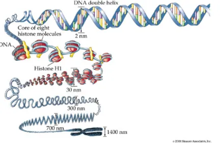
Figure 1.1: Schematic representation of various DNA compaction levels. Details are described in the text. Image adapted from Sinauer Associates, 2001.
1.3 Chromatin composition
Genome sizes vary greatly among eukaryotic organisms. For example the genome of the bakers yeast Saccharomyces cerevisiae consists of ~12.5 million basepairs (bp), whereas the whisk fern (Psilotum nudum) has its genomic information stored in 2.5x10¹¹ bp which is 20.000 times the genome size of S.cerevisiae. Since DNA is a linear molecule, a prime
Introduction question is how it can be packed in a space-saving way but be concurrently kept flexible enough to allow recombination, replication, transcription or repair. Given the human genome with more than 6.4 billion basepairs (bp) per diploid cell, some 2 meters of DNA molecule have to fit into each cell nucleus. To accomplish this, different levels of DNA compaction have evolved. DNA in eukaryotes does not exist as a naked molecule but is organized within an array of proteins, predominantly histones, into a structure called chromatin. The basic structural unit of chromatin is the nucleosome. 146 bp of DNA are wrapped in 1.75 turns around an octamer of histone proteins which consists of two copies each of histones H2A, H2B, H3 and H4 (Luger, et al., 1997).
Histones are small basic proteins of 103 to 230 amino acids and are highly conserved throughout the eukaryotic world. A nucleosome is present every 200±40 bp and because of its electron microscopic appearance this structure is termed “beads on a string”. However there are several higher-order compaction levels. Arrays of nucleosomes are proposed to fold into a fiber of 30 nm diameter upon incorporation of the linker histone H1. Chromatin which is not actively transcribed exists predominantly in the condensed 30 nm fiber form, whereas actively transcribed chromatin is thought to assume the “beads on a string” form. Higher order compaction levels include association of loops of the 30 nm fiber to a scaffold of non-histone proteins (Paulson and Laemmli, 1977). Folding of this scaffold into a helix and further packing of this helical structure produces the highly condensed structure characteristic of metaphase chromosomes (Fig. 1.1). Therefore accessibility of the genetic information is necessarily connected to mechanisms that either work on chromatin or are influenced by chromatin. Two main enzymatic activities can be distinguished that regulate chromatin access: chromatin remodeling complexes that help to move or remove nucleosomes, and chromatin modifying complexes that modify histones.
1.4 Mechanisms that alter chromatin properties
1.4.1 Chromatin remodeling
Biochemical and genetic experiments showed that nucleosomes are repressive for transcription (Laybourn and Kadonaga, 1991; Workman, et al., 1991). Since promoter elements and other transcriptional enhancers on the DNA level can be occupied by
Introduction nucleosomes, several mechanisms have evolved that provide access to DNA without biochemically modifying structural components of chromatin. Factors involved are called chromatin remodeling factors and are defined by their ability to either move or remove nucleosomes along a particular DNA sequence or to create a state of altered histone-DNA interaction (reviewed in (Becker and Horz, 2002) and (Sif, 2004)). Principally, all chromatin remodeling complexes use the energy of ATP hydrolysis to loosen the contact between DNA and histones. The first identified chromatin remodeler was the SWI/SNF complex of S.cerevisiae (Hirschhorn, et al., 1992). Nucleosome displacement by Swi2/Snf2 occurs by sliding or tracking nucleosomes along the DNA (Whitehouse, et al., 1999). How this is carried out from a mechanistic point of view is not entirely clear but the RSC complex (remodels the structure of chromatin), a close Swi2/Snf2 homologue, is thought to break DNA–histone contacts, generating a ‘wave of DNA’ that propagates around the nucleosome (Saha, et al., 2002). Besides their roles in transcriptional control there is strong evidence that they are also involved in replication, repair, and recombination and can interact with histone acetyltransferases, histone deacetylases or histone methyltransferases that biochemically modify chromatin.
1.4.2 Chromatin modifications
Chromatin modifications can occur either on DNA or on histones in their chromosomal context. Some of these modifications are associated with transcriptional activation whereas others lead to transcriptional repression. The fact that some modifications are reversible creates an additional layer of flexibility beyond the DNA sequence level. DNA methylation directly acts on DNA and is widely conserved among eukaryotes. Methylation of cytosine residues within CpG islands on gene promoters is a primary epigenetic event that acts to suppress gene expression (reviewed in (Bird, 2002)). DNA methylation accounts for the specific repression of genes in differentiated cells but also for the stable silencing of transposable elements (Lippman, et al., 2004).
Other chromatin modifying events target histones and include acetylation, methylation, phosphorylation, ubiquitination, sumoylation and ADP-ribosylation. Histones consist of a globular core domain and N- or C-terminal tails. When assembled into nucleosomes, the histone tails protrude unordered from the nucleosome, exposing 20-35 residues (Luger, et al., 1997). Histone tails as well as the histone core are subject to a large number of
Introduction posttranslational modifications. It seems likely that nearly every histone residue that is accessible to solvent may be a target for posttranslational modification.
A universal epigenetic mark in eukaryotic genomes is the acetylation of lysine residues at the ε-NH3+ position of the side chain. It is carried out by protein complexes called histone acetyltransferases (HATs). HATs can either act genomewide or on a local basis which is also dependent on factors that recruit these enzymes to the histones.
Histone acetylation is a reversible process and accordingly histone deacetylases (HDACs) have been isolated. Like histone acetyltransferases, HDACs have different specificities and can act either globally or locally. For example in S.cerevisiae Rpd3 deacetylates histones on a genomewide basis (Vogelauer, et al., 2000) thereby deacetylating lysine residues of H3 and H4 N-termini, whereas Sir2 is only found at transcriptionally silent regions and specifically removes acetylgroups of H4 K16, thus antagonizing the HAT Sas2 which acetylates the same residue (Suka, et al., 2002). Homologues of Sir2, the Hst proteins Hst1-4 (homologue of Sir two) have been identified and described (Brachmann, et al., 1995; Derbyshire, et al., 1996).
Hst1 and Hst2 are active histone deacetylases and share substrate specificity with Sir2 (Sutton, et al., 2001). Interestingly, overexpression of Hst1 can partially restore HMRa silencing in a sir2∆ strain (Brachmann, et al., 1995). Hst1 is even able to totally restore HMRa silencing in this strain background when targeted to HMR-E (Rusche and Rine, 2001;
Sutton, et al., 2001). So far, wild-type Hst1 has been found to act in transcriptional repression of meiotic genes during mitosis (this will be discussed in more detail in chapter 1.8.2).
Generally, hyperacetylated histones are associated with transcriptionally active chromatin whereas hypoacetylated histones correlate with heterochromatic regions. The process of acetylation and deacetylation is highly dynamic and some of these modifying complexes antagonize each other in a steady state equilibrium. For instance Sir2 acts at telomeric histones, whereas Sas2 targets them outside the telomeric region. Disruption of the Sir2/Sas2 equilibrium leads to either spreading of K16 deacetylation telomere-distal or spreading of K16 acetylation telomere-proximal (Kimura, et al., 2002; Suka, et al., 2002).
Histone acetylation may also affect DNA replication since the human MYST family HAT HBO1 interacts with the replication licencing factor Mcm2 (Burke, et al., 2001) and with human Orc1 (Iizuka and Stillman, 1999). Likewise, histone deacetylation has been implicated in negative regulation of replication. For example, in yeast the deletion of the HDAC Rpd3 leads to early activation of late origins (Aparicio, et al., 2004; Vogelauer, et al., 2002). Also,
Introduction deletion of the heterochromatin associated HDAC Sir2 can positively influence the activity of a subset of replication origins (Pappas, et al., 2004).
Another important histone modification is histone methylation which can occur both on the tails and the core of the protein. Lysine and arginine residues are targets of histone methyl transferases (HMT) and a variety of these modification have been implicated in transcription control. For example methylation of K4 and K36 at histone H3 is associated with transcriptional activity, whereas methylation of K9 at histone H3 is involved in the formation of stable repressive heterochromatin (Peters, et al., 2001). Notably the ε-NH3+ group of lysine can be either mono-, di- or trimethylated, adding an additional layer of complexity to control processes. Histone methylation is also reversible (Cuthbert, et al., 2004; Shi, et al., 2004;
Wang, et al., 2004) and therefore similar dynamics in this type of modification are expected as with histone acetylation.
Interestingly histone modifications do not occur independently but can influence each other.
Modification of one specific amino acid residue can induce or inhibit other histone modifications either in the immediate vicinity or on neighboring histones (Lo, et al., 2001;
Ng, et al., 2002). In addition, other events like chromatin remodeling are controlled by modifications on histones. For example the activity of Chd1, part of the SAGA and SLIK chromatin remodeling complexes, is dependent on methylation of K4 at histone H3 (Pray- Grant, et al., 2005). The growing number of histone modifications that influence important cellular processes has lead to the proposal of a “histone code”, which may have universal regulative function (Jenuwein and Allis, 2001).
1.5 Silencing in Saccharomyces cerevisiae
The yeast Saccharomyces cerevisiae has been one of the prime model organisms to study heterochromatin for many years. Heterochromatic regions are found in three domains of the yeast genome consisting of the two silent mating type loci HMLα and HMRa, the telomeres and the rRNA encoding DNA (rDNA). These domains have many features in common with heterochromatin of higher organisms as seen in position effect variegation in Drosophila or X chromosome inactivation in humans. In this respect silenced areas of the yeast genome are replicated late in S-phase, contain hypoacetylated nucleosomes and are generally restrictive to DNA modifying enzymes. Also numerous proteins involved in heterochromatin formation of
Introduction yeast have homologues in higher eukaryotes that are involved in the same processes.
Silencing in yeast has a function in regulating the expression of genes and moreover, it protects repetitive sequences as found at the telomeres and the rDNA locus from homologous recombination. While the involvement of the Sir2 protein in the process is common to all three silenced regions, the mechanisms of recruitment are different.
1.5.1 Silencing at the HM loci
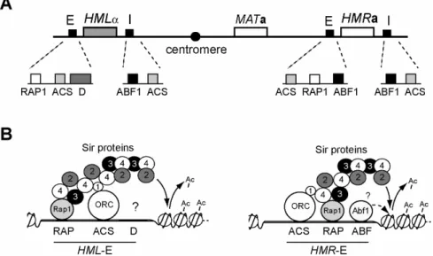
The HM loci are part of the mating type determination system of S.cerevisiae. Yeast cells can either assume the a or the α mating type and only haploids of different mating types are able to mate and form diploids. The mating type is determined by alternative alleles of a single locus located close to the center of chromosome III called the mating type locus (MAT) (Fig.
1.2A). The MATα allele determines cells of the α mating type and the MATa allele gives rise to cells of the a mating type. Both of these alleles contain genes for regulatory proteins that control the expression of factors which specify the functional differences between the two cell types (reviewed in (Herskowitz, et al., 1992)). In naturally occuring S.cerevisiae populations individual cells can interconvert their mating type from a to α or vice versa as frequently as once per generation in a gene conversion event catalyzed by the protein HO endonuclease. In laboratory strains, this conversion is abolished due to deletion of this endonuclease. Two additional copies of mating type information, the cryptic mating type loci, serve as donors during the HO endonuclease mating type conversion and are located on the same chromosome close to the telomeres (Fig. 1.2A). Situated left of the centromere is HML (homothallic mating left) that carries α mating type information, while HMR (homothallic mating right) is located to the right of the centromere and contains a mating type information.
Since simultaneous expression of opposite mating type factors is a cellular signal for diploidy and thus sterility, both HM loci are transcriptionally silenced and embedded in a heterochromatic region.
A number of trans-acting proteins as well as cis-acting regulatory DNA sequences called silencers that flank the HM loci are responsible for heterochromatin formation. In deletion experiments on HMRa, loss of the upstream silencer lead to complete derepression of HMRa, whereas loss of the downstream silencer had no apparent effect on silencing but sensitized it for additional mutations. Therefore, the former was termed E for essential and the latter I for important (Brand, et al., 1985) (Fig. 1.2A). In contrast, either the E- or the I-silencer at HMLα
Introduction are able to achieve silencing in the absence of the other (Mahoney and Broach, 1989).
Silencers are about 150bp in length and contain protein binding sites for the proteins Rap1 (repressor-activator protein 1), Abf1 (autonomous replicative sequence (ARS) binding factor 1) and ORC (origin recognition complex). While Rap1 and Abf1 binding site occurrence and composition is variable, ORC binding sites are present at each silencer (Fig. 1.2A).
Interestingly, each of these proteins has distinct roles in the cell elsewhere, Rap1 and Abf1 being general transcription factors (Planta, et al., 1995) and ORC being a protein complex important for replication initiation (reviewed in (Bell and Dutta, 2002)). At the silencers their function is to serve as anchor sites for the superordinate Sir-family proteins which ultimately create heterochromatin across the region.
Fig. 1.2: Schematic representation of mating-type loci and silencers
(A) Localization of the MAT locus and the silenced HM loci on chromosome III. Each of the HM loci is flanked by one E and one I silencer which is composed of binding sites for ORC, Rap1 and Abf1. (B) Nucleation of heterochromatin at HML-E and HMR-E. Sir-proteins bind the anchor proteins at the silencer and spread across the region thereby creating heterochromatin.
The composition of binding sites within the silencers has been well studied in the past years.
However, a sequence of approximately 100bp termed the D element in close vicinity to the Rap1 and ORC binding site was identified at the HML-E silencer, but has not been molecularly characterized (Mahoney, et al., 1991) (Fig. 1.2A). Natural silencers are redundant in function and therefore, deletion of any one of the binding sites still allows repression, whereas deletion of any two of these sites abolishes silencing (Mahoney and Broach, 1989).
Introduction At HML-E the D element showed the same redundancy upon deletion with simultaneous deletion of the Rap1 or the ORC binding site in the absence of HML-I (Mahoney, et al., 1991). Therefore a binding site for another, yet unidentified silencing protein might be contained within the D element of HML-E. The identification of this factor was the goal of this thesis.
One interesting issue is how the binding sites for three factors with independent roles in the cell can create a silencer element. The observation of a redundant function within silencers lead to the current view that while each of these proteins has some affinity for one or more Sir proteins only the close juxtapostion of two or three of these factors can create a sufficiently high local concentration of Sir proteins to sustain silencing. Consistent with this model, arrays of multiple Rap1 binding sites recruit Sir proteins to telomeres (Cockell, et al., 1995; Hecht, et al., 1996) and can create artificial silencers (Stavenhagen and Zakian, 1994).
The Sir proteins are the major effectors of heterochromatin formation. Sir3 and Sir4 (and to a limited extent Sir1) consitute structural components of heterochromatin while Sir2 contributes to the process via its activity as an NAD+ (nicotin adenine dinucleotide) dependent histone deacetylase. Sir2 targets and deacetylates K9 and 14 of H3 and K16 of H4 in vitro (Imai, et al., 2000) and Sir2, Sir3 and Sir4 are essential for the silencing process (Rine and Herskowitz, 1987). Sir1 is not essential but aids in the establishment of silenced regions in that it acts as a bridging factor between ORC and Sir4 (Triolo and Sternglanz, 1996). The establishment of a silenced region is thought to occur in a stepwise process of Sir protein polymerization across the region (Hoppe, et al., 2002) (Fig. 1.2B). The anchor proteins Rap1, Abf1 and ORC are bound to the silencers and assemble the nucleation sites. Sir2 and Sir4 form heterodimers and bind to them via interaction of Sir4 with Sir1 and Rap1. Sir3 can bind the silencers independently via interactions with Rap1 and Sir4. Sir2 which is now localized to the immediate vicinity of nucleosomes starts to deacetylate lysine residues at the tails of histones H3 and H4. Exposure of unacetylated histone tails leads to binding of Sir3 and Sir4 thus recruiting more Sir2/Sir4 complex to this locus (Rusche, et al., 2002). These processes of ongoing deacetylation and polymerization are completed when the Sir proteins emanating from both silencers meet. Although important for the establishment of silenced HM loci the role of the Abf1 binding sites at any of the HM silencers except HML-E is not clear since so far Abf1 was not found to interact with any of the other proteins implicated in silencing.
However, Abf1 has the ability to alter chromatin organization (Venditti, et al., 1994) and
Introduction could therefore aid in silencing by allowing easier loading of other silencing proteins (Miyake, et al., 2002).
Heterochromatin nucleating from the silencers generally spreads to both sides across the region. To limit it to the designated areas, boundary elements have evolved that block this spreading. For example actively transcribed genes can constitute such a barrier (Bi, 2002;
Donze, et al., 1999) since mutations in the respective promoter region or in polymerase factors abolish this barrier function. This could be due to parts of a stably bound polymerase complex forming a physical obstacle to the spreading of heterochromatin. Also the enzymatic activity of HATs or chromatin remodeling factors at the promoter can counteract the propagation of histone deacetylation (Oki, et al., 2004; Suka, et al., 2002). However, spreading of heterochromatin can also be unidirectional as it is the case at the HML-I silencer which causes heterochromatin to spread only in direction of the α-genes (Bi, et al., 1999).
Interestingly all silencers can also confer autonomous replication to plasmids but only HMR-E and -I are true chromosomal origins (Dubey, et al., 1991). This discrepancy is explained by the fact that the HML silencers, although capable of initiating replication, are passively replicated by an early initiating replication origin in the vicinity (Sharma, et al., 2001). ORC and to a lesser extent Abf1 and Rap1 binding are responsible for the bimodular ability of the silencers to nucleate silencing and to initiate replication (Fox, et al., 1995).
1.5.2 Silencing at the telomeres and the rDNA locus
Due to the fact that telomeres constitute the extreme end of the chromosome and contain a single stranded DNA overhang, they are subject to degradation or fusion with telomeres of other chromosomes. However, this is prevented by the creation of heterochromatin in these domains. The mechanistics of heterochromatin formation are similar to those of the HM loci.
The telomere ends consist of tandem C1-3A/TG1-3 repeats that are free of nucleosomes and are bound by Rap1 at 10-20 copies per telomere (Gilson, et al., 1993; Wright, et al., 1992). Here, it is the multitude of Rap1 copies that constitutes anchor sites for the recruitment of the Sir2/Sir4 heterodimer and Sir3, which ultimately leads to the same concerted event of histone deacetylation and Sir complex spreading as described for the HM loci (Luo, et al., 2002).
Thereby heterochromatin can spread up to 3kb inwards and silence reporter genes inserted at telomere proximal locations. Telomeric C1-3A/TG1-3 repeats are also able to silence reporter genes when placed elsewhere in the genome although a higher repeat copy number is
Introduction necessary (Stavenhagen and Zakian, 1994). This is due to the fact that natural telomeres contain an additional silencing anchor, an ORC and an Abf1 binding site within a sequence of
~500bp called the CoreX. This region is located subtelomerically as part of a larger X repeat element which is present at most telomeres. Silencing at natural telomeres is discontinuous and is enhanced around subtelomeric CoreX elements. Since Rap1 and ORC are in close proximity at the HM silencers to establish stable silencing, a current hypothesis for telomeric silencing is that Rap1-Sir clusters contact the ORC-Sir complex at CoreX by forming a foldback loop structure (Strahl-Bolsinger, et al., 1997).
Silencing at rDNA is different from that of telomeres and the HM loci. The S.cerevisiae locus coding for ribosomal RNA (rDNA locus) consists of a 9.1kb sequence that is repeated 100 to 200 times (Petes and Botstein, 1977). Each rDNA repeat encodes 35S rDNA, which is the precursor to the 25S, 18S, and 5.8S rRNA and is transcribed by polymerase I, and the 5S rRNA which is transcribed by polymerase III. These two genes are separated by the nontranscribed spacers NTS1 and NTS2. Only a fraction of the rDNA genes are transcribed at a given time and the majority remains silenced by the action of Sir2 (Smith and Boeke, 1997).
Sir2 in this context does not act via Sir4 in but is part of the nucleolar RENT (regulator of nucleolar silencing and telophase) complex (Straight, et al., 1999). RENT also contains Net1, which is required for association of Sir2 with the rDNA, and Cdc14, a phosphatase required for mitotic exit. However the mechanistic aspects of rDNA silencing are still unclear.
1.6 Replication initiation
In S.cerevisiae and Drosophila a shared feature between replication initiation and some types of heterochromatin formation is the fact that both require ORC binding at their nucleation sites (Foss, et al., 1993; Huang, et al., 1998; Pak, et al., 1997). Moreover, ORC is essential for replication initiation in all eukaryotes studied to date (Bell and Dutta, 2002). This chapter gives a general overview on this topic with a particular interest in origins of replication as nucleation sites for replication. The first part addresses the composition of replication origins in yeast and metazoa (chapter 1.6.1) while the second part aims to outline the events that occur at the origin during the time of replication initiation (chapter 1.6.2).
Introduction
1.6.1 Origins of replication
Origins of replication as starting points for DNA replication are scattered across the genome in eukaryotes. The origin number varies from about 300-400 in Saccharomyces cerevisiae (Raghuraman, et al., 2001; Wyrick, et al., 2001) to an estimated ten thousand in humans (Gilbert, 2001). While in S. cerevisiae a DNA consensus sequence (the ORC binding site) specifies origins of replication, to date no specific DNA sequence could be assigned to general origin function in any of the other eukaryotes tested.
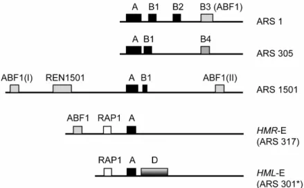
An origin of budding yeast spans a sequence of 150 to 200 base pairs and is able to support autonomous replication of plasmids. These autonomously replicating sequences (ARS) do not share obvious homology to each other except for an essential consensus sequence of 11 basepairs (WTTTAYRTTTW). This sequence called the A element or the ARS consensus sequence (ACS) is essential but not sufficient for full origin function (Celniker, et al., 1984).
Notably, an expanded 17 basepair ACS (EACS: WWWWTTTAYRTTTWGTT) has been described that more effectively predicts in vivo ARS elements (Theis and Newlon, 1997). The A element is bound by the origin recognition complex (ORC) in a sequence specific manner (Diffley and Cocker, 1992). Flanking the A element there are one or more B elements which are important but not essential for ARS activity (Marahrens and Stillman, 1992). ARS1, possibly the best studied yeast origin contains three B elements (Marahrens and Stillman, 1992) (Fig. 1.3). The B1 element is closest to the ACS and cooperates in ORC binding (Lee and Bell, 1997). B2 is required for loading of the MCM (minichromosome maintenance) complex (Wilmes and Bell, 2002; Zou and Stillman, 2000), and B3 is bound by Abf1 (Diffley and Stillman, 1988). The B2 element often overlaps with DNA unwinding elements (DUEs) that are presumably melted during replication initiation (Matsumoto and Ishimi, 1994). Abf1 is an accessory factor for origin function at a subset of chromosomal replication origins (Eisenberg, et al., 1988; Rhode, et al., 1992). Abf1 sites are found in several origins, and in three of the four HM silencers (Kimmerly, et al., 1988).
Analysis of other yeast ARS elements suggest that many origins may share at least the conserved A and B1 elements that form the ORC binding site (Fig. 1.3). Apart from that, additional B elements are very variable in size and location. Also highly individual sequence elements have been found like the REN1501 enhancer, an ARS element present at ARS1501 which is important for full origin function (Raychaudhuri, et al., 1997).
Introduction
Fig. 1.3: Comparison of ARS elements in S.cerevisiae
ARS 1 as described by (Marahrens and Stillman, 1992); ARS305 (Huang and Kowalski, 1996), the B4 element is of unknown function; ARS1501 (Raychaudhuri, et al., 1997); HMR-E (ARS317) an inefficient ARS (Rivier and Rine, 1992); HML-E (ARS301) (Mahoney, et al., 1991).
Asterisk: ARS301 does not initiate replication in its chromosomal context.
In budding yeast prediction of ARS sites on a DNA sequence level is difficult since the occurrence of an ACS alone is not sufficient for ARS activity and B elements do not share sequence homology. There are more than 10 000 matches of the ACS in the yeast genome, but only 300-400 among them serve as an ARS site (Breier, et al., 2004). One posibility to identify new origins is to test the origin activity of an ARS containing DNA sequence in individual experiments (Fangman and Brewer, 1991; Stinchcomb, et al., 1979). However, the majority of origins has been identified based on prediction methods that addressed additional hallmarks for origin activity. One large scale experiment searched for simultaneous in vivo presence of ORC and the MCM complex because these two complexes are present on active origins (see chapter 1.6.2) (Wyrick, et al., 2001). In another study the genomewide replication timing profile of S.cerevisiae was created using density transfer experiments. Regions that doubled their DNA sequences earlier than the surrounding area were presumed to be in vivo origins of replication (Raghuraman, et al., 2001). Although these two approaches greatly advanced the knowledge of origin location, more work is necessary to clarify why particular ACS containing sequences are origins and others are not. A number of recent studies suggest that in addition to an ARS sequence many more factors affect the activity of an origin (see also chapter 1.7).
Introduction In other eukaryotes ARS sequences are less well defined. While origins in fission yeast are loosely determined by their high A-T content (Clyne and Kelly, 1995; Okuno, et al., 1999), metazoan origins cannot be identified on DNA sequence level. Instead of that in metazoans sites of replication initiation can appear both in small defined sequence areas (Toledo, et al., 1998) or large initiation zones of 10 to 50kb (Dijkwel, et al., 2002). Still the question remains why the origins in more complex eukaryotes are not specified by a certain sequence element.
An explanation may come from the fact that origins are generally distributed in non- transcribed intergenic regions, most probably to avoid undesired interference of the replication and the transcription machinery. In budding yeast with its heavily transcribed genome there would be a selective pressure for origins to locate to nontranscribed regions. An advantage to target replication initiation to those regions would therefore be the evolution of specific DNA sequences as markers for nucleation sites of replication (Brewer, 1994).
Metazoans with significantly larger genomes but unproportionally more untranscribed regions would not need such a specific sequence since a stochastic distribution of nucleation sites would still lead to sufficient origin spacing. This hyphothesis can also explain the findings of distinct initiation loci and initiation zones in metazoans. Most solitary origin sites have been identified within loci containing multiple genes (Abdurashidova, et al., 2000; Aladjem, et al., 1998; Toledo, et al., 1998). By contrast, broad initiation zones consisting of multiple inefficient origins are observed at loci where there are large intergenic regions (Dijkwel, et al., 2002; Ina, et al., 2001; Little, et al., 1993).
1.6.2 Events during replication initiation
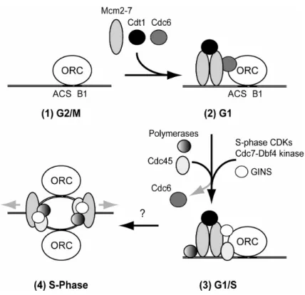
Principally every origin has the potential to initiate replication once per cell cycle. In vivo only a subset of origins initiates per S-phase. This is because some origins can assemble proteins important for initiation faster than others. They are called early origins and replication forks emanating from them migrate across sites containing later origins before these have a chance to fire. The creation of an active origin can be divided into four different phases (Fig. 1.4):
The first is the binding of ORC to the ACS within an origin (Fig. 1.4 (1)) (Bell and Stillman, 1992). In S.cerevisiae ORC remains bound to the origins throughout the cell cycle probably to mark them as sites of replication initiation (Aparicio, et al., 1997; Diffley, et al., 1994).
Introduction
Fig. 1.4: Different phases of replication initiation. Details are described in the text.
In the second phase, ORC promotes assembly of a multiprotein complex called the pre- replicative complex (pre-RC) at origins (Fig. 1.2, (2)) (Diffley, et al., 1994). Pre-RC formation starts during G1 phase and requires minimally ORC, Cdc6, Cdt1 and the MCM (Mcm2-7) complex (Bell and Dutta, 2002). The pre-RCs license origins to initiate replication once per cell cycle and disassemble after the origin has started replicating in S phase. During pre-RC formation Cdc6 directly interacts with ORC and is required together with Cdt1 to recruit the MCM complex, a putative DNA helicase (Bell and Dutta, 2002; Lei and Tye, 2001).
In the third phase, additional proteins join the pre-RC immediately prior to replication initiation. These include Cdc45, the recently described four subunit GINS complex and the replicative DNA polymerases (Fig. 1.2, (3)) (Takayama, et al., 2003). Cyclin-dependent kinase (CDK) activity is required for Cdc45 to associate with the pre-RC (Zou, et al., 1997), and the association of Cdc45 with origins coincides with their time of activation - late origins recruit Cdc45 later in S-phae than earlier origins (Aparicio, et al., 1999). The Cdc7-Dbf4 kinase also associates with chromatin just prior to S-phase and interacts with ORC (Duncker, et al., 2002; Weinreich and Stillman, 1999). The following activation of the putative MCM
Introduction helicase and the Polα-primase involves at least 20 polypeptides that must assembly coordinately within the chromatin context of hundreds of origins throughout the S.cerevisiae genome.
The fourth phase, origin initiation, signals the beginning of S-phase, and involves localized DNA unwinding at the origin and the start of DNA polymerization. The mechanism of origin unwinding and the initiation of DNA synthesis are poorly understood in eukaryotes and might occur simultaneously with the association of some of the factors outlined in phase three. As the replication starts, Cdc45 and MCM become associated with replication forks. To avoid that origins initiate replication repeatedly within one S-phase, ORC, the MCM complex and Cdc6 are prevented from re-establihing pre-RCs. This is achieved by the activity of CDK, which is upregulated during S and G2/M phase (Drury, et al., 2000; Labib, et al., 1999;
Nguyen, et al., 2001). Therefore CDK has the dual role to activate the pre-RC by promoting the binding of Cdc45 and simultaneously prevent new pre-RC assembly until the next G1 phase.
In summary, replication initiation at a given origin is a complex process requiring multiple proteins binding in several steps. It is conceivable that each individual step could be enhanced or inhibited by particular chromatin environments.
1.7 Regulation of replication initiation
There are many more factors besides origin structure that determine origin activity. Therefore the efficiency and timing of replication initiation for a given origin is always a sum of these factors that influence each other. The following chapter gives an overview on events that have been implicated in the regulation of replication initiation:
Chromosome localization
In yeast a number of observations suggest that the localization of an origin within the chromosome is important for its activity. For example placing a large 15kb fragment containing the late initiating ARS501 to a plasmid renders it to an early active origin (Ferguson, et al., 1991). Conversely, placing the effective and early initiating origin ARS1 near ARS501 on the chromosome causes ARS1 to fire as late as ARS501. Interestingly, ARS501 is located 27kb away from the right telomere of chromosome V, suggesting that
Introduction telomeres can exert a negative position effect on origin activity. In fact, whole genome replication timing experiments revealed that origins located proximal to telomeres (~40kb or less) are often activated later in S-phase (Raghuraman, et al., 2001).
However, not only the position relative to a telomere but also to other chromosomal regions determine the time of origin activity. For example a cluster of late initiating origins at chromosome XIV (ARS1411-ARS1414) is located more than 150kb away from telomeres.
ARS1412 and ARS1413 sites function as early origins on plasmids but a considerable amount of surrounding DNA is necessary to recapitulate the late origin activation time (Friedman, et al., 1996). This indicates that large chromosomal areas distinct from the ARS sites are required to determine different activation times.
Chromatin structure
The telomere binding Ku protein complex (Ku) was shown to control the late activation of ARS501 and other telomere proximal origins (Cosgrove, et al., 2002). Ku is required for the telomere position effect (TPE) in S.cerevisiae (Mishra and Shore, 1999) but the effect of Ku on ARS501 firing is independent of Sir proteins (Cosgrove, et al., 2002). Thus, a specialized chromatin domain established by Ku might be responsible for modulating the timing of origins over a long distance. One role of Ku is to tether telomeres to the nuclear envelope (Laroche, et al., 1998) and perinuclear position appears to correlate with late replication in mammalian cells (Heun, et al., 2001). This lead to the theory that Ku influences the activity of origins by locating them in a nuclear compartment containing chromatin modifying factors that can establish a late-activation domain (Gilbert, 2001).
The Sir protein complex also affects origin activity by establishing a specialized chromatin structure. The telomeric X elements contain an inactive ARS and serve along with telomeric repeats as nucleation sites for Sir dependent heterochromatin formation at the telomeres (see chapter 1.5.2). Deletion of Sir3 revealed that telomeric sequence of chromosome V replicates earlier because of earlier initiation of a nearby ARS and activation of the inactive X element ARS (Stevenson and Gottschling, 1999).
However the regulation of origin activity by the Sir proteins appears not only to be restricted to regions that are repressed by Sir-mediated heterochromatin. A recent study found that deletion of the HDAC Sir2 rescued the general replication defect of a cdc6-4 mutation (Pappas, et al., 2004). This could be caused by a loss of deacetylase activity around certain
Introduction origins. Interestingly, direct targeting of the HAT Gcn5 to the late initiating origin ARS1412 could significantly advance its timing of initiation (Vogelauer, et al., 2002).
Also deletion of the Rpd3, a global histone deacetylase, advanced replication of the entire genome (Vogelauer, et al., 2002). This was caused by a preferential advancement of late firing origins that also showed an increased acetylation status. It will be interesting to learn whether acetylation of origin regions coincides generally with a facilitated assembly of active origins.
Nucleosome positioning
While the influence of nucleosome movement and positioning in transcriptional regulation is well established, a picture is emerging that these events are also involved in the regulation of origin activity (Lipford and Bell, 2001; Simpson, 1990; Thoma, et al., 1984). At ARS1 the binding of Abf1 and ORC results in a nucleosome free zone across the origin and mutations in the Abf1 binding site can lead to nucleosome repositioning and reduced origin function (Venditti, et al., 1994). Similarly, forced movement of ARS1 into the central core of a nucleosome significantly reduced its origin activity (Simpson, 1990). Interestingly, if a nucleosome that normally constitutes one boundary of ARS1 is missing, origin function of ARS1 is also impaired. However, not the ability of ORC but of the MCM complex (Mcm2-7) to bind the origin is compromised. This points towards the influence of nucleosome positioning in events like late pre-RC formation or replication elongation (Lipford and Bell, 2001).
Origin activity is not only dependent on the close binding of nucleosome positioning factors but can also be regulated by factors binding to DNA within a distance of several hundred basepaires. For example the presence of multiple binding sites for the protein Mcm1 in a domain up to 600bp upstream of the telomeric ARS120 was shown to be important for full origin activity (Chang, et al., 2004). Since the binding of Mcm1 induces a 66° bend in the DNA (Acton, et al., 1997; West and Sharrocks, 1999) a theory is that multiple Mcm1 molecules in this domain induce a loop-like tertiary structure that could help to exclude nucleosomes from the area thus providing an environment for pre-RC assembly. Alternatively this tertiary complex could limit the region where ORC can bind to enhance its specificity for the ACS. Other DNA binding factors also regulate origin activity. For example stability of
Introduction plasmids carrying the ARS HMR-E as sole origin is compromised if a binding site for Rap1 within HMR-E is mutated (Kimmerly, et al., 1988).
Given the importance of the correct position of a nucleosome, protein complexes that can move or remove nucleosomes might also play a role in origin activity. Several chromatin remodelling factors like the chromatin accessibility complex (CHRAC) in Drosophila or SWI/SNF of S.cerevisiae and humans have been implicated in origin activity (Alexiadis, et al., 1998; Flanagan and Peterson, 1999). For example the human SWI/SNF complex interacts with the human papilloma virus E1 replication protein and is involved in efficient replication of papilloma virus DNA (Lee, et al., 1999). Also mutations in the budding yeast SWI/SNF remodeling complex compromises plasmid stability of plasmids carrying some origins (Flanagan and Peterson, 1999). Interestingly, while ARS1 activity is unaffected in this SWI/SNF mutant, a deletion of its Abf1 binding site renders this ARS1 dependent on SWI/SNF suggesting that Abf1 and SWI/SNF have redundant roles in the modulation of origin activity.
1.8 Proteins investigated in this work
The following two chapters provide information on the proteins Sum1, Hst1 and Rfm1 that had been the focus of this study. Since Sum1 is implicated in multiple cellular roles, more detailed sub-chapters will individually refer to these roles.
1.8.1 Sum1 Sum1 in silencing
More than a decade before the wild-type function of the protein Sum1 was described, a mutant allele of SUM1, SUM1-1 puzzled researchers in the field of transcriptional silencing.
In a screen for mutant suppressors of a loss of function allele of SIR2, a strong suppressing candidate allele was isolated. Since the screen was initially carried out with a SIR2 mutant called MAR1-1, the identified allele received the name SUM1 (suppressor of mar) (Klar, et al., 1985). Silencing at the HM loci can be disrupted or compromised by mutating or deleting any member of the SIR family genes (Fig. 1.5A). Surprisingly the identified mutant allele SUM1- 1 was able to reestablish silencing of both HM loci in any sir mutant (Klar, et al., 1985;
Laurenson and Rine, 1991). It also suppressed several other mutations that impair HM locus
Introduction repression like mutations in HHF2, a gene for histone H4 or deletional weakening of the HMR-E silencer. Interestingly the ability of SUM1-1 to repress HMLα in sir2∆ was decreased by 100fold in comparison to HMRa and SUM1-1 required either HMR-E or -I for full suppression of HMRa (Laurenson and Rine, 1991; Sutton, et al., 2001). SUM1-1 was also not able to overcome the telomeric derepression effect that occurs in a sir2∆ strain or upon introduction of the rap1-17 mutant allele, while wild-type repression of telomeric silencing could be slightly improved (Chi and Shore, 1996).
Fig. 1.5: Sum1-1 can establish silencing at HMRa independently of the Sir-proteins
(A) Schematic representation of Sir mediated silencing of HMRa. (B) Schematic representation of Sir-independent silencing of Sum1-1 via Rfm1 and the HDAC Hst1.
Also notable was the finding that SUM1-1 mutants exhibit reduced viability and a significantly increased chromosome loss rate compared to wild-type cells. Cloning of the SUM1-1 gene revealed a single missense mutation at codon 974 in the predicted 1062 aa of the gene SUM1, which resulted in a threonine-to-isoleucine change and was responsible for the dominant SUM1-1 suppressor phenotype (Chi and Shore, 1996). It was later found that Sum1-1 as well as Sum1 recruit the Sir2 homologue Hst1 (homologue of Sir two) via a bridging factor Rfm1 (repression factor of MSEs) (McCord, et al., 2003; Rusche and Rine, 2001; Sutton, et al., 2001; Xie, et al., 1999). In addition Sum1-1 considerably enhanced an interaction with ORC at the silencer and therefore recruited the HDAC Hst1 to the silencer. It was hypothezised that the strong ORC-Sum1-1 interaction enabled Hst1 to deacetylate
Introduction histone tails at HMRa analogous to Sir2. Since Hst1 as well as Sum1-1 were not only found at the silencer but also across the entire HMRa locus it was assumed that these proteins were able to establish an alternative repressive structure that could spread along a chromosome like the Sir-protein complex (Rusche and Rine, 2001; Sutton, et al., 2001) (Fig. 1.5B). Interaction of Sum1-1 and ORC was probably via the N-terminus of Orc1 since a mutant allele of Orc1 that misses the 235 N-terminal amino acids eliminated the ability of Sum1-1 to silence the HM loci (Rusche and Rine, 2001).
So far, wild-type Sum1 has not been implicated in silencing since overexpression of SUM1 did not lead to the SUM1-1 phenotype and this phenotype was decreased if SUM1 was coexpressed from plasmids. Therefore it was hypothesized that the SUM1-1 mutation was not of increased function (hypermorphic) but rather of new function (neomorphic) (Chi and Shore, 1996). Additional investigations revealed that Sum1 was localized to the nucleus and was neither essential for normal growth nor transcriptional repression at the telomeres or the HM loci. In sum1∆ strains only a very mild derepression effect was observed at HMRa in a sensitized assay where the ADE2 gene replaced HMRa and the E silencer was compromised by deletion of the ACS or the Abf1 binding site (Chi and Shore, 1996).
Sum1 in gene specific repression and meiosis
Although the mutant allele SUM1-1 was a repressor of HM silencing defects, the wild-type gene product appeared not to be required for silencing. Instead, Sum1 was found to be a repressor for a number of middle sporulation specific genes during vegetative growth and in the early and late phase of sporulation (Lindgren, et al., 2000; Pak and Segall, 2002; Pak and Segall, 2002; Pierce, et al., 2003; Xie, et al., 1999). Sporulation specific genes can broadly be divided into early, middle or late categories based on the timing of their expression (Mitchell, 1994). In this respect middle sporulation specific genes are expressed as cells exit prophase, enter the nuclear division and assemble spores. More than 150 genes are induced around that time (Chu, et al., 1998). Activation of these genes is carried out by the Ndt80 transcription factor that binds to a conserved sequence termed the middle sporulation element (MSE) found in many middle sporulation gene promoters (Chu, et al., 1998; Ozsarac, et al., 1997). The MSEs of a subset of these genes is also bound by Sum1 during vegetative growth and in the early and late stages of meiosis which leads to their repression. This repression is brought about through Sum1´s recruitment of the histone deacetylase Hst1 (Xie, et al., 1999).
Introduction Comparison of sequence requirements for MSE binding revealed very similar but distinct consensus sequences for Sum1 (DSYGWCAYWDW) and Ndt80 (VNDNCRCAAW), so Ndt80 binding sites can be contained within Sum1 binding sites. Also subtle basepair changes within the MSE can alter the individual affinities of Sum1 or Ndt80 to the respective MSE site (Pierce, et al., 2003). Notably only a subset of middle sporulation genes was derepressed in sum1∆ strains, and some sporulation unrelated genes were affected (Pierce, et al., 2003).
However, Sum1-repressed middle sporulation genes are further divided into two subclasses:
one class whose repression is also dependent on Hst1 and Rfm1 and the other which is only Sum1 dependent (McCord, et al., 2003). The reason why not all MSE containing genes were derepressed in a sum1∆ strain is probably because some Sum1 repressed genes require additional activation by the meiosis specific Ndt80 transcription factor while others are not a target of Sum1. Also at some genes additional regulatory sequences such as URS1 (upstream regulatory sequence 1) are present. URS1 is mitotically bound by the Ume6-Rpd3-Sin3 repressor complex that prevents expression in a sum1∆ strain (Kadosh and Struhl, 1997; Pak and Segall, 2002). This, and the fact that Ndt80 itself contains MSE sites which are partly targets of Sum1, points towards a carefully controlled execution programm for meiotic genes (Pak and Segall, 2002).
1.8.2 Hst1 and Rfm1
All eukaryotic species examined to date have multiple homologues of Sir two (HSTs), which share a highly conserved globular core domain. In S.cerevisiae the family of HST proteins consists of four members HST1 to HST4 (Brachmann, et al., 1995; Derbyshire, et al., 1996).
Yeast Hst1 is the closest relative to Sir2, showing 63% overall identity and 82% identity in the conserved core. Like Sir2 it exhibits NAD+ dependent deacetylase activity on K16 of histone H4 (Sutton, et al., 2001). Disruption of HST1 has no phenotype regarding mechanisms in which SIR2 has a role, namely, regional silencing of HMLα (Brachmann, et al., 1995) or in rDNA recombination (Derbyshire, et al., 1996). However HST1 overexpression can partially restore HMRa silencing in a sir2∆ strain (Brachmann, et al., 1995). The other HST members have varying silencing phenotypes. For example the cytosolic HST2 which also shows NAD+
dependent deacetylase activity improves rDNA silencing but reduces telomeric silencing when overexpressed, probably by competing with Sir2 for a limiting ligand (Perrod, et al.,
Introduction 2001). HST3 and HST4 double mutants are defective in telomeric silencing and chromosome maintenance (Brachmann, et al., 1995).
Cellular Hst1 occurs in two different complexes: in the SET3 complex (Set3C) and in complex with Rfm1 and Sum1 (Pijnappel, et al., 2001; Xie, et al., 1999). Set3C consists of Set3, Snt1, YIL112w, Sif2, Cpr1 and two histone deacetylases, Hos2 and Hst1. It acts as a meiosis specific repressor for a set of genes that are expressed at the early middle- and middle stage of meiosis (Chu, et al., 1998; Pijnappel, et al., 2001). The repressive properties of Set3C are dependent on Hos1 and Hst1 but Hst1 is only weakly associated with Set3C (Pijnappel, et al., 2001). The second complex is the Hst1-Rfm1-Sum1 complex. As pointed out above, Hst1 almost exclusively interacts with Sum1 through Rfm1 (McCord, et al., 2003). It represses many but not all of the middle sporulation genes that are bound by Sum1 during vegetative growth (Xie, et al., 1999). Hst1 is also the enzymatically active component responsible for histone deacetylation and silencing at the HM loci in the SUM1-1 mutant (Rusche and Rine, 2001; Sutton, et al., 2001).
Interestingly, Hst1 as an NAD+ dependent deacetylase is also involved in biosynthesis and maintenance of cellular NAD+ levels. Hst1 has a relatively low affinity to NAD+ and represses via Sum1 key factors for the de novo synthesis of NAD+. When NAD+ levels decrease, the repressive properties of Hst1 diminish and repression at these genes is abrogated so that NAD+ biosynthesis can take place (Bedalov, et al., 2003). By this simple feedback loop maintenance of particular cellular NAD+ levels are ensured.
Introduction
1.9 Outline of this thesis
Silencers flanking the HM loci are among the determinants for the establishment of a transcriptionally silent domain across these regions. It has been shown that they consist of a set of binding sites for proteins that in turn can recruit the Sir-family proteins to this loci. Sir proteins are subsequently able to polymerize across the region, thereby deacetylating histone tails and creating heterochromatin. Thus, silencers provide the targeting sites for heterochromatin nucleation.
While the HMR-E silencer had been thoroughly mapped in the past, one sequence element within the HML-E silencer escaped molecular characterization. Deletional studies at HML-E had discovered a Rap1, an ORC binding site and a sequence of 93bp termed the D element.
Deletion of each of these sites individually lead to minor derepression of HMLα . Deletion of any combination of two of these sites however resulted in total loss of HMLα silencing (Mahoney, et al., 1991). This lead to the assumption, that the D element contained a binding site for a yet unidentified silencing protein.
The aim of this study was to identify a protein that binds the D element. Specifically, we found a minimal sequence element within the D element that was essential for D function.
This element spanned 14 basepairs and was termed D2 element. We further identified a factor, the transcriptional repressor Sum1, that acted genetically via the D element. Using in vitro binding assays we showed that Sum1 bound the D element and that this binding was mediated via D2. In vivo assays further confirmed this finding, suggesting that Sum1 is the assumed factor that aids in the establishment of silencing at HML-E.
All silencers can act as origins of replication if replaced on plasmids. We found that Sum1 was necessary for full origin function of HML-E but not HMR-E and other origins lacking a Sum1 binding site. We (and others) found that sum1∆ was synthetically lethal with a mutation in the second largest ORC subunit, orc2-1 (Suter, et al., 2004). Since this allele has severely reduced genomewide replication initiation efficiency (Foss, et al., 1993), this and the effect of Sum1 on origin activity of HML-E pointed towards a general relevance for Sum1 in replication initiation. This predicts a larger number of origins that are bound and affected by Sum1. Using the datasets of two in vivo binding studies (Lee, et al., 2002; Wyrick, et al., 2001), we identified a number of origins, that were bound by ORC and Sum1. Examination of several of these origins revealed that their activity on plasmids was indeed dependent on
Introduction
SUM1. Furthermore we found for a subset of these origins that full activity in their chromosomal context was also dependent on Sum1.
As Sum1 in its role as transcriptional repressor often acts in concert with the HDAC Hst1, we tested the influence of hst1∆ on the activity of the origins of our dataset. Like Sum1, Hst1 also was able to positively regulate the activity of these origins. Since hst1∆, like Sum1, also was synthetically lethal with orc2-1 (Suter, et al., 2004), this pointed towards an important function of Sum1 and Hst1 in replication initiation for a subset of origins in the genome.
In summary, we provide evidence for two novel findings: (1) Sum1 is binding the D2 sequence within the D element and aids in the establishment of heterochromatin at HML. (2) Sum1 and Hst1 are general replication factors that are important for the activity for a subset of origins.
Materials and Methods
2 Materials and Methods
2.1 E.coli strains
TOP 10: F- mcrA ∆(mrr-hsdRMS-mcrBC) φ80lacZ∆M15 lacX74 recA1 araD139 (ara- leu)7697 galU galK rpsL (StrR ) endA1 nupG
(Invitrogen, chemically competent)
DH5alpha: F- φ80lacZ∆M15 ∆(lacZYA-argF)U169 recA1 endA1 hsdR17(rk-, mk+) phoA supE44 thi-1 gyrA96 relA1 λ-
(Invitrogen, chemically competent) BL 21 CodonPlus
(DE3) RIL: F- ompT hsdS(rB-mB-) dcm+ Tetr gal λ(DE3) endA Hte [argU ileY leuW Camr] (Stratagene, chemically competent)
2.2 Growth conditions and media
E.coli strains used for plasmid amplification were cultured according to standard procedures (Sambrook, et al., 1989) at 37°C in Luria-Bertani (LB) medium supplemented with either 100 µg/ml ampicillin or 50 µg/ml kanamycin. Media for the growth of S.cerevisiae were as described previously (Sherman, 1991). YM (yeast minimal) medium was supplemented as required with 20 µg/ml for adenine, uracil, tryptophan and histidine or 30 µg/ml leucine and lysine. YM + 5-FOA (5-fluoro-orotic acid) medium contained 5-FOA at 1mg/ml and 2%
glucose. Strains were grown at 30°C unless otherwise noted.
2.3 Yeast strain construction Yeast strains
Yeast strains used in this study are given in Table 2.1. Strains were generated either by direct deletion or by chromosomal integration of the sequence area of interest. Alternatively, strains were derived from crosses between strains from the laboratory stock. Strain AEY3474 was generated by transformation of AEY1558 with AflII digested pAE223. Expression of the tagged Orc2 protein was confirmed by Western blotting using anti-polyhistidine antibody (Sigma). To generate strain AEY3552, pAE1163 was linearized in HML with Bsu36I and AEY2 was transformed to uracil prototrophy. Yeast transformations were according to (Klebe, et al., 1983) or by the lithium acetate procedure according to (Ito, et al., 1983).
Gene disruption
Gene knockouts were performed with the kanMX or HIS3MX module according to the guidelines for EUROFAN (Wach, et al., 1997; Wach, et al., 1994). Knockout strains were verified by PCR analysis. For genomic introduction of HML-E mutated alleles, the two step gene replacement technique was used. In brief the entire HMLα locus was replaced by the