Function of constitutive I!B Kinase 2 activation in the intestinal epithelium of mice
Inaugural-Dissertation zur
Erlangung des Doktorgrades
der Mathematisch-Naturwissenschaftlichen Fakultät der Universität zu Köln
vorgelegt von Katerina Vlantis aus Mönchengladbach
Köln, 2010
Berichterstatter: Prof. Dr. Manolis Pasparakis Prof. Dr. Thomas Langer
Tag der mündlichen Prüfung: 28.01.2011
Zusammenfassung
Die NF-!B Signalkaskade stellt eine Verbindung zwischen Entzündungsprozessen und Krebs dar. Der NF-!B Signalweg wird durch Stress-assoziierte Reize wie Pathogen-assoziierte Molekulare Muster (PAMPs), inflammatorische Zytokine und UV-Licht initiiert. Da die Aktivierung dieses Signalweges zur Expression von anti- apoptotischen Molekülen, inflammatorischen Signalkomponenten, sowie anti- bakteriellen Peptiden führt, stellt NF-!B einen zentralen Regulator von entzündlichen Prozessen dar. Darüber hinaus weisen verschiedene Krebsarten eine erhöhte NF-!B Aktivität auf, die sich nicht nur auf Tumor-infiltrierende Immunzellen beschränkt, sondern auch in den Krebszellen selber aufzufinden ist. Es wird vermutet, dass die Wirkung von NF-!B in Tumorzellen den Zelltod verhindert und die Zellproliferation fördert. Dahingegen induziert NF-!B in Zellen des Immunsystems die Sekretion von Zytokinen und Wachstumsfaktoren, wodurch Tumorwachstum und Angiogenese gefördert werden. Allerdings ist bisher nicht geklärt worden, ob die Aktivierung von NF-!B die Entstehung von Tumoren durch zellinterne Tumor-fördernde Wirkungen in nicht transformierten Zellen zu initiieren vermag. In dieser Arbeit wird gezeigt, dass die Expression einer konstitutiv aktiven Form der zur Aktivierung des kanonischen NF-!B Signalwegs notwendigen zentralen Kinase IKK2 (IKK2ca) in intestinalen Epithelzellen von Mäusen zu einer spontanen Entzündung sowohl des Dick- als auch des Dünndarms führte. Die konstitutive Aktivierung von IKK2/NF-!B im Darmepithel resultierte in einer Rekrutierung und Aktivierung von Immunzellen und Fibroblasten.
Darüber hinaus erhöhte die Expression von IKK2ca im Darmepithel die Sensitivität gegenüber DSS-induzierter Colitis und steigerte die Tumorrate in chemisch- sowie Apc-Mutation-initiiertem Darmkrebs. Intestinale Epithelzellen, die konstitutiv aktive IKK2 exprimierten, zeigten eine erhöhte Proliferationsrate, eine Stabilisierung von "- catenin und eine verstärkte Transkription von Stammzell-assoziierten Faktoren.
Hierbei ist zu betonen, dass die Expression von IKK2ca im Darmepithel von Mäusen zur spontanen Entwicklung von Colon- und Dünndarmtumoren führte. Die in dieser Dissertation vorgelegten Daten implizieren, dass die konstitutive Aktivierung von IKK2/NF-!B im Darmepithel ausreichend ist um sowohl durch zellinterne Faktoren, als auch durch die Erzeugung eines Tumor-förderlichen, entzündlichen Milieus, eine Tumorgenese nicht nur zu unterstützten, sondern auch zu induzieren.
Abstract
The NF-!B pathway has been proposed to provide a link between inflammation and carcinogenesis. NF-!B signalling is activated by various stress-associated stimuli such as pathogen associated molecular patterns (PAMPs), inflammatory cytokines and UV-light. Since NF-!B activation induces the expression of anti-apoptotic molecules, proinflammatory mediators and anti-microbial peptides, it is a central regulator of inflammatory processes. Moreover, various cancers exhibit increased activation of NF-!B, not only in tumour infiltrating immune cells, but also in cancer cells themselves. In tumour cells NF-!B is believed to act mainly to prevent tumour cell death and promote proliferation, whereas in immune cells NF-!B functions to induce secretion of cytokines and growth factors to sustain tumour growth and angiogenesis. However, so far it has not been addressed whether activation of NF-
!B signalling is sufficient to initiate tumour development through cell-intrinsic tumour- promoting effects in pre-malignant cells. Here we show that expression of constitutively active IKK2, the central kinase inducing canonical NF-!B activation, specifically in intestinal epithelial cells of mice results in spontaneous inflammation of the colon and the small intestine. Constitutive activation of IKK2/NF-!B in the intestinal epithelium induced recruitment and activation of inflammatory cells and stromal fibroblasts. Furthermore, expression of IKK2ca in the intestinal epithelium rendered mice more susceptible to DSS-mediated colitis and strongly enhanced tumour development in chemically induced and Apc-mutation mediated tumourigenesis. IKK2ca-expressing IECs exhibited increased proliferative activity, stabilisation of "-catenin and elevated expression levels of stem cell associated factors. Importantly, expression of IKK2ca in intestinal epithelial cells was sufficient for spontaneous tumour development, both in the colon and the small intestine of aged mice.
Therefore results presented in this thesis imply that constitutive activation of IKK2/NF-!B in the intestinal epithelium is sufficient to promote and induce intestinal tumourigenesis through cell-intrinsic mechanisms, as well as through creating a tumour-promoting, inflammatory microenvironment.
Contents
Abbreviations 8
1. Introduction
1.1 NF-!B signalling 11
1.2 The gastrointestinal tract and anatomy of the intestinal epithelium 15
1.3 Wnt signalling pathway 19
1.4 Murine models of colitis and intestinal cancer 22
1.5 NF-!B in intestinal disease 23
1.6 Cre/LoxP conditional gene targeting 26
1.7 Project description 26
2. Material and Methods
2.1 Material 28
2.1.1 Chemicals 28
2.1.2 Material for mouse work 28
2.1.3 Material for Histology 28
2.1.4 Material for Biochemistry 28
2.1.5 Molecular Biology Reagents and Equipment 29
2.1.6 Laboratory equipment 29
2.1.7 Cell culture 29
2.1.8 Software 30
2.1.2 Buffers and Solutions 30
2.1.2.1 Washing buffers 30
2.1.2.2 Buffers and solutions for immunostainings 30
2.1.2.3 Preparation of protein extracts 31
2.1.2.4 Buffers and solutions used for Western Blot analysis 32 2.1.2.5 Buffers and solutions used for EMSA 33 2.1.2.6 Buffers used for DNA extraction and genotyping PCRs 34 2.2 Methods
2.2.1 Animal handling and mouse experiments 35
2.2.1.1 Mouse maintenance 35
2.2.1.2 Generation of conditional mice 35
2.2.1.3 Endoscopy 35
2.2.1.4 Dextran sulfate sodium (DSS)-induced colitis 36 2.2.1.5 AOM/DSS-induced colorectal cancer protocol 36
2.2.1.6 AOM-induced tumour protocol 37
2.2.1.7 Sacrifice of mice 37
2.2.1.8 Tissue processing 37
2.2.2 Histology 38
2.2.2.1 Preparation of intestinal tissue for histological evaluation 38 2.2.2.2 Haematoxylin and Eosin staining of intestinal tissue sections 38
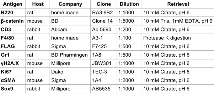
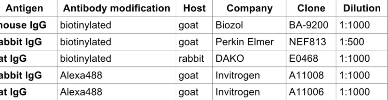
2.2.2.3 Immunostainings 38
2.2.3 Biochemical analysis 40
2.2.3.1 Isolation of intestinal epithelial cells (IECs)
from colon and small intestine 40 2.2.3.2 Preparation of IEC protein extracts 40 2.2.3.3 Preparation of cytoplasmic and nuclear protein extracts 41 2.2.3.4 Assessment of protein concentration by Bradford assay 41
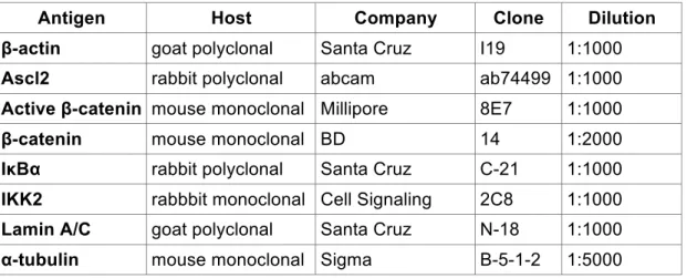
2.2.3.5 Western Blot analysis 41
2.2.4. Molecular Biology 43
2.2.4.1 Electrophoretic mobility shift assay (EMSA) 43
2.2.4.2 Extraction of RNA 43
2.2.4.3 cDNA synthesis 44
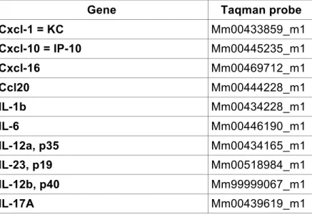
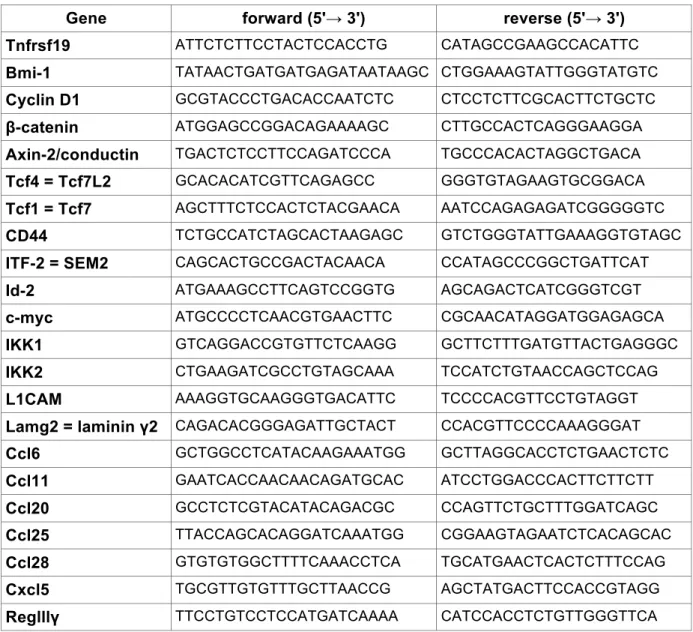
2.2.4.4 Quantitative real time PCR 45
2.2.4.5 Preparation of genomic DNA from tail biopsies 47
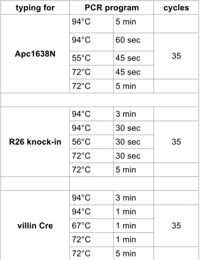
2.2.4.6 Genotyping PCRs 47
2.2.4.7 Agarose gel electrophoresis 48
2.2.5 Cell Biology 49
2.2.5.1 Culture of human colon cancer cell lines 49 2.2.5.2 Transfection of human colon cancer cells 49
2.2.6 Statistics 49
3. Results
3.1 Constitutive IKK2/NF-!B activation in IECs results in focal inflammation of the
colon and the small intestine 50
3.2 Expression of IKK2ca in the intestinal epithelium increases sensitivity to DSS- induced colitis and strongly enhances AOM-induced colon cancer
development 59
3.3 Strong cooperativity between Wnt- and IKK2/NF-!B signalling in intestinal
tumourigenesis 65
3.4 Spontaneous intestinal tumour development accompanied by strong
inflammation in aged IKK2caIEChom mice 71
3.5 Increased stabilisation and activation of "-catenin in the intestine of young
IKK2caIEChom mice 77
3.6 Perturbation of stem cells in the intestine of young IKK2caIEChom mice 82 3.7 Activation of non-immune cells and DNA damage in the intestine of
IKK2caIEChom mice 84
4. Discussion
4.1 What is the role of constitutive IKK2/NF-!B activity
in the intestinal epithelium? 88
4.2 Cooperation between Wnt- and NF-!B signalling in intestinal tumour
development 93
4.3 Impact of IKK2ca-expression on "-catenin stability and activity in IECs 94 4.4 Effect of IKK2ca-expression in IECs on intestinal epithelial stem cells 96
5. Concluding Remarks 98
Bibliography 99
Acknowledgements 111
Erklärung 112
Curriculum Vitae 113
Attachment (publication) 117
Abbreviations
ABC Avidin-Biotin-Complex
AOM Azoxymethane
APC Adenomatous Polyposis Coli Ascl2 Achaete scute like 2
Bmi-1 polycomb ring finger oncogene
BSA Bovine Serum Albumin
CAC Colitis Associated Cancer
CD Crohn´s Disease
cDNA complementary DNA
CRC Colorectal Cancer
Cre Causes recombination
DAB Diaminobenzidine
DAPI 4'-6-Diamidino-2-phenylindole DLK1 Drosophila homolog delta like 1 DMEM Dulbecco´s Modified Eagle Medium DNA Desoxyribonucleic acid
dNTPs desoxyribonucleotides DSS Dextrane Sulfate Sodium DTT Dithiothreithol
ECL Enhanced Chemiluminescence EDTA Ethylene Diamine Tetraacetate
eGFP enhanced GFP
EMSA Electrophoretic Mobility Shift Assay
FCS Fetal Calf Serum
GFP Green Fluorescent Protein
GI Gastrointestinal Tract
H&E Hematoxylin and Eosin
het heterozygous
hom homozygous
HRP Horseradish peroxidase IBD Inflammatory Bowel Disease IEC Intestinal Epithelial Cell
IF immunofluorescent
IHC immunohistochemistry
I.p. intraperitoneal
i!B# Inhibitor of NF-!B alpha IKK2 Inhibitor of !B Kinase 2
IKK2ca Inhibitor of !B Kinase 2 constitutively active
IL Interleukin
IRES Internal Ribosomal Entry Site IVC Individual Ventilated Cages
l liter
Lgr5 Leucine rich G-protein coupled receptor 5 LoxP Locus of X-over P1
MEFs Murine Embryonic Fiboblasts
MEICS Mean Endoscopic Index of Colitis Severity
ml milliliter
$l microliter
MMP Matrix Metalloprotease
mRNA messenger RNA
NEMO Nuclear Factor-kappa B essential modulator NF-!B Nuclear Factor-kappa B
NGS Normal Goat Serum
NP-40 Nonident P40
Olfm4 Olfactomedin 4
PCR Polymerase chain reaction PBS Phosphate-buffered saline
PFA Paraformaldehyde
RNA Ribonucleic Acid
ROS Reactive Oxygen Species
rpm rounds per minute
RT-PCR Real-Time PCR
SDS Sodium Dodecyl Sulfate
SI Small Intestine
sFL loxP flanked Stop cassette
#-SMA alpha Smooth Muscle Actin
TA Transit Amplifying
TBE Tris Boric acid EDTA buffer
TBS Tris buffered saline
TE Tris EDTA buffer
Tg transgenic
TLR Toll-Like Receptor TNF Tumour Necrosis Factor TNFR1 TNF Receptor 1 (p55)
TnfRsF19 TNF Receptor superfamily member 19
UC Ulcerative colitis
v/v volume per volume
vil villin
WT wild-type
w/v weight per volume
1. Introduction
1.1 NF-!B signalling
The Rel/NF-!B family of transcription factors in mammals consists of five members:
RelA or p65, RelB, c-Rel, p50 (NF-!B1) and p52 (NF-!B2) (Hayden and Ghosh, 2008). NF-!B molecules consist of a Rel-homology domain (RHD) situated at the N- terminus that is essential for formation of homo- and heterodimers and is required for DNA binding (Hayden and Ghosh, 2008). Nuclear NF-!B dimers bind to specific DNA consensus sites in the promoter regions of NF-!B target genes. p65, RelB and c-Rel posses a transcription activation domain (TAD) located in the C-terminal part of the protein and therefore homo- and heterodimers containing one of these family members can mediate transcriptional activation of NF-!B target genes (Hayden and Ghosh, 2008). In contrast, homodimers of p50 or p52 or dimers composed of a p50 and a p52 subunit are not able to activate transcription of NF-!B target genes and have instead been suggested to suppress target gene expression.
In unstimulated cells NF-!B target gene expression is prevented by proteins of the inhibitor of NF-!B (I!B) family consisting of I!B#, I!B" and I!B%, BCL3, and I!B& or I!BNS, as well as p100 and p105, which are precursor forms of p52 and p50, respectively (Hayden and Ghosh, 2008). Ankyrin repeats of I!Bs mediate binding to NF-!B molecules, which masks the nuclear localisation signals (NLS) of NF-!B. This way, the I!Bs prevent nuclear accumulation of NF-!B. In addition, I!B# contains a nuclear export signal (NES) that exports I!B-NF-!B complexes back into the cytoplasm (Hayden and Ghosh, 2008).
For NF-!B activation, I!Bs have to be degraded to release NF-!B dimers and allow their nuclear accumulation. The activation of NF-!B is under control of the so-called IKK-complex, which is composed of two kinases, I!B kinase 1 (IKK1 or IKK#) and I!B kinase 2 (IKK2 or IKK") and the regulatory subunit NF-!B essential modulator (NEMO or IKK') (Hayden and Ghosh, 2008). The latter is devoid of kinase activity, but nevertheless is essential for the activation of the IKK-complex. IKKs phosphorylate I!B# on two particular serine residues, Ser32 and Ser36. This phosphorylation pattern generates a phospho-degron signal that marks I!B# for K48- linked polyubiquitination by the "TrCP-SCF complex and is followed by its proteasomal degradation (Hayden and Ghosh, 2008). Free NF-!B dimers are then
able to translocate to the nucleus, where they can induce transcription of NF-!B target genes.
Two distinct pathways for the activation of NF-!B have been described. The classical or canonical NF-!B pathway is triggered by inflammatory cytokines such as TNF and IL-1, by bacterial and viral products through Toll-like receptors (TLRs) and intracellular nucleotide-binding and oligomerization domain (NOD) like receptors (NLRs), by irradiation, reactive oxygen species (ROS) and other stress factors. The canonical pathway leads to nuclear translocation of NF-!B dimers mainly consisting of p65, p50 and c-Rel and subsequent activation of genes coding for proinflammatory cytokines and chemokines, adhesion molecules and anti-apoptotic proteins. The non- canonical or alternative NF-!B pathway induces the processing of p100 to p52 (Hayden and Ghosh, 2008). Alternative NF-!B signalling is mainly induced upon ligation of receptors belonging to the TNF receptor superfamily, such as the receptor for BAFF (B-cell activating factor belonging to the TNF family, Tnfsf13b) or lymphotoxin-! receptor (Vallabhapurapu and Karin, 2009). Induction of the alternative pathway also results in transcriptional upregulation of cytokines and chemokines but additionally controls the organisation of lymphoid tissues and the development and survival of lymphoid cells. Processing of p100 is induced by phosphorylation of two particular serine residues, Ser866 and Ser870, in the C-terminal part of themolecule.
This processing of p100 requires NIK-mediated activation of IKK1 and occurs independently of IKK2 or NEMO (Hayden and Ghosh, 2008).
The generation of mice lacking specific subunits of the IKK-complex allowed investigation of the distinct functions of each individual IKK subunit (Pasparakis et al., 2006; Xiao et al., 2001). Studies with cells lacking NEMO showed that NEMO is indispensable for canonical NF-!B signalling and in the absence of this subunit no canonical NF-!B activation takes place. Similar experiments revealed that IKK2 is mainly responsible for activating NF-!B in the canonical pathway, though deletion of IKK2 does not completely block NF-!B activation. Residual NF-!B activity is mediated by IKK1, since the deletion of both kinases leads to complete blockade of NF-!B function, as it is the case in the absence of NEMO. IKK1 was shown to mediate phosphorylation of p100 at two particular serine residues in the C-terminal part of the protein, leading to its partial proteolysis, which gives rise to p52 (Xiao et al., 2001). Thus, while canonical NF-!B activation mainly depends on IKK2, non- canonical NF-!B activation depends solely on IKK1.
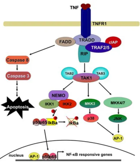
Depending on the NF-!B activating signal, different pathways are triggered and different upstream molecules are in charge for activating the IKK complex. IKK activation downstream of TNF stimulation will be described in further detail as an example of a signal transduction pathway leading to NF-!B activation. Upon ligation of TNF to TNFR1 the latter induces intracellular signalling cascades triggering programmed cell death (apoptosis) on the one hand but also the induction of the NF-
!B pathway and the mitogen activated protein kinase (MAPK) pathways leading to activation of JNK, p38 and ERK on the other hand.
Figure 1 Activation of the NF-!B signalling cascade downstream of TNFR1
Upon ligation of TNF to TNFR1 adapter molecules are recruited to the cytoplasmic part of the receptor. Various ubiquitination and phosphorylation events result in activation of NF-!B and MAPK signalling and the subsequent upregulation of pro-inflammatory and anti-apoptotic molecules. In the absence of NF-!B and MAPK activation, TNF signalling results in apoptosis. For details see text.
Upon binding of TNF to TNFR1, a number of proteins required for the activation of the TNF-induced signalling cascade are recruited to the plasma membrane. Through its cytoplasmic protein-protein interaction domain named death domain (DD), the central adapter TNFR1-associated death domain (TRADD) protein is bound to the
receptor. Furthermore receptor interacting protein 1 (RIP1) kinase, TNF receptor associated factors TRAF2 and TRAF5, which are both endowed with ubiquitin ligase activity, as well as the cellular inhibitors of apoptosis cIAP1 and cIAP2 become part of this receptor-proximal signalling complex (Varfolomeev and Vucic, 2008; Vlantis and Pasparakis, 2010). Through the generation of knockout cells for these components of the TNF-signalling cascade it has been shown that TRADD, RIP1 and TRAF2/5 are important for robust activation of NF-!B downstream of TNFR1 (Ea et al., 2006; Ermolaeva et al., 2008; Tada et al., 2001).
Ligation of TNFR1 by TNF results in polyubiquitination of RIP1, establishing a platform for the recruitment and activation of kinase molecules required for the induction of NF-!B and MAPK pathways. Analysis of knockout cells has revealed that polyubiquitination of RIP1 depends on the ubiquitin ligases TRAF2 and TRAF5 (Tada et al., 2001). Furthermore cIAP1 and cIAP2 were shown to be important for signalling through TNFR1 (Varfolomeev et al., 2008). K63-linked polyubiquitination of RIP1 has turned out to be of major importance for the activation of TNF-induced signalling and the downstream activation of NF-!B and MAPK signalling (Adhikari et al., 2007; Ea et al., 2006). It has been suggested that these K63-linked polyubiquitin chains on RIP1 form a docking site for the so called TAK1 signalling complex composed of Transforming growth factor-"-activated kinase1 (TAK1) and the TAK binding proteins TAB1 and TAB2 (Ea et al., 2006). TAB1 and TAB2 are considered to be responsible for binding to ubiquitin chains of RIP1. Furthermore recruitment of the IKK-complex is also mediated by ubiquitinated RIP1 (Ea et al., 2006). TAK1 has been proposed to be responsible for phosphorylation and therefore activation of IKKs and upstream kinases of the MAPK pathway (Wang et al., 2001).
Though the described signalling model had found wide acceptance, a recent study showed that the lack of RIP1 did not prevent activation of NF-!B downstream of TNFR1, suggesting that RIP1 is not required for the activation of NF-!B (Wong et al., 2010). This raises the possibility that either the role of RIP1 is cell type specific or that other molecules can compensate for the loss of RIP1 and activate the downstream pathway.
1.2 The gastrointestinal tract and anatomy of the intestinal epithelium
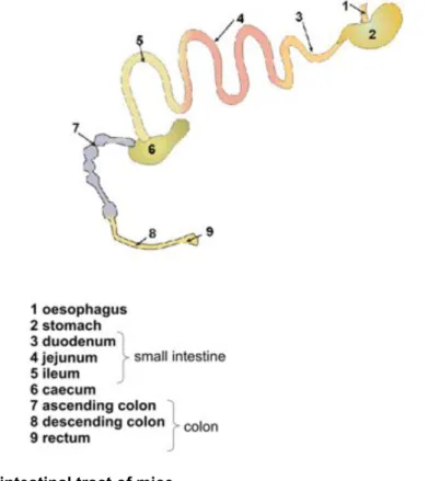
The intestinal tract consists of the small intestine, composed of (from cranial to caudal) the duodenum, jejunum and ileum and is followed by the further distally located large intestine or colon. The main function of the small intestine is digestion of food and absorption of nutrients. In the colon, in addition to the nutrient absorption, water is reabsorbed from the faeces. Furthermore the intestine harbours a large number of immune cells and therefore represents a major part of the immune system.
Figure 2 Gastrointestinal tract of mice
Schematic representation of the gastrointestinal tract of mice. Figure adapted from NIAID.com
A cross section through the intestine reveals the following structures from the outside to the inside. The outer part of the intestine is formed by two muscle layers, the inner circular muscle and the outer longitudinal muscle layer, which together are responsible for peristalsis, regulated tissue contractions that propagate the intestinal contents further distally. The muscle layers are followed by the submucosa, which harbours big blood and lymphatic vessels as well as nerves embedded in an irregular connective tissue. The innermost layer, the mucosa, can be separated into three parts: the intestinal epithelium that lines the intestinal lumen, the lamina propria harbours the immune cells of the digestive tract in a loose connective tissue and the muscularis mucosa, a thin smooth muscle layer that separates the mucosa from the submucosa.
The intestinal epithelium forms a single cell layered barrier that separates cells in the lamina propria from luminal contents. This barrier prevents the contact of mucosal immune cells by commensal bacteria and various antigens residing in the lumen.
Disturbance of epithelial integrity leads to an encounter of immune cells with immune reactive components of the intestinal lumen, resulting in an inflammatory response that impairs intestinal immune homoeostasis. Indeed, patients suffering from inflammatory bowel disease (IBD) such as Ulcerative Colitis (UC) or Crohn’s Disease (CD) display a disturbance in the intestinal epithelial barrier function (McGuckin et al., 2009; Roda et al., 2010). Therefore, maintaining intestinal epithelial integrity is essential for the health of an individual.
Although the intestinal tract is lined by a single cell-layered epithelium composed of intestinal epithelial cells (IECs), the epithelium of the small intestine and the colon each display a characteristic architecture and are composed of various kinds of specialised epithelial cells. The mucosa of the small intestine is organised into protrusions that project into the lumen, the villi, and mucosal invaginations called the crypts of Lieberkühn (see Fig. 3) (Reya and Clevers, 2005). The presence of villi enlarges the surface area where absorption of nutrients takes place. Crypts harbour the proliferating epithelial compartment that consists of stem cells and the transit amplifying (TA) cells that fuel the villi with new IECs. In contrast to the small intestine, the surface of the colonic mucosa is flat and does not possess villi (Fig. 3) (Reya and Clevers, 2005). However, also the colonic epithelium is organised in crypts with the stem cell compartment at the crypt bottom and the adjacent TA region. The intestinal epithelium is one of the most actively regenerating tissues of the mammalian body, in which the whole epithelium is renewed every 3 to 5 days (Brittan and Wright, 2004;
Radtke and Clevers, 2005). New IECs derive from stem cells at the crypt bottom that divide only once per day and give rise to new stem cells and TA cells. The TA cells undergo cell divisions more frequently (every 12-16 hours) and are responsible for replenishment of the epithelium (Marshman et al., 2002). TA cells remain in the crypt for about two days and divide four to five times before they undergo cell cycle arrest and migrate towards the apical part of the crypt (van der Flier and Clevers, 2009).
During this migratory phase epithelial precursors start differentiating into one of the four intestinal epithelial cell types, namely the absorptive enterocytes and three secretory cell types: goblet cells, enteroendocrine cells and in the small intestine also Paneth cells (van der Flier and Clevers, 2009). Enterocytes or columnar epithelial
cells are the most abundant cell type in the epithelium and make up more than 80%
of all IECs (van der Flier and Clevers, 2009). Enterocytes possess a brush border at the apical side and are responsible for the uptake of nutrients. Goblet cells are the most frequent cell type of the secretory lineage. Goblet cells secrete mucins and trefoil factors and thus produce the protective mucus layer that prevents tissue damage from luminal contents and hinders bacteria to get into direct contact with IECs (McGuckin et al., 2009; van der Flier and Clevers, 2009). Enteroendocrine (or neuroendocrine) cells can be found throughout the intestinal tract and these cells coordinate intestinal functions by secreting peptide hormones such as gastrin, secretin, serotonin and substance P, which regulate secretion of digestive juices and contraction of intestinal muscles.
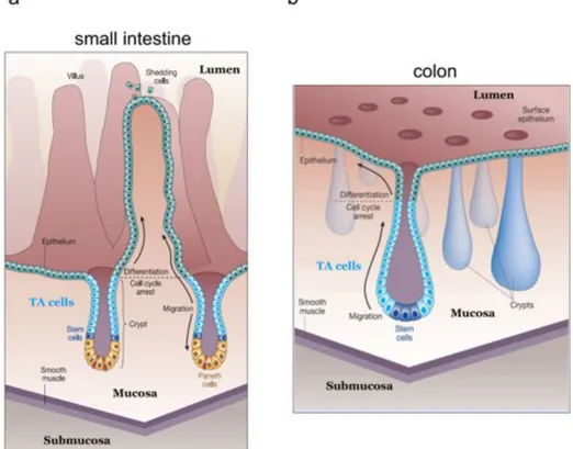
Figure 3 Tissue architecture of the small intestine and colon.
a. The surface area of the small intestinal mucosa is increased due to formation of protrusions, called villi. A single cell-layered epithelium separates the mucosa from the intestinal lumen. Stem cells and proliferating precursors (TA cells) of intestinal epithelial cells (IECs) are located in epithelial invaginations, the crypts of Lieberkühn. Paneth cells are located at the crypt bases in between stem cells (stem cells depicted in blue and red).
b. The mucosal surface of the colon is flat, though crypts are arranged in the same way as in the small intestine and harbour stem cells and TA cells but are devoid of Paneth cells.
Figure adapted from Reya and Clevers, 2005
Paneth cells reside at the base of small intestinal crypts and with a life expectancy of more than three weeks they are the most long-lived cells of the epithelium after stem cells (Bjerknes and Cheng, 1981). Paneth cells play an important role in the
regulation of the bacterial outgrowth in the intestinal tract by producing antimicrobial peptides such as lysozyme and defensins (Wehkamp and Stange, 2006).
In contrast to Paneth cells, which upon differentiation migrate towards the crypt base, differentiating IECs move towards the apical part of the mucosa (Reya and Clevers, 2005). When terminally differentiated IECs have reached the tip of small intestinal villi or the apical part of the colonic mucosa, they are shed off into the lumen. This is regulated through a specific form of cell death, termed anoikis, which is initiated by detachment from the basal membrane (Gilmore, 2005).
Although it was known that the stem cells of the intestinal epithelium are located at the lower part of the crypts in both the colon and the small intestine, the exact position and identity of this cell type remained elusive for a long time due to the lack of specific markers reliably labelling epithelial stem cells. However, recent reports have provided evidence that intestinal crypts harbour two types of stem cells with different kinetics in cell division and different localisation (Li and Clevers, 2010). Stem cells at the base of the crypt, also known as crypt-base columnar cells, are actively cycling with a dividing frequency of about once per day (Barker et al., 2007). These cells produce Ki67 and express Lgr5 and Ascl2, two molecules proposed to label this stem cell compartment (Barker et al., 2007; van der Flier et al., 2009). The second population of stem cells is located further towards the lumen of crypts at the +4 position (4th cell from the crypt bottom) and is negative for Lgr5 or Ascl2 (Barker et al., 2007; van der Flier et al., 2009). These cells are cycling very rarely, they do not produce Ki67 and they are referred to as quiescent stem cells. Quiescent stem cells have been identified to exist also in other tissues, such as the hair follicle and the bone marrow (Li and Clevers, 2010). This quiescent stem cell compartment is thought to function as a backup system that only replicates under stress conditions, such as extensive epithelial damage upon irradiation, and under these circumstances gives rise to new actively cycling crypt base columnar cells (Li and Clevers, 2010;
Scoville et al., 2008). Although some molecules have been proposed, thus far no marker has been identified that exclusively labels the quiescent stem cell compartment. For instance, Bmi-1 is a chromatin associated factor expressed in +4 stem cells in the small intestine (Li and Clevers, 2010), but also Lgr5 positive base columnar cells were shown to express this protein (van der Flier et al., 2009).
Currently it is not clear, whether Lgr5 positive and Bmi-1-expressing cells represent fully overlapping cell populations. Importantly, the base columnar and quiescent stem
cell compartments were reported to function independently of each other. Indeed, deletion of Bmi-1 did not affect Ascl2 positive stem cells at the crypt base (van der Flier et al., 2009).
1.3 Wnt signalling pathway
It is well established that Wnt signalling is crucial for proper development and maintenance of the intestinal epithelium (Fevr et al., 2007; Korinek et al., 1998). The Wnt pathway is responsible for differentiation and proliferation of the intestinal epithelium, as well as for proper positioning of the different intestinal epithelial cell types along the crypt-villus axis through regulating the expression of the EphB/ephrin-B system (Batlle et al., 2002). In addition, the Wnt pathway is essential for maintaining intestinal epithelial stem cells, since deletion of Tcf4 leads to depletion of the stem cell compartment and early postnatal death in mice (Korinek et al., 1998). Induced deletion of "-catenin in the intestinal epithelium of adult mice disrupted intestinal homoeostasis and resulted in loss of intestinal functions due to loss of proliferating cells and stem cells (Fevr et al., 2007).
Along the basal-apical axis of the intestinal epithelium, Wnt signalling is highest at the stem cell containing crypt base and gradually decreases towards the luminal part of the mucosa. This is not only reflected in a declining expression of Wnt target genes, but also in a decrease of the ratio of cytoplasmic versus nuclear "-catenin abundance when IECs move towards the apical part of the crypt. These observations clearly show that the main driving force behind proliferation of IECs and maintenance of stem cells is the Wnt signalling pathway. This signalling cascade shows a high degree of conservation throughout the animal kingdom underscoring the importance of this pathway in developmental processes (Klaus and Birchmeier, 2008).
Activation of the transcription factor "-catenin is the central outcome of the Wnt signalling pathway (see Fig. 4). If not membrane-bound to adherens junctions via its association with E-cadherin, cytoplasmic "-catenin is targeted for proteasomal degradation at the so-called “destruction complex”. The destruction complex is composed of the scaffolding proteins adenomatous polyposis coli (APC) and Axin2/Conductin, which bind "-catenin and expose particular N-terminal serine and threonine residues of "-catenin for phosphorylation. Through phosphorylation of these target sites by casein kinase l (CK1) and glycogen synthase kinase 3"
(Gsk3"), "-catenin is targeted to the "-TrCP-SCF complex, which in turn is
responsible for polyubiquitination and proteasomal degradation of the molecule (Clevers, 2006). When a Wnt-ligand binds to the cognate receptor complex, composed of a Frizzled (Frz)-receptor and the low-density lipoprotein receptor- related protein 5/6 (LRP5/6) co-receptor, the destruction complex is reorganized due to relocalisation of Dishevelled (a protein acting upstream of "-catenin and GSK3") and Axin to the phosphorylated receptor complex at the cell membrane. Therefore, "- catenin is not phosphorylated at its N-terminus and thus accumulates in the cytoplasm and translocates to the nucleus, where it binds to transcription factors of the T cell factor/lymphocyte enhancer factor (TCF/LEF) family (Clevers, 2006). These dimers can induce transcription of Wnt target genes, which encode proteins involved in proliferation such as c-myc and Cyclin D1, but also proteins involved in tissue remodelling (matrix metalloprotease 7, MMP7) or cell positioning (EphB receptors), as well as proteins produced in stem cells (Lgr5/Gpr49, Ascl2, Sox9) (Batlle et al., 2002; Brabletz et al., 1999; Crawford et al., 1999; He et al., 1998; Shtutman et al., 1999; Tetsu and McCormick, 1999; van der Flier et al., 2009).
.
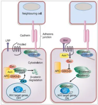
Figure 4 Wnt-signalling pathway
The Wnt-pathway is activated upon ligation of a Wnt-ligand to the cognate Frizzled-receptor complex.
This leads to recruitment of dishevelled and Axin2 to the receptor complex and thus to inactivation of the so-called destruction complex, comprising the tumour suppressor Apc, as well as Axin2 and the kinases CK1 and GSK3". When Wnt is present "-catenin can accumulate, translocate to the nucleus and activate TCF/LEF transcription factors and thereby induce expression of Wnt target genes. In the absence of Wnt signal cytoplasmic "-catenin is marked for ubiquitin-mediated degradation through phosphorylation by CK1 and GSK3" and thereby its accumulation and the transcription of target genes are prevented. Figure adapted from Reya and Clevers, 2005
Because of the obvious role for "-catenin activation in the differentiation and proliferation of IECs, it is clear that deregulation of this pathway might have deleterious consequences for intestinal homoeostasis. Indeed, both in humans and in mice mutations associated with intestinal carcinogenesis often affect components of the Wnt signalling cascade, leading to an enhancement of cytoplasmic and nuclear accumulation of "-catenin and thus uncontrolled production of Wnt target genes.
Mutations leading to activation of the Wnt pathway often affect APC, resulting in the formation of a non-functional APC molecule that cannot exert its functions and ensure degradation of cytoplasmic "-catenin (Fodde et al., 2001). Mutations in Apc are very common in both hereditary cancer syndromes like familial adenomatous polyposis coli (FAP), in which over 95% of FAP patients were shown to carry Apc mutations and in spontaneous intestinal cancers, with more than 80% of spontaneous colon cancers harbouring mutant forms of Apc (Laken et al., 1999). The function of APC as a true tumour suppressor protein was proven in mice lacking APC in IECs or carrying a mutated Apc allele, as such mice spontaneously develop intestinal tumours (see further). These observations indicate that deregulation of the Wnt pathway might be an early event in intestinal tumour formation and underscore the need for controlling Wnt signalling for the maintenance of intestinal homoeostasis and to prevent tumour development (Bjerknes and Cheng, 1981; Fodde et al., 2001).
1.4 Murine models of colitis and intestinal cancer
Various colitis models have been established in mice that mimic human IBD. Apart from genetic disease models such as IL-10 knockout mice or TNF(ARE animals that develop spontaneous intestinal disease, chemically inducible inflammatory models have been established (Kontoyiannis et al., 1999; Kuhn et al., 1993; Rennick et al., 1995). For instance, supplying mice with dextran sulfate sodium (DSS) in the drinking water results in inflammation of the distal colon that is similar to disease characteristics in UC patients (Cooper et al., 1993; Okayasu et al., 1990). So far the exact mechanisms through which DSS induces colonic inflammation are not completely understood. DSS acts as an irritant on the colonic epithelium, resulting in mucosal ulceration and translocation of luminal contents into the mucosa, triggering an inflammatory response. Disease severity induced by DSS ingestion has been shown to depend on the bacterial microflora. Mice held under germ-free conditions show enhanced sensitivity to DSS colitis, implying that bacteria exert a protective role in this epithelial erosion based colitis model (Kitajima et al., 2001).
DSS can be administered following an intraperitoneal injection with the pro- carcinogen Azoxymethane (AOM) in order to induce colonic tumour formation. AOM is hydroxylated in the liver by cytochromes P450 to an intermediate compound (methylazoxymethanol, MAM) and then transported in bile to the intestine. Factors derived from commensal bacteria are required for the generation of the alkylating methyldiazonium that induces DNA mutations through alkylation of Guanine residues at the O6 position (Greten et al., 2004; Neufert et al., 2007). Therefore, AOM induces DNA damage specifically in the intestinal epithelium of the colon (Neufert et al., 2007;
Tanaka et al., 2003). In order to accelerate proliferation of cells with AOM-induced mutations, multiple cycles of DSS are administered to the mice interrupted by a regeneration phase, in which mice receive normal drinking water. In this way, DSS invokes a chronic colitis associated with damage to the mucosal epithelium and regenerative proliferation of intestinal epithelial cells (Suzuki et al., 2006).
To study AOM-induced tumour formation that does not depend on inflammation- mediated tumour promotion, AOM is repetitively applied in weekly doses without the following DSS administration and tumour formation is assessed usually 30 weeks after the first AOM injection (Neufert et al., 2007).
In addition to chemically induced cancer models, also genetic mouse models for intestinal carcinogenesis have been established. For instance, mouse models
carrying mutations that occur in human colorectal cancer patients such as mutations in the Apc tumour suppressor gene were generated.
In this respect, Apcmin mice carry a point mutation in codon 850 in the Apc gene that leads to production of a truncated Apc polypeptide (Moser et al., 1990; Su et al., 1992). Apcmin mice develop multiple small intestinal polyps mimicking intestinal polyposis in FAP patients. Apcmin animals exhibit a very aggressive phenotype with 100-200 polyps growing in their small intestines and therefore exhibit a reduced life expectancy (Moser et al., 1990). A different Apc mutation in Apc1638N mice results in a milder disease manifestation (Fodde et al., 1994). In these mice a truncated version of the molecule, comprising the amino-terminal 1638 amino acids of Apc is produced, which is achieved through interruption of the coding sequence by a NEO- cassette (Fodde et al., 1994). Apc1638N mice only develop four to six small intestinal polyps in the very proximal part of the small intestine (duodenum and jejunum) at an age of about five to nine months (Fodde et al., 1994). When mice are left to age for one year, they were reported to show lesions also in the colon and the stomach (Fodde et al., 1994). In all mouse APC-models lesions occur only upon loss of heterozygocity for the wild-type Apc allele in tumour initiating epithelial cells.
In the AOM/DSS tumour protocol mutations affecting "-catenin that render the molecule resistant to degradation are very frequent (Takahashi et al., 2000;
Takahashi and Wakabayashi, 2004). Therefore mutations leading to activation of Wnt signalling are involved in different intestinal tumour models, indicating that perturbations of the Wnt pathway are an early event in intestinal tumourigenesis.
1.5 NF-!B in intestinal disease
A number of recent studies have demonstrated the importance of functional NF-!B signalling in the intestinal epithelium for maintaining tissue homoeostasis. Deletion of NEMO specifically in intestinal epithelial cells (NEMOIEC-KO) triggers spontaneous development of chronic colitis in mice (Nenci et al., 2007). NEMOIEC-KO mice display scattered apoptotic events in the epithelium already at young age, which may result in a disturbance of the intestinal epithelial barrier leading to colonic inflammation.
This hypothesis is supported by the observed translocation of commensal bacteria into the colonic mucosa, associated with recruitment of immune cells that secrete various cytokines and chemokines. Mice lacking either TAK1 or p65 specifically in IECs confirmed the essential role for NF-!B activation in the intestinal epithelium as
demonstrated by NEMOIEC-KO mice. TAK1IEC-KO mice exhibited severe intestinal disease affecting the colon and the small intestine and resulted in death within the first day after birth (Kajino-Sakamoto et al., 2008). Already during embryogenesis (at embryonic day 18), though the intestinal morphology still appeared normal, TAK1IEC-KO mice displayed increased apoptosis of IECs and transcription levels of inflammatory mediators were elevated. Since deletion of TAK1 blocks activation of NF-!B and MAPK signalling, it is not surprising that TAK1IEC-KO mice display a more severe phenotype than NEMOIEC-KO mice. While NEMOIEC-KO mice only lack canonical NF-!B activity and can induce production of anti-apoptotic and pro- inflammatory molecules through activation of MAPK signalling, TAK1IEC-KO mice cannot.
Furthermore, 10-15% of mice lacking the NF-!B subunit p65 specifically in the intestinal epithelium developed intestinal bleeding few days after birth and died shortly after weaning age (Steinbrecher et al., 2008). However, 85-90% of p65IEC-KO mice remained disease free throughout adulthood and did not display histological abnormalities in the intestinal tract. This milder phenotype of p65IEC-KO mice compared to NEMOIEC-KO animals most probably can be explained by the remaining activity of other NF-!B transcription factors in IECs. Indeed, whereas deletion of NEMO completely blocks activation of canonical NF-!B signalling, loss of p65 in the intestinal epithelium only partially impairs NF-!B dependent gene transcription.
However, when challenged with DSS, p65IEC-KO animals exhibited more severe colitis development than control littermates (Steinbrecher et al., 2008). This observation underscores the importance of NF-!B signalling in IECs for maintaining intestinal homoeostasis under stress conditions.
In accordance with the idea that partial inhibition of NF-!B signalling does not result in impaired intestinal homoeostasis under basal conditions, mice lacking IKK2 in IECs (IKK2IEC-KO mice) did not exhibit a spontaneous phenotype (Greten et al., 2004).
Only IKK2IEC-KO mice with an additional deletion of IKK1 in IECs (IKK2, IKK1IEC-KO) developed a colonic disease similar to NEMOIEC-KO animals (Nenci et al., 2007), indicating that under basal conditions residual NF-!B activation in IECs mediated by IKK1 is sufficient to maintain intestinal homoeostasis. However, IKK2IEC-KO mice were more sensitive to high doses of DSS-administration, arguing that full NF-!B signalling capacity in IECs is necessary to respond appropriately during DSS-triggered colitis (Greten et al., 2004).
Together, these above described studies show that NF-!B signalling in IECs is essential for maintaining epithelial integrity under steady state levels, during which low levels of NF-!B activity appear to suffice for IEC maintenance. However, to control colitis severity in chemically induced colitis high levels of epithelial NF-!B activation are necessary.
Apart from studying the role of NF-!B activation during intestinal inflammation, mice carrying conditional alleles of IKK2 have also been used to investigate the role of NF-
!B activation in intestinal tumourigenesis. In the AOM/DSS-induced model of colon cancer, IKK2IEC-KO mice exhibited reduced colorectal carcinogenesis. Since the lack of IKK2 in the intestinal epithelium led to increased IEC apoptosis upon DSS treatment, the authors suggested that cells carrying AOM-induced carcinogenic mutations were eliminated and therefore could not give rise to tumours (Greten et al., 2004). Thus, epithelial IKK2 activity was suggested to contribute to AOM/DSS- induced cancer development through its anti-apoptotic effect in early initiated tumour cells. In contrast to IKK2IEC-KO mice, mice lacking IKK2 in myeloid cells (IKK2myeloid-KO) did not reveal a differential tumour incidence but instead harboured significantly smaller colonic tumours than control animals. This indicates that IKK2 in myeloid cells promotes tumour growth rather then tumour initiation (Greten et al., 2004). Lack of IKK2 in myeloid cells reduced the production of cytokines and chemokines during AOM/DSS treatment. Since cytokines and chemokines are thought to act as growth factors for tumour cells (Coussens and Werb, 2002), this observation suggests that shortage of these inflammatory factors in IKK2myeloid-KO mice impairs tumour growth (Greten et al., 2004).
1.6 Cre/LoxP conditional gene targeting
Conditional gene targeting by Cre/LoxP mediated recombination allows cell type specific analysis of gene function in mice. In addition, the Cre/LoxP technique permits to investigate the function of genes that are essential for mouse development and of which the corresponding conventional full body knockout mouse does not reach adulthood (Rajewsky et al., 1996; Sauer and Henderson, 1988). To generate a cell type specific knockout of a particular allele, this allele is flanked by specific 34 bp sequences, the so-called LoxP-sites, in the same orientation. Alleles with these LoxP-flanked sequences are referred to as ‘floxed’ alleles. Mice carrying these floxed alleles are then crossed to mice expressing a bacteriophage P1-derived Cre recombinase transgene under the control of a cell type specific promoter. This Cre recombinase binds to and mediates recombination between the LoxP sites, resulting in excision of the DNA flanked by the LoxP sites specifically in the cell type producing Cre.
The Cre-LoxP system can also be used in order to induce the expression of a transgene only in a specific cell type. This is achieved when the coding sequence of this transgene is preceded by a stop-signal that is flanked by LoxP-sites, thereby preventing its expression. Cre-mediated recombination between the LoxP-sequences removes the Stop-signal and allows expression of the transgene specifically in the cell type in which Cre is present.
1.7 Project description
Genetic mouse models, in which NF-!B signalling was impaired specifically in the intestinal epithelium, have shown that activation of NF-!B in intestinal epithelial cells protects the intestine during inflammatory processes (Greten et al., 2004; Nenci et al., 2007; Steinbrecher et al., 2008; Vallabhapurapu and Karin, 2009). A complete blockade of canonical NF-!B activity in intestinal epithelial cells resulted in severe chronic colitis, demonstrating that basal levels of functional NF-!B signalling are required to preserve intestinal immune homoeostasis (Nenci et al., 2007). Partial inhibition of NF-!B signalling by deletion of either IKK2 or p65 in IECs rendered mice more sensitive to DSS-induced colitis (Greten et al., 2004; Steinbrecher et al., 2008), indicating that NF-!B activity in IECs is essential to cope with inflammation associated tissue damage. Indeed, IKK2-deficient IECs displayed increased apoptosis upon DSS treatment, suggesting a cytoprotective effect of NF-!B activation in IECs under inflammatory conditions. On the other hand, the anti-apoptotic effect of
NF-!B activation in IECs promotes intestinal tumour development, as lack of IKK2 in intestinal epithelial cells reduced tumour incidence in AOM/DSS-induced cancer experiments due to enhanced cell death of carcinogen exposed IECs (Greten et al., 2004). These loss-of-function approaches in mice indicate that though NF-!B activation in IECs preserves intestinal homoeostasis and therefore is protective during excessive intestinal inflammation, at the same time it acts as a tumour promoter in intestinal tumourigenesis.
Considering these findings we decided to use a gain-of-function approach to investigate in vivo the outcome of increased NF-!B activity in intestinal epithelial cells. Therefore, we applied Cre/LoxP mediated recombination to generate mice that express a constitutively active IKK2 molecule selectively in intestinal epithelial cells.
With this mouse model we wanted to address the role of constitutive IKK2/NF-!B activation specifically in the intestinal epithelium in mucosal homoeostasis, inflammatory disease and intestinal tumourigenesis.
2. Material and Methods 2.1 Material
2.1.1 Chemicals
Chemicals and compounds were purchased from Sigma & Aldrich, AppliChem, Merck, Roth, MP, VWR, Bayer, Ratiopharm, Dako, Vector Laboratories, GE Healthcare, Thermo Scientific, Li-Cor Biosciences
2.1.2 Material for mouse work
High resolution mini-endoscope, Coloview with Xenon light source, Karl-Storz (Tuttlingen, Germany)
Ketamin 10 mg/ml, Ratiopharm Rompun 2%, Bayer Healthcare Azoxymethane (AOM), Sigma
Dextran Sulfate Sodium (DSS), MW 36000-50000, MP Haemoccult sensa, Beckmann Coulter
Syringes, Braun and Injection Needles, Terumo and Braun
2.1.3 Material for Histology Tissue retriever 2100, PickCell Tissue processor, Leica TP1020 Rotary Microtome, Leica RM2255
Modular tissue embedding center, Leica EG1150 and Leica EG1150 C Fluorescent microscope, Leica DM5500
ABC Kit Vectastain Elite (Vector, PK 6100) Avidin/Biotin Blocking Kit (Vector, no. SP-2001)
Liquid DAB Substrate Chromogen System (DakoCytomation, Code K3466) Fluoromount-G, SouthernBiotech
Glas slides, Menzel
2.1.4 Material for Biochemistry Homogenizer Precellys 24, PeqLab 2 ml tubes for homogenisation, Peqlab
1,4 mm Zirconium oxide beads for tissue homogenisation, Peqlab
Gel casting system and SDS-Page system, Biorad Powersupply, Biorad
Protease Inhibitor Cocktail complete mini EDTA free, Roche PhosphoStop, phosphatase inhibitor, Roche
Bradford reagent, Biorad
Protein Marker PeqGold Protein Marker V, PeqLab PVDF membranes Immobilon-P, Millipore
Films Hyperfilm ECL, Amersham Odyssey detection system
2.1.5 Molecular Biology Reagents and Equipment Trizol reagent, Invitrogen
RNA extraction RNeasy mini kit, Qiagen RNase-free DNase set, Qiagen
SuperScriptlll cDNA synthesis Kit, Invitrogen
RT Cycler ABI HT 7900 Cycler, Applied Biosystems
Power SYBR® Green PCR Master Mix, Applied Biosystems TaqMan® Gene Expression Master Mix, Applied Biosystems MicroAmp® Optical Adhesive Film, Applied Biosystems
MicroAmpTM Optical 384-Well Reaction Plate, Applied Biosystems
2.1.6 Laboratory equipment Centrifuges, Eppendorf and Haereus Thermomixer, Eppendorf
PCR-cyclers, Biorad DNA Engine, Biometra and Eppendorf DNA ladder, Peqlab
Primers, Invitrogen and Metabion Bio Photometer, Eppendorf NanoDrop ND8100, PeqLab
QIAfilter plasmid purification kit, Qiagen
2.1.7 Cell culture DMEM (Gibco)
TrypLETM Express (Gibco)
100x Penicillin (10000 U/ml)/Streptomycin (10000 $g/ml) (Gibco)
100x L-Glutamine (200 mM) (Gibco) 100x Sodiumpyruvate (100 mM) (Gibco) Fetal Calf Serum (PAN)
PBS (without Ca2+ and Mg2+) (Gibco) Lipofectamine Reagent 2000 (Invitrogen)
Plastic ware for cell culture from BD Falcon, Millipore, TPP, Corning Tubes Eppendorf 1,5 ml and 2 ml reaction tubes
8 x 0,2 ml tube stripes Biozym
2.1.8 Software
Photoshop CS3, Leica microscopy Software Leica application suite, Prism Graph, Microsoft Office, OpenOffice Sun Microsystems, EndnoteX2
2.1.2 Buffers and Solutions 2.1.2.1 Washing buffers
PBS (1x) pH 7,3
NaCl 137 mM
KCl 2,7 mM
Na2HPO4 – 7H2O 4,3 mM KH2PO4 1,4 mM
TBS (1x) pH 7,5
Tris-Base 24,2 g
NaCl 80 g
2.1.2.2 Buffers and solutions for immunostainings
Endogenous peroxidase blocking buffer (for IHC) NaCitrate 0,04 M
Na2HPO4 0,121 M
NaN3 0,03M
H2O2 3 % (v/v)
TEX Protease K buffer pH 8.0 (for protease mediated antigen retrieval)
Tris-base 50 mM
EDTA 1 mM
Triton X-100 0,5% (v/v)
2.1.2.3 Preparation of protein extracts
High salt RIPA lysis buffer (for preparation of total cell extracts) HEPES (pH 7,6) 20 mM
NaCl 350 mM
MgCl2 1 mM
EDTA 0,5 mM
EGTA 0,1 mM
Glycerol 20% (v/v)
1% Nonident P-40, Protease inhibitor and Phosphatase inhibitors were added prior to use.
Cytoplasmic and nuclear protein extraction buffers Buffer A (hypotonic lysis buffer)
HEPES (pH 7.6) 10 mM
KCl 10 mM
MgCl2 2 mM
EDTA 0.1 mM
After the swelling step 0,8% (v/v) Nonident P-40 was added.
Protease inhibitor and Phosphatase inhibitors were added prior to use
Buffer C (high salt nuclear lysis buffer) HEPES (pH 7.8) 50 mM
KCl 50 mM
NaCl 300 mM
EDTA 0.1 mM
Glycerol 10 % (v/v)
Protease inhibitor and Phosphatase inhibitors were added prior to use
2.1.2.4 Buffers and solutions used for Western Blot analysis
Tris-glycine electrophoresis buffer Tris-Base 25 mM
Glycine 250 mM
SDS 0,1% (w/v)
SDS-polyacrylamid gel 10% resolving gel (for 20 ml)
H2O 7,9 ml
30% acrylamide mix 6,7 ml 1,5 M Tris (pH 8,8) 5,0 ml 10% (w/v) SDS 0,2 ml 10% (w/v) APS 0,2 ml
TEMED 0,012 ml
5% stacking gel (for 10 ml)
H2O 6,8 ml
30% acrylamide mix 1,7 ml 1 M Tris (pH 6,8) 1,25 ml 10% (w/v) SDS 0,1 ml 10% (w/v) APS 0,1 ml
TEMED 0,01 ml
Transfer Buffer (for semidry transfer) Tris-Base 25 mM
Glycine 192 mM
Methanol 20% (v/v) pH 8,3-8,5
Blocking buffer
PBS + 0,1% (v/v) Tween-20 + 5% (w/v) nonfat dry milk
primary antibody dilution buffer
TBS + 0,1% (v/v) Tween-20 + 5% (w/v) BSA or 5% (w/v) nonfat dry milk
5x Laemmli loading buffer
Tris-HCl (pH 6,8) 250 mM
SDS 10% (w/v)
Glycerol 50% (v/v)
Bromphenolblue 0,01% (w/v)
"-Mercaptoethanol 10% (v/v) (can be added prior to use)
Home made ECL solution Stock solutions
Tris-HCl (pH 8,5) 100 mM
Luminol 250 mM (in DMSO)
Paracoumaric acid (PCA) 90 mM (in DMSO)
H2O2 30% (v/v)
Solution 1
10 ml of 10 mM Tris-HCl (pH 8,5) with 2,5 mM Luminol (100 $l of stock) and 400 $M PCA (44 $l of stock).
Solution 2
10 ml of 10 mM Tris-HCl (pH 8,5) with 7 $l 30% (v/v) H2O2 (5,4 mM).
The two solutions were mixed and applied on the membrane.
2.1.2.5 Buffers and solutions used for EMSA
1x TBE
Tris-base 89 mM
Boric acid 89 mM
EDTA 2 mM
Nondenaturing gels (for 25 ml)
H2O 19,25 ml
10x TBE 1,25 ml
30% Acrylamide/Bisacrylamide mix 4,14 ml
10% (w/v) APS 315 $l
TEMED 40 $l
10x EMSA binding buffer
Tris-HCl (pH 7,5) 100 mM
NaCl 500 mM
DTT 10 mM
2.1.2.6 Buffers used for DNA extraction and genotyping PCRs
Tail lysis Buffer
Tris-HCl (pH 8,5) 100 mM
EDTA 5 mM
NaCl 200 mM
SDS 0,2 % (w/v)
0,1 mg Proteinase K (10mg/ml in 50 mM Tris, pH 8.0) per 500 $l lysis buffer was added prior to use.
TE buffer
Tris-HCl (pH 8) 10 mM
EDTA (pH 8) 1 mM
10x TAG buffer
Tris-base (pH 8,5) 200 mM
KCl 500 mM
TAE Buffer (25x) for 10 l
Tris-Base 1210 g
EDTA (pH 8.0) 500 ml of 0,5 M solution Acteic acid 285.5 ml
DNA loading buffer
15 % (w/v) Ficoll 400 was resolved in distilled water at ~50 °C. Orange G was added till the colour turned red
2.2 Methods
2.2.1 Animal handling and mouse experiments
2.2.1.1 Mouse maintenance
Mice were housed in individually ventilated cages (IVC) in a specific pathogen free (SPF) mouse facility and in a conventional animal facility in the Institute for Genetics at the University of Cologne. Mice had permanent access to regular chow diet (Teklad Global Rodent 2018, Harlan) and acidified water. Animals were kept at a regular 12 hours light and 12 hours dark cycle.
For breeding male and female mice were set together at a minimum age of 6 weeks.
Litters were weaned at 3 weeks of age and marked with an eartag. Tail biopsies were taken at the same time for isolation of genomic DNA and genotyping
Care of all mice was within institutional animal care committee guidelines. All animal procedures were conducted in accordance with European, national and institutional guidelines and protocols and were approved by local government authorities (Bezirksregierung Köln, Cologne, NRW, Germany).
2.2.1.2 Generation of conditional mice
To generate mice that express constitutively active IKK2 specifically in the intestinal epithelium R26IKK2casFL (Sasaki et al., 2006) mice were bred with villin Cre transgenics (Madison et al., 2002) (R26IKK2ca sFL; villin Cre mice). To generate double mutant mice Apc1638N (Fodde et al., 1994) were crossed to R26IKK2ca sFL;
villin Cre mice. All mice were maintained in a C57BL/6 background.
For all experiments littermates only carrying loxP-flanked alleles served as control mice.
2.2.1.3 Endoscopy
Mice were anaesthetized by intraperitoneal injection of 200 $l per 20 g bodyweight a Ketamine/Rampun mixture. The distal colon was analysed with a high-resolution mouse mini-endoscope, denoted Coloview, from Karl Storz. For imaging of the distal colon area the endoscope was gently inserted into the anus. Constant airflow induced dilation of the intestine and allowed an unperturbed view on the colonic mucosa. Disease index determined via endoscopy was composed of 5 scoring
features indicative for intestinal disease severity, each of which was scored ranging from 0 to 3 with 0 being healthy and 3 indicating severe inflammation, as has been described before (Becker et al., 2005). The sum of the following parameter scores results in murine endoscopic index of colitis severity (MEICS), an index between 0 and 15, allowing the evaluation of disease severity. The characteristics scored during examination were stool consistency, vascularisation pattern, fibrin abundance, thickening of the bowel wall and surface appearance of the mucosa. Furthermore the existence of tumours, ulcers and other intestinal lesions could be assessed through endoscopic examination.
After endoscopy mice were allowed to recover from anaesthesia or were immediately killed.
2.2.1.4 Dextran sulfate sodium (DSS)-induced colitis
In order to induce colonic inflammation resembling UC in human IBD patients, a well- established protocol of Dextran sulfate sodium (DSS) feeding was conducted (Cooper et al., 1993; Okayasu et al., 1990). Mice were fed DSS ad libidum in the drinking water. DSS (1,5% w/v or 2% w/v) was dissolved in millipore water and autoclaved prior administration. During the course of DSS experiments mice were monitored on a daily basis to assess bodyweight, stool consistency and intestinal bleeding. In order to monitor intestinal inflammation and damage of the colonic mucosa, stool samples of each animal were collected every day. Fresh stool pellets were spread on Heamoccult test sheets (Beckmann Coulter) in order to visualize occult blood. Blood scores from 0 to 4 were determined as follows: no blood in stool:
score 0, occult blood visualized with Haemoccult kit: score 1, blood in stool visible in stool when spread: score 2, stool pellet covered by blood: score 3, gross bleeding from anus: score 4. Stool consistency scores from 0 to 4 were determined by the following scoring criteria: regularly formed stool pellets: score 0, still formed but softer stool pellets: score 1, enlarged, soft stool pellets: score 2, spread unformed stool:
score 3, heavy diarrhoea with stool spread around the anus: score 4. The sum of blood score and stool consistency scores results in the total stool score.
2.2.1.5 AOM/DSS-induced colorectal cancer protocol
To induce colorectal colitis associated cancer (CAC), mice were undertaken an established chemically induced inflammation dependent cancer protocol (Neufert et al., 2007; Tanaka et al., 2003). On day zero of the treatment mice received an
intraperitoneal injection with 10 mg/kg bodyweight Azoxymethane (AOM) diluted in sterile PBS. In order to induce acute intestinal inflammation, on the same day, drinking water was replaced by water supplemented with DSS. During the first 2 weeks of the experiment mice were weighed daily and stool was analysed for diarrhoea and occult blood as described for DSS treatment. During the course of the treatment and at the end of the experiment mice were endoscopised in order to monitor disease and tumour development in vivo. Mice were sacrificed 9 weeks after the AOM injection and tissue samples were processed for histological evaluation.
2.2.1.6 AOM-induced tumour protocol
To induce colorectal cancer independently of inflammation driven tumour promotion, mice were repetitively injected with AOM without additional DSS feeding (Neufert et al., 2007). Mice received 10 mg/kg bodyweight AOM diluted in sterile PBS i.p. once per week for 5 consecutive weeks. Mice were monitored regularly to monitor health status.
2.2.1.7 Sacrifice of mice
Mice were euthanized by cervical dislocation.
2.2.1.8 Tissue processing
After cervical dislocation the abdominal cavity was opened by a longitudinal incision.
The large and small intestines were removed by cutting out the colon in close proximity to the anus and the small intestine was cut after the pylorus. The intestines were placed into PBS and cleaned from mesentery and fat. The intestinal contents were carefully squeezed out with scissors and pieces of the gastrointestinal tract were taken for histological examination and RNA extraction.
For examination of bigger parts of intestinal tissue the “swiss-roll technique” was applied. For this the whole colon was opened longitudinally and the contents were removed. The tissue was then placed with the mucosa facing down on a dish. With the help of injection needles the tissue was rolled up lengthwise to form a so-called
“swiss-roll” with the muscle tissue facing to the inside of the roll and the mucosa outwards. Small intestine of adult mice was cut into three pieces and individual rolls were prepared from these segments. About 3 to 5 mm long pieces of intestinal tissue were used for preparation of cross sections.
2.2.2 Histology
2.2.2.1 Preparation of intestinal tissue for histological evaluation
For histological examination tissue was fixed in histology cassettes over night at 4°C in 4% (w/v) phosphate buffered paraformaldehyde. The samples were passed through an increasing concentrations of ethanol for tissue dehydration (for 2 h each in 30% (v/v), 50% (v/v), 70% (v/v), 96% (v/v) and 2x in 100% (v/v) ethanol), were kept for two times 2 h each in xylol and then transferred to paraffin in order to be embedded in paraffin blocks. Intestinal tissue was sectioned with a microtome at 3 to 4 $m thickness and stained with haematoxylin and eosin for histological evaluation.
2.2.2.2 Haematoxylin and Eosin staining of intestinal tissue sections To de-wax tissue sections, glass slides were placed in xylol for 20 min, then put for 2 min in each of the following ethanol-solutions for tissue rehydration: 100% (v/v) ethanol (or 100% (v/v) isopropanol), 95% (v/v) ethanol, 75% (v/v) ethanol and then in PBS for 5 min. Slides were washed in tap water for 1 min and placed into Meyers' Haematoxylin for 2 min. After being kept in tepid tap water for maximally 20 s the slides were incubated in tap water for another 15 min at room temperature to blue, followed by a short incubation in deionised water. Samples were placed in Eosin staining solution for 1 min and the excessive staining removed by washing the slides for 6 to 7 times in tap water. Samples were placed for 2 min in each 75% (v/v), 96%
(v/v) and 100% (v/v) ethanol to dehydrate and were subsequently cleared in xylol.
Coverslips were mounted with Entellan.
2.2.2.3 Immunostainings
For immunofluorescent and immunohistochemical (IHC) staining, slides were de- waxed in xylol and transferred through an ascending series of ethanol solutions as described under H&E staining procedure. After washing twice for 5 min in tap water for IHC endogenous peroxidase activity was blocked for 15 min at room temperature in endogenous peroxidase blocking buffer. Thereafter slides were washed three times for 5 min in tap water and antigen unmasking in form of heat induced epitope retrieval was performed, either in a pressure steam cooking device, where samples were heated for 20 min to 120°C or over night at 80°C in citrate buffer, pH 6 supplemented with 0,05% (v/v) Tween-20. Alternatively (for cytoplasmic and nuclear visualisation of !-catenin) retrieval was performed in TE pH 9 (10 mM Tris, 1 mM