*For correspondence:
erhard.strohm@ur.de
Present address:†Evolutionary Ecology, Institute of Organismic and Molecular Evolution, Johannes Gutenberg University, Mainz, Germany
Competing interests:The authors declare that no competing interests exist.
Funding:See page 27 Received:16 November 2018 Accepted:05 May 2019 Published:11 June 2019 Reviewing editor: Marcel Dicke, Wageningen University,
Netherlands
Copyright Strohm et al. This article is distributed under the terms of theCreative Commons Attribution License,which permits unrestricted use and redistribution provided that the original author and source are credited.
Nitric oxide radicals are emitted by wasp eggs to kill mold fungi
Erhard Strohm1*, Gudrun Herzner1, Joachim Ruther2, Martin Kaltenpoth1,3†, Tobias Engl1,3†
1Evolutionary Ecology Group, Institute of Zoology, University of Regensburg,
Regensburg, Germany;2Chemical Ecology Group, Institute of Zoology, University of Regensburg, Regensburg, Germany;3Insect Symbiosis Research Group, Max Planck Institute for Chemical Ecology, Jena, Germany
Abstract
Detrimental microbes caused the evolution of a great diversity of antimicrobial defenses in plants and animals. Insects developing underground seem particularly threatened. Here we show that the eggs of a solitary digger wasp, the European beewolfPhilanthus triangulum,emit large amounts of gaseous nitric oxide (NO) to protect themselves and their provisions, paralyzed honeybees, against mold fungi. We provide evidence that a NO-synthase (NOS) is involved in the generation of the extraordinary concentrations of nitrogen radicals in brood cells (~1500 ppm NO and its oxidation product NO2). Sequencing of the beewolfNOSgene revealed no conspicuous differences to related species. However, due to alternative splicing, the NOS-mRNA in beewolf eggs lacks an exon near the regulatory domain. This preventive external application of high doses of NOby wasp eggs represents an evolutionary key innovation that adds a remarkable novel facet to the array of functions of the important biological effector NO.
DOI: https://doi.org/10.7554/eLife.43718.001
Introduction
Microbes pose a major threat to the health of all animals and plants. These have responded by evolving a great diversity of defenses including hygienic behaviors (Gilliam et al., 1983), antimicro- bial chemicals (Herzner et al., 2013;Vilcinskas et al., 2013;Gross et al., 2008), complex immune systems (Hooper et al., 2012; Iwasaki and Medzhitov, 2010), and defensive symbioses (Kaltenpoth et al., 2005;Flo´rez et al., 2017). Besides such pathogenic effects, many bacteria and fungi are severe, but often neglected, competitors of animals for nutrients, thus prompting the evo- lution of mechanisms to preserve food sources (Janzen, 1977;Rozen et al., 2008).
Some animals are particularly prone to suffer from microbial attack due to (1) high abundance of potentially harmful microbes in their environment, (2) a microbe-friendly microclimate and/or (3) lim- ited defense mechanisms. The progeny of many insect species develop under warm and humid con- ditions in the soil, where they are exposed to a high diversity of bacteria and fungi. Moreover, compared to adult insects, immature stages, in particular eggs, have usually reduced abilities to pre- vent microbial infestation due to, for example, a thin cuticle or an inability to groom (Wilson and Cotter, 2013;Tranter et al., 2014). The situation is even aggravated when eggs and larvae have to develop on limited amounts of provisions that are susceptible to attack by ubiquitous and fast grow- ing putrefactive bacteria and mold fungi (Janzen, 1977;Arce et al., 2013).
Such hostile conditions prevail in nests of subsocial Hymenoptera like the European beewolfPhi- lanthus triangulum (Hymenoptera, Crabronidae). The offspring of these solitary digger wasps develop in subterranean brood cells provisioned by the female wasps with paralyzed honeybee workers (Apis mellifera, Apidae, Hymenoptera) (Strohm and Linsenmair, 2000) (Figure 1A). The beewolf egg is laid on one of the bees, the larva hatches after three days, feeds on the bees for six
RESEARCH ARTICLE
to eight days, then spins a cocoon and either emerges the same summer or hibernates. The warm and humid microclimate in the brood cell promotes larval development but also favors fast growing, highly detrimental fungi (Engl et al., 2016). Without any countermeasures the provisions will be completely overgrown by mold fungi within three days (Figure 1B), and the beewolf larva becomes infested by fungi or starves to death (Strohm and Linsenmair, 2001;Herzner et al., 2011a).
We have previously documented two adaptations that beewolves have evolved to counter the detrimental effects of fungi on their brood. First, beewolf females reduce molding of the larval provi- sions by coating the paralyzed bees with ample amounts of unsaturated hydrocarbons (Herzner et al., 2007). This embalming changes the physicochemical properties of the preys’ surface causing reduced water condensation on the bees (Herzner and Strohm, 2007). Due to the depriva- tion of water, germination and growth of fungi is delayed by two to three days (Herzner et al., 2011b). Second, during the long period of overwintering in their cocoons beewolf larvae are pro- tected by antibiotics on their cocoons (Kaltenpoth et al., 2005;Kroiss et al., 2010). Prior to ovipo- sition, beewolf females apply a secretion containing symbioticStreptomycesbacteria to the ceiling of the brood cell. The secretion is taken up by the larvae and incorporated into the silk threads of their cocoons. There, the bacteria produce several antibiotics that effectively protect the cocoon and, thus, the larvae against fungus infestation (Kroiss et al., 2010;Engl et al., 2018).
Despite the considerable effect of prey embalming, when removed from brood cells at least 50%
of embalmed bees showed fungus infestation within six days after oviposition (Strohm and Linsen- mair, 2001). Since in natural brood cells only around 5% of the progeny succumb to mold fungi
eLife digest
Humans use heat, cooling, and freezing to protect their foods from mold and bacteria. Many animals, including a wasp called the European beewolf, have also developed ways to store and preserve food. Female beewolves hunt honeybees. After paralyzing a bee, the beewolf takes the body into an underground chamber and lays an egg on it. When a larva hatches from the egg, it feeds on the bee. The warm, humid conditions in the chamber provide ideal conditions in which larvae can develop, but also encourage mold and bacteria to grow.Previous research has uncovered two methods used by beewolves to fight off mold. In 2007, researchers discovered that the female beewolf coats her bee prey with a layer of fats. This prevents water loss and keeps the outside of the bee dry so that mold spores cannot grow. In 2010, a further study showed that the female beewolf grows helpful bacteria inside her antennae and transfers some to her young. The bacteria produce antibiotics that protect the larvae and their cocoons from mold. But these two strategies alone cannot explain the high survival rate of beewolf young. This suggests that the beewolves have at least one more strategy to prevent mold from growing.
Now, Strohm et al. – including some of the researchers involved in the 2007 and 2010 studies – show that beewolf eggs emit high levels of a gas called nitric oxide, which reacts with oxygen to form nitrogen dioxide. Nitrogen dioxide is part of the air pollution generated by cars and is harmful to many species in high concentrations. Nitric oxide also plays an important role for many
biochemical processes in virtually all organisms, albeit in very low concentrations. The beewolf eggs produce comparatively huge amounts of this gas to fumigate their brood chambers and protect themselves and their food from mold.
Strom et al. then investigated how the eggs produce nitric oxide. The eggs appear to use the same enzyme that some other organisms use to produce nitric oxide. However, the wasp version of the enzyme contains a small modification that might explain why the eggs can produce the gas in such large amounts.
Learning more about how beewolves evolved different anti-mold strategies could help researchers to develop new antimicrobial treatments for medical applications. In addition, it is not yet clear how the wasp eggs survive in high concentrations of nitric oxide and nitric dioxide.
Inflammation and some human diseases produce nitric oxide, killing nearby cells. Understanding how the beewolf eggs survive could therefore help to treat these cells or protect them from damage.
DOI: https://doi.org/10.7554/eLife.43718.002
Research article Ecology Evolutionary Biology
(Strohm and Linsenmair, 2001), we searched for an additional antimicrobial defense mechanism that takes effect during the early stages of beewolf development.
Here we report on a unique antifungal strategy that is employed by beewolf eggs to defend themselves and their provisions against mold fungi. Employing bioassays we discovered that beewolf eggs emit a strong antifungal agent that we identified as the gaseous radical nitric oxide (NO). We characterize the amount, time course and temperature dependence of emission and show that syn- thetic NO exerts a similar effect as the gas emitted by beewolf eggs. Furthermore, we tested whether there was an interaction of the gas emitted by the eggs and the embalming of the prey by beewolf females. Using histological methods, inhibition assays, and gene expression analysis, we elu- cidate a biosynthetic pathway for NOsynthesis in beewolf eggs. To explore the evolutionary back- ground of this remarkable antimicrobial strategy, we sequenced the relevant gene and mRNA. Our findings reveal a novel function of the eminent and widespread biological effector NOin providing an extended immune defense to the producer by sanitizing its developmental microenvironment.
Results
Emission of an antifungal volatile by beewolf eggs
Thorough examination of beewolf nests in observation cages (Strohm and Linsenmair, 1994) revealed that within 24 hr after oviposition, a conspicuous pungent smell occurred that was clearly emanating from the eggs and disappeared by the time the larvae hatched. We hypothesized that this smell was due to an antifungal agent. When paralyzed honeybees from completed beewolf brood cells were incubated individually, bees carrying an egg showed significantly delayed fungus growth compared to bees without egg over the period from oviposition to cocoon spinning (Kaplan Meier survival analysis, Breslow test, day 0–11: Chi square = 12, df = 1, p=0.001;Figure 2A). This difference was also significant for the period from oviposition to the hatching of the larvae (day 0–3:
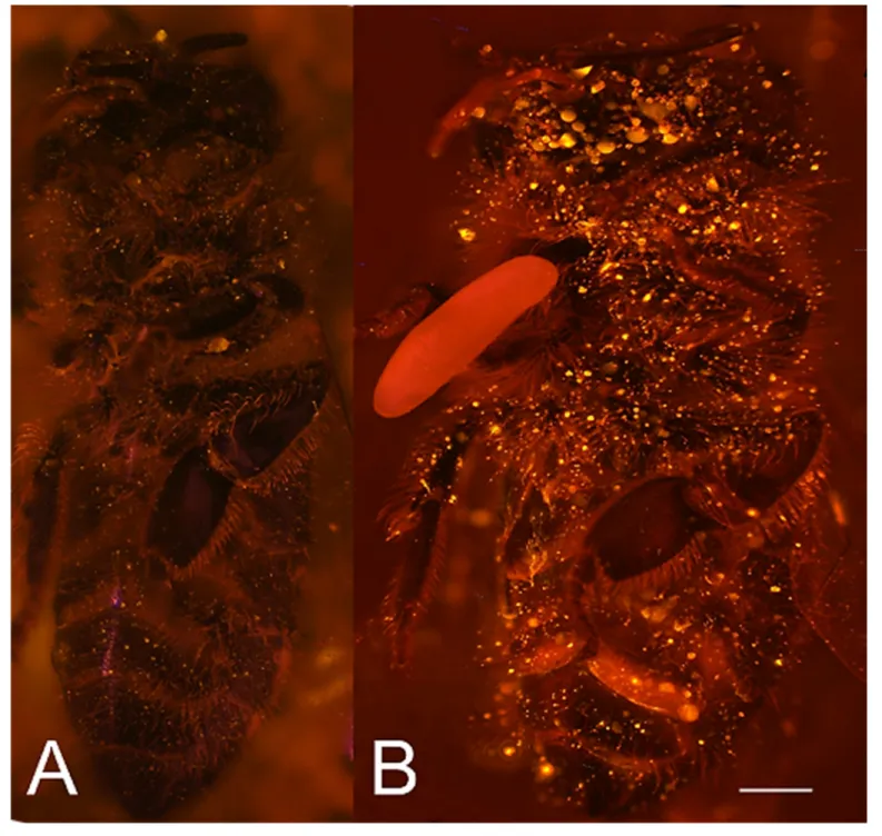
Chi square = 9.5, df = 1, p=0.002), suggesting that this effect is not due to possible antifungal mech- anisms of the larvae but that it is mediated by the egg. Considering the distinctive odor that Figure 1.Paralyzed honeybees under different conditions. (A) Brood cell of the European beewolf with two bees, one carrying an egg, in an
observation cage. (B) Honeybee paralyzed by a beewolf female but immediately removed and kept in an artificial brood cell, heavily overgrown by mold fungi that have already developed conidia. Scale bar = 5 mm.
DOI: https://doi.org/10.7554/eLife.43718.003
Research article Ecology Evolutionary Biology
emanated from the eggs, we tested whether the antifungal effect is caused by a volatile agent. Two experiments supported this assumption. First, provisioned bees without wasp eggs that were kept in artificial brood cells together with bees carrying an egg (but without physical contact) showed signif- icantly delayed fungal growth compared to control bees that were kept alone (Breslow test, day 0–
11: Chi square = 7.6 df=1, p=0.006; day 0–3: Chi square = 9.1, df = 1, p=0.003;Figure 2B). Second, when one of the most abundant mold species from infested beewolf brood cells, the fast growing Aspergillus flavus(Engl et al., 2016), was exposed to the volatiles presumably emanating from bee- wolf eggs on nutrient agar for three days, its growth was entirely inhibited, whereas it thrived in con- trols (for all observation times 24 hr, 48 hr, 72 hr: binomial test: N = 20, p<0.001, Figure 3).
Notably, when the beewolf larvae were removed from the assays shortly after hatching (three days after oviposition), no fungal growth occurred in the exposed areas during another three days. A simi- lar experiment showed that paralyzed honeybees alone did not show any antifungal activity (Appen- dix 1: Additional data 1). Analogous bioassays with five other fungal strains (A. flavusstrain B, Mucor circinelloides, Penicillium roqueforti, Candida albicansandTrichophyton rubrum) revealed that in all cases fungus growth was likewise completely inhibited in the area that was exposed to volatiles from beewolf eggs, whereas the fungi thrived in the respective control areas (for each strain: N = 8, bino- mial test: p<0.01). We conclude that beewolf eggs release a volatile compound with broad spectrum fungicidal properties.
Identification of the antifungal volatile
The odor emanating from the eggs was similar to that of strong oxidants like chlorine, ozone and nitrogen dioxide (subjective evaluation of several observers, [Mu¨cke and Lemmen, 2010]). In fact, Figure 2.Onset of fungal growth on paralyzed honeybees taken fromPhilanthus triangulumnests and kept in artificial brood cells. The fraction of bees showing first signs of fungal growth is shown as a function of days since oviposition. (A) Honeybees that either carried an egg (dashed line) or not (solid line) (N = 22 each, hazard ratio = 0.29, 95% confidence interval: 0.13–0.64). (B) Honeybees that were either kept alone (solid line) or shared a brood cell with a bee carrying an egg (dashed line) (N = 16 each, hazard ratio = 0.39, 95% confidence interval: 0.17–0.9).
DOI: https://doi.org/10.7554/eLife.43718.004 The following source data is available for figure 2:
Source data 1.Effect of egg on fungus growth.
DOI: https://doi.org/10.7554/eLife.43718.005
Research article Ecology Evolutionary Biology
the generation of a strong blue coloration when placing a beewolf egg into the lid of a reaction tube filled with a iodide/starch solution ([Jander and Blasius, 1971], see iodometry in Materials and methods) revealed the existence of an oxidant in the headspace of beewolf eggs.
There are few gaseous oxidants that might be considered, in particular chlorine, ozone and nitrogen dioxide. Ozone has, to our knowledge, not been described to be synthesized by organisms. Molecu- lar chlorine has been reported as an intermediate in some organisms but its occurrence seems to be restricted to phagocytosis (Hazen et al., 1996). The most likely candidate was the radical nitrogen dioxide (NO2
), because there is a plausible way for it being generated by wasp eggs: Insect embryos synthesize small amounts of nitric oxide (NO) as signaling effectors for developmental pro- cesses (Andersen et al., 2013). If such odorless NOwas emitted from the egg, it would spontane- ously react with oxygen (Soegiarto et al., 2003;Mur et al., 2011) to yield the strong-smelling NO2
. Moreover, belonging to the reactive nitrogen species (RNS), NOand NO2
show considerable anti- mycotic activity (Fang, 1997;Lai et al., 2011) that would explain the observed fungicidal effect of beewolf eggs. Hence, we hypothesized that eggs synthesize and emit NOthat reacts with the oxy- gen in brood cells to NO2
thus generating the pungent smell and the antimycotic activity.
We tested whether beewolf eggs produce and emit NOand/or NO2
by conducting a series of experiments. First, headspace samples of confined beewolf eggs were subjected to the Griess assay, the standard procedure for the specific detection of NOand NO2
(Tsikas, 2007). The emerging red color of the resulting azo dye (Figure 4—figure supplement 1) clearly indicated the presence of NO/NO2
. To visualize the emission of NOfrom beewolf eggs, we sprayed a solution of an NOspe- cific fluorescent probe, Diaminorhodamin-4M AM (DAR4M-AM), onto prey bees carrying freshly laid eggs. The small droplets of the DAR4M-AM solution on the bees showed a clear fluorescence Figure 3.Bioassay demonstrating the inhibitory effect of a beewolf egg againstAspergillus flavus. Two areas on the agar were covered by caps of a volume similar to natural beewolf brood cells. One cap, the control (C), was empty, while the experimental cap (E) contained a fresh beewolf egg attached to the ceiling of the cap. The caps were removed and the picture was taken after 24 hr of incubation at 25˚C. The control area (C) shows dense whitish fungal hyphae similar to the surroundings. However, the area that was exposed to the volatiles from a beewolf egg (E) shows bare agar, indicating that the growth of this aggressive fungus was entirely inhibited. Scale bar = 2.5 cm.
DOI: https://doi.org/10.7554/eLife.43718.006
Research article Ecology Evolutionary Biology
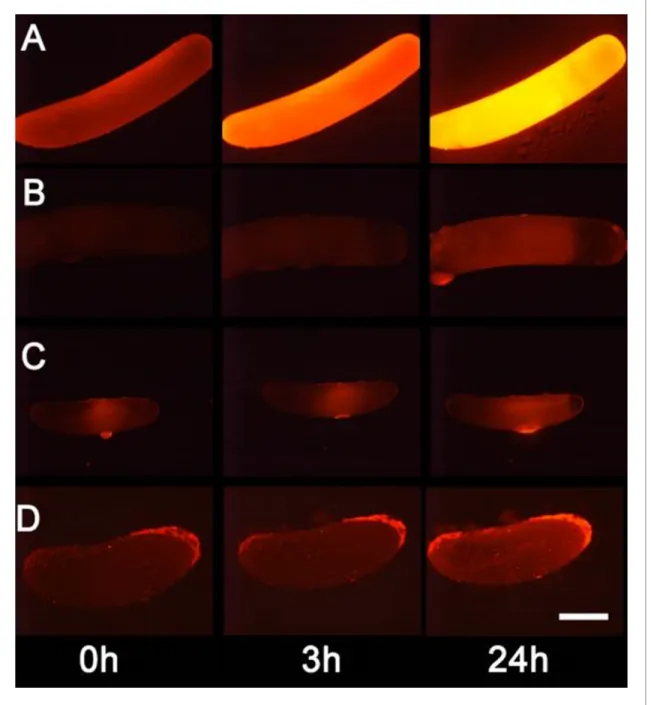
around the egg that increased over several hours (Figure 4B). No such effect was seen on control bees without eggs (Figure 4A). Moreover, beewolf eggs injected with the DAR4M-AM solution showed a strong fluorescence that peaked about one day after oviposition (N = 45,Figure 5A). The same treatment yielded only weak fluorescence in the eggs of two other Hymenoptera (the Emerald cockroach wasp, Ampulex compressa, N = 9, and the Red mason bee, Osmia bicornis, N = 12;
Figure 5C and D) and in newly hatched beewolf larvae (N = 4, Figure 5—figure supplement 1).
Autofluorescence of beewolf eggs injected with buffer only (N = 10) was negligible (Figure 5B).
These findings strongly imply that beewolf eggs produce and release NO.
Amount and time course of NO
emission
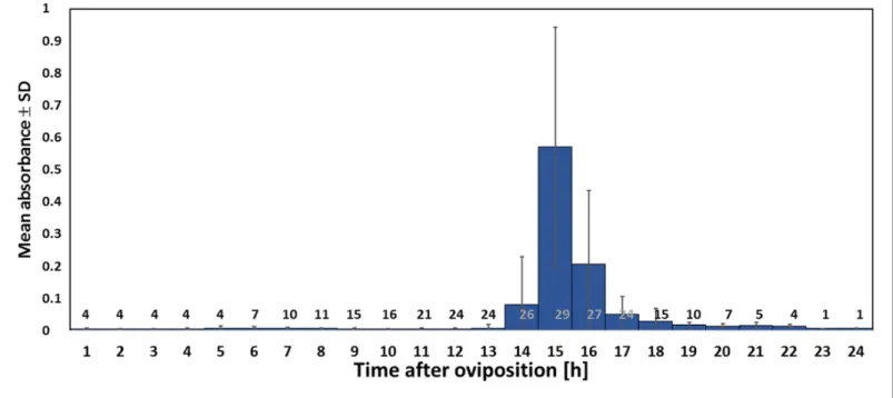
Using iodometry, we determined that a beewolf egg (volume: 4.1±0.5 mm3, N = 16) emits on aver- age 0.25±0.09mmol NO(N = 233). The rate of NOproduction was initially very low, but increased to a distinct peak 14–15 hr (at 28˚C) after oviposition (Figure 6); around 90% of NO emission occurred within a two-hour period. Assuming no loss due to reactions or leaking out of the confined space of brood cells (volume 3.2±0.9 cm3, N = 250), the nitrogen oxides would accumulate to aver- age concentrations of 1690±680 ppm. The timing of the onset of NOemission was strongly tem- perature dependent (Figure 6—figure supplement 1), with higher temperatures resulting in an earlier NOproduction (temperature coefficient Q10= 2.74).
Antifungal effect of synthetic NO
To test whether the observed antifungal effect of the gas that is emitted by beewolf eggs was due to NO, we conducted an experiment using synthetic NObut otherwise emulated natural conditions as closely as possible. Artificial brood cells containing a bee without egg were injected with either synthetic NOto generate a peak concentration of 1500ppm or with nitrogen as controls. There was a significant delay in the onset of fungus growth on the bees exposed to synthetic NOas compared to controls (Figure 7, Breslow test: Chi square = 13.3, df = 1, p<0.0001).
Combined effect of NO
emission and prey embalming
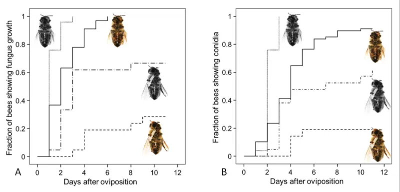
Since both, NOemission and embalming of the prey bees by beewolf females take effect during the first days after oviposition, we assessed the antifungal effects of these defense mechanisms alone and in combination. Bioassays with bees in artificial brood cells that were either embalmed or not and carried an egg or not revealed that the onset of fungal growth differed significantly among treatment groups (Figure 8: Kaplan Meier survival analysis, Breslow test: Chi square = 69.6, df = 3, p<0.001). Pairwise comparisons showed that, on average, fungus growth was first detected on hon- eybees that had not been embalmed and did not carry an egg, second were prey items that were embalmed but did not carry and egg, third were not embalmed honeybees carrying an egg and least susceptible were embalmed honeybees with an egg (Breslow test for all pairwise comparisons:
Chi square8.6, df = 1, p0.003). The timing of conidia formation followed the same pattern (Bre- slow test: Chi square = 67.4, df = 3, p<0.001; for all pairwise comparisons: Chi square4.5, df = 1, p0.034;Figure 8).
Synthesis of NO
in Beewolf eggs
Eukaryotes synthesize NO from the amino acid L-arginine by the enzyme nitric oxide synthase (NOS) (Ro¨szer, 2012) which is highly conserved also in insects (Regulski and Tully, 1995). The exceptional level of NOemission of beewolf eggs raised the question of whether they employ the same pathway or have evolved a different mechanism. First, using histological staining, we assessed evidence for and site of NOS activity in beewolf eggs during the time of peak NOemission. The fix- ation insensitive nicotinamide-adenine-dinucleotide phosphate (NADPH) -diaphorase assay resulted in strong blue staining only in embryonic tissue (Figure 9), thus indicating NOS activity. Second, by employing reverse transcription and real time quantitative PCR, we revealed that the temporal expression pattern of NOS-mRNA showed a clear peak around 19–20 hr after oviposition (Fig- ure 10). For this experiment, the eggs were kept at 25˚C, so the timing of peak NOS expression cor- responds to the timing of peak NOemission at this temperature (Figure 6—figure supplement 1).
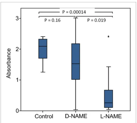
Third, to directly test for the involvement of NOS we injected beewolf eggs with Nw-nitro-L-arginine methylester (L-NAME), a NOS-inhibiting analog of L-arginine (Willmot et al., 2005). This treatment
Research article Ecology Evolutionary Biology
Figure 4.Visualization of NOemission by beewolf eggs using fluorescence imaging. (A) Honeybee from a brood cell without an egg and (B) honeybee with egg. Both bees were sprayed with a solution of the NOspecific fluorescence probe DAR4M-AM. Only the droplets on the bee with the egg (B) show a bright yellow and orange fluorescence indicating the presence of NO. Images are composites of multiple pictures of the x/y plane and z-axis.
Scale bar = 1 mm.
DOI: https://doi.org/10.7554/eLife.43718.007
The following figure supplement is available for figure 4:
Figure supplement 1.Results of the Griess assay with beewolf eggs.
DOI: https://doi.org/10.7554/eLife.43718.008
Research article Ecology Evolutionary Biology
caused a significant decrease in NOemission, whereas the non-inhibiting enantiomer D-NAME had no such effect (Figure 11).
Figure 5.Detection of nitric oxide (NO) in beewolf eggs. Newly laid eggs of beewolves,Philanthus triangulum, of the cockroach waspAmpulex compressaand of the Red Mason bee,Osmia bicorniswere injected with the NOsensitive fluorescence probe DAR4M-AM. Control beewolf eggs were injected with phosphate buffer. Images were obtained by fluorescence microscopy 0, 3 and 24 hr after injection. Row (A) DAR4M-AM injected beewolf egg showing strong increase in fluorescence; (B) Buffer-injected control beewolf egg showing the level of autofluorescence; (C) DAR4M-AM injected egg ofA. compressa;(D) DAR4M-AM injected egg ofO. bicornis. Scale bar: 1 mm.
DOI: https://doi.org/10.7554/eLife.43718.009
The following source data and figure supplement are available for figure 5:
Source data 1.Eggs injected with DAR4M-AM.
DOI: https://doi.org/10.7554/eLife.43718.011
Figure supplement 1.Detection of nitric oxide (NO) in beewolf larva.
DOI: https://doi.org/10.7554/eLife.43718.010
Research article Ecology Evolutionary Biology
The beewolf NOS-gene and NOS-mRNA in eggs
In contrast to vertebrates, most invertebrates appear to have only one type of NOS (Rivero, 2006;
Whitten et al., 2007). Considering the high level of NOproduction in beewolf eggs, we hypothe- sized that beewolves have more than oneNOSgene or that the NOS responsible for the NOsyn- thesis in beewolf eggs might exhibit considerable changes in enzyme structure compared to the NOS of related species. Sequencing of theNOS-gene(s) ofP. triangulum(Pt-NOS) revealed only one Pt-NOScopy in the beewolf genome comprising 9.36 kbp with 25 exons (Figure 11—figure supple- ment 1). A phylogenetic analysis of the resulting amino acid sequence revealed a high similarity to the NOS of the closely related bees (Apidae,Figure 11—figure supplement 2). However, mRNA sequencing showed that, in contrast to adult beewolves and honeybees, the NOS-mRNA of beewolf eggs (3.72 kbp) lacks exon 14 comprising 144 bp. In the NOS-mRNA of adult beewolves this exon is located between the binding domains for calmodulin and flavin mononucleotide (FMN) (Figure 11—
figure supplement 1).
Discussion
Fighting pathogens is of outstanding importance for any organism and has driven the evolution of a great diversity of antimicrobial defenses. Internal immune systems have been extensively docu- mented especially in vertebrates (Akira et al., 2006; Hirano et al., 2011) but also in insects (Lemaitre and Hoffmann, 2007; Siva-Jothy et al., 2005), including insect eggs (Gorman et al., 2004). However, comparatively little is known about external antimicrobial strategies that provide Figure 6.Timing of NOemission from beewolf eggs (kept at 28˚C). The photometrically determined absorbance at 590 nm (mean±SD) is shown as a function of time after oviposition for iodide-starch solutions successively exposed to beewolf eggs for one hour. Sample size (number of eggs measured) at each one hour interval is indicated above the x-axis.
DOI: https://doi.org/10.7554/eLife.43718.012
The following source data and figure supplements are available for figure 6:
Source data 1.Timing of NO emssion.
DOI: https://doi.org/10.7554/eLife.43718.014
Figure supplement 1.Start of NOemission (h after oviposition) as a function of temperature.
DOI: https://doi.org/10.7554/eLife.43718.013
Figure 6—figure supplement 1—source data 1.Start of NO emission.
DOI: https://doi.org/10.7554/eLife.43718.015
Research article Ecology Evolutionary Biology
protection for the own body, for the progeny, or for food. Mechanical grooming is an important mechanism to remove microbes (Zhukovskaya et al., 2013). There are some reports on the applica- tion of antimicrobial secretions on the body surface by adult insects (Wilson and Cotter, 2013;
Otti et al., 2014) or inside a host by larvae of a parasitoid wasp (Herzner et al., 2013). Carrion bee- tles preserve the larval food, buried carcasses, by application of antimicrobials (Degenkolb et al., 2011) and by controlling the microbiome on the carcasses (Shukla et al., 2018). Females of some insect species deposit antimicrobial chemicals (Vander Meer and Morel, 1995; Marchini et al., 1997) or antibiotics producing symbiotic bacteria (Flo´rez et al., 2017; Flo´rez et al., 2015) onto their eggs and ant workers can counter microbial infestation of the brood by applying venom (Tragust et al., 2013). Recently, the employment of volatile antimicrobials by insects as a means of external defense has gathered some interest (Gross et al., 2008; Gross and Schmidtberg, 2009;Weiss et al., 2014;Lopes et al., 2015).
Like other insects that develop in the soil, beewolves are particularly menaced by a diverse and unpredictable range of detrimental microbes. In fact, beewolf progeny and their provisions are under severe threat from fast growing mold fungi (Strohm and Linsenmair, 2001). The development of beewolf progeny from oviposition to cocoon spinning lasts about 11 days and is, thus, rather fast.
So even a few days delay in fungus growth provides a considerable benefit for the larvae. Beewolves have evolved at least three very different antimicrobial defenses that provide an effective, coordi- nated, and long-term protection against a broad spectrum of microbes during the whole develop- ment. First, throughout the long period of winter diapause prior to emergence progeny are protected by antibiotics on their cocoons that are produced by symbiotic Streptomyces bacteria (Kaltenpoth et al., 2005;Kroiss et al., 2010; Kaltenpoth et al., 2014). Second, during the early egg and larval stages, molding of the provisions is retarded by an embalming of the honeybees with Figure 7.Onset of fungal growth (time after onset of experiment) on honeybees that were not embalmed in artificial brood cells. Brood cells were either injected with synthetic NOto a concentration of 1500ppm (solid line) or were injected with nitrogen (dashed line) (N = 20 each, hazard ratio = 0.41, 95% confidence interval: 0.198–
0.845).
DOI: https://doi.org/10.7554/eLife.43718.016 The following source data is available for figure 7:
Source data 1.Effect of synthetic nitric oxide on fungus growth.
DOI: https://doi.org/10.7554/eLife.43718.017
Research article Ecology Evolutionary Biology
lipids by the mother wasp (Strohm and Linsenmair, 2001). Third, as shown here, the emission of gaseous nitrogen oxide radicals by the beewolf egg results not only in delay of molding but in killing of detrimental fungi in their immediate environment thus, at least partly, eliminating this major threat.
Figure 8.Fungus growth on honeybees of four different treament groups. Timing of occurrence of (A) fungal hyphae and (B) conidia on paralyzed honeybees that were (1) not embalmed by beewolf females and did not carry an egg (n = 25, colorless bee, point line), (2) embalmed but did not carry an egg (n = 68, colored bee, solid line), (3) not embalmed but carried an egg (n = 21, colorless bee with egg, dash-point line) or (4) embalmed and carried an egg (n = 21, colored bee with egg, dashed line). SeeAppendix 1—table 2for hazard ratios.
DOI: https://doi.org/10.7554/eLife.43718.018 The following source data is available for figure 8:
Source data 1.Combined effect of embalming and fumigation.
DOI: https://doi.org/10.7554/eLife.43718.019
Figure 9.Micrograph of a longitudinal section of a beewolf egg fixed 15–16 hr after oviposition showing fixation insensitive NADPH-diaphorase activity. Strong blue staining in the embryonic tissue indicates the presence of reduced nitroblue tetrazolium demonstrating NOS activity (c = cuticle, s = serosa, e = embryo, a = amnion, ac = amnion cavity, scale bar = 1 mm, image composed from two separate photos of the left and right parts of the egg.).
DOI: https://doi.org/10.7554/eLife.43718.020
Research article Ecology Evolutionary Biology
The emission of a gaseous agent by beewolf eggs to their confined brood cells is an ideal way to sanitize such intricately structured surfaces as the bodies of honeybees and the rough walls of the brood cell. NOseems to be a most suitable gaseous agent because it can obviously be produced by beewolf eggs in amounts that effectively kill mold fungi in their brood cell. Such volatile sanitation mechanisms that provide a front-line defense against microbes (Gross et al., 2008;Weiss et al., 2014;Lopes et al., 2015) will mostly be inconspicuous and might turn out to be a wider theme in nature.
Exact quantification of nitrogen oxides (NOand NO2
) in beewolf brood cells on a micro scale or with time has not yet been accomplished. Brood cells are located in rather compact fine grained sandy soil with some moisture. Moreover, the walls of the nest burrows and the brood cells are cov- ered with a layer of hydrocarbons (Kroiss et al., 2009) that might provide an additional barrier.
Thus, brood cell walls are neither very porous nor are they sealed. Accordingly, the concentration of NOand NO2
in the brood cell will decrease but at a slow rate. By the time the larvae hatch (three days after oviposition) the smell of NOhas vanished, indicating that the nitrogen oxides have disap- peared or at least decreased considerably, explaining why the larvae remain unaffected without actually being resistant to NO. However, even assuming some loss during the two hour period of peak NOproduction the estimated maximum concentration of nitrogen oxides (NO and NO2
) in beewolf brood cells (probably around 1500 ppm or 60mmol/l) considerably exceeds the concentra- tions observed in animal tissues (mostly lower than 0.1mmol/l [Wink et al., 2011], 0.85–1.3mmol/l in muscle tissue [Vaughn et al., 1998]). The maximum concentration in beewolf brood cells might be even higher than what is used in medical applications against multiple drug resistant bacteria (200 ppm NO[Ghaffari et al., 2006]) or in antifungal treatment of fruit (50–500 ppm NO[Lazar et al., 2008]) and is far beyond permissible exposure limits for humans (e.g. for the USA: 25 ppm for NO, 5 ppm for NO2
[Administration USOSaH, 2014]).
Synthetic NOapplied to artificial brood cells at a concentration of 1500ppm, the estimated con- centration of nitrogen oxides in natural brood cells, significantly delayed fungus growth on bees.
Since there was oxygen available in the brood cell, NOwas oxidized to NO2
similarly to natural brood cells. The effect size (NOtreatment vs control,Figure 7: hazard ratio = 0.41, 95% confidence Figure 10.Gene expression of NOS relative to ß-actin in beewolf eggs at different times after oviposition and in freshly hatched larvae. Two trials were conducted, each with 25 pooled eggs or larvae per time interval. Mean ratios of NOS-mRNA to ß-Actin-mRNA are shown (with standard deviations), as determined by Q-RT-PCR.
DOI: https://doi.org/10.7554/eLife.43718.021 The following source data is available for figure 10:
Source data 1.NOS gene expression.
DOI: https://doi.org/10.7554/eLife.43718.022
Research article Ecology Evolutionary Biology
interval 0.198–0.845) was slightly lower than in a comparable experiment with eggs as the source of the antifungal gas (data for unembalmed bees from the experiment of a combined effect of embalming and emissions from the egg:Appendix 1—table 2: hazard ratio = 0.22, 95% confidence interval 0.1–0.47). However, since the confidence intervals of the hazard ratios are mutually overlap- ping, there is no evidence for a significant difference in the antifungal effect of 1500ppm synthetic NOand the gas emitted by beewolf eggs. Although we cannot exclude that small amounts of other active volatiles are released by beewolf eggs, we thus conclude that the antifungal effect of brood cell fumigation by beewolf eggs is predominantly or exclusively due to NOand its oxidation product NO2
.
Notably, the combination of prey embalming with unsaturated hydrocarbons that reduces con- densation of water on the bees (Herzner and Strohm, 2007) and brood cell fumigation with NO/ NO2
seems to affect fungal growth beyond either of these antimicrobial measures alone. One possi- ble explanation is based on the fact that NOand NO2
dissolve in water (confirmed by the spraying of a bee with a fluorescent dye,Figure 4) to yield nitric acid and nitrous acid, with the latter being a potent antibacterial agent (Gao et al., 2015). Although embalming reduces the amount of water Figure 11.Effect of NOS inhibition on NOproduction. Amount of NOand/or NO2
emanating from non-injected beewolf eggs (control; N = 14) and those injected with D-NAME (a non-inhibiting enantiomer of L-NAME, N = 9) or L-NAME (a NOS inhibiting L-arginine analog, N = 14). The photometrically determined absorbance at 590 nm is shown for iodide-starch solutions that were exposed for 24 hr to the headspace of eggs of the indicated treatment group (shown are median, quartiles and range, * indicates an outlier, included in the analysis). P-values are for Holm-corrected Mann-Whitney U-tests.
DOI: https://doi.org/10.7554/eLife.43718.023
The following source data and figure supplements are available for figure 11:
Source data 1.NOS inhibition.
DOI: https://doi.org/10.7554/eLife.43718.026
Figure supplement 1.Structure of thePt-NOSgene indicating position and length of exons.
DOI: https://doi.org/10.7554/eLife.43718.024
Figure supplement 2.Consensus tree obtained from Bayesian analysis of NOS amino acid sequences from five orders of insects (distinguished by different colors), including the NOS sequences ofP. triangulumeggs (lowermost entry).
DOI: https://doi.org/10.7554/eLife.43718.025
Research article Ecology Evolutionary Biology
condensation on the prey bees, some very small droplets occur. The concentration of nitrous acid in these droplets will be considerably higher than in the larger droplets that would occur without embalming. Thus, fungal germs that might have survived the NO/NO2
atmosphere are not only impaired by limited availability of water, but the accessible water might be toxic for them. Moreover, due to the solubility of NOand NO2
, abundant water droplets on the bees could reduce the con- centration of these radicals in the brood cell, thus lessening their antimicrobial effect. The reduction of water on the bees due to prey embalming could thus help to keep fumigation effective. The com- bination of prey embalming and fumigation, thus, has a twofold effect. Many fungi will be killed by the NO/NO2
. The remaining spores will encounter unfavorable conditions that slow-down germina- tion and growth so that the majority of larvae are able to consume most of their provisions without severe competition by mold fungi. Once larvae have spun their cocoon the antibiotics that are pro- duced by the symbiotic bacteria take over protection until emergence (Kaltenpoth et al., 2005;
Kroiss et al., 2010;Engl et al., 2018). Within this multifaceted antimicrobial strategy of beewolves, brood cell fumigation might be the most important component since it takes effect at a very early developmental stage and, thus, provides beewolf offspring with a decisive head start over the fast growing mold fungi.
NO is an ancient biological effector of immense importance for all kinds of organisms ranging from prokaryotes to higher plants and animals (Ro¨szer, 2012;Moroz and Kohn, 2007). Owing to its high diffusibility across biomembranes and specific chemical properties, this gaseous radical plays a crucial role in a multitude of biological processes (Ro¨szer, 2012;Moroz and Kohn, 2007). In verte- brates, NOis synthesized from L-arginine by three different isoforms of NOS that are encoded by different genes (Ro¨szer, 2012;Moroz and Kohn, 2007). Low levels of NO (<1mmol/l) are produced by constitutive NOS (cNOS) isoforms (endothelial eNOS, neuronal nNOS) and have signaling functions, for example in neuronal development and in the regulation of vascular tone in vertebrates.
Higher NOconcentrations (1–10mmol/l, [Thomas et al., 2003]) are generated by an inducible NOS (iNOS). At such levels NOis highly cytotoxic (Thomas et al., 2003), making it a powerful antimicro- bial (Fang, 1997;Lai et al., 2011), for example in macrophages (Ro¨szer, 2012). However, overpro- duction of NO due to inflammatory processes (Filipovic´ et al., 2010) or certain diseases (e.g.
Alzheimer’s disease, [Lu¨th et al., 2002]) may cause harmful side-effects (Pacher et al., 2007) and even septic shock (Titheradge, 1999). Moreover, NOmight affect carcinogenesis and tumor pro- gression in a positive as well as in a negative way (Burke et al., 2013).
In living tissues, NOis usually removed within seconds by reacting with the heme group of mole- cules such as oxyhemoglobin (Beckman and Koppenol, 1996;Wink et al., 2011) (very low concen- trations may still persist for hours [Moroz and Kohn, 2007]). In brood cells, there is enough oxygen (670ml) to support the metabolism of the egg and of the paralyzed bee as well as the oxidation of NO(for more details see Appendix 1: Additional discussion 1). In air, the autooxidation to NO2
is comparatively slow so that NOmay persist (depending on its concentration) for several seconds to minutes (Mur et al., 2011;Wink et al., 2011) or even hours (Soegiarto et al., 2003). Thus, the NO emitted by beewolf eggs might directly affect fungi, for exapmle by damaging DNA (Lai et al., 2011;Jones et al., 2010) or by reacting with the heme group of enzymes like cytochrome P450 and cytochrome c oxidase, thus inhibiting these crucial components of the mitochondrial respiratory chain (Thomas et al., 2003;Feelisch, 2008;Canessa and Larrondo, 2013). Yet, most of the antimi- crobial activity of NOis attributed to indirect effects via reactive nitrogen species (RNS), in particular nitrogen oxides (NO2
, N2O3) and peroxynitrite (ONOO-, upon reaction with superoxide) (Thomas et al., 2003). NO2
, has been reported to be severely cytotoxic, for example by nitration of tyrosine residues and oxidation of proteins and lipids (Fang, 1997;Bogdan, 2001).
A beewolf egg of approximately 5 mg emits 0.25mmol NOwithin a period of about 2.5 hr, or 20.000mmol/kg*h, a value that is about four orders of magnitude higher than reported baseline lev- els of NOsynthesis in humans (0.15 -~4.5mmol/kg*h [Castillo et al., 1996], rats (0.6–9mmol/kg*h [Wu et al., 1999]) and plants (Arabidopsis thaliana, 0.36–3 mmol/kg*h [Zeidler et al., 2004]), and even considerably higher than in lipopolysaccharide (LPS)-activated macrophages (~800mmol/kg*h, estimated fromWu et al., 1999). To investigate whether a NOS was involved in NOproduction in beewolf eggs we conducted three experiments. First, a specific histochemical assay indicated NOS activity in embryonic tissue but not in other parts of the egg. Second, quantitative PCR revealed ele- vated expression of the NOS gene at the time of peak NOproduction. Finally, competitive inhibi- tion of NOS by L-NAME caused a significant reduction in NOproduction. While each of the three
Research article Ecology Evolutionary Biology
results might have alternative explanations (e.g. L-NAME might not be a perfectly specific NOS inhibitor [Peterson et al., 1992]), taken together these findings provide strong evidence that a NOS, located in the embryonic tissue, is involved in NOproduction of beewolf eggs.
Searching for possible adaptations that might accomplish this extremely high rate of NOproduction the beewolf NOS gene was sequenced. Only one beewolf NOS (Pt-NOS) gene was found. The derived amino acid sequence did not reveal considerable differences compared to the NOS of the closely related bees (Figure 11—figure supplement 2). Thus, there is no evidence for extensive evolutionary changes with regard to the gene itself. Moreover, the structure of the Pt- NOS gene is largely homologous to other insects, for example Anopheles stephensi mosquitoes (Luckhart et al., 1998).
However, in contrast to adult beewolves, the NOS-mRNA in beewolf eggs lacks exon 14 (144 bp, Figure 11—figure supplement 1). Such alternative splicing that results in different NOS-mRNAs, including the deletion of exons (but others than in beewolves), has been documented inA. stephensi in response toPlasmodium infection (Luckhart and Li, 2001). Moreover, NOS splice variants may result in organ-specific enzymes in other organisms (Ro¨szer, 2012). Presumably, beewolf eggs pro- duce smaller amounts of another NOS splice variant to support signaling functions in the developing embryo.
In adult beewolves, the exon missing in the NOS-mRNA of eggs is located between the binding domains for calmodulin and FMN. Since calmodulin is believed to be responsible for NOS regulation (Smith et al., 2013) the deletion of an adjacent part might affect the control of NOS activity in bee- wolf eggs. Thus, the alternative splicing might enable the production of such large amounts of NO. Notably, compared to the cNOS (comprising eNOS, and nNOS) the inducible NOS isoform of verte- brates (iNOS) that generates higher concentrations of NO to combat microbes lacks a section of about 40 amino acids (120 bp) near the FMN domain. Interestingly, this section is thought to be responsible for autoinhibition of the cNOS (Salerno et al., 1997) and its lack enhances NOproduc- tion by the iNOS. The conspicuous similarity between vertebrate iNOS and the NOS in beewolf eggs with regard to the length of the missing section and its position might suggest a convergent modification to achieve a NOS with high synthetic capacity. Whereas vertebrates have evolved another gene, beewolf eggs might accomplish a similar effect by alternative splicing of the mRNA.
The possible loss of regulation of the NOS and the pattern ofPt-NOSexpression in the eggs sug- gest that in beewolf eggs the activity of the enzyme is regulated by gene expression like the NOS in PlasmodiuminfestedA. stephensi(Luckhart et al., 1998) and the iNOS of vertebrates (Wong et al., 1996;Morris Jr, 1999). However, in contrast to these caes, in beewolf eggs expression of thePt- NOSseems not to be induced by immunostimulants but to occur obligatorily at a certain stage in the development of the beewolf embryo. While we cannot exclude that there is an additional, yet unknown, pathway of NOproduction in beewolf eggs, we hypothesize that the NOS and in particu- lar its alternative splice variant plays a significant role in brood cell fumigation by beewolf eggs.
However, even the combined effect of prey embalming and brood cell fumigation does not pro- vide perfect protection as fungus infestation still causes larval mortality in 5% of the brood cells in the field (Strohm and Linsenmair, 2001). Some fungal spores might survive under the bees because they were screened against the gas. Another possibility, namely that strains of the ubiquitous mold fungi that are the main causes of molding in beewolf brood cells (Engl et al., 2016), have evolved resistance against the toxic effects of NO/NO2
seems rather unlikely. Ultimately, there will be only weak selection for resistance at all since beewolf brood cells are certainly a rare habitat for the ubiq- uitous mold fungi (Engl et al., 2018). Moreover, there will be no repeated exposure of the same fun- gal strains to fumigation that would be required to favor the evolution of resistance. While there are examples for detoxification of lower concentrations of NO(mainly by scavengers like flavohemoglo- bins) in different fungi, including species ofAspergillus(Martins et al., 2011;Zhou et al., 2009), the NO/NO2
levels emitted by beewolf eggs are very high and likely affect several very basic biochemi- cal processes, thus making the evolution of an effective resistance unlikely.
While brood cell fumigation clearly retards molding of larval provisions, the antimicrobial effect of NOand NO2
might harm the symbioticStreptomycesbacteria that beewolf females apply to the brood cell prior to egg laying (Kaltenpoth et al., 2005;Kroiss et al., 2010). Since the symbiotic bacteria are important for the survival of larvae in the cocoon and are vertically transmitted from beewolf mothers to their daughters (Kaltenpoth et al., 2014), a considerable number of symbionts have to survive the brood cell fumigation. At the moment we can only speculate how the bacteria
Research article Ecology Evolutionary Biology
can survive. Conceivably, because of strong selection due to specialization and repeated exposition, the symbiotic bacteria have evolved mechanisms to cope with the high concentrations of NO/NO2
(Poole, 2005; Wareham et al., 2018). Possibly, the fumigation slowly evolved after the establish- ment of the symbiosis; thus bacteria might have been able to gradually evolve resistance. Moreover, the bacteria are applied to the ceiling of the brood cell, which might reduce negative effects of the nitrogen oxides since these are heavier than air (Lide, 1995) and will accumulate in the lower part of the brood cell. Additionally, the bacteria are embedded in copious amounts of a secretion consisting of mostly unsaturated hydrocarbons (Kaltenpoth et al., 2009) that might shield the bacteria from the fumigants. Finally, host- and/or symbiont derived antioxidants in the hydrocarbon matrix could detoxify NOand NO2
and protect the symbioticStreptomycesbacteria.
How could brood cell fumigation with high concentrations of NO/NO2
have evolved? Generally, it has been assumed that the primary purpose of NOwas signaling at low concentrations and that the antimicrobial functions of higher concentrations are derived (Fang, 2004). Assuming a similar scenario for beewolves, small amounts of NOthat were originally produced for developmental pro- cesses (Andersen et al., 2013) might have accidentally been released into the confines of the sub- terranean brood cell and slightly affected the germination or growth of fungi by interfering with regulatory processes (Ro¨szer, 2012;Wang and Higgins, 2005). Given the severe threat posed by microbes, such initial benefits would have caused strong selection for elevated NOemission by the eggs. This would have considerably increased progeny survival and might have allowed ancestral beewolves to nest in an expanded range of habitat types, including nesting sites with high risk of microbial infestation, or to exploit highly susceptible but readily available prey species. Brood cell fumigation with large doses of NOthus represents a key evolutionary innovation. Since NOis used as an antimicrobial in the immune systems of many animals (Bogdan et al., 2000), its deployment as an antifungal gas can be viewed as an innate, externalized immune defense of beewolf eggs. Such externalized components of the immune system have recently been recognized as important and possibly widespread antimicrobial measures (Otti et al., 2014).
The clear benefit of brood cell fumigation, however, is probably accompanied by substantial costs in terms of energy and biochemical resources (Rivero, 2006). NOis synthesized from L-arginine, an amino acid that is an important constituent of many proteins and biochemical pathways (Morris Jr, 2000) and it is an essential amino acid for most insects (Barbehenn et al., 1999;Payne and Loomis, 2006) (e.g. phytophagous insects [Berenbaum, 1995], mosquitos [Uchida, 1993], aphids [Sasaki and Ishikawa, 1995;Akman Gu¨ndu¨z and Douglas, 2009], butterflies [Erhardt and Ruster- holz, 1998; O’Brien et al., 2003], true bugs [Mesquita et al., 2015], parasitoid wasps [Thomp- son, 1976; Barrett and Schmidt, 1991], bees [de Groot, 1952; Weiner et al., 2010]). Thus, beewolves have either evolved the capacity to synthesize L-arginine or female beewolves have to provide each egg with sufficient L-arginine for both brood cell fumigation and embryogenesis.
Moreover, NO synthesis by NOS requires the cofactors flavin adenine dinucleotide (FAD), FMN, (6R-)5,6,7,8-tetrahydrobiopterin (BH4) and NADPH (Fo¨rstermann and Sessa, 2012), thus competing with other metabolic pathways in the developing beewolf embryo.
One of the most remarkable aspects of our study is that the embryos inside the egg survive the high concentrations of toxic nitrogen oxides during synthesis and emission as well as after its release to the brood cell. This is all the more surprising since beewolf larvae that were accidentally exposed to the gas emitted by eggs died (Strohm, unpublished observations). The synthesis and emission of such high amounts of NOlikely requires a number of concomitant adaptations that protect beewolf embryos against the cytotoxic effects of high concentrations of NOand NO2
. One possibility is the employment of carrier molecules to transfer NOto the egg shell. In blood sucking hemipterans, for example, nitrophorins carry NOto its release site to dilate blood vessels (Davies, 2000). The mech- anistic basis of NOtolerance of beewolf eggs is of particular interest, since excessive production of NOdue to inflammatory processes (Guzik et al., 2003) or certain diseases (e.g. Alzheimer’s disease, [Lu¨th et al., 2002;Pacher et al., 2007; Calabrese et al., 2007;Pautz et al., 2010]) might cause severe pathological complications in humans. Thus, understanding how beewolf eggs avoid the toxic effects of NOmight inspire the development of novel medical applications.
Our findings reveal a surprising adaptation in a mass-provisioning digger wasp to cope with the threat of pathogen infestation in the vulnerable egg and larval stages. Sanitizing the brood cell envi- ronment by producing high amounts of NO significantly enhances the survival of immatures by reducing fungal growth on their provisions. Given that mass-provisioning and development
Research article Ecology Evolutionary Biology
underground are widespread ecological features among digger wasps and bees and considering the difficulties of detecting volatiles in subterranean nests, such gaseous defenses might be more wide- spread and as yet underappreciated. In addition to revealing new perspectives on antimicrobial strategies in nature and amplifying the biological significance of NO, beewolves offer unique oppor- tunities to elucidate general questions on the evolution and regulation of NOS as well as the produc- tion of and resistance to high concentrations of NO.
Materials and methods
Key resources table Reagent type (species)
or resource Designation
Source or
reference Identifiers
Additional information Biological
sample
European beewolf, Philanthus triangulum
Field caught or laboratory reared F1 of field caught females Biological
sample
Emerald cockroach wasp,
Ampulex compressa
Laboratory reared
Biological sample
Red mason bee, Osmia bicornis
Field caught
Biological sample
Aspergillus flavus Strain I: Isolated from beewolf brood cells, Strain II: Department of Hygiene and Microbiology of the University Hospital, Wu¨rzburg, Germany
na
Biological sample
Penicillium roquefortii
Department of Hygiene and Microbiology of the
University Hospital, Wu¨rzburg, Germany
na
Biological sample
Candida albicans Department of Hygiene and Microbiology of the
University Hospital, Wu¨rzburg, Germany
na
Biological sample
Trichophyton rubrum Department of Hygiene and Microbiology of
the University Hospital, Wu¨rzburg, Germany
na
Sequence- based reagent
Adapter + PolyT 3’RACE, Molecular cloning protocol
SeeSupplementary file 1
Sequence- based reagent
Adapter 3’RACE, Molecular
cloning protocol
See
Supplementary file 1
Sequence- based reagent
polyT Reverse
transcription protocol
See
Supplementary file 1
Sequence- based reagent
NOS_qPCR_F2 P. triangulum, this paper
See
Supplementary file 1
Sequence- based reagent
NOS_qPCR_R2 P. triangulum, this paper
See
Supplementary file 1
Sequence- based reagent
Actin_qPCR_F1 Apis mellifera,Gryllus bimaculatus, P. triangulum,
this paper
See
Supplementary file 1
Sequence- based reagent
Actin_qPCR_R1 A. mellifera, G. bimaculatus, P. triangulum, this paper
See
Supplementary file 1
Continued on next page
Research article Ecology Evolutionary Biology
Continued Reagent type (species)
or resource Designation
Source or
reference Identifiers
Additional information Sequence-
based reagent
NOS860fwd2 A. mellifera,
D. melanogaster, Anopheles stephensi, Rhodnius prolixus, Manduca sexta, this paper
See
Supplementary file 1
Sequence- based reagent
NOS1571rev1 A. mellifera,
D. melanogaster,A. stephensi,R. prolixus, M. sexta, this paper
See
Supplementary file 1
Sequence- based reagent
NOS_seq_F1_deg A. mellifera, Nasonia vitripennis, this paper
See
Supplementary file 1
Sequence- based reagent
NOS_seq_R1_deg A. mellifera,N. vitripennis, this paper
See
Supplementary file 1
Sequence- based reagent
NOS_seq_5-F1 P. triangulum, this paper
See
Supplementary file 1
Sequence- based reagent
NOS_seq_5-R1 P. triangulum, this paper
See
Supplementary file 1
Sequence- based reagent
NOS_seq_5-F2 P. triangulum, this paper
See
Supplementary file 1
Sequence- based reagent
NOS_seq_5-R2 P. triangulum, this paper
See
Supplementary file 1
Sequence- based reagent
NOS_seq_5-F3 P. triangulum, this paper
See
Supplementary file 1
Sequence- based reagent
NOS_seq_5-F6 P. triangulum, this paper
See
Supplementary file 1
Sequence- based reagent
NOS_seq_3-F1 P. triangulum, this paper
See
Supplementary file 1
Sequence- based reagent
NOS_seq_3-R1 P. triangulum, this paper
See
Supplementary file 1
Sequence- based reagent
NOS_seq_3-F2 P. triangulum, this paper
See
Supplementary file 1
Sequence- based reagent
NOS_seq_3-R2 P. triangulum, this paper
See
Supplementary file 1
Sequence -based reagent
NOS_seq_3-F3 P. triangulum, this paper
See
Supplementary file 1
Sequence- based reagent
NOS_seq_3-F6 P. triangulum, this paper
See
Supplementary file 1
Sequence- based reagent
NOS_RT_R1 P. triangulum,
this paper
See
Supplementary file 1
Commercial assay or kit
Griess assay.
Merck Spectroquant
Merck,
Darmstadt, Germany
114776
Continued on next page
Research article Ecology Evolutionary Biology
Continued Reagent type (species)
or resource Designation
Source or
reference Identifiers
Additional information Commercial
assay or kit
peqGOLD total RNA Kit
peqLab,
Erlangen, Germany
732–2867
Commercial assay or kit
GeneRacer Kit Invitrogen, Carlsbad, CA, USA
L1502-01
Commercial assay or kit
BioScript One- Step RT-PCR-Kit
Bioline, London, UK BIO-65033
Commercial assay or kit
peqGOLD Taq- DNA-Polymerase
peqLab, Erlangen, Germany
01–1030
Commercial assay or kit
SensiMixPlus SYBR Mit
Quantace/Bioline, London, UK
QT615-05
Commercial assay or kit
Epicentre MasterPure Complete DNA and RNA purification Kit
Epicentre, now Lucigen, Middleton, WI, USA
MC85200
Commercial assay or kit
innuPREP RNA Mini Kit
Analytik Jena, Jena, Germany
845-KS-2040050
Commercial assay or kit
PeqGOLD Mid- Range PCR System
peqLab,
Erlangen, Germany
PEQL02-3020_P
Chemical compound, drug
DNase I Fermentas, Lithuania
Now Thermo Fisher Scientific, Germany
EN0525
Chemical compound, drug
Oligo-dT primer Fermentas, Lithuania Now Thermo Fisher Scientific, Germany
na
Chemical compound, drug
L-NAME Hydrochloride
Axxora Deutschland, Lo¨rrach, Germany
ALX-105–004 M250
Chemical compound, drug
D-NAME Hydrochloride
Axxora Deutschland, Lo¨rrach, Germany
ALX-105–003 G005
Chemical compound, drug
DAR-4M AM Axxora Deutschland,
Lo¨rrach, Germany
ALX-620–069 M001
Chemical compound, drug
4-Nitro-m-Xylol Merck,
Darmstadt, Germany
8415470025
Chemical compound, drug
NADPH- Tetranatriumsalz
Carl-Roth,
Karlsruhe, Germany
AE14.1
Tools Eppendorf
Microinjector with Femtotips II
Eppendorf, Hamburg, Germany
930000043
Tools Axiophot II
Fluorescence microscope
Zeiss, Jena, Germany
Tools Nikon DS-2 Mv Nikon, Tokyo, Japan
Tools Uvikon 860
spektrophotometer
Kontron,
Augsburg, Germany
Tools Cryostat
microtome CM3000
Leica,
Wetzlar, Germany
Tools Eppendorf
Realplex Cycler
Eppendorf, Hamburg, Germany Continued on next page
Research article Ecology Evolutionary Biology