Mak and Swaminathan: Assessment of vitamin BI, B2 and B6 status 213 J. Clin. Chem. Clin. Biochem.
Vol. 26, 1988, pp. 213-217
© 1988 Walter de Gruyter & Co.
Berlin · New York
Assessment of Vitamin BI, B
2and B
6Status by Coenzyme Activation of Red Cell Enzymes Using a Centrifugal Analyser
By Y. T. Mak and R. Swaminathan
Department of Chemical Pathology, Prince of Wales Hospital, The Chinese University of Hong Kong, Shatin, Hong Kong
(Received July 13, 1987/January 20, 1988)
Summary: We describe a fully automated method for the assessment of vitamin B1?
B
2and B
6status using a centrifugal analyser. The activation of the red cell enzymes transketolase, glutathione reductase and aspartate aminotransferase
1) by their respective coenzymes were measured in freshly prepared haemolysate. The enzyme catalytic activities in the sample were measured with (maximal activity) and without (basal activity) the coenzyme, and the percentage activation was calculated. The between run precision for red cell transketolase, glutathione reductase and aspartate aminotransferase were 8.5%, 10.3% and 9.5% respectively. When whole blood was stored at room temperature for 6 hours, red cell aspartate aminotransferase activity significantly decreased (n = 10, p < 0.05). There were no significant changes in the activities of the other two enzymes.
For a group of 30 healthy young subjects, the mean (standard deviation) values for the percentage activation of transketolase, glutathione reductase and aspartate aminotransferase were 11.9% (7.3), 35.1% (19.1) and 85.3% (18.0), respectively. The vitamin status of a group of 86 pregnant women was assessed by this method;
2.3%, 8.1% and 8.1%, respectively, of the pregnant women showed a higher percentage activation for transketolase, glutathione reductase and aspartate aminotransferase than that found in the young subjects.
Both groups correlated well with respect to the basal activity and the percentage activation of each enzyme.
Basal activity was inversely proportional to the percentage activation. It is therefore suggested that the basal activity can be used as a second criterion in the assessment of vitamin status.
Introduction Materials and Methods
The nutritional survey of a population requires the Instrument and reagents
analysis of a large number of specimens. Vitamin B The kit for aspartate aminotransferase was obtained from Baker assessment by in vitro coenzyme activation of red cell Instrument Ltd. (Allentown PA, USA). Glycerol phosphate . , ' dehydrogenase/tnose phosphate isomerase, glutathione and enzymes is known to be widely accepted (1, 2) and thiamine pyrophosphate were from Boehringer Mannheim manual spectrophotometric methods are well estab- GmbH (Mannheim, Germany). Tris was from BDH Chemical lished (3-5). However they are time consuming and Co- (?°°le> England). All other chemicals were from Sigma
• 1-1 r i. ji- / ^ i? · Chemical Co. (St. Louis, MO, USA). All analyses were per- not suitable for handling a large number of specimens. formed on a Cobas Bio centrifugal analyser (Roche Diagnostic, Here we describe an automated version of these meth- Basle, Switzerland).
ods. Specimen
1) Enzymes: Blood was collected into heparinized tube and centrifuged 1. Aspartate aminotransferase, I,-Aspartate: 2-oxoglutarate within 2 hours. After separation of plasma, 1 ml of cells was aminotransferase (EC 2,6.1.1) transferred into a 5 ml tube and 4ml of cold isotonic magne- 2. Glutathione reductase, NAD(P)H: glutathione oxidored- sium chloride solution (osmolality = 290 mosmol/kg) was uctase (EC 1.6.4.2) added. The tube was inverted gently several times then centri- 3. Transketolase, Sedoheptulose-7-phosphate: D-glyceralde- fuged at 1000 g for 10 minutes. The clear supernate was dis- hyde-3-phosphateglycolaidehydetransferase(EC2.2.1.1) carded, and the washing was repeated twice. After the final J. Clin. Chem. Clin. Biochem. / Vol. 26,1988 / No. 4
214
Mak and Swaminathan: Assessment of vitamin Bj, B2 and B6 status.washing, two O.I ml aliquots of cells were stored at -20°C.
Analysis was usually done within 1 week. Before analysis, haemolysate was prepared by adding 1 ml of distilled water to each lube followed by vigorous mixing, haemolysates were kept on ice during the assay of enzyme activities.
Procedures
Previously reported procedures were modified for the auto- mated method (3, 4). All reagents were freshly prepared except the buffers.
Transkeiolase1)
The reagent for the Cobas Bio was 100 mmol/1 Tris-HCl buffer, pH 7.6, containing 16.4 mmol/1 of ribose-5-phosphate, 154 μηιοΐ/ΐ of NADH, with or without 360 μηιοΐ/ΐ of thiamine pyrophosphate. Glycerol phosphate dihydrogenase/triosephos- phate isomerase suspension was diluted 4 times with tris buffer and used as the start reagent. The Cobas Bio program is shown in table 1.
Glutathione reductase*)
Phosphate buffer, 80 mmol/1, pH 7.4, containing 2.22 mmol/1 glutathione (oxidizing form), 1.78 mmol/1 EDTA, with or with- out 11.1 μιηοΐ/ΐ flavin adenine dinucleotide, was used as the reagent for the Cobas Bio. Start reagent was 3 mmol/1 of NADPH in 10 g/1 sodium bicarbonate. The Cobas Bio program is shown in table 1.
Aspartate aminotransferase1)
The aspartate aminotransferase kit from Baker Instrument Ltd.
was used by reconstituting each vial with 2 ml of the diluent supplied. This solution was used as the start reagent for the Cobas Bio. The reagent was 80 mmol/1 Tris-HCl buffer, pH 7.8, with or without 25 μιηοΐ/ΐ pyridoxal-5-phosphate. The Cobas Bio program is shown in table 1.
Calculation
Enzyme catalytic activity per gram of haemoglobin (Hb) is calculated according to the following equation.
Transketolase:
Activity (U/g Hb) ΔΑ
minΔΑ min
χ 1000 χ χ 2894 χ
180 101 Hb(g/l)
1 l1
6.22 Hb (g/l)
Aspartate aminotransferase l glutathione reductase:
Activity (U/g Hb)
Δ A 260 l l
_ x 8360 χ -j--.
min Hb (g/l)
The factors, 2894 and 8360, were incorporated into Cobas Bio programs for transketolase and aspartate aminotransferase/
glutathione reductase, respectively.
Percentage activation is calculated according to the following formula.
Percentage activation
activity with vitamin — activity without vitamin
activity without vitamin χ 100.
Haemoglobin was determined by adapting the cyanmethae- moglpbin method (6) to the Cobas Bio. The program is shown in table 1. The reagent was 500 mg/1 of potassium ferricyanide and 125 mg/1 of potassium cyanide in 2.5 g/1 of sodium bicar- bonate solution. Haemoglobin standard (Sigma 525-18) was prepared by reconstitution with an appropriate amount of reagent.
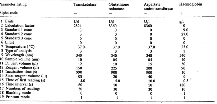
Tab. 1. Cobas Bio program for the three red cell enzymes and haemoglobin.
Parameter listing Alpha code
1 Units
2 Calculation factor 3 Standard 1 cone 4 Standard 2 cone 5 Standard 3 cone 6 Limit
7 Temperature ( °C) 8 Type of analysis 9 Wavelength (nm) 10 Sample volume (nm) 1 1 Diluent volume (μΐ) 12 Reagent volume (μΐ) 13 Incubation time (s) 14 Start reagent volume (μΐ) 1 5 Time of first reading (s) 16 Time interval (s) 17 Numbers of readings 18 Blanking mode 19 Printout mode
Transketolase
U/l2894 00 00 37.03 34010 15012 99008 605.0 300
1
Glutathione reductase U/l8360
00 00 37.03 34005 22510 90020 105.0 300
1
Aspartate aminotransferase U/l8360
00 00 37.03 34005 20015 90040 10.010 300
1
Haemoglobin +
g/10 27.09.0
00 25.01 54010
5090 100 . 1800.5
101 1
Mak and Swaminathan: Assessment of vitamin BI, B2 and B6 status 215 Result and Discussion
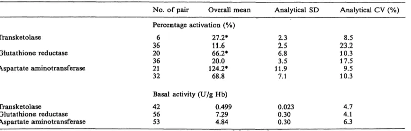
Precision
Precision of the method was evaluated by determi- nation of the analytical standard deviation (SD) and the analytical coefficient of variation (CV) (7) from duplicate analysis results of specimens in separate batches (tab. 2). The CVs for basal activities were between 4.1 and 6.3%, which are similar to those previously reported (3, 4). For percentage activation, the CV was calculated for normal and low values.
The CVs of percentage activation for all 3 enzymes were between 8.5 and 10.3% at a mean value near the lower limit of normal (tab. 4).
Stability
Stability was evaluated in both washed cells and whole blood. The stability of washed cells was studied by comparing the percentage activation of enzymes after storage at —20 °C for a week; the results are shown in table 3 A. There was no significant change (by paired t-test) in percentage activation after one week of storage.
Secondly, we studied the stability of enzymes in whole blood stored at room temperature. Ten fresh speci- mens were divided into 2 portions; one was washed immediately and one was left at room temperature for 6 hours. The results are shown in table 3 B. Only the aspartate aminotransferase percentage activation showed a small but statistically significant decrease after 6 hours at room temperature, while the other two enzymes were stable. Therefore, in our protocol, cells were washed within 2 hours of blood collection to ensure minimal deterioration of enzyme activity.
Control study
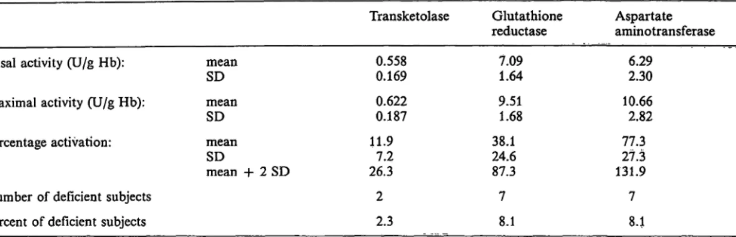
Blood samples were obtained from a group of 30 healthy subjects (medical students and laboratory staff, age 20 — 40) and assessed by the above method (tab. 4). The mean and range of results obtained are comparable to those reported in the literature (2, 3, 8). If the mean percentage activation plus 2 SD is taken as the lower limit of normal, a percentage activation of greater than 26.5, 73.3 and 121.4% for transketolase, glutathione reductase and aspartate aminotransferase, respectively, can be considered to indicate deficiency of the respective vitamins.
Tab. 2. Precision study.
Transketolase
Glutathione reductase Aspartate aminotransferase
No. of pair Percentage 366
2036 2132
Overall mean activation (%)
27.2*
66.2*11.6 124.2*20.0 68.8
Analytical SD
2.52.3 6.83.5 11.97.1
Analytical
23.28.5 10.317.5 10.39.5
CV (%)
Transketolase
Glutathione reductase Aspartate aminotransferase
Basal activity (U/g Hb) 4256
53
0.499 7.294.84
0.023 0.300.30
4.74.1 6.3
* Values near the lower limit of normal (see tab. 4)
Tab. 3 A. "Stability of washed cell aliquots stored at —20 °C for Tab. 3B. Stability of the 3 red cell enzymes in whole blood, one week.
Percentage activation, mean (SD)
No. of pairs Day 0 Day 7
Trans- ketolase 4214.1 (8.4) 12.9 (7.7)
Glutathione reductase 40.3 (29.2)56 40.6 (29.7)
Aspartate amino- transferase 5389.0(40.80) 92.5 (37.8)
Time stored at room temperature
O h6 h
Percentage activation, mean (SD) Trans-
ketolase 12.3 (5.7) 13.9(9.0)
Glutathione reductase 40.7(17.5) 39.6(17.8)
Aspartate amino- transferase 84.2 22.3)*
80.1 (22.1)*
* p < 0.05, by paired t-test J. Clin. Chem. Clin. Biochem. / Vol. 26,1988 / No. 4
216
Mak and Swaminathan: Assessment of vitamin B,, B2 and B6 status.Tab. 4. Basal activity, maximal activity and percentage activation of the 3 red cell enzymes in 30 healthy young adults.
BasaJ activity (U/g Hb):
Maximal activity (U/g Hb):
Percentage activation:
meanSD
mean - 2 SD meanSD
meanSD
mean + 2 SD
Transketolase 0.557 0.134 0.289 0.618 0.126 11.97.3 26.5
Glutathione reductase
7.781.57 4.65 10.22 1.73 35.119.1 73.3
Aspartate aminotransferase
6.431.39 3.65 10.00 2.07 85.418.0 121.4
Tab. 5 A. Basal activity, maximal activity and percentage activation of the 3 red cell enzymes in 86 pregnant women.
Basal activity (U/g Hb):
Maximal activity (U/g Hb):
Percentage activation:
Number of deficient subjects Percent of deficient subjects
meanSD meanSD meanSD
mean + 2 SD
Transketolase 0.558 0.169 0.622 0.187 11.97.2 26.3 2 2.3
Glutathione reductase
7.091.64 9.511.68 38.124.6 87.3 7 8.1
Aspartate aminotransferase
2.306.29 10.66 2.82 77.327.3 131.9 7 8.1 Tab. 5 B. Percentage activation of the three enzymes in preg-
nant women.
Percentage activation Transketolase
Glutathione reductase Aspartate aminotransferase
33.0, 31.5
101.2, 73.6, 87.3, 93.6 110.8, 105.9, 82.9 122.2, 130.2, 124.2, 129.1 130.2, 129.2, 133.5
Pregnant women study
Blood samples from a group of 86 pregnant women were assessed (tab. 5 A). Using the criterion men- tioned above, 2 (2.3%), 7 (8.1%) and 7 (8.1%) out of the 86 subjects were found to have abnormally high percentage activation for vitamin B
l5B
2and B
6, respectively. Individual values of these percentage ac- tivations are given in table 5 B.
Basal activity
It has been suggested that vitamin deficiency is usually associated with a low basal activity due to the low levels of apoenzyme (9). Therefore we correlated the basal activity with percentage activation by Spearmen rank for the 3 different vitamins. The correlation
coefficient showed (tab. 6) a significant correlation in both groups for glutathione reductase and aspartate aminotransferase, and in the control group for trans*
ketolase.
If the mean basal activity minus 2 SD of the control group is taken as the lower limit of normal, then a basal activity of less than 0.289, 4.65 and 3.65 U/g Hb (tab. 4) for transketolase, glutathione reductase and aspartate aminotransferase, respectively, can be considered to indicate deficiency of the respective vitamins. Using this criterion, 0 (0%), 1 (1.2%) and 9 (10.5%) of the 86 pregnant women were identified as abnormal for vitamin B
l9B
2and B
6, respectively.
Tab. 6. Correlation of percentage activation and basal activity by Spearman rank.
Transketolase
Glutathione reductase Aspartate aminotransferase
Control group (ii = 30) 0.411*
0.720*
0.512*
Pregnant group (n = 86) 0.046**
0.641***
0.814***
* p < 0.01
** p > 0.01
*** p < 0.001
Mak and Swaminathan: Assessment of vitamin Bt, B2 and B6 status 217
If both the percentage activation and basal activity
limits are considered, then only 0 (0%), 1 (1.2%) and 4 (4.7%) of the pregnant women would be classified as abnormal for vitamin BI, B
2and B
6, respectively.
Assessment of vitamin status using solely percentage activation, which reflects the functional aspect of vi- tamin (1), is controversial (9, 10). Because of our demonstration of good correlation between basal ac- tivity and percentage activation, it is suggested that
the mean minus 2 SD of the basal activity should be used as a second criterion, in addition to percentage activation, in the assessment of vitamin status. Use of both criteria would give a more specific and sen- sitive assessment. Further investigations are in prog- ress.
The analysis of all 3 red cell enzymes in 39 samples can be performed in one day. The method is therefore suitable for use in nutritional surveys.
References
1. Truswell, A. S. (1985) Br. Med. J. 297, 1258-1262.
2. Clemens, R. A. & Brown, R. C. (1986) Food Technology 40,71-81.
3. Baynoumi, R. A. & Rosalki, S. B. (1976) din. Chem. 22, 327-335.
4. Williams, D. G. (1976) Clin. Biochem. P, 252-255.
5. Vuilleumier, J. P., Keller, H. E., Rettenmaier, R. & Hun- ziker, R (1983) Int. J. Vit. Nutr. Res. 53, 359-370.
6. Dacie, J. V. & Lewis, S. M. (1984) Practical Hematology.
Churchill Livingstone Inc., Edinburgh.
7. Williams, D. L., Nunn, R. R & Marks, V. (1978) Clinical Biochemistry Vol. I: Analytical Aspects, pp. 421 —480, Hei- nemann Med. Books Ltd., London.
8. Kemm, J. R. & Ancill, R. J. (1985) Vitamin Deficiency in The Elderly. Blackwell Scientific Publications, Oxford.
9. Ryle, P. R. & Thomson, A. D. (1984) In: Clinical Biochem- istry of Alcoholism (Rosalki, S. B., ed.) pp. 188-224, Chirchill Livingstone Inc., Edingurgh.
10. Grandal, N., Torp-Pedersen, K., Hanel, H., Kirstensen, M., Thomsen, A. C. & Norgard, G. (1985) Internat. J. Vit.
Nutr. Res. 55, 399-403.
Professor R. Swaminathan
Department of Chemical Pathology Prince of Wales Hospital
The Chinese University of Hong Kong Shatin, New Territories
Hong Kong
J. Clin. Chem. Clin. Biochem. / Vol. 26,1988 / No. 4