Microsolvation of Molecules in Superfluid Helium Nanodroplets Revealed by Means of Electronic Spectroscopy
Tobias Premke, Eva-Maria Wirths, Dominik Pentlehner, Ricarda Riechers, Rudolf Lehnig, Alexander Vdovin and Alkwin Slenczka
Journal Name: Frontiers in Chemistry
ISSN: 2296-2646
Article type: Original Research Article
Received on: 02 Apr 2014
Accepted on: 25 Jun 2014
Provisional PDF published on: 25 Jun 2014
www.frontiersin.org: www.frontiersin.org
Citation: Premke T, Wirths E, Pentlehner D, Riechers R, Lehnig R, Vdovin A and Slenczka A(2014) Microsolvation of Molecules in Superfluid Helium Nanodroplets Revealed by Means of Electronic
Spectroscopy. Front. Chem. 2:51. doi:10.3389/fchem.2014.00051 Copyright statement: © 2014 Premke, Wirths, Pentlehner, Riechers, Lehnig, Vdovin and
Slenczka. This is an open-access article distributed under the terms of the Creative Commons Attribution License (CC BY). The use, distribution or reproduction in other forums is permitted, provided the original author(s) or licensor are credited and that the original publication in this journal is cited, in accordance with accepted academic practice. No use, distribution or reproduction is permitted which does not comply with these terms.
This Provisional PDF corresponds to the article as it appeared upon acceptance, after rigorous peer-review. Fully formatted PDF and full text (HTML) versions will be made available soon.
1
Microsolvation of Molecules in Superfluid Helium Nanodroplets Revealed by Means of Electronic Spectroscopy
Tobias Premke1, Eva-Maria Wirths1, Dominik Pentlehner3, Ricarda Riechers4, Rudolf Lehnig2, Alexander Vdovin5, Alkwin Slenczka1,∗
1Institute for Physical and Theoretical Chemistry, Faculty for Chemistry and Pharmacy, University of Regensburg, Regensburg , Germany,
2 BASF the Chemical Company, Ludwigshafen, Germany,
3 OSRAM Opto Semiconductors GmbH, Regensburg, Germany,
4 Carl Zeiss AG, Oberkochen, Germany,
5 Philips International B.V., Amsterdam, Netherlands Correspondence*:
Alkwin Slenczka
Institute for Physical and Theoretical Chemistry, Faculty for Chemistry and
Pharmacy, University of Regensburg, Universit ¨a tsstrasse 31, Regensburg, 93053, Germany, alkwin.slenczka@chemie.uni-regensburg.de
ABSTRACT
2
The empirical model explaining microsolvation of molecules in superfluid helium droplets
3
proposes a non-superfluid helium solvation layer enclosing the dopant molecule. This model
4
warrants an empirical explanation of any helium induced substructure resolved for electronic
5
transitions of molecules in helium droplets. Despite a wealth of such experimental data, quanti-
6
tative modeling of spectra is still in its infancy. The theoretical treatment of such many-particle
7
systems dissolved into a quantum fluid is a challenge. Moreover, the success of theoretical acti-
8
vities relies also on the accuracy and self-critical communication of experimental data. This will
9
be elucidated by a critical resume of our own experimental work done within the last ten years.
10
We come to the conclusion that spectroscopic data and among others in particular the spectral
11
resolution depend strongly on experimental conditions. Moreover, despite the fact that none of
12
the helium induced fine structure speaks against the empirical model for solvation in helium
13
droplets, in many cases an unequivocal assignment of the spectroscopic details is not possible.
14
This ambiguity needs to be considered and a careful and critical communication of experimental
15
results is essential in order to promote success in quantitatively understanding microsolvation in
16
superfluid helium nanodroplets.
17
Keywords: electronic spectroscopy, molecules, molecular complexes, microsolvation, helium droplets, zero phonon line, phonon
18 19 wing
1 INTRODUCTION
One of the first helium induced fine structures reported for electronic spectroscopy in superfluid helium
20
droplets was a doublet splitting of all zero phonon lines (ZPL) accompanied by a phonon wing (PW)
21
with an unexpected spectral shape for tetracene (Tc) as dopant species Hartmann et al. (1998). After
22
the first purely empirical Hartmann et al. (2001) and later also theoretically founded Whitley et al.
23
(2009) attempt to explain the doublet splitting, a new theoretical model has recently been presented,
24
namely, coherent quantum states of the helium solvation layer covering the dopant surfaceWhitley et al.
25
(2011). With the implementation of some empirically justified modifications, this new theoretical model
26
appeared to agree with one particular experimental spectrumKrasnokutski et al.(2005) chosen from the
27
wealth of experimental spectra published so far for Tc in helium droplets Krasnokutski et al. (2005);
28
Poertner et al. (2001); Lindinger et al. (2004, 2006). Shortly later, a new experimental paper puts
29
the new theoretical approach into questionPoertner et al. (2012). There, a remarkable additional fine
30
structure present only for the second line of the doublet of Tc provides evidence for different physical
31
origins of the two peaks in the doublet. Moreover, the signal was found to depend on the size of the
32
helium droplets. For very large droplets (N > 107), the fine structure has gradually vanished and a
33
new asymmetric peak without a fine structure grows in, however, slightly shifted to the blue. The same
34
shift was observed for the first unstructured line in the doublet. As reported already in Ref. Poertner
35
et al. (2001), the full resolution of the fine structure requires a very well collimated droplet beam in
36
combination with a single mode cw dye-laser as used in Ref.Poertner et al.(2012). A pulsed multimode
37
laser as used in Ref. Krasnokutski et al. (2005) does not allow for the resolution of these details. The
38
presence of a non-superfluid helium solvation layer has already been deduced from the first rotationally
39
resolved infrared (IR) spectrum recorded for SF6 in helium dropletsHartmann et al.(1995). In contrast
40
to vibrational or rotational excitations, electronic excitations exhibit a rather strong coupling to the helium
41
environment. This coupling generates the PW which reveals the spectrum of elementary excitations of the
42
helium environment. As the model of a non superfluid helium solvation layer justifies all the helium
43
induced fine structures recorded so far in electronic spectra the fine structures provide evidence for the
44
helium solvation layer. While this empirical model proposed about two decades ago is generally accepted,
45
a quantitative simulation of the helium induced fine structures has not be seen so far. The discussion on the
46
helium induced fine structure of Tc was the motivation for a critical presentation of our own experimental
47
work on electronic spectroscopy of molecules in superfluid helium droplets with the focus on empirical
48
explanations and interpretations as well as on the experimental conditions. As a result, there is no evidence
49
speaking against the empirical model of a dopant species surrounded by a non superfluid helium solvation
50
layer. However, the assignment for the helium induced fine structures is not as evident as presented in
51
many papers. Moreover, experimental conditions can easily hide important details of the helium induced
52
fine structure. This article aims to draw attention to these issues which play a key role for the quantitative
53
understanding of microsolvation in superfluid helium droplets.
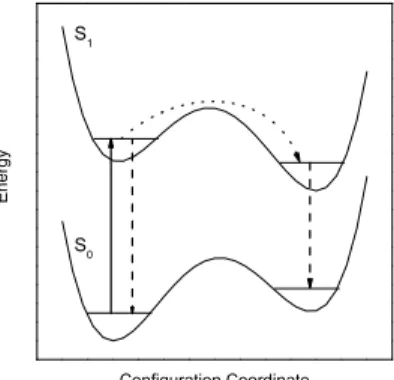
54
2 EXPERIMENTAL TECHNIQUE
The solubility for atoms and molecules in liquid helium is rather poor due to the fact that most substances
55
use to condense to the solid phase at the temperature of liquid helium. This problem has been overcome by
56
using helium droplets doped with single atoms or molecules which levitate freely in a vacuum chamber
57
Toennies and Vilesov (1998). Performing chemical or physical experiments with atoms or molecules
58
in superfluid helium droplets requires first the generation of droplets and secondly the doping of the
59
droplets with the system to be investigated. Both conditions have successfully been investigated in the
60
late eighties of the last century where an appropriate droplet source was combined with the well known
61
pick-up procedure for doping of rare gas clusters Toennies and Vilesov (1998); Gough et al. (1985,
62
1983);Lewerenz et al.(1993). The droplets are generated via adiabatic expansion of helium gas under
63
high pressure (20bar < p < 100bar) and pre-cooled to low temperatures (4K < T < 25K) through a
64
small orifice (5µm) into a vacuum chamber Toennies and Vilesov(1998). Depending on the stagnation
65
pressure and the nozzle temperature, helium droplets are generated with an average size from 103to 108
66
helium atomsToennies and Vilesov (2004b,a); Harms et al. (1998). Collimated to a droplet beam the
67
droplets pass a skimmer to get to a second high vacuum chamber. Alternatively, a pulsed valve is used in
68
order to generate a pulsed droplet beam. By maintaining similar gas flux the droplet density in the pulses
69
can be significantly increased which bears advantages when using pulsed lasers. The first pulsed droplet
70
source was a modification of a commercially available valve (General Valve No 9) Slipchenko et al.
71
(2002). Its performance depends critically on the nozzle shapeYang et al.(2005);Yang and Ellis(2008).
72
Much higher repetition rates up to 1 kHz and more confined pulses (20µs) are generated with a cryogenic
73
modification of the Even Lavie valve Pentlehner et al. (2009); Even et al. (2000). Typical expansion
74
conditions are a stagnation pressure between 50 and 100 bar, a nozzle temperature between 10 and 30
75
K and an orifice of 60 µm. As in the first case the droplet beam enters the detection chamber through a
76
skimmer with an opening diameter of 6 mm. In Regensburg two helium droplet machines are operated
77
one with a continuous flow source and the other with a pulsed Even Lavie valve. The two machines have
78
identical detection chambers where the droplet beam is first guided through a pick-up unit. It consists of an
79
oven for sublimation of solid samples and of a gas cell for gas phase samples. Both have an entrance and
80
exit aperture adjusted to the droplet beam axis. The oven is surrounded by a liquid-nitrogen cooled brass
81
cylinder in order to shield thermal radiation and cryo-pump effusing gas. About 10 cm downstream the
82
doped droplet beam is intersected perpendicularly by a laser beam. Perpendicular to both beam axes the
83
laser induced fluorescence is collected by a lens system and imaged onto photodetectors. Two detection
84
systems are mounted. One is a photo multiplier which records the integrated fluorescence. Secondly, the
85
fluorescence is dispersed by a grating spectrograph and imaged onto the chip of a CCD (charge coupled
86
device) camera. In the first case, the fluorescence is recorded as a function of the laser frequency which
87
results in a fluorescence excitation spectrum. In the second case, the laser is tuned to a particular resonant
88
absorption and a dispersed emission spectrum is recorded.
89
3 EXPERIMENTAL RESULTS
The signature of microsolvation is omnipresent in spectroscopy of molecules in helium droplets. In the
90
following, our own experimental work on electronic spectroscopy of molecules or molecular aggregates
91
inside superfluid helium nanodroplets will be reinvestigated with the focus on helium induced spectral
92
features and their consistent interpretation. The data emerge from numerous experiments which can be
93
separated into three groups. The first deals with the very detailed study of one particular dopant species.
94
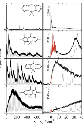
The second outlines comparative studies of related molecular compounds, and the final group deals with
95
photo-chemistry inside superfluid helium droplets.
96
3.1 ELECTRONIC SPECTROSCOPY OF PHTHALOCYANINE INSIDE SUPERFLUID HELIUM DROPLETS
With the aim to use helium droplets as a host system to study photochemistry of cold molecules by spe-
97
ctroscopic meansLehnig et al.(2009), our first experimental result drew our attention to the fundamental
98
problem of microsolvation or, in other words, the helium induced spectroscopic featuresPentlehner et al.
99
(2011). The corresponding dopant to helium interaction is revealed for example by a PW, a red shifted
100
dispersed emission spectrumLehnig and Slenczka(2003), or by a helium-induced fine structure as repor-
101
ted already for the first such spectra Hartmann et al.(2001, 1996b, 1998). Such spectroscopic features
102
are also characteristic for photochemical processes. Therefore, we have studied microsolvation by means
103
of fluorescence excitation and dispersed emission spectra first for phthalocyanine (Pc), a photochemically
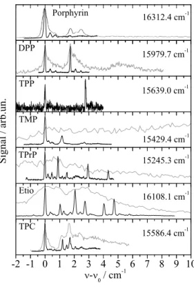
104
inactive dopant species with fortunate excitation energy, oscillator strength, and fluorescence quantum
105
yield of the S0-S1transition. Moreover, at that time its electronic spectroscopy was well known in the gas
106
phase Fitch et al. (1978, 1980, 1979, 1981), in solid matrices Bondybey and English (1979); Huang
107
et al. (1982) and also in helium droplets Hartmann (1997). In addition to the fluorescence excitation
108
spectrum and numerous dispersed emission spectra, our study included pump-probe spectra and the inve-
109
stigation of the saturation behavior. The particular experimental data revealed Pc to be surrounded by a
110
rather rigid helium solvation layer. The entire complex moves freely inside the superfluid helium dro-
111
plet. The experimental observables were as follows. The major discrepancy of the fluorescence excitation
112
spectrum to the gas phase data was a solvation shift of the S0-S1electrionic transition of -42 cm−1Har-
113
tmann(1997);Hartmann et al.(2002);Lehnig et al.(2004). Otherwise, vibronic transitions appeared
114
to be very sharp (∆ν < 1 cm−1 with almost identical vibrational frequency as in the gas phase. The
115
asymmetric line shape at the electronic origin with a line width in the order of 0.1 cm−1reflects precisely
116
the size distribution of the droplet beam and can be used to determine the size distribution for subcritical
117
expansion in the continuous flow droplet sourceDick and Slenczka (2001);Slenczka et al.(2001). For
118
droplet sizes beyond106 helium atoms the asymmetry vanishes while the solvent shift passes a maxi-
119
mum and decreases with further increasing droplet size. For droplets with more than107 helium atoms a
120
fine structure appears which can be fitted by the rotational envelop calculated for the well known almost
121
symmetric top Hamiltonian of Pc however with increased moments of inertia as to be expected from the
122
additional mass of the helium solvation layerLehnig et al.(2004);Pentlehner et al.(2009). And as to be
123
expected, the phonon wing (PW) shows a spectral structure which reveals the presence of non-superfluid
124
heliumHartmann et al.(2002);Lehnig and Slenczka(2005);Lehnig et al.(2007). These details speak
125
for the dopant molecule to be dissolved inside the droplet. Moreover, the dopant molecule is surrounded
126
by a non-superfluid helium solvation layer.
127
As revealed by the doubling of the entire dispersed emission spectrum, the S0-S1electronic excitation
128
of Pc in helium droplets transfers the excited Pc-helium complex into a metastable configuration which
129
partly relaxes prior to radiative decayLehnig and Slenczka(2003, 2004a,b) (cf. Fig. 1). The correspon-
130
ding branching ratio correlates with the additional excitation energy put into the vibrational degrees of
131
freedom of the solvation complexLehnig and Slenczka(2003, 2004a). Any excitation energy exceeding
132
the electronic origin fully dissipates into the helium droplet prior to radiative decayLehnig and Slenczka
133
(2003). In the case of Pc the amount of dissipating energy promotes relaxation of the helium solvation
134
layer. A detailed analysis of homogeneous line widths of numerous vibronic transitions did not show any
135
correlation with the vibrational excess excitation energy. This was taken as evidence for an intermediate
136
step preceding energy dissipation into the helium droplet, most probably internal vibrational redistribution
137
Pentlehner et al.(2011). The radiative decay of the relaxed complex leads to a metastable configuration
138
in the electronic ground state (cf. Fig. 1). As revealed by pump-probe experiments, the metastable configu-
139
ration in S0relaxes to the global minimum configuration with a rate constant of only 200 kHzPentlehner
140
et al.(2011). All these findings fit to the model of a Pc-helium solvation complex which undergoes a
141
photoinduced cycle as depicted in Fig. 1. The increased moments of inertia together with the very sharp
142
resonances in the dual emission spectra provide evidence for a helium solvation layer exhibiting a well
143
defined configuration (which means localized helium atoms). The relaxation of the helium solvation layer
144
which leads to the second emission spectrum is accompanied by an increase of the helium induced red
145
shift from 42cm−1to 52.8cm−1which corresponds to an increase of 26%Lehnig and Slenczka(2003).
146
Electronic excitation causes in the first place a change of the electron density distribution. In the case of
147
Pc this change is of negligible influence on the intramolecular nuclear configuration or binding conditions.
148
This is revealed by the close similarity of fluorescence excitation and dispersed emission spectra. Howe-
149
ver, the helium solvation layer which is soft compared to the dopant molecule may follow the change of the
150
electron density distribution. Vice versa, the change of the electron density distribution becomes observa-
151
ble by helium induced spectroscopic features. The electron density distribution is an important quantity
152
for modeling helium induced spectroscopic features. This quantity may not be properly implemented
153
when using pair potentials as done for example inWhitley et al.(2005).
154
In order to learn more about the helium solvation layer we have added Ar atoms and, thus, designed Pc-
155
Arn clusters inside helium dropletsLehnig et al.(2007). Thereby, we stay with the same chromophore,
156
namely Pc. The Ar atoms can be seen as a part of a Pc-Arn cluster dissolved in helium droplets or as
157
part of the solvation layer surrounding the Pc dopant. In a sequential order a single Pc molecule and prior
158
or afterwards a certain amount of Ar atoms were doped into the helium droplets as previously reported
159
for Tc-Arn clusters Hartmann et al.(1998). Thereby, Pc-Arn clusters are formed and cooled down to
160
0.37 K for all degrees of freedom within pico-seconds. As described in Hartmann et al. (1998) each
161
individual sharp transition in the fluorescence excitation spectrum can be assigned to a particular cluster
162
stoichiometry. Doping Ar atoms prior to Pc favors complexes of one Pc molecule attached to the surface
163
of a solid Arn-cluster while the inverse doping sequence favors complexes of one Pc molecule inside an
164
Arn-cluster. In the case complexes consisting of a large planar molecule (such as Pc) and only very few
165
Ar-atoms we speak in the first case of single-sided and in the latter case of double sided Ar-occupancy.
166
Pump-probe spectraHartmann et al.(1998) or dispersed emission spectraLehnig et al.(2007) allow to
167
identify configurational isomers of the clusters. Using the latter technique, three complex configurations
168
were identified for the Pc-Ar cluster exhibiting Ar-induced red shifts of 15 cm−1, 4 cm−1and 1.6 cm−1,
169
respectively. The vibrational fine structure of the most abundant cluster was identical to bare Pc in helium
170
droplets and its Ar-induced red shift of 15 cm−1 was identical as reported for the corresponding gas
171
phase experiment Cho and Kim(2000). This speaks for a complex configuration with an Ar atom just
172
above the center of theπ-conjugated ring close to the center of mass of Pc, a position coincident with the
173
global minimum of the Pc-Ar pair potential which amounts to roughly 680 cm−1 Cho and Kim(2000);
174
Lehnig et al.(2007). Upon vibronic excitation with excess energy of only 128 cm−1put into a low energy
175
vibrational mode of this Pc-Ar cluster (which is less than 20%of the dissociation energy of the isolated
176
Pc-Ar cluster), emission of bare Pc could be recorded in addition to the cluster emission Lehnig et al.
177
(2007). Further dynamics upon electronic excitation has been observed for the Pc-Ar2 clusters. It is the
178
smallest cluster which allows for distinguishing single-sided and double-sided Ar-occupancy on the planar
179
Pc dopant as individually favored by the two pick-up sequences. For one of the most prominent signals
180
of a single-sided Pc-Ar2 cluster the dispersed emission spectrum recorded upon excitation at vibronic
181
transitions showed dual emission. In addition to the ordinary emission spectrum identical to that upon
182
excitation at the corresponding electronic origin, a second emission spectrum was observed matching in
183
the frequency position and the relative intensity distribution perfectly with the dispersed emission upon
184
excitation at the origin of a double-sided Pc-Ar2clusterLehnig et al.(2007).
185
At this point one may raise the question on the structure of the solvated clusters. Are we dealing with
186
Pc-Arn complexes surrounded by a helium solvation layer or may there be Ar atoms attached to the
187
helium solvation layer of Pc? In the first case Ar atoms are merged into the helium solvation layer while
188
in the second case the Pc-helium complex remains intact and the Ar atom is separated from the dopant
189
by the helium solvation layer. It is not only the small red shift of only 1.6 and 4 cm−1 not reported
190
for the gas phase experiment which provides evidence for the latter complex. It is also the emission of
191
bare Pc recorded upon excitation of a Pc-Ar cluster with an excess excitation energy of only 128 cm−1
192
(cf. previous paragraph) and the configurational modification from a single-sided to a double-sided Pc-Ar2 193
complex induced by electronic excitation which reveals a rather small binding energy as to be expected for
194
Pc and Ar shielded from each other by the helium layer. It should be noted that Pc-Ar clusters in helium
195
droplets exhibit a similar relaxation dynamics upon electronic excitation as depicted in Fig. 1 for bare
196
Pc in helium dropletsLehnig and Slenczka (2004a). As the change of the electron density distribution
197
accomplishes the relaxation of the helium solvation layer it may also afford the dissociation of the van der
198
Waals clusters inside helium droplets.
199
3.2 COMPARATIVE STUDIES OF RELATED MOLECULAR COMPOUNDS
While electronic spectra of Phthalocyanines show very sharp transitions, other dopant species have shown
200
surprisingly severe line broadening in the electronic spectra recorded in helium droplets. This may be
201
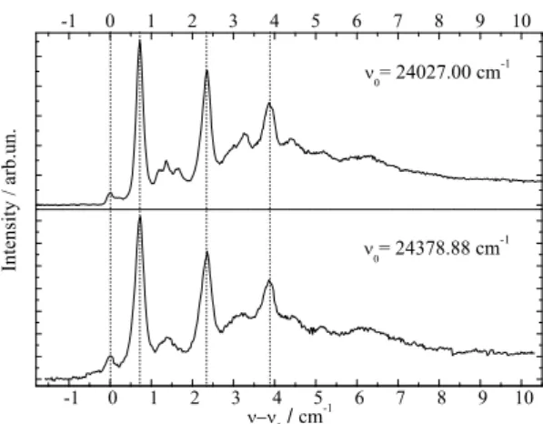
due to damping of vibrational excitations in particular of low energy and large amplitude modes or due
202
to perturbation of the change of the electron density distribution. Much information on helium induced
203
line broadening was provided by systematic investigations of a series of related dopant species. For three
204
molecular species namely PyrromethenePentlehner et al.(2011);Stromeck-Faderl et al. (2011), Por-
205
phyrinPentlehner et al.(2011);Riechers et al.(2013), and AnthracenePentlehner et al.(2011, 2010);
206
Pentlehner and Slenczka (2012, 2013) several derivatives have been investigated which differ in the
207
number and the species of substituents such as methyl, ethyl, propyl, phenyl, and cyano groups which
208
substitute hydrogen atoms in the periphery of the molecular compound. The main conclusions concer-
209
ning the influence of electronic and vibrational degrees of freedom will be outlined for each of the three
210
molecular species.
211
The series of Pyrromethene dye molecules includes derivatives such as 1,2,3,5,6,7-hexamethyl-8-
212
cyanopyrromethene-difluoroborat, 8-phenylpyrromethene-difluoroborat, and 1,3,5,7,8-pentamethyl-2,6-
213
diethylpyrromethen-difluoroborat. If one disregards intramolecular configurational variants of the sub-
214
stituted derivatives, the symmetry of the Pyrromethene derivatives listed in Fig. 2 is identical to the
215
non-substituted compound shown in the top panel. For all derivatives the substitution is accompanied
216
by extended progressions of torsional and/or bending modes which are well resolved in the gas phase
217
Stromeck-Faderl et al.(2011) (cf. Fig. 2 left panel grey lines). Extended progressions reveal different
218
equilibrium configuration of the substituents in both electronic states. When put into helium droplets, the
219
corresponding progressions look like the gas phase spectrum convoluted with a line broadening function
220
(cf. Fig. 2 left panel black lines)Pentlehner et al. (2011). It should be noted that in Fig. 2 the helium
221
induced solvent shift of the electronic spectra has been ignored in order to compare the vibrational fine
222
structure of both spectra. In contrast to the torsional mode progressions, the electronic origin remains
223
spectrally sharp (cf. Fig. 2 right panel red line). In some cases (second and bottom panel in Fig. 2) a
224
fine structure is recorded which could not be resolved in the gas phase. These observations provide clear
225
evidence for line broadening due to the damping of vibrational modes by the helium environment, a mech-
226
anism which leaves the electronic origin unaffected. Thus, in the case of the Pyrromethene derivatives the
227
vibrational degrees of freedom and in particular those of the substituents suffer from helium induced line
228
broadening while purely electronic excitation does notPentlehner et al.(2011);Stromeck-Faderl et al.
229
(2011).
230
The study of Porphyrin Riechers et al. (2013) includes derivatives such as 5,15-diphenylporphyrin
231
(DPP), 5,10,15,20-tetraphanylporphyrin (TPP), 5,10,15,20-tetramethylporphyrin (TMP), 5,10,15,20-
232
tetrapropylporphyrin (TPrP), and 2,7,12,27-tetraethyl-3,8,13,18-tetramethylporphyrin (Etio). In addition
233
5,10,15,20-tetraphenylchlorine (TPC) was investigated, which came as an impurity of the TPP sample
234
Riechers et al.(2013). Again, for all derivatives the molecular symmetry is conserved if one ignores the
235
configurational variants of the substituents. None of the Porphyrin derivatives shows signals which could
236
be attributed to an envelope or fully resolved progression of low energy modes representing torsional or
237
bending modes of the substituents. Obviously, the equilibrium configuration of the substitunets is main-
238
tained upon electronic excitation as is the nuclear configuration of the Porphyrin moietyRiechers et al.
239
(2013). In contrast to the spectra recorded by means of a pulsed dye laserLehnig et al.(2007);Lindinger
240
et al.(2001), the low photon flux and single mode radiation of a cw-dye laser allows to resolve a triple
241
peaked ZPL of Porphyrin as shown in the top panel of Fig. 3. The comparative presentation of the electro-
242
nic origins of the entire series of Porphyrin derivatives including the TPC compound allows to recognize
243
this triple peak feature with slight modification for all the Porphyrin derivatives shown in Fig. 3 (for more
244
details cf.Riechers et al.(2013)). For DPP the triple peak feature doubles as to be expected for the two
245
conformers differing in the sense of the tilt angle of the two phenyl substituent. Depending on the number
246
and species of substituents the number of different isomeric conformers increases as does the number of
247
intense peaks. Thus, the entire fine structure is interpreted as a congestion of the triple peak features of
248
the various configurational conformers. Obviously, this triple peak feature represents the basic signature
249
of microsolvation of Porphyrin derivatives in helium droplets. Severe line broadening can be induced by
250
strong saturation as obtained by the high photon flux of pulsed dye lasers. The corresponding spectra are
251
added as grey lines in Fig. 3. Similar as Phthalocyanine, Porphyrin exhibits exceptionally sharp electronic
252
and vibronic transitions which is ideal for resolving the helium induced fine structure. For both species
253
the vibrational fine structure of the electronic excitation of substituted compounds does not shows the
254
characteristic low energy torsional or bending modes of the substituents. The close similarity of the vibra-
255
tional fine structure of the fluorescence excitation spectrum and the dispersed emission spectra reveals a
256
negligible change of the electron density distribution upon electronic excitation to S1.
257
The third study investigates Anthracene derivatives. This study includes derivatives where substitu-
258
tion reduces the molecular symmetry. In the case of a single substituent inversion symmetry is lost and
259
the compound exhibits a permanent dipole moment. For bare Anthracene and additional four Anthra-
260
cene derivatives, namely 1-methyl-anthrazene (1MA), 2-methylanthracene (2MA), 9-methylanthracene
261
(9MA), and 9-phenylanthracene (9PA), the fluorescence excitation spectra are shown in Fig. 4Pentleh-
262
ner et al.(2011). Roughly, the vibrational mode pattern of bare Anthracene repeats very similar for all
263
four derivatives as indicated by the vertical dashed lines. Two of the derivatives do not exhibit low energy
264
progressions (1MA and 9MA) while the other two do (2MA and 9PA). As revealed by the presence of
265
low energy progressions, only for the latter two derivatives the equilibrium configuration changes upon
266
electronic excitation. For both species, the line widths of the low energy progressions are significantly
267
broadened (black lines) compared to the gas phase spectra (grey lines). In contrast to the Pyrromethene
268
derivatives, line broadening is present throughout the spectrum including the electronic origin. Thus, the
269
damping of low energy modes can’t justify the line broadening. The change of the equilibrium configura-
270
tion as expressed by the low energy progressions is induced by the electronic excitation and, thus, caused
271
by the change of the electron density distribution. Most likely this change acts not only on the intramole-
272
cular nuclear arrangement but also on the arrangement of the helium environment. The latter perturbation
273
may be the reason for line broadening. According to this mechanism, the change of the electron density
274
distribution is the driving force for intra- and intermolecular rearrangements which become effective on
275
the line widths in the electronic spectra of these two Anthracene derivatives. Further details of these spe-
276
ctra are discussed in Refs.Pentlehner et al.(2011, 2010);Pentlehner and Slenczka(2012, 2013). Thus,
277
the systematic investigation of Anthracene derivatives provides evidence for the change of the electron
278
density distribution being responsible for helium induced spectral features.
279
3.3 PHOTOCHEMISTRY INSIDE SUPERFLUID HELIUM DROPLETS
Our first approach to photochemistry in superfluid helium droplets was the study of the well known exci-
280
ted state intramolecular proton transfer (ESIPT) of 3-hydroxyflavone (3-Hf) and its counterpart in the
281
electronic ground state called back proton transfer (BPT)Sengupta et al.(1979). As depicted in the cen-
282
ter panel of Fig. 5, ESIPT and BPT are induced by electronic transition and, thus, by the change of the
283
electron density distribution in accordance with Born-Oppenheimer approximation. As demonstrated in
284
Ernsting and Dick (1989);Muehlpfordt et al.(1994);Ito et al. (1992) the homogeneous line width at
285
the electronic origin of the corresponding fluorescence excitation spectrum reveals the rate constant of
286
ESIPT given that other non-radiative decay paths of N* can be neglected. The homogeneous line width
287
of the corresponding transition in the dispersed emission spectrum is given by the rate constant for BPT
288
and the rate constant for the radiative decay of T*. The latter can be determined experimentally from the
289
readily observable radiative decay time. Since in the gas phase a hot tautomer is generated, congestion
290
of transitions of numerous quantum states of the tautomer prevents from resolving the homogeneous line
291
width of individual transitions in the dispersed emission spectrumIto et al.(1992). This problem can be
292
overcome by helium droplets as host system. The experiment may profit from the highly efficient dissipa-
293
tion of vibrational energy into the helium droplet. Thus, the cooling rate of the nuclear degrees of freedom
294
of the excited dopant molecule which exceeds the radiative decay rate allows to record dispersed emission
295
of a cold tautomer (T*). In fact, dispersed emission spectra of the tautomer showed vibrational fine stru-
296
cture, however, only Voigt-profiles with line widths of about 60 cm−1could be resolvedPentlehner et al.
297
(2011);Lehnig et al.(2009). Even more surprising, the electronic origin and the vibrational fine structure
298
in the fluorescence excitation spectrum nicely resolved in the supersonic jet experiment Ernsting and
299
Dick (1989) were entirely washed out in helium droplets Lehnig et al. (2009). Obviously, in this case
300
the electronic degree of freedom is responsible for the strong perturbation by the helium environment.
301
ESIPT as well as BPT are initiated by purely electronic transitions and, thus, by the change of the electron
302
density distribution. The electron density distributions of the four conformers are shown as contour plots
303
in Fig. 5. The corresponding dipole moment is emphasized by the red arrows, indicating its value and
304
direction. Compared to bending or tilting of a methyl or phenyl substituent, proton transfer requires even
305
stronger forces. It is inconceivable that changes of the molecular polarity as induced by electronic tran-
306
sitions of 3-Hf should proceed without severe perturbation of the helium environment. As in the case of
307
2MA and 9PA, it appears to be the change of the electron density distribution which perturbs the helium
308
environment and, thus, induces severe line broadening in the electronic spectra.
309
The possibility to design molecular complexes with well defined stoichiometry and the option to distin-
310
guish even isomeric variants of such complexes allows to study the influence of solvents on photophysical
311
processes on a molecular level. Since the influence of polar or protic solvents on the ESIPT of 3-Hf is well
312
knownSengupta et al.(1979);Ito et al.(1992), we have investigated 3-Hf-(H2O)nclusters in helium dro-
313
pletsLehnig et al.(2009). In a gas phase experiment it was shown that a single water molecule suffices to
314
suppress ESIPT entirelyIto et al.(1992). More recent gas phase experiments come to the conclusion that
315
at least two H2O molecule are needed to block ESIPT Bartl et al.(2008, 2009). In contrast, the helium
316
experiment unequivocally reveals that one or two water molecules do not affect the 100%efficiency of
317
ESIPT. This was revealed by dispersed emission spectra showing exclusively the signal of the tautomer
318
Lehnig et al.(2009). Only for an average amount of 4 or 5 water molecules a signal contribution of the
319
normal form N* of 3-Hf could be recorded in helium droplets. All may depend on the configuration of
320
the 3-Hf-(H2O)n clusters present in the various experiments. According to our calculations which were
321
performed without the helium environment (which means under gas phase conditions) only one stable
322
configuration of a 3-Hf-H2O complex was found. For this complex the water molecule is merged into the
323
proton transfer coordinate. For this complex concerted proton transfer proceeds under similar energetic
324
conditions as for bare 3-HfPentlehner et al.(2011). For the complex with two water molecules one can
325
imagine the same 3-Hf-H2O configuration with one additional water molecule attached or a chain of two
326
water molecules inserted into the proton transfer coordinate. According to our calculations both confi-
327
gurations allow for concerted proton transfer under energetic conditions similar to bare 3-HfPentlehner
328
et al.(2011). Obviously, calculations of ESIPT for the water complexes without the helium environment
329
(which means for gas phase conditions) are in contradiction to the experimental observations under gas
330
phase conditions. However, they are in agreement with the experimental observations in helium droplets.
331
Recent data recorded in helium droplets from deuterated samples of bare 3-Hf and in addition from all
332
possible combinations of deuterated and protonated samples of 3-Hf and water molecules have shown
333
identical ESIPT behavior as for the purely protonated 3-Hf. At this point one may raise the question on
334
the complex configuration in the helium droplet experiment. The missing influence of one or two water
335
molecules on the ESIPT may indicate that the 3-Hf molecule is shielded by the helium solvation layer.
336
Thus, the water molecules are separated by the helium layer and ESIPT remains unaffected. Only for an
337
average amount of 4 or 5 water molecules the shielding by the helium layer is overcome. Alternatively, the
338
helium environment may favor exclusively those configurations which allow for concerted proton transfer
339
for the 3-Hf-water complex with less than fife water molecules. Finally it should be noted that both the
340
tautomeric and the normal emission (the latter observed for clusters with more than 4 H2O molecules)
341
were spectrally very broad. In the case of ESIPT and BPT of 3-Hf and of its clusters with water, the expe-
342
rimental observations of severe line broadening were counterintuitive. Again, the possible mechanism
343
may be the change of the electron density distribution which simultaneously drives the proton transfer
344
and perturbs the helium environment. The latter explains line broadening.
345
4 DISCUSSION
Electronic spectroscopy provides insight into microsolvation in superfluid helium droplets. Detailed infor-
346
mation is revealed by the spectral fine structure of the ZPL and of the accompanying PW. The electronic
347
spectrum of Glyoxal reflects what is expected for a molecule when doped into a superfluid helium droplet.
348
The ZPL reveals the rotational fine structure of an asymmetric top rotor while the PW reflects the spectral
349
structure of elementary excitations of superfluid heliumHartmann et al.(1996a);Poertner et al.(2002).
350
However, in this respect Glyoxal is exceptional. All other molecules or molecular complexes investigated
351
so far show a ZPL which is either single peaked or exhibits a helium induced fine structure other than
352
free rotation in a quantum fluid. The PW comes up with a spectral shape in the range from very broad
353
and unstructured to rather narrow in the width consisting of a series of peaks sometimes as sharp as the
354
ZPL. Empirically these features are easily justified by the also empirical model of a non-superfluid helium
355
solvation layer covering the surface of the dopant species. Consequently, we deal with a helium solva-
356
tion complex dissolved into a superfluid helium nanodroplet. Thus, the PW may consist of excitations
357
of the helium solvation layer with possibly rather sharp transitions (known as van der Waals modes) in
358
addition to excitations of the helium droplet body, both coupled to electronic excitation of the dopant
359
species. A helium induced fine structure of the ZPL is explained by the presence of more than only one
360
configuration of the helium solvation complex. Thus, the spectral position and spectral shape which are
361
similar for the fine structure of the ZPL and van der Waals modes are not anymore the discriminating
362
criteria of ZPL against PW. Consequently, other criteria need to be established in order to provide an
363
unequivocal assignment of the helium induced spectral features. As shown also for Glyoxal Hartmann
364
et al. (1996a) in many cases the oscillator strength of the ZPL exceeds that of the PW which becomes
365
effective in a different saturation behavior of both signals. And in contrast to the ZPL at the electronic
366
origin, the PW exhibits only red shifted emission because of the dissipation of the phonon energy prior
367
to radiative decay. Vice versa, a coincidence of the origin in the dispersed emission spectrum with the
368
excitation frequency is an unequivocal criterion for the ZPL. This criterion confirmed the presence of two
369
different species responsible for the doublet splitting in the ZPL of TcPentlehner and Slenczka(2012)
370
and also to identify the number of isomeric configurations of Pc-Ar clusters designed in helium droplets
371
Lehnig et al.(2007). If ZPL and PW are merged into a single helium induced fine structure (as shown for
372
example in Fig. 3 of this manuscript) the problem in the assignment of ZPL and PW is in the first place
373
the missing of the phonon gap which separates the PW of superfluid helium from the preceding ZPL.
374
Secondly, electronic excitation accompanied by a significant change of the shape of the dopant species
375
may lead to an oscillator strength of the PW dominating over the ZPL as reported in Ref. Loginov et al.
376
(2005). The change in the shape of the dopant species can either be a nuclear rearrangement or a change
377
in the electron density distribution or both. Thirdly, transitions of metastable configurational variants of a
378
helium solvation complex do not necessarily exhibit oscillator strengths which all exceed that of the PW.
379
Finally, the electronic excitation of such complexes may further reduce the configurational stability. Thus,
380
even without the presence of excess excitation energy the excited complex may undergo relaxation prior
381
to radiative decay. In this case, even a ZPL may show red shifted emission. In summary, the ZPL may
382
show spectroscopic features such as high saturation threshold and red shifted emission which are usually
383
taken as evidence for a PW. Vice versa, the PW may come up with rather sharp spectral features similar as
384
the ZPL which are assigned to van der Waals modes of the helium solvation complex. Thus, experimental
385
criteria to distinguish the PW and ZPL in electronic spectra of molecules in helium droplets do not allow
386
to discriminate van der Waals modes as part of the PW against a ZPL of a metastable solvation complex.
387
As demonstrated for the Porphyrin derivatives in Fig. 3, saturation broadening may hide the helium
388
induced fine structure entirely. While saturation broadening is a technical problem which can be avoided,
389
line broadening induced by the dopant to helium interaction is an intrinsic problem for the application
390
of helium droplet spectroscopy. Established as HENDI spectroscopy Callegari et al.(2001) with many
391
expectations, the limiting factors need to be discussed and, thereby, might even be turned into a prospect.
392
This will be emphasized in the following discussion by some example spectra. The electronic origin of
393
bare Porphyrin is a prototype for the problem caused not only by saturation broadening but in addition
394
for the problem to distinguish ZPL and PW. The oscillator strength revealed by the saturation behavior
395
and the spectral position were the criteria supporting the assignment of the ZPL and PWHartmann et al.
396
(2002). However, the experimental observations taken as evidence for an assignment of the PW do not
397
exclude an alternative assignment to ZPLs of configurational variants of a solvation complex. Similar
398
ambiguities need to be considered for the signals assigned to the PW of Mg-PcLehnig et al.(2004) or Pc
399
Lehnig et al.(2007). The problem of saturation broadening is nicely exemplified at the ZPL of Porphyrin
400
which consists of a fully resolvable triple peak feature when recorded under appropriate experimental
401
conditions (cf. Fig. 3 top panel). The same ZPL has previously been identified as singly peaked already
402
under moderate saturation conditionsLindinger et al. (2001). The problem of saturation broadening is
403
nicely demonstrated for the entire series of Porphyrin derivatives. It need to be mentioned that in addition
404
to pure saturation broadening the growing intensity of the PW may finally hide the ZPL entirely.
405
A remarkable example in this context is the electronic origin of TPC shown in the bottom panel of
406
Fig. 3. Within the first 10 cm−1 the signal can be separated into three parts. The first part is the signal
407
within the first 1 cm−1 showing what was identified as the triple peak feature characteristic for the ZPL
408
of Porphyrins in helium droplets Riechers et al. (2013). The leading intense peak exhibits a line width
409
of only 0.05 cm−1). The second part beyond 1 cm−1 consists of a series of similarly sharp peaks (cf.
410
black line in the bottom panel of Fig. 3) which all exhibit a reduced oscillator strength compared to the
411
ZPL. The third contribution exhibits the smallest oscillator strength and, therefore, can only be recorded
412
upon severe saturation of the first two parts. The grey spectrum in the bottom panel of Fig. 3 recorded
413
for high photon flux shows the third part in overlap with the second part and preceded by the first part
414
the latter two with severe saturation broadening. The third part fulfills characteristic criteria of a PW of
415
the helium droplet body such as low oscillator strength, frequency gap to the ZPL, and spectrally broad
416
shape. As discussed above the analysis of the second signal part can not discriminate an assignment to
417
ZPLs of configurational variants of the helium solvation complex against van der Waals modes of the
418
non-superfluid solvation layer. The saturated spectrum plotted as grey line in the bottom panel of Fig. 3
419
shows the ZPL still spectrally separated from the other two - now - congested signal parts. Upon further
420
increased photon flux all three signal parts merge all into a single peak about 10 cm−1 in width. Such
421
a spectrum is shown in Fig. 13 of Ref.Callegari and Ernst(2011). Besides the problem of identifying
422
the correct dopant species, the interpretation of this spectrum modified by severe saturation broadening
423
leads to conclusions on the properties of the dopant species which are clearly refuted by high resolution
424
spectroscopy.
425
In this context two additional examples need to be discussed which are found in the literaturePei et al.
426
(2007);Carcabal et al.(2004). Both underline the problem of ambiguity in the assignment of PW and
427
ZPL and the problem of saturation broadening. It concerns Aluminum-Chloro-Phthalocyanine (AlCl-Pc)
428
Pei et al.(2007) and PeryleneCarcabal et al.(2004), whose electronic origins measured in our laboratory
429
are shown in Figs. 6 and 7, respectively. In Fig. 6 dispersed emission is added in the spectral range below
430
-2 cm−1while for Perylene a vibronic transition is added in the lower panel of Fig. 7. Despite the different
431
dopant species, both spectra are dominated by a surprisingly similar triple peak series. However, as the
432
two dopant species are different, the analysis of the two fine structures reveals also very different results.
433
By the help of dispersed emission spectra, the AlCl-Pc spectrum was found to represent two different
434
solvation complexes as indicated by the grey and black combs. Both complexes show almost identical fine
435
structure in the excitation dominated by a series of three peaks. The different intensity of the two signaly
436
may reflect the difference in the abundance of the two solvation complexes. The frequency shift of both
437
systems of about 0.7 cm−1 is also reflected by the corresponding dispersed emission spectra as indicated
438
by the combs in Fig. 6. The red shift of the emission of 8.5 cm−1 reveals the relaxation of the solvation
439
complex configuration prior to radiative decay. AlCl-Pc is an example for red shifted emission even upon
440
excitation at the ZPL at the electronic origin. When measured with the high peak power of a pulsed dye
441
laser (certainly not for the purpose of resolving the helium induced spectral signature) much of the fine
442
structure remains hidden (cf. Pei et al.(2007)). In contrast to AlCl-Pc, the entire fine structure resolved
443
for Perylene exhibits only one common emission spectrum as shown in Lehnig and Slenczka(2005).
444
The origin of the emission coincides with the first tiny peak shown at the origin of the wavenumber scale
445
in the upper panel of Fig. 7. When recorded with increased photon flux, all the tiny resonances in between
446
the dominant trio as well as the leading tiny are missing. Consequently, the real origin is missing which
447
causes a false assignment of the electronic origin (cf. Carcabal et al.(2004). Despite all the additional
448
information gained from high resolution excitation spectra and dispersed emission spectra an assignment
449
to either a series of ZPL of variants of a solvation complex or to van der Waals modes of the solvation
450
complex remains open for the fine structure of both molecular dopant species.
451
The issue of configurational variants as dicussed for a single dopant surrounded by a helium solvation
452
layer includes van der Waals complexes designed inside superfluid helium droplets. It addresses in par-
453
ticular small complexes consisting of a single chromophore and less than 10 additional particles such as
454
rare gas atoms (other than He) or small molecules as published for Tc-Xn (X; rare gas, H2O, and D2O)
455
Lindinger et al. (2006); Hartmann et al. (1998) or Pc-Arn complexes Lehnig et al. (2007). For ele-
456
ctronic excitation the relaxation of a metastable configuration prior to radiative decay and the observation
457
of van der Waals modes need to be considered. Consequently, we are facing the same ambiguity in the
458
assignment of ZPL and PW. Moreover, the presence of a helium solvation layer may support cluster con-
459
figurations which are entirely absent in the gas phase. Besides the promotion of metastable sites by the
460
helium environment and the low temperature, we need to consider a complex configuration where the
461
noble gas atoms or small molecules reside on top of the helium solvation layer instead of being directly
462
attached to the chromophore. Cluster signals with negligible spectral shift with respect to the bare chro-
463
mophore and drastically reduced dissociation energies as compared to the gas phase provide evidence
464
for such complexesLehnig et al.(2007). In the ultimate case multiple particle doping may thus produce
465
numerous individual particles inside one helium droplet shielded from each other by a helium solvation
466
layer. In contrast to the formation of a large cluster inside the helium droplet this phenomenon is addressed
467
as foamPrzystawik et al.(2008);Goede et al.(2013).
468
While line broadening as a result of saturated transitions is an avoidable problem, line broadening cau-
469
sed by the dopant to helium interaction is a limiting factor for spectroscopic experiments in superfluid
470
helium droplet and in particular for electronic spectroscopy. As was known from the very beginning, low
471
energy and large amplitude vibrational modes are usually efficiently damped by the helium environment
472
Hartmann(1997). As shown by the series of Pyrromethene dye molecules such a damping may become
473
a limiting factor compared to gas phase studies at much higher temperatures. However, this mechanism
474
does not affect the electronic origin which may show up with better spectral resolution and more details as
475
in the gas phase (cf. Fig. 2. In addition to this type of vibrational modes the influence of electronic degrees
476
of freedom constitutes a limiting factor. As revealed by the series of Anthracene derivatives the change of
477
the electron density distribution constitutes a severe perturbation of the surrounding helium which finally
478
causes line broadening. The entire field of intramolecular photochemical processes induced by electro-
479
nic excitation is driven by significant changes of the electron density distribution. As exemplified by the
480
ESIPT and BPT of 3-Hf, the accompanying perturbation of the helium environment prevents resolution
481
of any fine structure within the electronic transition. According to our ongoing investigations of isomeri-
482
zation reactions this problem appears to be a real limitation. The influence of the change of the electron
483
density distribution brings us back to Pc the first example discussed in the previous section. The doubling
484
observed in the dispersed emission of Pc is a remarkable spectral signature and a quantifiable response
485
to the change of the electron density distribution of Pc upon the S0-S1 transition. In contrast to the total
486
vanishing of any fine structure, such spectroscopic signatures show the power of molecular spectroscopy
487
in helium droplets to study the electron density distribution of molecules and its change upon excitation
488
quantitatively.
489
Finally, recent experiments on free rotation inside superfluid helium droplets in the time domain revealed
490
surprising results. While innumerable experiments provide beautiful rotationally resolved IR spectra of
491
molecules in helium droplets the observation of rotational recurrences of a coherent superposition of mole-
492
cular rotor states as induced by non-adiabatic alignment revealed the absence of any coherencePentlehner
493
et al.(2013b,a). These experiments are continued in Aarhus and will provide additional information on
494
the dopant to helium interaction which determines the quantitative understanding of microsolvation in
495
superfluid helium droplets.
496
5 CONCLUSIONS
Superfluid helium droplets serving as cryogenic matrix revolutionized high resolution matrix isolation
497
spectroscopy. IR spectra in helium droplets revealed unique properties such as free rotation of the dopant,
498
an ambient temperature of only 0.37 K and the possibility to design cold clusters with well defined stoi-
499
chiometry Choi et al. (2006). Moreover, helium droplets immediately found a broad reception for the
500
investigation of elementary chemical processesSlenczka and Toennies(2008). Besides a triumphal pro-
501
cession into many fields covering physical chemistry and chemical physics, spectroscopy of molecules
502
doped into superfluid helium droplets provides insight into an exceptional weak dopant to helium intera-
503
ction and into the phenomenon of superfluidity on an atomic scale. Despite the weakness of the dopant
504
to helium interaction, electronic spectroscopy of molecules in helium droplets reveals very pronounced
505
features in particular in electronic spectra. Of particular interest for the study of microsolvation are the
506
fine structure imprinted into the ZPL and the PW. Sometimes these structures suffer from line broadening.
507
While saturation broadening can easily be avoided line broadening due to damping of low energy and large
508
amplitude motions is an intrinsic problem of matrix isolation spectroscopy. According to the variety of
509