Experimental treatment of stroke in spontaneously hypertensive rats by CD34
+and CD34
-cord blood cells
Experimentelle Schlaganfallbehandlung mittels CD34
+- und CD34
-- Nabelschnurblutzellen in spontan hypertensiven Ratten
Abstract
Human umbilical cord blood as a source of stem cells has recently been reported in experimental treatment of cerebral disorders. However, little
Johannes Boltze
1Ina Kowalski
1is known about the nature of cells and cellular mechanisms leading to
Kathrin Geiger
2neurofunctional improvement. Here we investigated the potential of
Doreen Reich
1separated CD34+ versus CD34- human umbilical cord blood cells (HUCBC) to promote functional recovery following stroke. The experi-
Albrecht Gunther
3ments were performed in spontaneously hypertensive (SH) rats, known
for a risk profile comparable to stroke patients.
Christian Buhrle
4Dietmar Egger
5After three weeks of behavioral training in the RotaRod and Beamwalk
test arrays, stroke was induced by permanent middle cerebral artery
Manja Kamprad
1occlusion (MCAO). For cell therapy, 1x10⁶cryopreserved cells were ad-
Frank Emmrich
1ministered systemically between 8 and 10 hours after MCAO. The beha- vioral tests were performed together with a neurological severity score
(mNSS) until day 29 to assess neurofunctional disabilities. Nearly 1 Institute for Clinical
Immunology and Transfusion complete functional remission was observed with both subpopulations
CD34+as well as CD34-cells. To localize cells histologically, they were Medicine, University of Leipzig, Germany labeled with a fluorescence dye (CFSE) before injection. Again, after
administration of CD34+as well as CD34-cells, CFSE labelled cells were 2 Institute of Pathology, Department for found that accumulated in the border zone between the central necrosis
of the ischemic lesion and functional brain tissue, thus indicating active Neuropathology, University of Dresden, Germany
attraction towards the lesion for both cell populations. Immunohistology with anti-CD68 and antibodies to human neuronal markers (NF-L,
3 Department of Neurology, University of Leipzig, Germany
chromogranin) indicated an accumulation of human and rat monocytes in the border zone of the lesion while neuronal cells of human origin could not be detected in host brains.
4 Max-Planck-Institute for Neurological Research, Cologne, Germany Keywords:behavioral test, cord blood, middle cerebral artery occlusion,
stem cell, stroke, transplantation
5 VITA34 Inc., Leipzig, Germany
Zusammenfassung
Obwohl humanes Nabelschnurblut als Quelle von Stammzellen für ex- perimentelle Therapien von Erkrankungen des Zentralnervensystems derzeit intensiv untersucht wird, ist noch wenig über die zellulären Prozesse bekannt, die der funktionellen Verbesserung von Ausfaller- scheinungen zugrunde liegen. In der vorliegenden Studie untersuchten wir das Potenzial humaner Nabelschnurblutzellen, funktionelle Verbes- serungen nach einem experimentellen Schlaganfall zu unterstützen.
Die Experimente wurden mit spontan hypertensiven (SH) Ratten durchgeführt, deren metabolische Grunderkrankungen dem Risikoprofil menschlicher Schlaganfallpatienten entsprechen.
Nach drei Wochen der Konditionierung für die Verhaltenstests RotaRod und Beamwalk wurde ein experimenteller Schlaganfall durch permanen- te Okklusion der mittleren Hirnarterie (middle cerebral artery occlusion, MCAO) ausgelöst. Im Zuge der Zelltherapie wurden 1x10⁶kryokonser- vierte CD34+- oder CD34--Zellen 8 bis 10 Stunden nach Verschluss der rechten mittleren Hirnarterie intravenös appliziert. Die Verhaltenstests
wurden zusammen mit der Erhebung des modified neurological severity score (mNSS) bis 29 Tage nach Eintritt des experimentellen Schlagan- falls durchgeführt, um die Entwicklung neurofunktionaler Defizite im Verlauf beurteilen und quantifizieren zu können. Dabei wurde eine an- nähernd komplette Rückbildung von Ausfallerscheinungen bei Gabe beider Zellpopulationen beobachtet. Um transplantierte Zellen lokalisie- ren zu können, wurden diese mit dem Fluoreszenzfarbstoff CFSE unmit- telbar vor der systemischen Zellgabe markiert. Sowohl nach der Gabe von CD34+- als auch CD34--Zellen konnten CFSE-markierte Zellen in der Grenzzone zwischen zentraler Kolliquationsnekrose und funktionellem Hirngewebe detektiert werden. Immunhistologische Untersuchungen mit anti-CD68 Antikörpern gegen neuronale Marker (NF-L, Chromogranin) zeigten eine verstärkte Ansammlung von Zellen monozytärer Natur, während neuronale Zellen humanen Ursprungs nicht identifiziert werden konnten.
Schlüsselwörter:Verhaltenstest, Okklusion der mittleren Hirnarterie, Stammzelle, Schlaganfall, Transplantation
Introduction
Cell transplantation after stroke has been used to either substitute stem cells for neuronal differentiation or to promote the endogenous regenerative potential and plasticity of the brain with the therapeutic goal being restoration of functional neuronal networks. However, the use of embryonic or fetal grafts in humans is restrict- ed by ethical considerations, severe logistical problems, and because intracerebral administration of homologous embryonic stem cells may lead to teratocarcinomas as demonstrated in rodents [11]. Therefore, alternative stem cell sources for treatment of neurological disorders have been evaluated in rat models, among them human bone marrow [7] and human umbilical cord blood (HUCB, [23]).
HUCB mononuclear cells are easy to obtain and show extensive proliferative capacity in vitro [34]. Similar to the percentage observed in bone marrow, 1%-2% of mononuclear HUCB cells express CD34, the surface marker molecule of hemopoietic stem cells [2]. Sanchez- Ramos et al. [26] and Bicknese et al. [3] demonstrated, that HUCB-derived cells express neuronal markers when cultivated in media known to promote expansion and differentiation of neuronal precursors. Consequently, HUCB cells were used for treatment of experimental neurological diseases in animal models like traumatic brain injury [22], paraplegia caused by spinal cord injury [27], amyotrophic lateral sclerosis [14], and stroke [6].
The therapeutic benefit in stroke treatment was demon- strated by intravenous administration of HUCB cells en- riched for CD34+cells after middle cerebral artery occlu- sion (MCAO) in Wistar rats [6].
However, because of the heterogeneity of HUCB mono- nuclear cells, it is the question whether or not the classic- al hemopoietic stem cell characterized by the surface marker CD34 (as used in the study of Chen et al. [6]) is responsible for the therapeutic benefit. To evaluate whether CD34 is an appropriate marker to describe the cell population responsible for the therapeutic benefit upon experimental stroke in rats, we prepared by separ-
ation with magnetic beads highly enriched CD34+ cells and used them in rat MCAO in comparison to cell prepar- ations which were depleted from CD34+cells. RotaRod, Beamwalk, and mNSS, a modified neurological score system [6], were used to evaluate frequently the neuromo- toric capability of the animals. In order to localize the cells in tissue sections the cells were labeled prior to injection intracellularly with the fluorescence dye carboxyfluores- cein succinimidyl ester (CFSE) according to Parish [24].
When our experiments were started by establishing the MCAO model we found the dimension of functional defi- cits in Wistar rats not very suitable for the evaluation of subtle distinctions in the efficiency of treatment protocols.
Therefore, further experiments were performed with spontaneously hypertensive (SH) rats. During maturation these animals develop an idiopathic hypertension, hyperinsulinemia, hypertriglyceridemia and hypercholes- terinemia associated with reduced cerebral blood flow [28] very similar to the risk profile of stroke patients. Le- sions are larger and more consistent as with normotens- ive rats [10], [31]. Permanent instead of transient surgical occlusion was performed in our MCAO model since the majority of stroke events in humans (85%) is caused by thrombembolism [8]. Another important parameter is the time window selected for therapy. We performed cell in- jection 8-10 hours after experimental MCAO which is earlier than in other studies [6], [33] in order to intervene in the early remodelling phase following the ischemic in- sult.
In this study, we could demonstrate a nearly complete recovery from functional deficits caused by permanent MCAO in spontaneously hypertensive rats by intravenously administered HUCB cells. Furthermore, human CD34- cells from HUCB were as sufficient for successful treat- ment of stroke in SH rats than CD34+ cells. Both cell populations were found accumulated in the border zone of the lesion. CD34-cells comprise more than 98% of HUCB cells.They are much easier to obtain than purified CD34+ cells and may provide an important source for
treatment of stroke in future cell based therapies in hu- mans.
Materials and methods
Human cord blood (HUCB) sources
Samples of HUCB were obtained from several hospitals in Leipzig, Germany. They were taken in accordance to ethical prescripts immediately after delivery. Mononuclear cells were isolated as described below and stored in the gaseous phase of liquid nitrogen. Cryopreserved samples of cord blood were obtained from the cord blood bank VITA34 Inc., and the cells were separated after thawing the frozen HUCB samples.
HUCB cell preparation
The mononuclear cell fraction of pooled cord blood samples was separated using Ficoll-Paque PLUS (Amer- sham Biosciences, Uppsala, Sweden) density gradient centrifugation according to the manufacturers protocol.
Cells were stored by freezing after addition of 5.5% di- methyl sulfoxide (DMSO, Serumwerke Bernburg Inc., Bernburg, Germany) in small drops according to standard operation procedures using a computer-controlled freez- ing device (Sylab, Austria). Prior to transplantation, cryo- preserved HUCB cell samples were thawed rapidly in a water bath at 37°C, transferred to a tube containing a mixture of 10 ml RPMI 1640 Medium (Biochrom Inc., Berlin, Germany), 2 ml DNAse I and 60 µl 0.5M MgCl2 (Sigma, Germany) at 37°C. Cells were centrifuged at 1250 rpm for 5 minutes at room temperature, the super- natant was removed and the pellet resuspended in 8 ml phosphate buffered saline (PBS, pH 7.4; Biochrom Inc.) and 2 ml DNAse I for a second washing step. Cell viability was then assessed by dye exclusion using a 0.4% Trypan Blue solution and a Neubauer counting chamber in order to adjust the cell number in the samples to viable cells.
Only samples with a viability above 90% were used for further processing. Thereafter, cells were centrifuged and resuspended in a final volume of 300 µl PBS per 108total cells.
Enrichment or depletion of CD34
+cells
The enrichment or depletion of CD34+cells was performed using microbead-conjugated anti-CD34 antibodies (CD34 Progenitor Cell Isolation Kit), LS+/VS+columns and a Su- perMACS magnetic cell separator (all Miltenyi-Biotech Inc., Bergisch Gladbach, Germany) according to manufac- turers instructions. Briefly, 100 µl of FcR blocking was added per 108cells to inhibit unspecific Fc receptor-me- diated binding. Samples were incubated with microbead- conjugated anti-CD34 antibody solution (clone QBEnd/10 against epitope II) for 30 minutes at 6°C. Cells were se- lected using a LS+/VS+column and a SuperMACS magnet- ic cell separator. CD34- cells remaining in the column
were washed out and collected in a separate tube. A representative fraction of separated cells was incubated with a conjugated non-competing anti-CD34 antibody (dilution 1:100, Immunotech, clone 581 against CD34 epitope III) for further FACS analysis.
Sample analyses
In order to characterize the cell samples, flow cytometric analyses were performed using a FacsCalibur™ Scanner equipped with the CellQuest™ software (both Becton- Dickinson, Franklin Lakes, USA). Integral cells were de- termined above the level of 200 arbitrary units on the sideward scatter scale (FSC-H). Events below this level were regarded as cell fragments according to control ex- periments.
Cell labeling with carboxy
fluoresceindiacetate succinimidyl ester (CFSE)
For localization in histological sections, HUCB cells were labeled in vitro before injection with 0.1% CFSE (Molecular Probes Inc., Eugene, USA) for 10 minutes in RPMI 1640 medium at 37°C. Labeling efficiency was verified by cytofluorometric analysis and the samples were only taken for further experiments when at least 99% of the cells showed an increase in fluorescence intensity at least by a factor of 100. Cell viability was again calculated by the Trypan Blue method and a total amount of 1x10⁶viable CD34+ or CD34-cells was injected i.v. per animal. The distribution of CFSE-labeled cells was examined by UV microscopy in tissue sections as described below.
Animals
All experimental procedures were approved by the Exper- imental Animal Committee of the Regional Council of Leipzig. 48 male spontaneously hypertensive (SH) rats (Charles River Laboratories), weighting 220 g to 275 g were used in the experiments. Animals were maintained in individual home boxes at 12:12 hour light:dark cycle.
Light was turned on at 6:00 a.m. Rats were allowed to have access to food and waterad libitumand were kept at room temperature (23°C). Conditioning of animals was started at least 7 days after animal delivery to allow for adaption to the new environment.
HUCB cell injection
8 to 10 hours after MCAO-induced brain ischaemia, the animals received either HUCB cells as single cell suspen- sion or phospate buffered (pH 7.4) physiological saline (PBS) for control. Animals were shortly anaesthetized using a Plexiglas chamber flooded with CO2 and were rapidly transferred to an application desk on which the tail was fixed. A total amount of 1x10⁶ viable CD34+or
CD34-cells in 1 ml total fluid volume (PBS) were injected into a tail vein.
Permanent middle cerebral artery occlusion (MCAO)
Rats were initially anaesthetized with ketamine chloride 100 mg/kg body weight (Merial Ltd., Hallbergmoos, Ger- many), 10 mg/kg xylazine (Bayer Inc., Leverkusen, Ger- many) and 0.1 mg/kg atropine (Ratiopharm Inc., Ulm, Germany), delivered intraperitoneally. Then, the animals were kept in their home cages until the corneal reflex could not be triggered anymore as a sign of deep anaes- thesia. The head of the animal was then fixed on a tooth bar mounted in a flexible face mask allowing oxygen supply throughout the surgical procedure. The mask can be rotated up to 30 degrees around the longitudinal axis to turn the animal´s head to get better access to the middle cerebral artery (MCA). Animals were placed throughout the whole surgical procedure on a feedback controlled warming pad (Fine Science Tools Inc., Heidel- berg, Germany) which included measurement of pulse rate and blood oxygenation status (Nonin Medical Inc., Plymouth, USA). Both eyes were protected against drying with sterile eye ointment.
The MCA was occluded according to a method reported by Tamura [30]. A L-shaped incision was performed in the scalp and the musculus temporalis between eye and ear on the right side. Then, the head of the animal was turned to the left for easy access to MCA proximally to its origin at the circulus arteriosus. The zygomatic arch was carefully removed using a bone forceps. A subtemporal craniotomy was carried out using a dental drill and the dura mater was locally incised with a fine needle. Then the MCA was carefully elevated from brain surface by a hook mounted on a stereotactic frame. For permanent vessel blocking an electrically heated wire loop was used to disrupt and immediately occlude the MCA. The incisions were sutured using absorbable 4-0 surgical threads and the wound surface was temporally anaesthetized by lidocaine gel (2%, Farcopharma Ltd, Cologne, Germany).
Following surgery, animals were taken back to their home cages to recover. Postsurgical anaesthesia was provided by adding metamizole-Na (25 mg, Ratiopharm Inc., Ulm, Germany) and 5% glucose to the water storage can (40 ml) of each cage daily for one week.
Functional behavioral testing
All animals received a 3-week training on a set of behavi- oral tests before MCAO. The battery of functional tests comprised the computer controlled RotaRod system (TSE Systems Ltd., Bad Homburg, Germany), the beam walk test and the modified neurological severity score (mNSS, [6]).
The Rota Rod test was basically performed according to Dunham and Miya [9] and Jones and Roberts [17]. Rats were placed on a motor driven horizontal cylinder (the rod) in up to four individual chambers of the device. The
time animals are able to remain on the accelerating cy- linder was measured. The acceleration, final rotation speed and time of animal drop off were controlled and documented digitally. In our setting, rotation speed in- creased from 3 rpm to 60 rpm within 3 minutes. A trial was terminated if an animal fell or jumped off the rod or if the rat remained on the rod for 200 seconds. Animals were allowed to recover in their home cages for at least 3 minutes before a new trial was started. Mean duration (in seconds) was recorded from five individual trials.
In the beam walk test according to Hamm et al. [15], rats must move over a horizontally mounted bar (1 m in length, 14 mm in diameter) to enter their home boxes placed at the end of the beam. The time animals took to get over the bar was measured in five trials and the mean time was recorded. If the rat slipped of the beam and hung, a maximum time of 20 seconds was noted for this trial. If an animal fell off the bar, it received a 30-second malus.
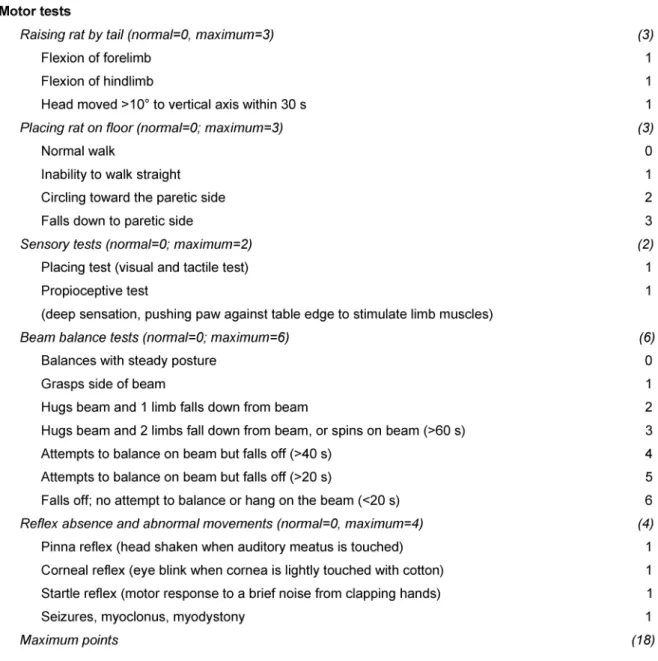
Table 1 shows the items for the modified neurological severity score (mNSS) according to Chen et al. [6]. Neuro- logical function including motoric and sensoric systems as well as reflexes and a balance test is graded on a nu- meric scale from 0 to 18 (0 for no disorders/normal function and a maximum lesion score of 18). If an animal was unable to perform a test or if the lack of a tested re- flex was noticed, a score point was given. Thus, the higher the score the more severe the ischemic lesion caused by MCAO. 1-6 mNSS score points were considered as mild, 7-12 score points as medium and 13-18 score points as severe injury.
A baseline measurement was taken one day prior to surgery. From the day following the surgical procedure, RotaRod and beam walk testing were performed daily up to 7 days post operation. Thereafter, tests were carried out every second day until day 29. All animals underwent the mNSS test at day 1, 4, 7, 11, 15, 19, 23, and 27.
Behavioral testing was conducted during the light phase and animals were not removed from their familiar envir- onment. All experiments were done by one researcher blinded to the experimental groups, and the sequence of tests was randomised between the animals each day.
Data are presented as absolute values in seconds (RotaRod and beam walk) or score points (mNSS).
Tissue preparation,
immunohistochemistry and histological analysis
After termination of behavioral tests animals were deeply anaesthetized with CO2and were perfused transcardially with physiological saline solution. The brains were re- moved immediately and evaluated for gross macroscop- ical pathology. 2 mm thick frontal slices of fresh rat brains were cut and then snap frozen on dry ice using precooled metal plates with polished surfaces. 10-15 µm thick serial sections throughout the whole brain were fixed in 8% ethanol 1/1 v:v for 15-30 minutes and then stained with either Hematoxylin/Eosin (HE) or Giemsa stain
Table 1: Detailed description of the items forming the modified neurological severity score (mNSS) according to Chen et al. [6].
The beam walk is commonly used to evaluate sensomotoric deficits in rats following MCAO.
(Merck, Haar, Germany), dehydrated and mounted in Eukitt (Merck). Sections designated for fluorescence mi- croscopy were mounted in non-fixed condition in aqueous medium (Aquamount; Polysciences, Warrington, USA).
Microscopy was done using a Zeiss Axioplan 2 microscope with Neofluar objectives and the Zeiss Fluorescence filter sets 02, 05, 06, 09, 15 and 26.
After morphologic differentiation between functional tis- sue versus colliquation necrosis or the border zone in HE- stained slices, borderlines between those areas were marked digitally. Then analysis of the following, corres- ponding fluorescence slices was performed after transfer of digital borderlines to the fluorescence image.
For further characterization, brain slices were incubated with mouse monoclonal antibodies against anti-human CD34, anti-human CD68, anti-human neurofilament-L fragment (NF-L, 70 kDa) antibodies, anti-human chromo- granin (all Dako Cytomation, Glostrup, Denmark) or anti- rat CD68 (Dako, Glostrup, Denmark) according to manu- facturers instructions.
Statistical analysis
All behavioral tests were evaluated for normality and equal variances. Analyses of significance were performed using the non-parametric Mann-Whitney test for compari- son of the groups. We also tested the balance of the
baseline variables in comparison of all groups to include any potential imbalances in the analysis of the treatment effect. Both, differences between treated groups and control animals as well as differences between CD34+ and CD34-treated subjects were evaluated for each be- havioral test.P-values of 0.05 or less were considered statistically significant. All data are shown as mean ± standard deviation (SD).
Results
Characterization of separated HUCB cells
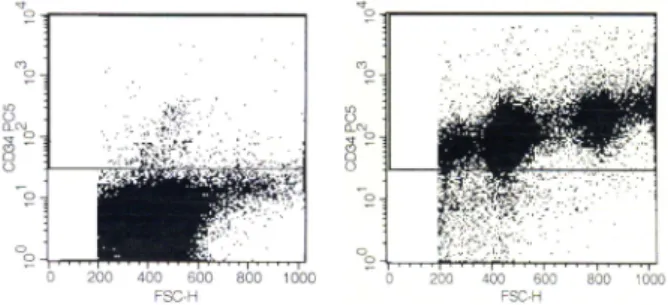
HUCB mononuclear cells were separated by anti-CD34 antibodies and magnetic beads as described above. Fig- ure 1 shows characterization of the cell preparations used for treatment studies. The right panel shows the CD34+ cell enriched population containing a mean proportion of 98.9% CD34+cells with a viability of >91% (5 independ- ent experiments). The enrichment factor amounted to more than 50 (mean of 1.8% CD34+ cells in regular HUCB). The left panel shows the preparation depleted from CD34+cells. The degree of depletion ranged from 3x to 6x i.e. from 0.1 to 0.5% CD34+cells, as determined in five independent experiments. Viability was >92% in each experiment.
Figure 1: FACS analysis of HUCB cells used for treatment. Left panel shows a CD34+cell depleted sample stained with an
anti-CD34 antibody (0.4% CD34+cells) directed to a non- competing CD34 epitope as compared to the antibody used for cell separation. Right panel shows a CD34+cell enriched
fraction (98.9%).
Neurofunctional recovery after cell therapy
Animals were randomly assigned to one of three groups.
Group 1 (n=18) received CD34+cells while group 2 (n=12) was injected with CD34-cells. Group 3 (n=18) was taken as a control receiving a sham injection of PBS.
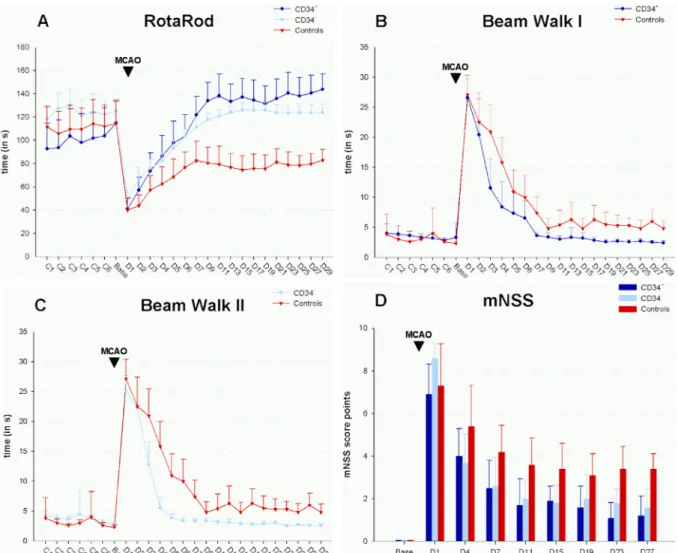
Rats receiving HUCB cells of both populations showed a remarkable reduction of functional and neurological defi- cits in each test with complete remission in the RotaRod and beam walk test as compared to baseline measure- ment (Figure 2A-D). Statistically significant differences (P<0.05) between animals treated with CD34+cells and untreateted controls were first observed at day 2 in the RotaRod (P<0.05, Figure 2A), at day 3 in the beam walk array (P<0.01, Figure 2B and Figure 2C) and at day 4 in the mNSS (P<0.05, Figure 2D). CD34-HUCB cell admin- istration became superior to saline injection at the third day in the RotaRod experiment (P<0.05) and the beam walk test (P<0.01) and at day 4 in the mNSS (P<0.05).
With each functional test a high level of statistical signi- ficance (P<0.00001) was reached in the plateau phase of functional recovery (see Figure 2). This level of highly significant difference was maintained at least from day 11 until the end of behavioural testing.
Comparing CD34+HUCB cells with CD34-HUCB cells, no statistically significant difference (P<0.05) was observed with regard to the beneficial effect of both populations (Figure 2A-D). In the RotaRod test, marginal differences were only observed at day 9 (P=0.03), day 11 (P=0.04) and day 29 (P=0.04). Beam walk data were marginally different only at day 6 (P=0.01) while in the mNSS test a marginal divergence could be noticed at day 1 (P=0.02).
In all cases, data obtained from animals that were admin- istered CD34-HUCB cells tended to a slightly less effective treatment as compared to the use of CD34+HUCB cells.
However, the null hypothesis, i.e. no statistical difference between CD34+versus CD34-HUCB cells, had to be ac- cepted (P>0.4) in each case.
An example of the sensomotoric performance of experi- mental animals 14 days after MCAO with and without cell transplantation as well as before MCAO is given in the video file attached. A cell-transplanted animal is able to pass the beam walk within 1.5 to 3.0 s like the healthy control animal without major stop overs or imbalances, while a non-transplanted MCAO subject clearly suffers from sensomotoric disabilities interrupting its walking frequently with the tendency to loose balance and to fall down.
Figure 2: Results of behavioral tests before and after permanent MCAO (▼). SH rats received 1x10⁶cells enriched or depleted from CD34+cells by magnetic bead separation (n=18 for CD34+cell treatment;n=12 for CD34-cell treatment) or 1 ml of PBS
for control (n=18) 8 to 10 hours following ischemia. Behavioral tests (RotaRod, A; beam walk, B and C) and the modified neurological severity score (D) were performed during the phase of conditioning, before (Base line) and after MCAO throughout
the evaluation phase. CD34+and CD34-treated animals showed statistically significant improvement of neurological deficits in the plateau phase starting at day 11 (P<0.0001 in all test arrays). For statistical details in the early phase (D2-D9) see
description in the text. Data are presented as absolute values ± SD.
Histological localization of HUCB cells in brain and other organs
For histological studies the cells were labeled with CFSE prior to injection as described above and serial tissue sections were prepared after termination of the animal experiments at day 29 after MCAO for investigation by UV microscopy. Control experiments were performed without using HUCB cells as shown in Figure 3 for HE- staining (Figure 3A) and the green fluorescence channel (Figure 3B) displayed in parallel sections, while CFSE- labeled HUCB mononuclear cells were used in the exper- iment shown in Figure 3C in the green channel versus the red channel (Figure 3D) in order to demonstate exclu- sion of autofluorescence. CFSE-labeled cells could be detected clearly (Figure 3E-H) upon intravenous adminis- tration of both preparations, CD34+and CD34-HUCB cells.
They were found accumulating in the border zone (Figure 3F and H), while no fluorescent cells were observed at
the contralateral side of lesion or in control animals (Fig- ure 3B).
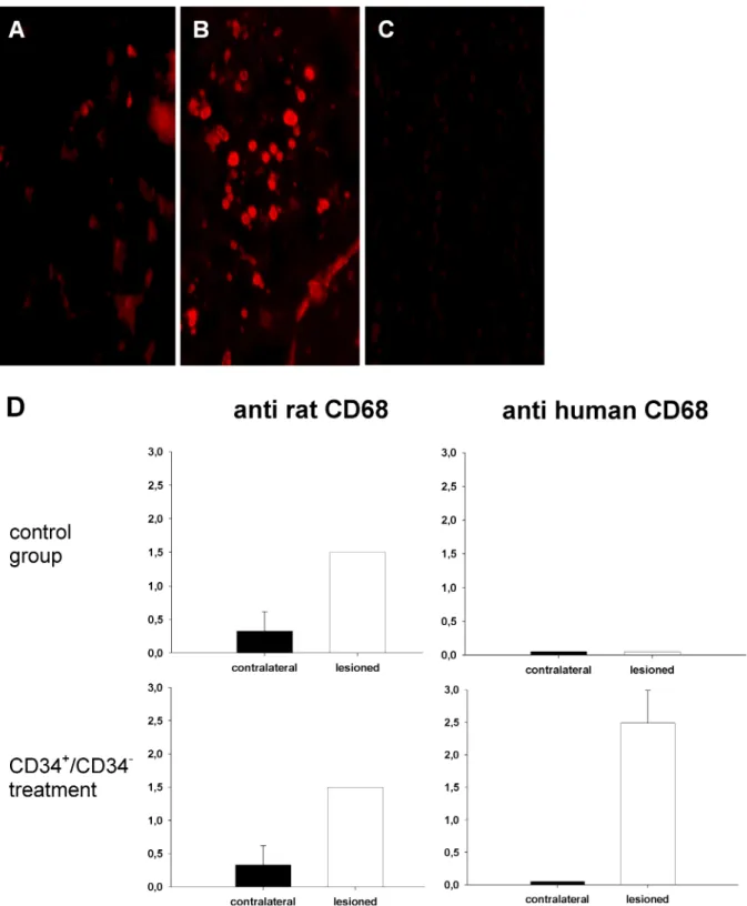
However, we were not able to identify neuronal cells of human origin by staining with antibodies against human NF-L or chromogranin as assessed four weeks after cell injection. Staining with anti-rat CD68 showed a moderate and diffuse activity of host microglial cells and monocytes in ischemic areas (Figure 4A and D). In both groups, an- imals transplanted with either human CD34+ or CD34- cells, an increase of human CD68+cells could be found in the border zone of the lesion (Figure 4B and D), while human CD68 could not be found in the contralateral hemisphere or in animals that were not transplanted with human cells (Figure 4C and D). Staining with anti-human CD34 antibodies suffered from diffuse fluorescence activity in most endothelial cells. After HUCB cell admin- istration, no tumor formation was found in any animal until termination of the experiment at day 29 post opera- tion.
Figure 3: Tracking of CFSE-labelled CD34+and CD34-HUCB cells in serial sections of SH in rat brains 29 days after MCAO. A:
control animal without cell therapy, HE staining; B: control animal, UV microscopy with green fluorescence; C: HUCB mononuclear cell treatment, green fluorescence, the borderline between the necrotic central area and the border zone to unaffected tissue is marked according to HE serial section; D: HUCB mononuclear cell treatment, red fluorescence; E: CD34+HUCB cell treatment, HE staining; F: CD34+HUCB cell treatment, green fluorescence (inverse colours); G: CD34-HUCB cell treatment, phase contrast
microscopy; H: CD34-HUCB cell treatment, green fluorescence. 400fold magnification in all images.
Figure 4: Detection of cells positive for rat or human CD68 in control animals and subjects that received CD34+/CD34-cell transplantation. Moderate and diffuse fluorescence signals from cells positive for rat CD68 occurs in the lesioned hemisphere of transplanted and control animals (A). A strong increase of fluorescence caused by cells positive for human CD68 cells could be detected in animals transplanted with both, CD34+and CD34-cells indicating a "monocyte fate" of administered cells (B).
Cells positive for human CD68 could not be detected in control rats (C) or in the contralateral hemisphere. D shows the level of fluorescence according to a score point system (0=none, 1=low, 2=moderate, 3=high fluorescence activity).
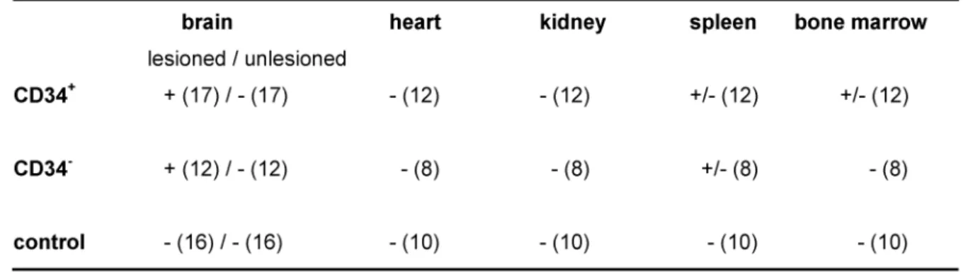
Tissue samples from heart, kidney, spleen and bone marrow of experimental animals were examined for CFSE- positive cells. Only few CFSE-positive cells were found in spleen and bone marrow of animals transplanted with CD34+cells (Table 2).
Table 2: Detection of CFSE-labeled cells in rat organs 29 days upon transplantation. Number of evaluated animals is given in brackets. + = high density of CFSE-positive cells; +/- = only few CFSE-positive cells were detected; - = no CFSE-positive cells
were detected in slices of harvested organs.
Discussion
The experimental model
In order to verify the benefit of new therapeutical proced- ures for future use in humans, it is important to design experimental animal models in close relation to clinical requirements. In many human patients suffering from stroke, the capillary system is damaged by long-term hy- pertension, hypertriglyceridemia and hypercholester- inemia. Therefore, we performed the experiments in SH rats instead of strains like Wistar or Sprague Dawley which were used in previous studies [6], [33]. According to Traystman [31], SH rats suffer from larger and more consistent ischemic lesions and showed less variable functional deficits within the experimental groups which could be confirmed by own experiences (data not shown).
Close relation to clinical reality for potential treatment protocols was also the reason to adopt the permanent artery occlusion instead of transient blood vessel block- age by a surgical filament [20]. The time frame for cell therapy (8-10 hours after MCAO) would be feasible in the clincal practice of a stroke unit considering even the most unfortunate delay until the patients had arrived and passed all diagnostic procedures.
In principle, the use of xenogeneic (human) cells might induce a local immune response in the (rat) host which could hypothetically interfere with treatment e.g. by re- leasing neurotropic cytokines. However, Chen et al. [6]
could demonstrate functional recovery by using HUCB cells in rat MCAO without immunosuppression. Their ex- perimental design and results were comparable to the study of Willing et al. [33] using cyclosporine A. Since neuroprotective effects have been described recently by immunosuppressive drugs [18] we decided to omit their use. Moreover, recent control experiments (data not shown) with an irradiated human T cell clone with irrele- vant specifity instead of HUCB cells showed no therapeut- ic effect at all. Because of irradiation at 5 Gy, T cells were not able to divide but remained viable in parallel in vitro culture for 29 days and were still able to produce TNF-α.
By this experiment, we wanted to show that a xenogeneic
immune reaction of the host towards cells of human origin was not responsible for the success of treatment.
Functional improvement in behavioral tests by CD34
-cells
In this study, cell therapy with HUCB-derived cells demonstrated a significant improvement of neuromotoric function in SH rats but CD34-cells were nearly as effective as CD34+cells. The number of remaining CD34+cells in the depleted preparation was below 0.5% i.e. less than 5000 cells, while the enriched preparation contained nearly 1 million viable CD34+cells. If CD34+cells would be the cellular entity responsible for the therapeutic be- nefit there should be a clear distinction between both preparations in their functional potential.
Very recently, Taguchi et al. [29] have reported that HUCB- derived CD34-cells in contrast to CD34+cells were not able to promote angiogenesis in SCID mice upon experi- mental focal cerebral ischemia. However, their cells were administered much later i.e. after 24 hours and persistent focal motor deficits were not observed in their model. Our results indicate that other cells than CD34+cells seem to be involved. Interestingly, a cell population isolated from adult human brain and characterized as CD133+, CD34-and CD45-could expand and differentiate effect- ively within the brain of neonatally immunodeficient mice [32]. Despite that the cells in the study of Uchida et al.
[32] were described as central nervous system stem cells, it is a possibility that more immature stem cells, described by another denominator, such as CD133, were respons- ible for the therapeutic benefit. Gallacher et al. [13]
demonstrated that CD133+, CD34-cord blood cells are capable of forming heamtopoietic cells in vitro as well as in NOD/SCID mice and these immature cells may be re- sponsible for the therapeutic effect seen in our CD34- cells.
The number of 106cells injected systemically is relatively small as compared to the mass of ischemic tissue. Signi- ficant functional recovery could be detected at least at day 4 in the mNSS test, while the maximum improvement of neuromotoric functions was reached at day 11 and was maintained until the end of study. It seems to be
unlikely that such a small number of cells integrates, develops appropriate neuronal connections, restores functional cerebral networks and leads to a conspicuous behavioural improvement within such a relatively short period of time. However, these cells may initiate or accel- erate endogenous regeneration processes.
In general, an ischemic cerebral lesion consists of a central colliquation necrosis surrounded by the penumbra, an area of incomplete i.e. reversible cell damage [21].
The penumbra, as the “tissue at risk”, is a border zone dividing the regions of total ischemic cell destruction from those containing unaffected and functional tissue. This area is the main therapeutic target for treating stroke by regeneration and cellular restoration [12].
Upon intravenous administration, CFSE-labeled cells ac- cumulated in a border zone comprising the brain area remaining from the penumbra after 4 weeks. Moreover, we found HUCB cells in the ipsilateral hemisphere of le- sion and only very few CFSE-labeled HUCB in the contralat- eral ones as shown first by Chen et al. [6]. We were not able to detect cells of human origin expressing neuronal markers indicating that neurofunctional restoration is not related to neurogenesis from the transplanted cells. The detection of cells being positive for human CD68, a cellu- lar marker for monocytes and macrophages, demon- strated that human cells take part in local processes of organisation of the lesion and the surrounding tissue.
Findings of Borlongan et al. [5] indicate that trophic and/or neuroprotective factors produced by transplanted cells are mere important for neurofunctional restoration than the generation of new neurons.
Here, we could demonstrate that this accumulation was not selective for CD34+. The accumulation indicates chemotactic attraction of transplanted cells towards the lesion or topic expression of adhesion molecules with the same result: focal accumulation of migrating cells close to the ischemic lesion. Ischemic brain tissue expresses chemotactic proteins like MCP-1 [19] and microvascular endothelial cells significantly upregulate the expression of ICAM 1, vascular adhesion molecule-1 (VCAM) and E- selectin following acute ischemic cerebral injury as repor- ted by Zhang et al. [35] and Blann et al. [4]. It is important to note that the blood brain barrier becomes leaky from 3 hours to 4 days after stroke in rats [16], [1], [25] which may allow for unselected entry of migrating cells. How- ever, more work has to be done to investigate the mech- anisms of cell migration and recruitment of regenerative potential in detail.
In conclusion, we could demonstrate that CD34+depleted HUCB cells accumulate in the border zone of the ischemic lesion and are highly sufficient for successful treatment of an ischemic insult after permanent occlusion of the middle cerebral artery in SH rats showing a risk profile. This population of cells is much easier to obtain than purified CD34+ cells which are a rare fraction in HUCB. Because HUCB cells are widely available, easy to store and to handle, they may provide an important source for treatment of stroke in future cell based ther- apies.
Acknowledgements
The authors wish to thank Manuela Ackermann, Mandy Glona, Silke Lehnert, and Christine Schulz for technical support as well as Sigrid Weisheit and Petra Madaj-Sterba for their valuable help considering animal care. This work was financially supported by a grant given by the state of Saxony (SAB) and by the Interdiszipinäres Zentrum für Klinische Forschung (IZKF) project A 00 supported by the BMBF (Bundesministerium für Bildung und Forschung).
Abbreviations
APC - allophycocyanine
bFGF - basic fibroblast growth factor CD - cluster of differentiation
CFSE - carboxy fluoresceindiacetate succinimidyl ester CNS - central nervous system
CSA - cyclosporine A DMSO - dimethyl sulfoxide FITC - fluorescein isothiocyanate FSC-H - foreward scatter scale HE - hematoxylin/eosin HFS - high foreward scatter
HUCB - human umbilical cord blood ICAM - intercellular adhesion molecule LFS - low foreward scatter
LSS - low sideward scatter
MCA(O) - middle cerebral artery (occlusion) MCP-1 - monocyte chemoattractant protein-1 mNSS - modified neurological severity score NF-L - neurofilament L fragment
NOD/SCID - nonobese diabetic/severe combined immun- odeficiency
PBS - phosphate buffered saline PC5 - phycoerythrin-cyanin 5 PE - phycoerythrin
SH - spontaneously hypertensive VCAM - vascular adhesion molecule VEGF- vascular endothelial growth factor
Attachments
Available from
http://www.egms.de/en/gms/2008-3/000027.shtml 1. GMS-Experimental-cell-therapy-of-stroke.wmv
(38883.996 KB)
Example of the sensomotoric performance of experimental animals 14 days after MCAO with and without cell transplantation as well as before MCAO:
A cell-transplanted animal is able to pass the beam walk within 1.5 to 3.0 s like the healthy control animal without major stop overs or imbalances, while a non-transplanted MCAO subject clearly suffers from sensomotoric disabilities interrupting its walking frequently with the tendency to lose balance and to fall down.
References
1. Belayev L, Busto R, Zhao W, Ginsberg MD. Quantitative evaluation of blood-brain barrier permeability following middle cerebral artery occlusion in rats. Brain Res. 1996;739(1-2):88-96.
2. Bender JG, Unverzagt KL, Walker DE, Lee W, Van Epps DE, Smith DH, Stewart CC, To LB. Identification and comparison of CD34- positive cells and their subpopulations from normal peripheral blood and bone marrow using multicolor flow cytometry. Blood.
1991;77(12):2591-6.
3. Bicknese AR, Goodwin HS, Quinn CO, Henderson VC, Chien SN, Wall DA. Human umbilical cord blood cells can be induced to express markers for neurons and glia. Cell Transplant.
2002;11(3):261-4.
4. Blann A, Kumar P, Krupinski J, McCollum C, Beevers DG, Lip GY.
Soluble intercelluar adhesion molecule-1, E-selectin, vascular cell adhesion molecule-1 and von Willebrand factor in stroke.
Blood Coagul Fibrinolysis. 1999;10(5):277-84.
5. Borlongan CV, Hadman M, Sanberg CD, Sanberg PR. Central nervous system entry of peripherally injected umbilical cord blood cells is not required for neuroprotection in stroke. Stroke.
2004;35(10):2385-9.
6. Chen J, Sanberg PR, Li Y, Wang L, Lu M, Willing AE, Sanchez- Ramos J, Chopp M. Intravenous administration of human umbilical cord blood reduces behavioral deficits after stroke in rats. Stroke. 2001;32(11):2682-8.
7. Chopp M, Li Y. Treatment of neural injury with marrow stromal cells. Lancet Neurol. 2002;1(2):92-100.
8. Delcker A, Diener HC, Timmann D, Faustmann P. The role of vertebral and internal carotid artery disease in the pathogenesis of vertebrobasilar transient ischemic attacks. Eur Arch Psychiatry Clin Neurosci. 1993;242(4):179-83.
9. Dunham NW, Miya TS. A note on a simple apparatus for detecting neurological deficit in rats and mice. J Am Pharmacol Assoc.
1957;46(3):208-9.
10. Duverger D, MacKenzie ET. The quantification of cerebral infarction following focal ischemia in the rat: influence of strain, arterial pressure, blood glucose concentration, and age. J Cereb Blood Flow Metab. 1988;8(4):449-61.
11. Erdo F, Buhrle C, Blunk J, Hoehn M, Xia Y, Fleischmann B, Focking M, Kustermann E, Kolossov E, Hescheler J, Hossmann KA, Trapp T. Host-dependent tumorigenesis of embryonic stem cell transplantation in experimental stroke. J Cereb Blood Flow Metab.
2003;23(7):780-5.
12. Fisher M, Ratan R. New perspectives on developing acute stroke therapy. Ann Neurol. 2003;53(1):10-20.
13. Gallacher L, Murdoch B, Wu DM, Karanu FN, Keeney M, Bhatia M. Isolation and characterization of human CD34(-)Lin(-) and CD34(+)Lin(-) hematopoietic stem cells using cell surface markers AC133 and CD7. Blood. 2000;95(9):2813-20.
14. Garbuzova-Davis S, Willing AE, Zigova T, Saporta S, Justen EB, Lane JC, Hudson JE, Chen N, Davis CD, Sanberg PR. Intravenous administration of human umbilical cord blood cells in a mouse model of amyotrophic lateral sclerosis: distribution, migration, and differentiation. J Hematother Stem Cell Res. 2003;12(3):255- 70.
15. Hamm RJ, Pike BR, O'Dell DM, Lyeth BG, Jenkins LW. The rotarod test: an evaluation of its effectiveness in assessing motor deficits following traumatic brain injury. J Neurotrauma. 1994;11(2):187- 96.
16. Hatashita S, Hoff JT. Brain edema and cerebrovascular permeability during cerebral ischemia in rats. Stroke.
1990;21(4):582-8.
17. Jones BJ, Roberts DJ. A rotarod suitable for quantitative measurements of motor incoordination in naive mice. Naunyn Schmiedebergs Arch Exp Pathol Pharmakol. 1968;259(2):211.
18. Kaminska B, Gaweda-Walerych K, Zawadzka M. Molecular mechanisms of neuroprotective action of immunosuppressants- -facts and hypotheses. J Cell Mol Med. 2004;8(1):45-58.
19. Kim JS. Cytokines and adhesion molecules in stroke and related diseases. J Neurol Sci. 1996;137(2):69-78.
20. Koizumi J, Yoshida Y, Nakazawa T, Ooneda G. Experimental studies of ischemic brain edema, I: a new experimental model of cerebral embolism in rats in recirculation can be introduced in the ischemic area. Jpn J Stroke. 1986;8:1-8.
21. Lassen NA, Vorstrup S. Ischemic penumbra results in incomplete infarction: is the sleeping beauty dead? Stroke. 1984;15(4):755- 8.
22. Lu D, Sanberg PR, Mahmood A, Li Y, Wang L, Sanchez-Ramos J, Chopp M. Intravenous administration of human umbilical cord blood reduces neurological deficit in the rat after traumatic brain injury. Cell Transplant. 2002;11(3):275-81.
23. Newman MB, Davis CD, Kuzmin-Nichols N, Sanberg PR. Human umbilical cord blood (HUCB) cells for central nervous system repair. Neurotox Res. 2003;5(5):355-68.
24. Parish CR. Fluorescent dyes for lymphocyte migration and proliferation studies. Immunol Cell Biol. 1999;77(6):499-508.
25. Rosenberg GA, Estrada EY, Dencoff JE. Matrix metalloproteinases and TIMPs are associated with blood-brain barrier opening after reperfusion in rat brain. Stroke. 1998;29(10):2189-95.
26. Sanchez-Ramos JR, Song S, Kamath SG, Zigova T, Willing A, Cardozo-Pelaez F, Stedeford T, Chopp M, Sanberg PR. Expression of neural markers in human umbilical cord blood. Exp Neurol.
2001;171(1):109-15.
27. Saporta S, Borlongan CV, Sanberg PR. Neural transplantation of human neuroteratocarcinoma (hNT) neurons into ischemic rats.
A quantitative dose-response analysis of cell survival and behavioral recovery. Neuroscience. 1999;91(2):519-25.
28. Swislocki A, Tsuzuki A. Insulin resistance and hypertension:
glucose intolerance, hyperinsulinemia, and elevated free fatty acids in the lean spontaneously hypertensive rat. Am J Med Sci.
1993;306(5):282-6.
29. Taguchi A, Soma T, Tanaka H, Kanda T, Nishimura H, Yoshikawa H, Tsukamoto Y, Iso H, Fujimori Y, Stern DM, Naritomi H, Matsuyama T. Administration of CD34+ cells after stroke enhances neurogenesis via angiogenesis in a mouse model. J Clin Invest. 2004;114(3):330-8.
30. Tamura A, Graham DI, McCulloch J, Teasdale GM. Focal cerebral ischaemia in the rat: 1. Description of technique and early neuropathological consequences following middle cerebral artery occlusion. J Cereb Blood Flow Metab. 1981;1(1):53-60.
31. Traystman RJ. Animal models of focal and global cerebral ischemia. ILAR J. 2003;44(2):85-95.
32. Uchida N, Buck DW, He D, Reitsma MJ, Masek M, Phan TV, Tsukamoto AS, Gage FH, Weissman IL. Direct isolation of human central nervous system stem cells. Proc Natl Acad Sci U S A.
2000;97(26):14720-5.
33. Willing AE, Lixian J, Milliken M, Poulos S, Zigova T, Song S, Hart C, Sanchez-Ramos J, Sanberg PR. Intravenous versus intrastriatal cord blood administration in a rodent model of stroke. J Neurosci Res. 2003;73(3):296-307.
34. Yamaguchi M, Hirayama F, Murahashi H, Azuma H, Sato N, Miyazaki H, Fukazawa K, Sawada K, Koike T, Kuwabara M, Ikeda H, Ikebuchi K. Ex vivo expansion of human UC blood primitive hematopoietic progenitors and transplantable stem cells using human primary BM stromal cells and human AB serum.
Cytotherapy. 2002;4(2):109-18.
35. Zhang RL, Chopp M, Zaloga C, Zhang ZG, Jiang N, Gautam SC, Tang WX, Tsang W, Anderson DC, Manning AM. The temporal profiles of ICAM-1 protein and mRNA expression after transient MCA occlusion in the rat. Brain Res. 1995;682(1-2):182-8.
Corresponding author:
Prof. Frank Emmrich
Institute for Clinical Immunology and Transfusion Medicine, University of Leipzig, Johannisallee 30, 04103 Leipzig, Tel.: +49 341 97 25500, Fax: +49 341 97 25509 frank.emmrich@medizin.uni-leipzig.de
Please cite as
Boltze J, Kowalski I, Geiger K, Reich D, Gunther A, Buhrle C, Egger D, Kamprad M, Emmrich F. Experimental treatment of stroke in spontaneously hypertensive rats by CD34+and CD34-cord blood cells. GMS Ger Med Sci. 2008;3:Doc09.
This article is freely available from
http://www.egms.de/en/gms/2008-3/000027.shtml
Received:2005-01-24 Published:2005-11-10
Copyright
©2008 Boltze et al. This is an Open Access article distributed under the terms of the Creative Commons Attribution License
(http://creativecommons.org/licenses/by-nc-nd/3.0/deed.en). You are free: to Share — to copy, distribute and transmit the work, provided the original author and source are credited.